Gallotannin inhibits the expression of chemokines and...
40
MOLPHARM/2005/012518 1 Gallotannin inhibits the expression of chemokines and inflammatory cytokines in A549 cells* Katalin Erdélyi, Andrea Kiss, Edina Bakondi, Péter Bai, Csaba Szabó, Pál Gergely, Ferenc Erdődi and László Virág Department of Medical Chemistry, Research Center for Molecular Medicine, Medical and Health Science Center, University of Debrecen, H-4026 Debrecen, Bem tér 18/B, Hungary (KE, AK, EB, PB, PG, FE, LV) Inotek Pharmaceuticals, 100 Cummings Center, Suite#419E, Beverly, MA 01915 (CS) Molecular Pharmacology Fast Forward. Published on June 23, 2005 as doi:10.1124/mol.105.012518 Copyright 2005 by the American Society for Pharmacology and Experimental Therapeutics. This article has not been copyedited and formatted. The final version may differ from this version. Molecular Pharmacology Fast Forward. Published on June 23, 2005 as DOI: 10.1124/mol.105.012518 at ASPET Journals on July 2, 2018 molpharm.aspetjournals.org Downloaded from
Transcript of Gallotannin inhibits the expression of chemokines and...
MOLPHARM/2005/012518
1
Gallotannin inhibits the expression of chemokines and inflammatory cytokines in A549 cells*
Katalin Erdélyi, Andrea Kiss, Edina Bakondi, Péter Bai, Csaba Szabó, Pál Gergely, Ferenc Erdődi and László Virág
Department of Medical Chemistry, Research Center for Molecular Medicine, Medical and
Health Science Center, University of Debrecen, H-4026 Debrecen, Bem tér 18/B, Hungary
(KE, AK, EB, PB, PG, FE, LV)
Inotek Pharmaceuticals, 100 Cummings Center, Suite#419E, Beverly, MA 01915
(CS)
Molecular Pharmacology Fast Forward. Published on June 23, 2005 as doi:10.1124/mol.105.012518
Copyright 2005 by the American Society for Pharmacology and Experimental Therapeutics.
This article has not been copyedited and formatted. The final version may differ from this version.Molecular Pharmacology Fast Forward. Published on June 23, 2005 as DOI: 10.1124/mol.105.012518
at ASPE
T Journals on July 2, 2018
molpharm
.aspetjournals.orgD
ownloaded from
MOLPHARM/2005/012518
2
Running title: Gallotannin inhibits cytokine and chemokine expression
All correspondence should be addressed to:
László Virág M.D., Ph.D.
Department of Medical Chemistry
Medical and Health Science Center
University of Debrecen
Bem tér 18/B
H-4026 Debrecen
Hungary
Phone: (36)-52-412-345; Fax: (36)-52-412-566; e-mail: [email protected]
Number of text pages: 33
Number of tables: 2 (+1 Supplemental table)
Number of figures: 8 (+1 Supplemental figure)
Number of references: 48
Number of words in the Abstract: 250
Introduction: 574
Discussion: 1193
List of nonstandard abbreviations used in the paper: ABTS 2,2’-Azino-bis-(3-
ethylbenzothiazoline-6-sulfonic acid; PAR poly(ADP-ribose); PARG PAR glycohydrolase;
PARP PAR polymerase; NFκB nuclear factor kappa B; AP-1 activator protein-1; PP1c
catalytic subunit of protein phosphatase 1; PP2Ac catalytic subunit of protein phosphatase
2A;
This article has not been copyedited and formatted. The final version may differ from this version.Molecular Pharmacology Fast Forward. Published on June 23, 2005 as DOI: 10.1124/mol.105.012518
at ASPE
T Journals on July 2, 2018
molpharm
.aspetjournals.orgD
ownloaded from
MOLPHARM/2005/012518
3
Abstract
Tannins are plant-derived, water-soluble polyphenols with wide-ranging biological activities.
The mechanisms underlying the anti-inflammatory effect of tannins are not fully understood and
may be due to inhibition of poly(ADP-ribose) (PAR) glycohydrolase (PARG), the main catabolic
enzyme of PAR metabolism. Therefore, we set out to investigate the mechanism of the anti-
inflammatory effect of GT in A549 cells with special regard to the role of poly(ADP-
ribosyl)ation. Using an inflammation-focused low density array and RT-PCR we found that GT
suppressed the expression of most cytokines and chemokines in cytokine-stimulated A549 cells
whereas the PARP inhibitor PJ-34 only inhibited few transcripts. Activation of the transcription
factors NF−kappaB and AP-1 was blocked by GT, whereas PJ-34 only suppressed NF−kappaB
activation but not AP-1 activation. GT also inhibited IκB phosphorylation and nuclear
translocation of NF−kappaB but PJ-34 had no effect on these upstream events. In the AP-1
pathway, GT treatment even in the absence of cytokines caused maximal phosphorylation of
JNK and c-Jun. GT also caused a low level phosphorylation of p38, ERK1/2, ATF-2 and CREB
but inhibited cytokine-induced phosphorylation of these kinases and transcription factors. GT
inhibited protein phosphatases 1 and 2A which may explain increased phosphorylation of MAP
kinases and their substrates. GT exerted potent antioxidant effect but failed to cause PAR
accumulation. In summary, the potent inhibitory effects of GT on the transcription of cytokine
and chemokine genes are not likely to be related to PARG inhibition. Inhibition of AP-1
activation and upstream signaling events may be responsible for the effects of GT.
This article has not been copyedited and formatted. The final version may differ from this version.Molecular Pharmacology Fast Forward. Published on June 23, 2005 as DOI: 10.1124/mol.105.012518
at ASPE
T Journals on July 2, 2018
molpharm
.aspetjournals.orgD
ownloaded from
MOLPHARM/2005/012518
4
Introduction
Tannins are water-soluble polyphenols that are widely distributed in the plant kingdom,
including food grains and fruits. To date more than a thousand different tannins have been
characterized and ordered into four major groups: 1) gallotannins, 2) ellagitannins, 3) complex
and 4) condensed tannins, with gallotannins and ellagitannins being the most widespread types.
The common structural elements of all tannins include one or more polyol units (mostly D-
glucose) and one or more polyphenols (gallic acid, 3,4,5 trihydroxyl benzoic acid).
The simplest hydrolyzable tannin, gallotannin is a mixture of polygalloyl esthers of glucose.
Gallotannin and other tannins have been shown to exert various biological effects ranging from
anti-inflammatory to anti-cancer and antiviral effects (Uchiumi et al., 1996; Van Molle et al.,
2000; Feldman et al., 2001; Mota et al., 1985; Fong et al., 1972). The mechanisms underlying the
anti-inflammatory effect of tannins include the scavenging of radicals (antioxidant effect)
(Hagerman et al., 1999) and inhibition of the expression of inflammatory mediators such as some
cytokines (Feldman et al., 2001), inducible nitric oxide synthase (iNOS) and cycloxygenase-2
(Lee et al., 2003). Most of these studies focused on the effects of tannins on immune cells with
special regard to mononuclear cells and macrophages and little is known about the possible
effects of tannins in epithelial cells.
Various classes of tannins have also been demonstrated to inhibit poly(ADP-ribose)
glycohydrolase (PARG) the catabolic enzyme of poly(ADP-ribose) metabolism. Poly(ADP-
ribosyl)ation is a posttranslational protein modification catalyzed by poly(ADP-ribose)
polymerase (PARP) enzymes with PARP-1 being responsible for more than 90% of the cellular
poly(ADP-ribosyl)ation capacity (Ame et al., 2004;Burkle, 2001). Activated PARP synthesizes
branching (ADP-ribose)n polymers from NAD+ and attaches the polymer to glutamate or
aspartate residues of suitable acceptor proteins including PARP-1 itself (automodification),
This article has not been copyedited and formatted. The final version may differ from this version.Molecular Pharmacology Fast Forward. Published on June 23, 2005 as DOI: 10.1124/mol.105.012518
at ASPE
T Journals on July 2, 2018
molpharm
.aspetjournals.orgD
ownloaded from
MOLPHARM/2005/012518
5
histones, DNA repair enzymes and transcription factors. Reversible poly(ADP-ribosyl)ation
regulates various cellular processes including transcription. The inhibitory effect of PARP
inhibitors on the transcription of inflammatory mediators such as cytokines, chemokines and
iNOS has been made responsible for the anti-inflammatory effects of PARP inhibition (for
review see Erdelyi et al., 2005). The role of PARG in this process, however, is not fully
understood. It is plausible to hypothesize that tannins may increase the amount of poly(ADP-
ribosyl)ated proteins in the cell and may thus modulate transcription.
Accelerated PAR metabolism has been implicated in various oxidative stress-related lung
diseases such as asthma, reperfusion injury, ARDS, asbestosis and shock (Virag, 2005). Many
studies have demonstrated that PARP inhibitors selectively regulated the expression of cytokines
and chemokines (chemotactic cytokines) in these and similar inflammatory disease models
(Virag, 2005). Reduced expression of chemokines and adhesion molecules may be responsible
for the reduced migration of inflammatory cells, the most common anti-inflammatory effect of
PARP inhibition as observed in animal studies (Hasko et al., 2002; Zingarelli et al., 1998). It is
not known, however, whether macrophages or parenchymal cells are the main targets of PARP
inhibitors in these diseases. Furthermore, the cellular effects of gallotannin in lung epithelial
cells have not yet been characterized.
Here we show that immune-stimulated A549 type II lung epithelial cells express many
chemokines and inflammatory cytokines. GT abolishes the expression of most
chemokines/cytokines whereas the potent PARP inhibitor PJ-34 only suppressed few transcripts.
We demonstrate that in A549 cells, GT (30 µM) acts at various levels of the signal transduction
cascade of the NFκB and AP-1 pathway without causing major perturbations in poly(ADP-
ribose) catabolism.
This article has not been copyedited and formatted. The final version may differ from this version.Molecular Pharmacology Fast Forward. Published on June 23, 2005 as DOI: 10.1124/mol.105.012518
at ASPE
T Journals on July 2, 2018
molpharm
.aspetjournals.orgD
ownloaded from
MOLPHARM/2005/012518
6
Materials and methods
Cell culture and treatments.
The A549 cell line was grown and maintained in a 5% CO2 incubator at 37 oC using RPMI-
1640 (Sigma-Aldrich Co., St. Louis, MO) containing 5% fetal bovine serum and
penicillin/streptomycin (Sigma-Aldrich Co., St. Loius MO). Cells were grown to confluency and
incubated in serum-free medium for 12 h prior to treatment. The PARP inhibitor PJ34 (10 µM)
(Inotek Corporation, Beverly, MA.) and the PARG inhibitor gallotannin (30 µM) (Fluka) were
added to the cultures 30 min prior to stimulation by recombinant human TNF-α (20 ng/ml) and
recombinant human IL-1β (5 ng/ml), both purchased from R&D Systems (Minneapolis, MN).
Expression profiling with low density arrays
Profiles of TNFα/IL-1β-induced gene expression were determined by using the protocol of
GEArray pathway-specific expression arrays from SuperArray (Superarray Bioscience Co.,
Frederick, MD). After treatment with TNF-α (20 ng/ml) and IL-1-β (5 ng/ml) for 4 h, total RNA
was isolated. RNA (2 µg) was reverse transcribed into cDNA with MMLV reverse transcriptase,
dNTP mix in the presence of RNasin ribonuclease inhibitor (both reagents were purchased from
Promega, Madison, WI). cDNA was PCR amplified with the Ampolabelling kit using
GEAprimer mix (supplied with the Ampolabelling kit) and dNTP mix containing biotin-16-
dUTP (Roche Hungary, Budaörs, Hungary). The resulting biotin-labeled cDNA probes were
hybridized to gene-specific cDNA fragments on the nylon membranes according to the
manufacturer’s instructions. Biotin was detected with streptavidine-alkaline phosphatase and
CDP-Star chemiluminescent substrate (supplied with the Superarray kit). The relative expression
level of each gene was determined with the Image J software by comparing the signal intensity of
each gene in the array after normalization to the signal of a housekeeping gene. Array
This article has not been copyedited and formatted. The final version may differ from this version.Molecular Pharmacology Fast Forward. Published on June 23, 2005 as DOI: 10.1124/mol.105.012518
at ASPE
T Journals on July 2, 2018
molpharm
.aspetjournals.orgD
ownloaded from
MOLPHARM/2005/012518
7
experiments were performed on 2 different experimental days and a minimum of twofold
difference obtained in both experiments were considered significant.
Reverse transciption and PCR.
Total RNA was isolated using SV Total RNA Isolation System (Promega Co, Madison WI)
according to the manufacturer’s instruction. Concentration and purity of the isolated RNA was
measured spectrophotometrically at 260 nm and 280 nm. Reverse transcription was performed
using MMLV-Reverse Transcriptase (Promega Co, Madison WI). A mix of 2 µg total RNA and
1 µl of Random Primers (Promega Co, Madison WI) was incubated for 5 min in a total volume
of 15 µl at 70oC and cooled on ice. After adding 5 µl of MMLV 5x Reaction Buffer (Promega
Co, Madison WI), 10 mM dNTPs, 1 µl Ribonuclease Inhibitor (Promega Co, Madison WI) and
finally 2 µl of MMLV Reverse Transcriptase (Promega Co, Madison WI) in a total volume of 25
µl, the reaction mix was incubated for an additional hour at 37oC.
PCR reactions were performed using RedTaq polymerase (Sigma-Aldrich Co., St. Loius, MO)
in reaction mixes containing 2.5 U polymerase, 10 nmol of each primer, 4-8 µl cDNA and PCR
buffers as supplied by the manufacturer, in a total volume of 50 µl. PCR primers used for the
analysis were designed based on sequences deposited in the UniGene database. Primer sequences
and sizes of the PCR products are listed in Table 1.
Nuclear extract preparation.
Nuclear protein extracts were prepared from cells grown to 90% confluence in a T-25 culture
flasks. All nuclear extraction procedures were performed on ice with ice cold reagents. Cells
were washed with PBS and harvested by scraping into 1 ml of PBS and pelleted at 5,000 rpm for
5 min. The pellet was resuspended in 400 µl of buffer „A” (10 mM HEPES pH: 7.9, 10 mM KCl,
0.1 mM EDTA, 0.1 mM EGTA, 1 mM DTT, 0.5 mM PMSF, protease inhibitors) and allowed to
swell on ice for 15 minutes. After adding Nonidet P-40 to a final concentration of 0.5% the cells
This article has not been copyedited and formatted. The final version may differ from this version.Molecular Pharmacology Fast Forward. Published on June 23, 2005 as DOI: 10.1124/mol.105.012518
at ASPE
T Journals on July 2, 2018
molpharm
.aspetjournals.orgD
ownloaded from
MOLPHARM/2005/012518
8
were vortex for 10 sec. After centrifugation at 10,000 rpm for 2 min the supernatant was
removed and the pellet was resuspended in 50 µl of buffer „B” (20 mM HEPES pH: 7.9, 420 mM
NaCl, 0.5 mM EDTA, 0.5 mM EGTA, 1mM DTT, 0.5 mM PMSF, protease inhibitors) and
incubated on ice for 20 min with occasional vortexing. Nuclear extracts were recovered after
centrifugation for 10 min at 10,000 rpm. Protein concentrations were determined with Coomassie
Plus Protein Assay reagent (Pierce Biotechnologie Inc., Rockford Illinois).
EMSA
The consensus NF-κB (5’-AGTTGAGGGGACTTTCCCAGG-3’) and AP-1 (5’-
CGCTTGATGACTCAGCCGGAA-3’) probes were obtained from Sigma-Aldrich Co., (Sigma-
Aldrich Co., St. Loius MO). The probes were labeled with Biotin 3’End DNA labeling kit
(Pierce Biotechnologie Inc., Rockford, IL) following the manufacturer’s instruction. Gel shift
assays were performed using LightShift Chemiluminescent EMSA kits (Pierce Biotechnologies
Inc., Rockford Illinois). Briefly, binding reactions containing 10 µg of nuclear extracts and 1
nmol oligonucleotide were performed for 30 min in binding buffer (2.5% glycerol, 0.05%
Nonidet P-40, 50 mM KCl, 5 mM MgCl2 1 mM EDTA, 10 mM Tris pH:7.6, 50 ng poly(dI.dC).
Protein-nucleic acid complexes were resolved using a non-denaturating polyacrilamide gel
consisting of 5% acrylamide (29:1 ratio of acrylamide/bisacrylamide) and run in 0.5x TBE (45
mM Tris-HCl pH 8,3; 45 mM boric acid, 1 mM EDTA) for 1 h at constant voltage (100V). Gels
were transferred to Bio Bond-Plus nylon membrane (Sigma-Aldrich Co., St. Loius MO) (80mA,
45 min). DNA was crosslinked to the membrane by UV cross linker. DNA was incubated in
blocking solution (supplied with the LightShift kit) followed by incubation of the membrane with
streptavidine-peroxidase. After extensive washing, signal was detected with chemiluminescence
solution (supplied with the kit).
Western blot analysis.
This article has not been copyedited and formatted. The final version may differ from this version.Molecular Pharmacology Fast Forward. Published on June 23, 2005 as DOI: 10.1124/mol.105.012518
at ASPE
T Journals on July 2, 2018
molpharm
.aspetjournals.orgD
ownloaded from
MOLPHARM/2005/012518
9
Cells were washed once in PBS and collected by scraping into 200 µl ice-cold lysis buffer
(62.5 mM Tris-HCl pH:6.8, 2% SDS, 10% glycerol, 50 mM DTT, 1mM PMSF, 1mM NaF, 1mM
Na3VO4, protease inhibitors). The extracts were further lysed with sonication and the supernatant
was collected after centrifugation. Protein concentrations were determined with the Coomassie
assay. Proteins (20 µg per lane) were separated in 10% SDS-PAGE and were transferred to
nitrocellulose membranes. Membranes were blocked with 5% non-fat dry milk in Tris-buffered
saline (TBS) for 90 min. Primary antibodies against NF-κB-p65 (polyclonal, Santa Cruz
Biotechnologies, Santa Cruz CA.), phospho-c-Jun (polyclonal, Santa Cruz Biotechnologies,
Santa Cruz, CA.), JNK/SAPK, phospho-JNK/SAPK (Thr183/Tyr185), phospho-IκB-
α (Ser32/36), phospho-p38MAPK (Thr180/Tyr182), phospho-ATF2 (Thr71) phospho-ERK1/2
and phospho-CREB (Cell Signaling Technology, Beverly MA) were applied overnight at 4oC.
After three washes in TBS containing 0.05% Tween-20 (TBST), secondary antibodies
(peroxidase-conjugated goat anti-mouse or anti-rabbit IgG, (Sigma-Aldrich Co., St. Loius MO)
were applied for 1 h. Blots were washed in TBST three times and once in TBS, incubated in
enhanced chemiluminescence reagent (Supersignal Chemiluminescent substrate, Pierce
Biotechnologies, Rockford, IL) and exposed to photographic film. Films were evaluated by
densitometry using Multi-Analysis software.
Detection of poly(ADP-ribose).
Poly(ADP-ribose) was detected by immunocytochemistry as described earlier (Burkle et al.,
1993) with slight modifications as follows. Cells were fixed in ice-cold 10% trichloroacetic acid
for 10 min, dehydrated by successive 5 min washes in 70%, 90%, and 100% ethanol at -20°C.
Coverslips were blocked in 5% horse serum diluted in PBS-Triton X-100 for 1 hour and were
then incubated overnight at 4°C with 10H monoclonal anti-poly(ADP-ribose) antibody
(Kawamitsu et al., 1983) diluted 1:10,000. After 5x5 min washes in PBS coverslips were
This article has not been copyedited and formatted. The final version may differ from this version.Molecular Pharmacology Fast Forward. Published on June 23, 2005 as DOI: 10.1124/mol.105.012518
at ASPE
T Journals on July 2, 2018
molpharm
.aspetjournals.orgD
ownloaded from
MOLPHARM/2005/012518
10
incubated with biotinylated horse anti-mouse IgG diluted 1:300 for 1 h at room temperature.
Excess antibody was removed by 5x5 min washes in PBS. Incorporated biotin was detected by
streptavidin-AlexaFluor-488 (Molecular Probes, Eugene, OR) diluted 1:100 in PBS- Triton X-
100 (30 min at room temperature). Coverslips were washed (4 x 5 min) with PBS- Triton X-100,
mounted in Antifade medium and viewed with a Zeiss Axiolab microscope. Pictures were taken
with a Zeiss Axiocam digital camera.
ABTS-assay.
The antioxidant capacity was determined using the standard ABTS+ decolorisation assay (Re
et al., 1999). 2,2’-Azino-bis-(3-ethylbenzothiazoline-6-sulfonic acid) (ABTS) (Sigma-Aldrich
Co., St. Loius MO) was used as a free radical provider and was generated by reacting this
compound (7.4 mM) with potassium persulphate (2.45 mM) overnight. The solution was diluted
with Glycine-HCl (50mM pH: 4.5) to obtain an absorbance of 1.5 at 414 nm. An aliquot (140 µl)
of the solution was added to 10 µl of sample into 96 well plate and the standard curve was
prepared using similar volume of L- ascorbic acid. All readings were taken after 30 min of
reaction time when the absorbance appeared to reach a plateau.
DHR-assay
Peroxynitrite scavenging effect was measured by monitoring the oxidation of DHR123
(Molecular Probes, Eugene Oregon) according to the method of (Kooy et al., 1994). DHR123 (5
µM) was diluted in 90mM NaCl, 50 mM Na2PO4 pH: 7.4, 5 mM KCl and was measured into
black 96 well plates (100 µl/well). Oxidation of DHR123 by peroxynitrite (final concentration:
50 µM) was measured with a microplate fluorescence spectrophotometer with excitation and
emission wavelengths of 485/527, respectively at room temperature in the presence or absence of
the test-compounds.
Phosphatase activity assay
This article has not been copyedited and formatted. The final version may differ from this version.Molecular Pharmacology Fast Forward. Published on June 23, 2005 as DOI: 10.1124/mol.105.012518
at ASPE
T Journals on July 2, 2018
molpharm
.aspetjournals.orgD
ownloaded from
MOLPHARM/2005/012518
11
The catalytic subunits of protein phosphatase 1 (PP1c) and 2A (PP2Ac) were prepared from
rabbit skeletal muscle and the two types of phosphatase were separetad by heparin-Sepharose
chromatography (Gergely et al., 1984). PP1c was purified from the heparin-Sepharose bound
fraction to homogeneity on an affinity column prepared by coupling the N-terminal PP1c binding
fragment of the myosin phosphatase target subunit to Sepharose matrix (Toth et al., 2000).
PP2Ac was further purified from the heparin-Sepharose flowthrough fractions on FPLC MonoQ
column. The activity of PP1c and PP2Ac was detrmined with 32P-labeled 20 kDa gizzard myosin
light chain (32P-MLC20) substrate as described earlier (Erdodi et al., 1995).
This article has not been copyedited and formatted. The final version may differ from this version.Molecular Pharmacology Fast Forward. Published on June 23, 2005 as DOI: 10.1124/mol.105.012518
at ASPE
T Journals on July 2, 2018
molpharm
.aspetjournals.orgD
ownloaded from
MOLPHARM/2005/012518
12
Results
Effects of GT and PJ-34 on the expression pattern of chemokines and cytokines in
immunostimulated A549 cells.
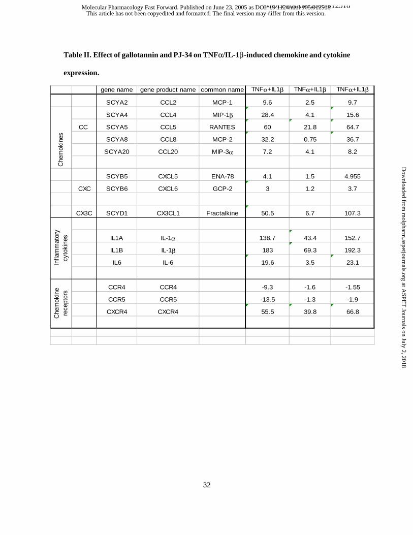
We have used a nylon-based, thematic low density array to investigate the effects of GT and
PJ-34 on the expression pattern of chemokines and inflammatory cytokines in A549 cells. Ten
positions on the arrays were occupied by positive controls (housekeeping genes) whereas six
positions contained no cDNA (blank) or plasmid DNA (negative control). The remaining
positions of the membrane contained 96 cDNAs of chemokines and inflammatory cytokines. Out
of these 96 genes, TNFα+IL-1β treatment significantly (minimum twofold induction) induced
the expression of 13 genes and suppressed the expression of 2 cytokine receptor genes (Table 2).
Pretreatment of cells with gallotannin significantly (by at least 50 %) reduced these alterations
with the exception of one chemokine (MIP-3a) and one chemokine receptor (CXCR4). PJ-34
significantly enhanced fractalkine expression and inhibited the downregulation of the chemokine
receptors CCR4 and CCR5. In order to confirm our results, we have also carried out RT-PCR
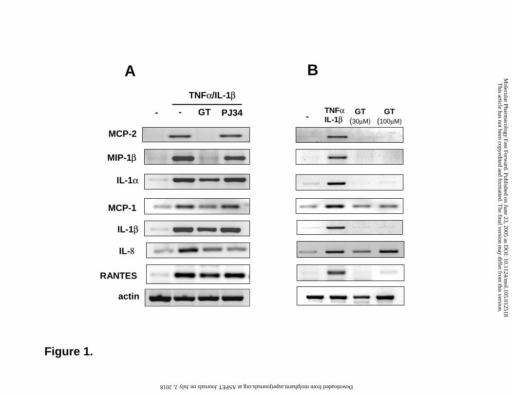
reactions for 7 genes which gave similar results (Fig 1A). In addition, the expression of IL-8, a
key neutrophil-recruiting chemokine which was not represented on the array, was also
investigated with RT-PCR and was found to be inhibited both by GT and by PJ-34. GT (30 µM)
alone did not induce any chemokines or cytokines tested (Fig 1B). [At very high concentration
(100 µM) GT induced IL-8 expression.] As NFκB and AP-1 are known to regulate the
expression of various inflammatory cytokines and chemokines, we have also investigated the
effects of GT and PJ-34 on the activation of these transcription factors.
Effects of GT and PJ-34 on NFκB activation.
This article has not been copyedited and formatted. The final version may differ from this version.Molecular Pharmacology Fast Forward. Published on June 23, 2005 as DOI: 10.1124/mol.105.012518
at ASPE
T Journals on July 2, 2018
molpharm
.aspetjournals.orgD
ownloaded from
MOLPHARM/2005/012518
13
The dimeric transcription factor NF- κB plays a central role in the transcriptional regulation of
inflammatory factors and is, in resting conditions, sequestered in the cytoplasm as an inactive
complex by its physical association with the inhibitor of NF- κB (I κB) (Schmitz et al., 2004).
Activation of NF-κB has been shown to occur through the activation of upstream protein kinases
[e.g. NIK (NF-κB-inducing kinase), MEKK1 (mitogen-activated protein kinase kinase kinase 1),
NAK (NF-κB-activating kinase)] phosphorylating the IKK (IκB-kinase) complex (Yamamoto
and Gaynor, 2004). Activation of this complex serves to mediate phosphorylation, ubiquitination
and degradation of IκB followed by nuclear translocation of NFκB.
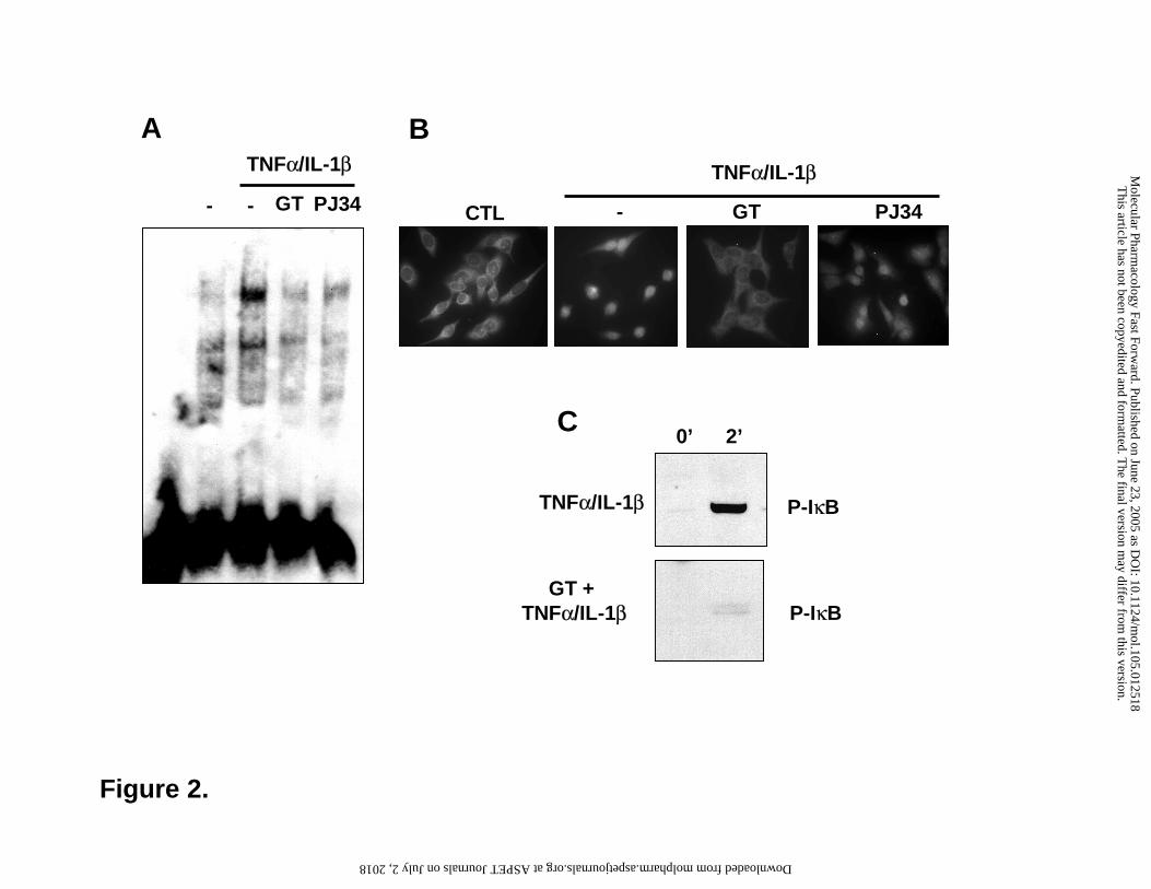
Treatment of A549 cells with TNFα/IL-1β induced NFκB activation as demonstrated by
EMSA analysis (Fig 2). Pretreatment of the cells with PJ-34 or GT markedly reduced the binding
of NFκB to its consensus oligonucleotide. TNFα/IL-1β-induced nuclear translocation of NFκB
was blocked by GT but was unaffected by PJ34 indicating that PARP inhibition by PJ-34 may
inhibit the DNA binding of the transcription factor. As for GT, we have also investigated IκB
phosphorylation, an event laying upstream in the NFκB pathway. GT abolished phosphorylation
suggesting that GT may inhibit the kinase cascade (IKK or upstream kinases).
Effects of GT and PJ-34 on AP-1 activation.
Activated protein-1 (AP-1) is a collection of dimeric transcription factors that belong to the
Jun, Fos, Maf and ATF subfamilies (Wisdom, 1999; Kyriakis, 1999). Depending on the
composition of the dimers they recognize either TPA response elements (TRE, 5'-TGAG/CTCA-
3') or c-AMP response elements (CRE, 5'-TGACGTCA-3'), which are located in the promoter
regions of genes encoding cytokines, chemokines, adhesion molecules and transcription factors.
Whereas the Jun/Fos heterodimers preferentially bind to the TRE elements, dimers containing
ATF2 (e.g ATF2/ATF2 or c-Jun/ATF2 dimers) bind to CRE.
We have observed a basal AP-1 activity as demonstrated by EMSA experiments using the
This article has not been copyedited and formatted. The final version may differ from this version.Molecular Pharmacology Fast Forward. Published on June 23, 2005 as DOI: 10.1124/mol.105.012518
at ASPE
T Journals on July 2, 2018
molpharm
.aspetjournals.orgD
ownloaded from
MOLPHARM/2005/012518
14
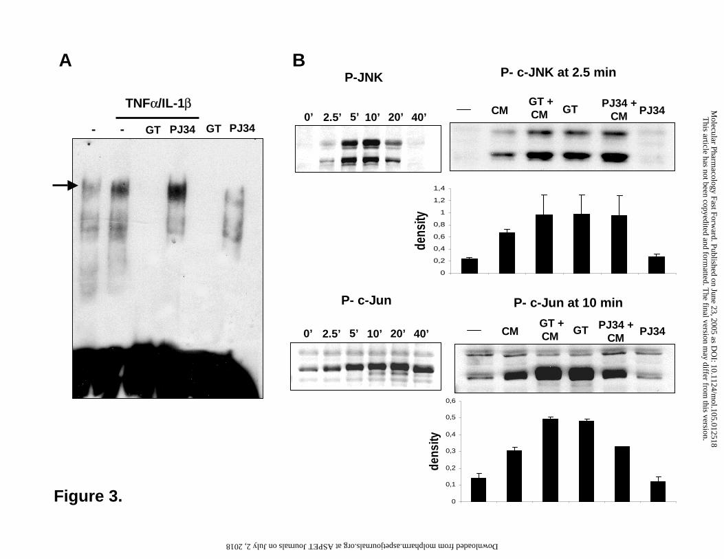
TRE consensus element (Fig 3). TNFα/IL-1β treatment triggered further AP-1 activation. GT
pretreatment abolished both basal and TNFα/IL-1β− induced AP-1 activation. PJ34 had no effect
on AP-1 activation.
Mitogen activated protein kinases JNK, p38 and ERK1/2 play key roles in cytokine-induced
signaling (Johnson and Lapadat, 2002). As formation of the c-Jun/c-Fos heterodimer of AP-1 is
induced by JNK-mediated phosphorylation of c-Jun (Kyriakis and Avruch, 2001), we have also
investigated the effects of GT and PJ-34 on these upstream events of the AP-1 pathway.
TNFα/IL-1β induced a rapid phosphorylation of JNK detectable as early as 2.5 min, peaking
between 5-10 min and fading 40 min after the cytokine treatment (Fig. 3). Surprisingly, GT
stimulated basal JNK phosphorylation that was not further increased by the cytokines. PJ-34 had
no effect on JNK phosphorylation. Phosphorylation of c-Jun has shown a prolonged pattern with
signals detectable even in unstimulated cells. Whereas PJ-34 had no effect on c-Jun
phosphorylation, GT treatment, even in the absence of cytokines, has induced maximal c-Jun
phosphorylation that was not further enhanced by TNFα/IL-1β. c-Jun can heterodimerize with
ATF-2 that is regulated mainly by p38-MAPK. Therefore we also sought to determine whether
GT and PJ-34 affect the p38-ATF-2 pathway. TNFα/IL-1β induced a rapid phosphorylation of
p38 which did not fade during the 40 min period tested (data not shown). PJ-34 had no effect on
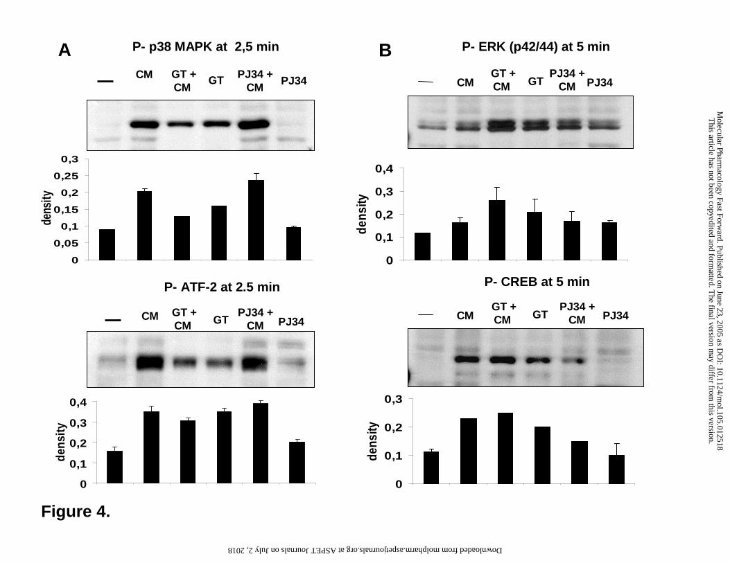
p38 phosphorylation (Figure 4A). Although GT alone caused a low level phosphorylation of p38,
the TNFα/IL-1β-induced signal was reduced by GT. Phosphorylation of ATF-2 was similarly
affected by the two drugs with no effect of PJ-34 and inhibition of cytokine-induced ATF-2
phosphorylation by GT (Figure 4A).
Although MAP kinase ERK1/2 is mainly involved in the regulation of cell proliferation
(Johnson and Lapadat, 2002) it has also been implicated in transcriptional regulation of
inflammatory mediators (Lecureur et al., 2005; Neff et al., 2003). One of the downstream events
This article has not been copyedited and formatted. The final version may differ from this version.Molecular Pharmacology Fast Forward. Published on June 23, 2005 as DOI: 10.1124/mol.105.012518
at ASPE
T Journals on July 2, 2018
molpharm
.aspetjournals.orgD
ownloaded from
MOLPHARM/2005/012518
15
in the activation of the ERK1/2 (and p38) pathway is the phosphorylation of the transcription
factor CREB (Yang et al., 2003). Phosphorylated CREB has been shown to be involved in the
transcriptional regulation of inflammatory mediators. We found that ERK1/2 and CREB are
regulated by GT the same way as seen with p38 and ATF-2 (Fig 4B). Whereas PJ34 had no
effect on the basal and cytokine-induced phosphorylation of ERK1/2 and CREB, GT induced the
phosphorylation of these proteins but inhibited their further activation by cytokines (Fig 4B).
Considering that GT increased the phosphorylation state of many proteins (JNK, c-Jun, p38,
ATF-2, ERK1/2 and CREB), we hypothesized that GT may interfere with protein phosphatase
activity. We have determined the effect of GT on the activities of protein phosphatases 1 and 2A
and found that GT inhibited both phosphatases in a concentration-dependent manner (Fig 5). GT
inhibited also the PP1 catalytic subunit associated with a regulatory subunit as assayed with
myosin phosphatase holoenzyme (not shown) indicating that regulatory subunits do not mask the
gallotannin binding site on the catalytic subunits.
Effect of GT on poly(ADP-ribosyl)ation
Based on the known PARG inhibitory effect of gallotannin, and the anti-inflammatory effect
of a recently developed non-tannin PARG inhibitor (Genovese et al., 2004) we have also
investigated the effect of GT on poly(ADP ribose) metabolism as a possible mechanism
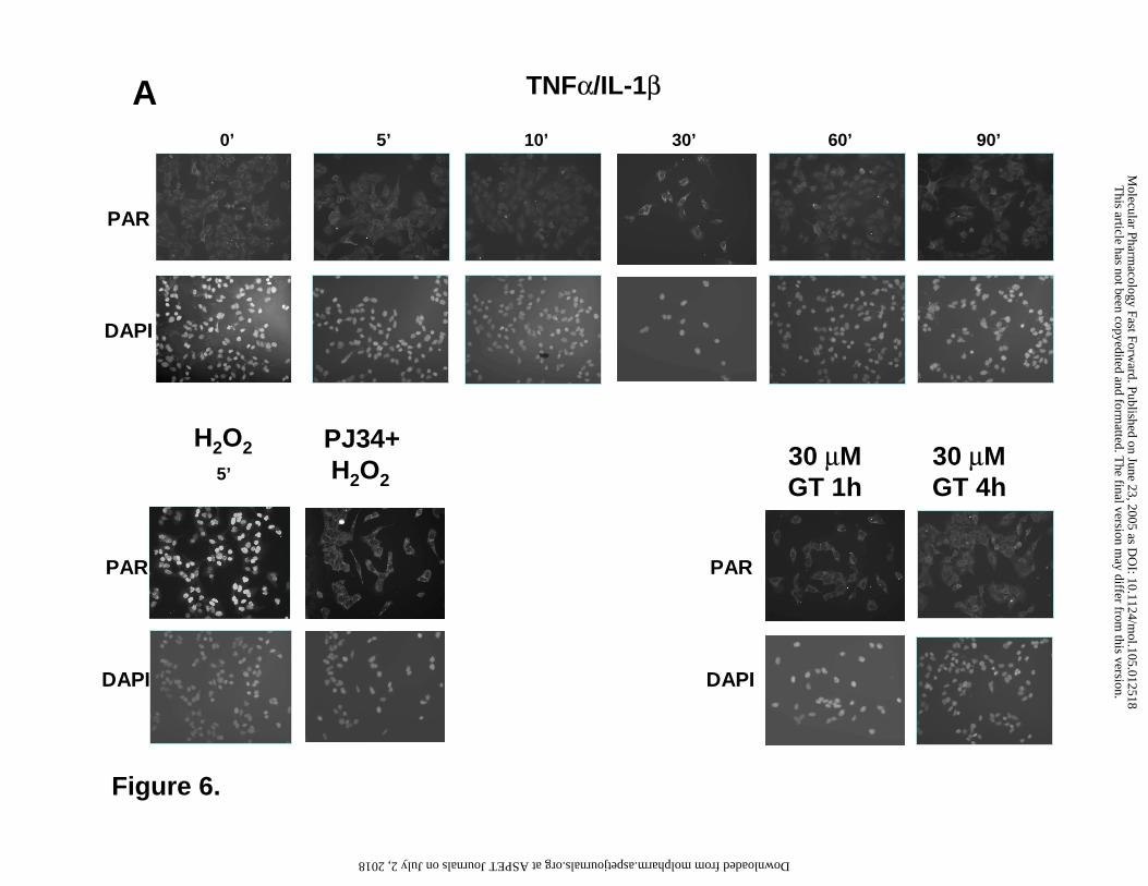
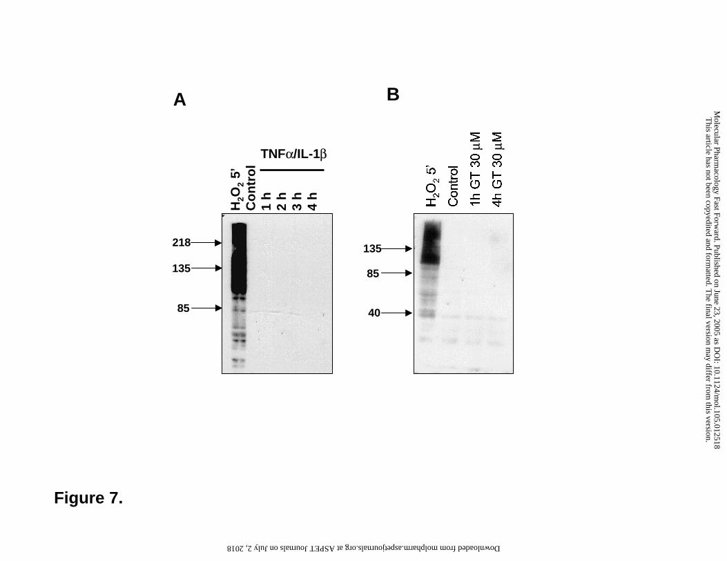
underlying the anti-inflammatory effect of GT. Treatment of the cells with the cytokines for
various time periods (5min-4h) caused no elevation in cellular PAR content as determined by
imunocytochemistry (Figure 6) or Western blotting (Figure 7) using the anti-PAR monoclonal
antibody. Hydrogen peroxide used as a positive control triggered PAR elevation in the nucleus as
demonstrated by immunocytochemistry (Figure 6). On Western blot, the lysates of hydrogen
peroxide-treated cells contained many positive bands with most immunopositivity found in the
region above 116 kDa (the molecular weight of PARP-1) corresponding to automodified PARP-1
This article has not been copyedited and formatted. The final version may differ from this version.Molecular Pharmacology Fast Forward. Published on June 23, 2005 as DOI: 10.1124/mol.105.012518
at ASPE
T Journals on July 2, 2018
molpharm
.aspetjournals.orgD
ownloaded from
MOLPHARM/2005/012518
16
(Figure 7). Treatment of cells with GT in the absence or presence of the cytokines caused no
elevation in the cellular PAR content.
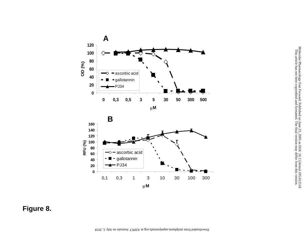
Antioxidant effects of GT
Another feature that could, at least in part, explain the effect of gallotannin on
cytokine/chemokine expression is the well-known antioxidant effect of tannins (Riedl and
Hagerman, 2001; Ho et al., 1999). Considering that both NFκB and AP-1 are redox-sensitive
transcription factors (Schulze-Osthoff et al., 1995), modification of the cellular redox state by
GT could be responsible for the described effect of GT. We have studied the radical-scavenging
effect of GT and PJ-34 in the ABTS decolorization assay. Using ascorbic acid as positive control
we have determined the ABTS scavenging effect of GT and PJ-34 (Figure 8A). In this assay, GT
displayed an even more potent radical scavenging effect as compared to ascorbic acid. PJ-34,
however, did not scavenge the radical (Figure 8A). We have also used a pathophysiologically
relevant oxidant, peroxynitrite. Peroxynitrite oxidizes dihydrorhodamine 123 into fluorescent
rhodamine. Addition of GT and ascorbic acid inhibited peroxynitrite induced DHR oxidation
with GT being the more potent antioxidant. PJ-34 had no effect (Figure 8B).
This article has not been copyedited and formatted. The final version may differ from this version.Molecular Pharmacology Fast Forward. Published on June 23, 2005 as DOI: 10.1124/mol.105.012518
at ASPE
T Journals on July 2, 2018
molpharm
.aspetjournals.orgD
ownloaded from
MOLPHARM/2005/012518
17
Discussion
Several laboratories have demonstrated that tannins exert potent anti-inflammatory effects.
Most cellular studies aiming at revealing the mechanism of these anti-inflammatory effects
utilized macrophages. In epithelial cells, however, the effect of tannins is not well characterized.
We hypothesized that PARG inhibition by gallotannin may contribute to this anti-inflammatory
effect as poly(ADP-ribose) polymerase-1 has been shown to regulate the expression of
inflammatory mediators.
In A549 lung epithelial cells our current study revealed no major role of poly(ADP-
ribosyl)ation as indicated by the lack of effect of PJ-34 on the expression of most chemokines
with the exception of IL-8, CCR4, CCR5 and fractalkine. This finding, however, does not
exclude the possibility that PARP-1 regulates inflammatory gene expression via protein-protein
interaction as previously demonstrated in experiments using PARP-1 knockout cells (Carrillo et
al., 2004; Ha et al., 2002; Ha, 2004). To investigate this possibility in epithelial cells, studies
using PARP-1 antisense or siRNA will be needed. PJ-34 has previously been shown to inhibit
chemokine expression in macrophages (Hasko et al 2002), a finding also confirmed by us (not
shown). This finding emphasizes the importance of cell type- and stimulus-dependent differences
in the requirement of PARP activity for transcriptional regulation.
In contrast to PJ-34, GT exerted a robust suppression of inflammatory gene expression. This
effect is not due to a general suppression of gene expression as GT has also prevented the
cytokine-induced downregulation of three chemokine receptors. Theoretically, the effects of GT
could be due to hyper-poly(ADP-ribosyl)ation of PARP-1 or other poly(ADP-ribose) acceptors
including the transcription factors NFκB and AP-1. De Murcia’s group identified deficient NF-
κB activation in PARP-1-/- mice (Oliver et al., 1999) and it was later proposed that PARP-1
physically interacts with the NF-κB-p50 but the DNA binding and catalytic activity of PARP-1
This article has not been copyedited and formatted. The final version may differ from this version.Molecular Pharmacology Fast Forward. Published on June 23, 2005 as DOI: 10.1124/mol.105.012518
at ASPE
T Journals on July 2, 2018
molpharm
.aspetjournals.orgD
ownloaded from
MOLPHARM/2005/012518
18
was found not to be required for the NF-κB coactivator function (Hassa et al. 2001). In certain
cellular systems, however, PARP inhibitors did inhibit NF-κB activation (Ha et al., 2002; Hasko
et al., 2002). Our data showing normal nuclear translocation but decreased DNA binding of NF-
κB in PJ-34-treated cells indicates that in cytokine-stimulated A549 cells, DNA binding of NF-
κB requires poly(ADP-ribosyl)ation. GT also blocked the NF-κB pathway. However, GT
targeted an event upstream of IκB phosphorylation. Nonetheless, the inhibition of NF-κB
activation by GT does not fully explain the marked effects of GT on cytokine/chemokine
expression, as PJ-34, that has also inhibited NF-κB, failed to affect cytokine expression.
Therefore, we have considered the possibility that GT also interferes with the activation of AP-1,
the other key transcription factor regulating inflammatory gene expression.
The redox-sensitive transcription factor AP-1 is composed of a mixture of heterodimeric
protein complexes derived from the Fos and Jun families. AP-1 heterodimers bind to DNA on a
serum response element with the 5'-TGA(C/G)TCA-3' sequence. AP-1 is regulated at the level of
both jun and fos gene transcription and by posttranslational modifications of their gene products.
Mitogen-activated protein kinases (MAPK) with special regard to Jun N-terminal kinases (JNK)
play key role in AP-1 activation by phosphorylating c-Jun (Kyriakis and Avruch, 2001; Johnson
and Lapadat, 2002). Zingarelli’s group reported increased basal JNK activity and c-Jun
phosphorylation but decreased AP-1 DNA binding in PARP-1 knock out cells (Zingarelli et al.,
2004). Interestingly, in A549 cells here we found similar effects with GT but PJ-34 had no major
effect on the AP-1 pathway.
Our current data suggest that AP-1 rather than NF-κB plays a key role in the regulation of
cytokine/chemokine gene expression in A549 cells. Interestingly, suppression of AP-1 DNA
binding by GT was paralleled by maximal activation (phosphorylation) of JNK and c-Jun even in
the absence of cytokines. To elucidate the mechanism by which GT "uncouples" phosphorylation
This article has not been copyedited and formatted. The final version may differ from this version.Molecular Pharmacology Fast Forward. Published on June 23, 2005 as DOI: 10.1124/mol.105.012518
at ASPE
T Journals on July 2, 2018
molpharm
.aspetjournals.orgD
ownloaded from
MOLPHARM/2005/012518
19
of JNK and c-Jun from DNA binding of AP-1 requires further investigation. It is possible that
GT triggers the JNK-c-Jun pathway by an unknown mechanism (e.g. by inhibiting protein
phosphatases) and, independently of this, it also interferes with the DNA binding of AP-1.
Decreased AP-1 DNA binding in GT-treated cells may result from the inhibition of the p38 -
ATF-2 pathway that is also important in the TNFα/IL-1β-induced inflammatory gene expression.
The MAP kinase ERK can also regulate inflammatory gene transcription by indirectly activating
CREB. The ERK-CREB pathway appears to be affected by GT similarly to the p38 pathway:
GT inhibited cytokine-induced activation of both ERK and CREB but GT alone caused a
moderate phosphorylation of these proteins.
Gallotannin-induced phosphorylation of MAP kinases and MAP kinase targets may be due to
interference of GT with protein phosphatases. Our data indicate that GT inhibits the catalytic
subunits of protein phosphatases 1 and 2A. This inhibitory activity could also be observed on the
phosphatase holoenzyme. PP1 and PP2A have been proposed to regulate the MAP kinase
pathways in various systems (Kim et al., 2003; Garcia et al., 2002). Therefore, inhibition of PP1
and PP2A by GT may contribute to the increased phosphorylation level of MAP kinases in GT-
treated cells. MAP kinase phosphatases also play a key role in dephosphorylation of MAP
kinases. Whether MAP kinase phosphatases are also inhibited by GT, remains to be seen.
We also sought to determine whether the transcriptional regulatory effect of GT is related to
PARG inhibition. Considering that a basal PARP activity is usually present in cultured cells
(Bakondi et al., 2002) we expected GT to cause PAR accumulation. Our data showing the lack of
PAR accumulation in GT-treated cells suggest that no major alterations of PAR metabolism
occur in response to GT treatment. This is in line with previous reports from Falsig et al. (Falsig
et al., 2004) demonstrating that GT inhibits PARG in a cell free assay but has no effect on PARG
activity in intact cells. Moreover, the cytokine exposure did not stimulate PAR synthesis, neither
This article has not been copyedited and formatted. The final version may differ from this version.Molecular Pharmacology Fast Forward. Published on June 23, 2005 as DOI: 10.1124/mol.105.012518
at ASPE
T Journals on July 2, 2018
molpharm
.aspetjournals.orgD
ownloaded from
MOLPHARM/2005/012518
20
in the absence nor in the presence of GT. In light of these data it appears unlikely, that PARG is
the major target of GT in our system. Furthermore, a GT concentration of 50 µM or higher was
previously shown to be required for PAR accumulation in cell lysates (Keil et al., 2004), whereas
the marked transcriptional inhibitory effects in our current study required lower concentrations.
Nonetheless, PAR accumulation on certain low abundance proteins may remain undetected in
western blots or immunocytofluorescent stainings and may be important for the regulation of
transcription. Recent generation of PARG-deficient mice (Cortes et al., 2004;Koh et al., 2004)
will certainly accelerate research on the role of PARG in transcriptional regulation.
Considering that both NFκB and AP-1 are regarded as redox sensitive transcription factors,
the antioxidant effect of GT may explain its effect on inflammatory gene expression. Our data
showing potent antioxidant effects of GT at relatively low concentrations (30 µM) support this
hypothesis.
Conclusion. As opposed to macrophages where GT appears to act as a proinflammatory
stimulus (Rohrbach et al., 1989; Rapizzi et al., 2004), in epithelial cells it acts as an anti-
inflammatory agent. This effect results from the inhibition of the AP-1 pathway and, to a lesser
extent, the NF-κB pathway. Unlike in macrophages, in A549 epithelial cells poly(ADP-
ribosyl)ation is not a crucial mechanism in the regulation of inflammatory gene expression and
PARG is not likely to be the target of GT in this system.
This article has not been copyedited and formatted. The final version may differ from this version.Molecular Pharmacology Fast Forward. Published on June 23, 2005 as DOI: 10.1124/mol.105.012518
at ASPE
T Journals on July 2, 2018
molpharm
.aspetjournals.orgD
ownloaded from
MOLPHARM/2005/012518
21
Acknowledgements
The authors wish to thank Dr. Shiao-Li Oei (Free University Berlin) for providing the 10H
hybridoma.
This article has not been copyedited and formatted. The final version may differ from this version.Molecular Pharmacology Fast Forward. Published on June 23, 2005 as DOI: 10.1124/mol.105.012518
at ASPE
T Journals on July 2, 2018
molpharm
.aspetjournals.orgD
ownloaded from
MOLPHARM/2005/012518
22
Reference List
Ame JC, Spenlehauer C and de M G (2004) The PARP Superfamily. Bioessays 26:882-893.
Bakondi, E., Bai, P., Szabo, E., Hunyadi, J., Gergely, P., Szabo, C., and Virag, L. (2002) Detection of Poly(ADP-ribose) Polymerase Activation in Oxidatively Stressed Cells and Tissues Using Biotinylated NAD Substrate. J.Histochem.Cytochem. 50:91-98.
Burkle A (2001) Physiology and Pathophysiology of Poly(ADP-Ribosyl)Ation. Bioessays 23:795-806.
Burkle A, Chen G, Kupper J H, Grube K and Zeller W J (1993) Increased Poly(ADP-Ribosyl)Ation in Intact Cells by Cisplatin Treatment. Carcinogenesis 14:559-561.
Carrillo A, Monreal Y, Ramirez P, Marin L, Parrilla P, Oliver F J and Yelamos J (2004) Transcription Regulation of TNF-Alpha-Early Response Genes by Poly(ADP-Ribose) Polymerase-1 in Murine Heart Endothelial Cells. Nucleic Acids Res 32:757-766.
Cortes U, Tong W M, Coyle D L, Meyer-Ficca M L, Meyer R G, Petrilli V, Herceg Z, Jacobson E L, Jacobson M K and Wang Z Q (2004) Depletion of the 110-Kilodalton Isoform of Poly(ADP-Ribose) Glycohydrolase Increases Sensitivity to Genotoxic and Endotoxic Stress in Mice. Mol Cell Biol 24:7163-7178.
Erdelyi K, Bakondi E, Gergely P, Szabo C and Virag L (2005) Pathophysiologic Role of Oxidative Stress-Induced Poly(ADP-Ribose) Polymerase-1 Activation: Focus on Cell Death and Transcriptional Regulation. Cell Mol Life Sci 62:751-759.
Erdodi F, Toth B, Hirano K, Hirano M, Hartshorne D J and Gergely P (1995) Endothall Thioanhydride Inhibits Protein Phosphatases-1 and -2A in Vivo. Am J Physiol 269:C1176-C1184.
Falsig J, Christiansen S H, Feuerhahn S, Burkle A, Oei S L, Keil C and Leist M (2004) Poly(ADP-Ribose) Glycohydrolase As a Target for Neuroprotective Intervention: Assessment of Currently Available Pharmacological Tools. Eur J Pharmacol 497:7-16.
Feldman KS, Sahasrabudhe K, Lawlor M D, Wilson S L, Lang C H and Scheuchenzuber W J (2001) In Vitro and In Vivo Inhibition of LPS-Stimulated Tumor Necrosis Factor-Alpha Secretion by the Gallotannin Beta-D-Pentagalloylglucose. Bioorg Med Chem Lett 11:1813-1815.
Fong HH, Bhatti W and Farnsworth N R (1972) Antitumor Activity of Certain Plants Due to Tannins. J Pharm Sci 61:1818.
Garcia L, Garcia F, Llorens F, Unzeta M, Itarte E and Gomez N (2002) PP1/PP2A Phosphatases Inhibitors Okadaic Acid and Calyculin A Block ERK5 Activation by Growth Factors and Oxidative Stress. FEBS Lett 523:90-94.
Genovese T, Di P R, Catalano P, Li J H, Xu W, Massuda E, Caputi A P, Zhang J and
This article has not been copyedited and formatted. The final version may differ from this version.Molecular Pharmacology Fast Forward. Published on June 23, 2005 as DOI: 10.1124/mol.105.012518
at ASPE
T Journals on July 2, 2018
molpharm
.aspetjournals.orgD
ownloaded from
MOLPHARM/2005/012518
23
Cuzzocrea S (2004) Treatment With a Novel Poly(ADP-Ribose) Glycohydrolase Inhibitor Reduces Development of Septic Shock-Like Syndrome Induced by Zymosan in Mice. Crit Care Med 32:1365-1374.
Gergely P, Erdodi F and Bot G (1984) Heparin Inhibits the Activity of Protein Phosphatase-1. FEBS Lett 169:45-48.
Ha HC (2004) Defective Transcription Factor Activation for Proinflammatory Gene Expression in Poly(ADP-Ribose) Polymerase 1-Deficient Glia. Proc Natl Acad Sci U S A 101:5087-5092.
Ha HC, Hester L D and Snyder S H (2002) Poly(ADP-Ribose) Polymerase-1 Dependence of Stress-Induced Transcription Factors and Associated Gene Expression in Glia. Proc Natl Acad Sci U S A 99:3270-3275.
Hagerman AE, Riedl K M and Rice R E (1999) Tannins As Biological Antioxidants. Basic Life Sci 66:495-505.
Hasko G, Mabley J G, Nemeth Z H, Pacher P, Deitch E A and Szabo C (2002) Poly(ADP-Ribose) Polymerase Is a Regulator of Chemokine Production: Relevance for the Pathogenesis of Shock and Inflammation. Mol Med 8:283-289.
Hassa PO, Covic M, Hasan S, Imhof R and Hottiger M O (2001) The Enzymatic and DNA Binding Activity of PARP-1 Are Not Required for NF-Kappa B Coactivator Function. J Biol Chem 276:45588-45597.
Ho KY, Huang J S, Tsai C C, Lin T C, Hsu Y F and Lin C C (1999) Antioxidant Activity of Tannin Components From Vaccinium Vitis-Idaea L. J Pharm Pharmacol 51:1075-1078.
Johnson GL and Lapadat R (2002) Mitogen-Activated Protein Kinase Pathways Mediated by ERK, JNK, and P38 Protein Kinases. Science 298:1911-1912.
Kawamitsu H, Hoshino H, Miwa M and Sugimura T (1983) Monoclonal Antibodies Against Poly(ADP-Ribose) Recognize Different Structures of Poly(ADP-Ribose). Princess Takamatsu Symp 13:41-7.:41-47.
Keil C, Petermann E and Oei S L (2004) Tannins Elevate the Level of Poly(ADP-Ribose) in HeLa Cell Extracts. Arch Biochem Biophys 425:115-121.
Kim HS, Song M C, Kwak I H, Park T J and Lim I K (2003) Constitutive Induction of P-Erk1/2 Accompanied by Reduced Activities of Protein Phosphatases 1 and 2A and MKP3 Due to Reactive Oxygen Species During Cellular Senescence. J Biol Chem 278:37497-37510.
Koh DW, Lawler A M, Poitras M F, Sasaki M, Wattler S, Nehls M C, Stoger T, Poirier G G, Dawson V L and Dawson T M (2004) Failure to Degrade Poly(ADP-Ribose) Causes Increased Sensitivity to Cytotoxicity and Early Embryonic Lethality. Proc Natl Acad Sci U S A 101:17699-17704.
Kooy NW, Royall J A, Ischiropoulos H and Beckman J S (1994) Peroxynitrite-Mediated
This article has not been copyedited and formatted. The final version may differ from this version.Molecular Pharmacology Fast Forward. Published on June 23, 2005 as DOI: 10.1124/mol.105.012518
at ASPE
T Journals on July 2, 2018
molpharm
.aspetjournals.orgD
ownloaded from
MOLPHARM/2005/012518
24
Oxidation of Dihydrorhodamine 123. Free Radic Biol Med 16:149-156.
Kyriakis JM (1999) Activation of the AP-1 Transcription Factor by Inflammatory Cytokines of the TNF Family. Gene Expr 7:217-231.
Kyriakis JM and Avruch J (2001) Mammalian Mitogen-Activated Protein Kinase Signal Transduction Pathways Activated by Stress and Inflammation. Physiol Rev 81:807-869.
Lecureur V, Ferrec E L, N'diaye M, Vee M L, Gardyn C, Gilot D and Fardel O (2005) ERK-Dependent Induction of TNFalpha Expression by the Environmental Contaminant Benzo(a)Pyrene in Primary Human Macrophages. FEBS Lett 579:1904-1910.
Lee SJ, Lee I S and Mar W (2003) Inhibition of Inducible Nitric Oxide Synthase and Cyclooxygenase-2 Activity by 1,2,3,4,6-Penta-O-Galloyl-Beta-D-Glucose in Murine Macrophage Cells. Arch Pharm Res 26:832-839.
Mota ML, Thomas G and Barbosa Filho J M (1985) Anti-Inflammatory Actions of Tannins Isolated From the Bark of Anacardium Occidentale L. J Ethnopharmacol 13:289-300.
Neff L, Zeisel M, Druet V, Takeda K, Klein J P, Sibilia J and Wachsmann D (2003) ERK 1/2- and JNKs-Dependent Synthesis of Interleukins 6 and 8 by Fibroblast-Like Synoviocytes Stimulated With Protein I/II, a Modulin From Oral Streptococci, Requires Focal Adhesion Kinase. J Biol Chem 278:27721-27728.
Oliver FJ, Menissier-De M J, Nacci C, Decker P, Andriantsitohaina R, Muller S, de La Rubia G, Stoclet J C and De Murcia G (1999) Resistance to Endotoxic Shock As a Consequence of Defective NF-KappaB Activation in Poly (ADP-Ribose) Polymerase-1 Deficient Mice. EMBO J 18:4446-4454.
Rapizzi E, Fossati S, Moroni F and Chiarugi A (2004) Inhibition of Poly(ADP-Ribose) Glycohydrolase by Gallotannin Selectively Up-Regulates Expression of Pro-Inflammatory Genes. Mol Pharmacol 66:890-898.
Re R, Pellegrini N, Proteggente A, Pannala A, Yang M and Rice-Evans C (1999) Antioxidant Activity Applying an Improved ABTS Radical Cation Decolorization Assay. Free Radic Biol Med 26:1231-1237.
Riedl KM and Hagerman A E (2001) Tannin-Protein Complexes As Radical Scavengers and Radical Sinks. J Agric Food Chem 49:4917-4923.
Rohrbach MS, Kreofsky T, Rolstad R A and Russell J A (1989) Tannin-Mediated Secretion of a Neutrophil Chemotactic Factor From Alveolar Macrophages. Potential Contribution to the Acute Pulmonary Inflammatory Reaction Associated With Byssinosis. Am Rev Respir Dis 139:39-45.
Schmitz ML, Mattioli I, Buss H and Kracht M (2004) NF-KappaB: a Multifaceted Transcription Factor Regulated at Several Levels. Chembiochem 5:1348-1358.
Schulze-Osthoff K, Los M and Baeuerle P A (1995) Redox Signalling by Transcription Factors NF-Kappa B and AP-1 in Lymphocytes. Biochem Pharmacol 50:735-741.
This article has not been copyedited and formatted. The final version may differ from this version.Molecular Pharmacology Fast Forward. Published on June 23, 2005 as DOI: 10.1124/mol.105.012518
at ASPE
T Journals on July 2, 2018
molpharm
.aspetjournals.orgD
ownloaded from
MOLPHARM/2005/012518
25
Toth A, Kiss E, Herberg F W, Gergely P, Hartshorne D J and Erdodi F (2000) Study of the Subunit Interactions in Myosin Phosphatase by Surface Plasmon Resonance. Eur J Biochem 267:1687-1697.
Uchiumi F, Maruta H, Inoue J, Yamamoto T and Tanuma S (1996) Inhibitory Effect of Tannic Acid on Human Immunodeficiency Virus Promoter Activity Induced by 12-O-Tetra Decanoylphorbol-13-Acetate in Jurkat T-Cells. Biochem Biophys Res Commun 220:411-417.
Van Molle W, Van den Berghe J, Brouckaert P and Libert C (2000) Tumor Necrosis Factor-Induced Lethal Hepatitis: Pharmacological Intervention With Verapamil, Tannic Acid, Picotamide and K76COOH. FEBS Lett 467:201-205.
Virag L (2005) Poly(ADP-Ribosyl)Ation in Asthma and Other Lung Diseases. Pharm Res 52:83-92.
Wisdom R (1999) AP-1: One Switch for Many Signals. Exp Cell Res 253:180-185.
Yamamoto Y and Gaynor R B (2004) IkappaB Kinases: Key Regulators of the NF-KappaB Pathway. Trends Biochem Sci 29:72-79.
Yang SH, Sharrocks A D and Whitmarsh A J (2003) Transcriptional Regulation by the MAP Kinase Signaling Cascades. Gene 320:3-21.:3-21.
Zingarelli B, Hake P W, O'Connor M, Denenberg A, Wong H R, Kong S and Aronow B J (2004) Differential Regulation of Activator Protein-1 and Heat Shock Factor-1 in Myocardial Ischemia and Reperfusion Injury: Role of Poly(ADP-Ribose) Polymerase-1. Am J Physiol Heart Circ Physiol 286:H1408-H1415.
Zingarelli B, Salzman A L and Szabo C (1998) Genetic Disruption of Poly (ADP-Ribose) Synthetase Inhibits the Expression of P-Selectin and Intercellular Adhesion Molecule-1 in Myocardial Ischemia/Reperfusion Injury. Circ Res 83:85-94.
This article has not been copyedited and formatted. The final version may differ from this version.Molecular Pharmacology Fast Forward. Published on June 23, 2005 as DOI: 10.1124/mol.105.012518
at ASPE
T Journals on July 2, 2018
molpharm
.aspetjournals.orgD
ownloaded from
MOLPHARM/2005/012518
26
Footnotes
*This work was supported by grants from the Hungarian Science Research Fund (OTKA T
35182, T37210, T43296), from the Hungarian Ministry of Education (BIO-2/2002) and from the
Hungarian Ministry of Health ETT 206/2003. L.V. is supported by a Széchenyi fellowship from
the Hungarian Ministry of Education.
This article has not been copyedited and formatted. The final version may differ from this version.Molecular Pharmacology Fast Forward. Published on June 23, 2005 as DOI: 10.1124/mol.105.012518
at ASPE
T Journals on July 2, 2018
molpharm
.aspetjournals.orgD
ownloaded from
MOLPHARM/2005/012518
27
Legends to the Tables and Figures
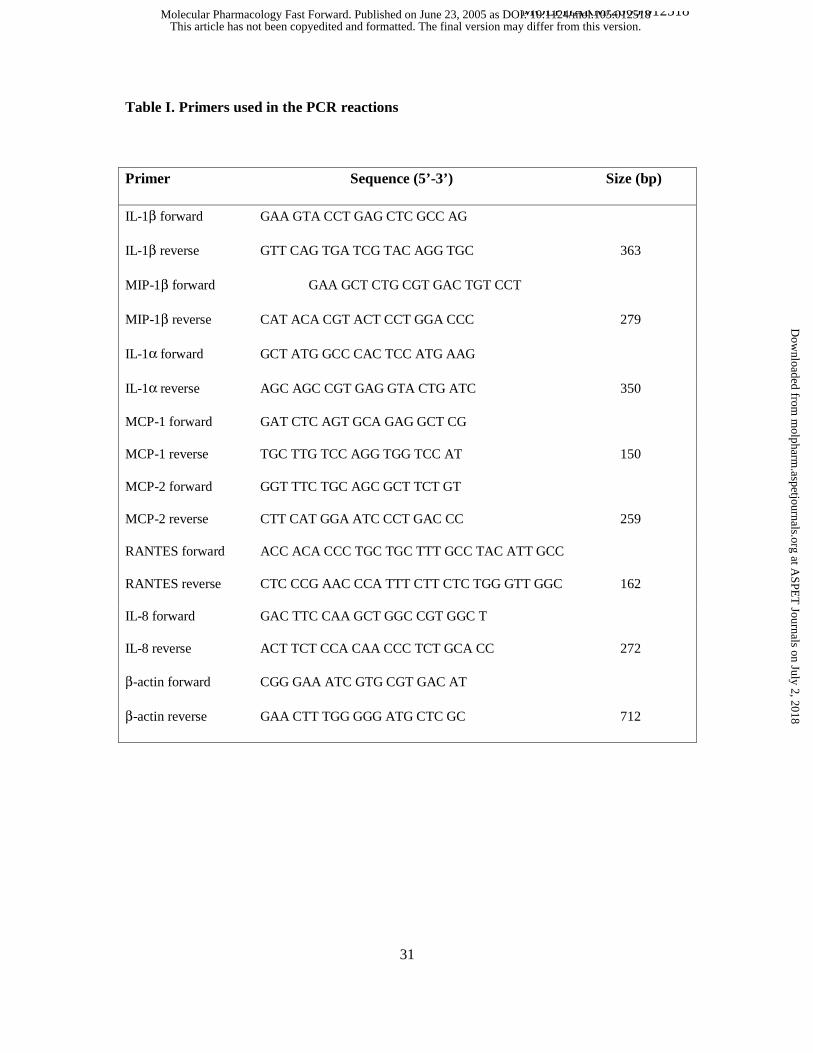
Table I. Sequence of primers used in the PCR reactions
Table II. Effect of gallotannin and PJ-34 on TNFα/IL-1β-induced chemokine and cytokine
expression. A549 cells were pretreated for 30 min with 30 µM gallotannin (GT) or 10 µM PJ-34
and were then stimulated with TNFα and IL-1β. After 4h, RNA was isolated and was reverse
transcribed into biotin-labelled cDNA which was then hybridized to Superarray membranes.
Biotin was detected with streptavidine peroxidase followed by ECL development. The spots
were analyzed by densitometry and a ratio of TNFα-IL-1β/control was calculated. A twofold
induction/suppression obtained in two independent experiments was considered significant and is
presented in the table. The effects of pretreatments was only considered significant if it was less
than 50% or more than 200% of the values obtained with cytokine stimulation only.
Figure 1. RT-PCR analysis of the expression of chemokines and cytokines in cytokine
stimulated A549 cells. Cells were pretreated for 30 min with 30 µM gallotannin (GT) or 10 µM
PJ-34 and were then stimulated with TNFα and IL-1β (panel Α). After 4h, RNA was isolated and
was reverse transcribed into cDNA. Specific transcripts were amplified with PCR. Panel B
shows the effect of GT (30 and 100 µM) on the expression of the same set of
chemokines/cytokines. Cytokine stimulation was used as positive control. The experiments were
repeated three times.
Figure 2. NFκB activation in A549 cells. Cells were pretreated for 30 min with 30 µM
gallotannin (GT) or 10 µM PJ-34 and were then stimulated with TNFα and IL-1β. After 1h,
nuclear extracts were prepared and binding of nuclear protein to NFκB consensus
This article has not been copyedited and formatted. The final version may differ from this version.Molecular Pharmacology Fast Forward. Published on June 23, 2005 as DOI: 10.1124/mol.105.012518
at ASPE
T Journals on July 2, 2018
molpharm
.aspetjournals.orgD
ownloaded from
MOLPHARM/2005/012518
28
oligonucleotide was studied with EMSA (A). Translocation of NFκB was demonstrated with
immunocytofluorescent staining 30 min after cytokine stimulation (B). Phosphorylation of IκB
was detected on Western blots using phosphopeptide specific antibody (C).
Figure 3. Activation of the JNK-c-Jun-AP-1 pathway in immunostimulated A549 cells.
Cells were pretreated for 30 min with 30 µM gallotannin (GT) or 10 µM PJ-34 and were then
stimulated with TNFα and IL-1β (CM = cytokine mix). After 2h, AP-1 activation was detected
with EMSA (A). Time courses for the cytokine-induced phosphorylation of JNK and c-Jun were
established in Western blot experiments using phosphopeptide specific antibodies (B). Effects of
gallotannin (GT) and PJ-34 were studied at selected time points as indicated (B). Bands were
evaluated by densitometry and density values of three independent experiments are presented as
mean ± SE of the mean. The total amounts of JNK and c-Jun were not different in the samples as
verified with non-phosphospecific antibodies (not shown).
Figure 4. Phosphorylation of p38, ATF-2, ERK and CREB in A549 cells. A549 cells were
pretreated for 30 min with 30 µM gallotannin (GT) or 10 µM PJ-34 and were then stimulated
with the cytokines TNFα and IL-1β (CM = cytokine mix). After 2.5-5 min, lysates were
prepared and phosphorylation of the proteins was detected in Western blots. Bands were
evaluated by densitometry and density values of three independent experiments are presented as
mean ± SE of the mean.
Figure 5. Effect of gallotannin on the activity of PP1c and PP2Ac. Gallotannin was
assayed on the phosphatase activity at concentrations of 0.5 µM, 2 µM, 10 µM and 50 µM.
Gallotannin was preincubated with PP1c or PP2Ac for 5 min and the reaction was initiated
This article has not been copyedited and formatted. The final version may differ from this version.Molecular Pharmacology Fast Forward. Published on June 23, 2005 as DOI: 10.1124/mol.105.012518
at ASPE
T Journals on July 2, 2018
molpharm
.aspetjournals.orgD
ownloaded from
MOLPHARM/2005/012518
29
by the addition of 32P-MLC20. Assays were performed at 30 °C and the 32Pi released from the
substrate was determined. Phosphatase activity of PP1c (○) or PP2Ac (●) in the absence of
gallotannin was taken as 100%. Values represent means ± SEM (n=7-9).
Figure 6. Lack of effect of 30 µM gallotannin on the poly(ADP-ribose) metabolism. Cells
were stimulated with TNFα and IL-1β or GT for various periods of times, as indicated.
Poly(ADP-ribose) was detected with immunofluorescent staining using the 10H anti-poly(ADP-
ribose) antibody. Hydrogen peroxide treatment (5 min) was used as a positive control. Nuclei
were stained blue with DAPI. Identical exposure times were used for all photographs taken.
Figure 7. Lack of effect of 30 µM gallotannin on the poly(ADP-ribose) metabolism. Cells
were stimulated with TNFα and IL-1β (A) or 30 µM GT (B) for various periods of times and
poly(ADP-ribose) was detected with Western blotting using the 10H anti-poly(ADP-ribose)
antibody. Hydrogen peroxide treatment (5 min) was used as a positive control.
Figure 8. Antioxidant effects of gallotannin. The general antioxidant effect of the indicated
concentrations of gallotannin and PJ-34 was determined in the ABTS scavenging assay as
described in the "Materials and methods" section. The peroxynitrite-specific scavenging activity
was determined in the dihydrorhodamine oxidation assay and results are given in relative
fluorescent units (RFU) as percent of control. Ascorbic acid was used as a positive control in
both assays. Mean ± SD of triplicate samples are shown.
Supplemental table I. Effect of gallotannin and PJ-34 on TNFα/IL-1β-induced chemokine
This article has not been copyedited and formatted. The final version may differ from this version.Molecular Pharmacology Fast Forward. Published on June 23, 2005 as DOI: 10.1124/mol.105.012518
at ASPE
T Journals on July 2, 2018
molpharm
.aspetjournals.orgD
ownloaded from
MOLPHARM/2005/012518
30
and cytokine expression. A549 cells were pretreated for 30 min with 30 µM gallotannin (GT) or
10 µM PJ-34 and were then stimulated with TNFα and IL-1β. After 4h, RNA was isolated and
was reverse transcribed into biotin-labelled cDNA which was then hybridized to Superarray
membranes. Biotin was detected with streptavidine peroxidase followed by ECL development.
The spots were analyzed by densitometry and a ratio of TNFα-IL-1β/control was calculated. A
twofold induction/suppression obtained in two independent experiments was considered
significant as indicated by the red/green color in the table. The effect of pretreatments with GT
or PJ34 is shown for genes that were significantly up or down regulated by the cytokines. Blue
and brown background indicates if pretreatments inhibited or enhanced, respectively the effects
of cytokine stimulation.
This article has not been copyedited and formatted. The final version may differ from this version.Molecular Pharmacology Fast Forward. Published on June 23, 2005 as DOI: 10.1124/mol.105.012518
at ASPE
T Journals on July 2, 2018
molpharm
.aspetjournals.orgD
ownloaded from
MOLPHARM/2005/012518
31
Table I. Primers used in the PCR reactions
Primer Sequence (5’-3’) Size (bp)
IL-1β forward GAA GTA CCT GAG CTC GCC AG
IL-1β reverse GTT CAG TGA TCG TAC AGG TGC 363
MIP-1β forward GAA GCT CTG CGT GAC TGT CCT
MIP-1β reverse CAT ACA CGT ACT CCT GGA CCC 279
IL-1α forward GCT ATG GCC CAC TCC ATG AAG
IL-1α reverse AGC AGC CGT GAG GTA CTG ATC 350
MCP-1 forward GAT CTC AGT GCA GAG GCT CG
MCP-1 reverse TGC TTG TCC AGG TGG TCC AT 150
MCP-2 forward GGT TTC TGC AGC GCT TCT GT
MCP-2 reverse CTT CAT GGA ATC CCT GAC CC 259
RANTES forward ACC ACA CCC TGC TGC TTT GCC TAC ATT GCC
RANTES reverse CTC CCG AAC CCA TTT CTT CTC TGG GTT GGC 162
IL-8 forward GAC TTC CAA GCT GGC CGT GGC T
IL-8 reverse ACT TCT CCA CAA CCC TCT GCA CC 272
β-actin forward CGG GAA ATC GTG CGT GAC AT
β-actin reverse GAA CTT TGG GGG ATG CTC GC 712
This article has not been copyedited and formatted. The final version may differ from this version.Molecular Pharmacology Fast Forward. Published on June 23, 2005 as DOI: 10.1124/mol.105.012518
at ASPE
T Journals on July 2, 2018
molpharm
.aspetjournals.orgD
ownloaded from
MOLPHARM/2005/012518
32
Table II. Effect of gallotannin and PJ-34 on TNFα/IL-1β-induced chemokine and cytokine
expression.
gene name gene product name common name TNFα+IL1β TNFα+IL1β TNFα+IL1β
SCYA2 CCL2 MCP-1 9.6 2.5 9.7
SCYA4 CCL4 MIP-1β 28.4 4.1 15.6
CC SCYA5 CCL5 RANTES 60 21.8 64.7
SCYA8 CCL8 MCP-2 32.2 0.75 36.7
SCYA20 CCL20 MIP-3α 7.2 4.1 8.2
SCYB5 CXCL5 ENA-78 4.1 1.5 4.955
CXC SCYB6 CXCL6 GCP-2 3 1.2 3.7
CX3C SCYD1 CX3CL1 Fractalkine 50.5 6.7 107.3
IL1A IL-1α 138.7 43.4 152.7
IL1B IL-1β 183 69.3 192.3
IL6 IL-6 19.6 3.5 23.1
CCR4 CCR4 -9.3 -1.6 -1.55
CCR5 CCR5 -13.5 -1.3 -1.9
CXCR4 CXCR4 55.5 39.8 66.8
Che
mok
ines
Infla
mm
ator
y cy
toki
nes
Che
mok
ine
rece
ptor
s
This article has not been copyedited and formatted. The final version may differ from this version.Molecular Pharmacology Fast Forward. Published on June 23, 2005 as DOI: 10.1124/mol.105.012518
at ASPE
T Journals on July 2, 2018
molpharm
.aspetjournals.orgD
ownloaded from
actin
MCP-2
MIP-1β
IL-1α
MCP-1
IL-1β
- - GT PJ34
TNFα/IL-1β
IL-8
RANTES
Figure 1.
A B
TNFαIL-1β
GT(30µM)
- GT(100µM)
This article has not been copyedited and form
atted. The final version m
ay differ from this version.
Molecular Pharm
acology Fast Forward. Published on June 23, 2005 as D
OI: 10.1124/m
ol.105.012518 at ASPET Journals on July 2, 2018 molpharm.aspetjournals.org Downloaded from
- - GT PJ34
TNFα/IL-1β
CTL - GT
TNFα/IL-1β
PJ34
0’ 2’
TNFα/IL-1β
GT + TNFα/IL-1β
Figure 2.
P-IκB
P-IκB
A B
C
This article has not been copyedited and form
atted. The final version m
ay differ from this version.
Molecular Pharm
acology Fast Forward. Published on June 23, 2005 as D
OI: 10.1124/m
ol.105.012518 at ASPET Journals on July 2, 2018 molpharm.aspetjournals.org Downloaded from
- - GT PJ34
TNFα/IL-1β
A B
GT PJ34
P-JNK
0’ 2.5’ 5’ 10’ 20’ 40’
CMGT +CM GT PJ34 +
CMPJ34
P- c-Jun at 10 minP- c-Jun
0’ 2.5’ 5’ 10’ 20’ 40’
Figure 3.
CM GT PJ34
P- c-JNK at 2.5 min
GT +CM
PJ34 +CM
0
0,2
0,4
0,6
0,8
1
1,2
1,4
CTL CM GT+CM GT PJ34+CM PJ34
dens
ity
0
0,1
0,2
0,3
0,4
0,5
0,6
CTL CM GT+CM GT PJ34+CM PJ34
dens
ity
This article has not been copyedited and form
atted. The final version m
ay differ from this version.
Molecular Pharm
acology Fast Forward. Published on June 23, 2005 as D
OI: 10.1124/m
ol.105.012518 at ASPET Journals on July 2, 2018 molpharm.aspetjournals.org Downloaded from
GT PJ34
P- ATF-2 at 2.5 min
CM GT +CM GT
PJ34 +CM PJ34
P- p38 MAPK at 2,5 min
Figure 4.
CM GT +CM
PJ34 +CM
P- ERK (p42/44) at 5 min
CM GT PJ34 GT +CM
PJ34 +CM
CM GT PJ34
P- CREB at 5 min
GT +CM
PJ34 +CM
0
0,1
0,2
0,3
CTL CM GT+CM GT PJ34+CM PJ34
dens
ity
0
0,05
0,1
0,15
0,2
0,25
0,3
CTL CM GT+CM GT PJ34+CM PJ34
dens
ity
0
0,1
0,2
0,3
0,4
CTL CM GT+CM GT PJ34+CM PJ34
dens
ity
0
0,1
0,2
0,3
0,4
CTL CM GT+CM GT PJ34+CM PJ34
dens
ityA B
This article has not been copyedited and form
atted. The final version m
ay differ from this version.
Molecular Pharm
acology Fast Forward. Published on June 23, 2005 as D
OI: 10.1124/m
ol.105.012518 at ASPET Journals on July 2, 2018 molpharm.aspetjournals.org Downloaded from
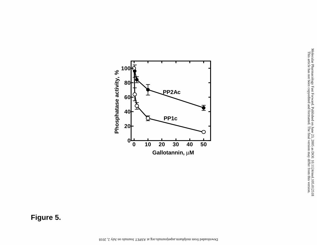
Figure 5.
0 10 20 30 40 500
20
40
60
80
100
Gallotannin, µM
Ph
osp
hat
ase
acti
vity
, %PP1c
PP2Ac
This article has not been copyedited and form
atted. The final version m
ay differ from this version.
Molecular Pharm
acology Fast Forward. Published on June 23, 2005 as D
OI: 10.1124/m
ol.105.012518 at ASPET Journals on July 2, 2018 molpharm.aspetjournals.org Downloaded from
0’ 10’ 30’ 60’ 90’
DAPI
PAR
TNFα/IL-1β
5’
5’
H2O2
Figure 6.
DAPI
PAR
PJ34+H2O2
30 µMGT 1h
30 µMGT 4h
A
DAPI
PAR
This article has not been copyedited and form
atted. The final version m
ay differ from this version.
Molecular Pharm
acology Fast Forward. Published on June 23, 2005 as D
OI: 10.1124/m
ol.105.012518 at ASPET Journals on July 2, 2018 molpharm.aspetjournals.org Downloaded from
H2O
25’
Co
ntr
ol
1 h
2 h
3 h
4 h
218
135
85
TNFα/IL-1β
Figure 7.
A B
H2O2
5’
Control
1h GT 30 µ
M
4h GT 30 µ
M
135
85
40
This article has not been copyedited and form
atted. The final version m
ay differ from this version.
Molecular Pharm
acology Fast Forward. Published on June 23, 2005 as D
OI: 10.1124/m
ol.105.012518 at ASPET Journals on July 2, 2018 molpharm.aspetjournals.org Downloaded from
0
20
40
60
80
100
120
0 0,3 0,5 3 5 30 50 300 500
µM
OD
(%
)
ascorbic acid
gallotannin
PJ34
0
20
40
6080
100
120
140
160
0,1 0,3 1 3 10 30 100 300
µM
RFU
(%
)
ascorbic acidgallotannin
PJ34
A
B
Figure 8.
This article has not been copyedited and form
atted. The final version m
ay differ from this version.
Molecular Pharm
acology Fast Forward. Published on June 23, 2005 as D
OI: 10.1124/m
ol.105.012518 at ASPET Journals on July 2, 2018 molpharm.aspetjournals.org Downloaded from