Haemoglobin Sickle D Punjab: - A Case Report - e-mjm.org · PDF fileHaemoglobin Sickle D...
2
42 Med J Malaysia Vol 69 No 1 February 2014 SUMMARY Haemoglobin S D-Punjab is a rare compound heterozygous haemoglobinopathy characterised by the presence of two β globin gene variants: β6(GAG→GTG) and β121(GAA→CAA). These patients’ clinical and haematological features mimic haemoglobin S disease. We describe the first case of doubly heterozygous HbSD-Punjab from Malaysia managed with regular blood transfusion at the age of one. This case highlights the propensity for occurrence of rare phenotypes within our multi-ethnic population and emphasises the importance of accurate genotyping to avoid erroneous counselling, and to plan an effective patient management strategy before complication evolves. KEY WoRDS: Haemoglobinopathies; Hb SD-Punjab; Malaysia INTRoDUCTIoN Haemoglobinopathies are characterised by structurally abnormal haemoglobin variants of the normal adult haemoglobin (HbA). Haemoglobin S, C, D and E are classical examples of β chain variants. Hb S (β6(A3), Glu→Val) variant has a worldwide distribution with highest gene frequencies seen in equatorial Africa, Quatif osases of eastern Saudi Arabia and parts of India. In Malaysia, Hb S variant is seen amongst the Malaysian Indians at a low frequency 1 . Hb S heterozygotes (β S /β A ) are usually asymptomatic but rarely may be associated with clinical and haematologic manifestation of significance. Homozygotes (β S /β S ), however, has enormous clinical importance. Haemoglobin D Punjab (also known as D-Los Angeles) is characterized by substitution of glutamic acid by glutamine at position 121 of the β-globin chain and is most commonly encountered in northwest India, Pakistan, and Iran; and is asymptomatic both in heterozygous and homozygous states. Co-inheritance of both haemoglobin S and haemoglobin D-Punjab, termed HbSD disease, may manifest with mild to moderate haemolytic anaemia resembling those of sickle cell anaemia. CASE REPoRT An Indian boy presented with a history of upper respiratory tract infection at the age of 4 months and was noted by his GP to be very pale (Hb of 5.5 gm/dL). He was then referred to a district hospital for further investigation and management. Red cell indices at the hospital revealed the following: Hb 6.5 gm/dL, RBC 2.3 X10 6 /dL, MCV 86.0fL, MCV 27.5pg and MCHC 32%. Blood smear showed marked anisopoikilocytosis with microcytic hypochromic red cells, dacrocytes, target cells, fragmented and polychromatic cells. A few sickle and boat shaped cells were also seen. Sickling test done was positive. The Hb electrophoresis done at pH 8.5 showed a variant band at haemoglobin S position along with a thick band at HbF position. A presumptive diagnosis of sickle cell disease (SCD) was then made. Further analysis on HPLC showed presence of Hb F and variant Hb peaks at the D (20.8%) and S (19.2%) windows. Hb A peak was virtually not visible. Based on this result the diagnosis of sickle cell/haemoglobin D-Punjab compound heterozygosity was made. At the age of one, the patient became transfusion dependent requiring two monthly blood transfusions. To establish a definitive molecular diagnosis, patient’s blood specimen was sent to our laboratory at Institute for Medical Research. His genomic DNA was tested for β-gene mutations prevalent in our multiethnic population. Having excluded the common point mutations by allele specific primer extension, β-globin gene sequencing was done to detect the presence of other mutations. Sequencing of the β-globin gene read from -100 bp to the 3’UTR showed the co-inheritance of a heterozygous change at codon 6 (GAG→GTG) and also a heterozygous mutation at codon 121 (GAA→CAA). Thus, the diagnosis of β S /β D-Punjab compound heterozygosity was confirmed. The parent’s blood samples were not sent to our institute for testing, therefore we were unable to fully determine the pattern of inheritance. The mother’s Hb analysis report was consistent with Hb S trait. However the father’s Hb analysis was to have been reanalysed. DISCUSSIoN Several variants of HbD such as Hb D Punjab, Hb D Iran, Hb D Ibadan and Hb D Bushman have been noted to co-inherit with HbS. With the exception of Hb D Punjab, compound heterozygous states of HbS with HbD variants were clinically innocuous. The substituted glutamine residue in Hb D Punjab interacts with Hb S to facilitate polymerisation during deoxygenation, and thus is presented perniciously with clinical and haematological features of variable degree of haemolytic anaemia resembling sickle cell anaemia 2 . Studies from India, Pakistan, Iran, UAE and Mexico have shown similar clinical presentations for HbSD-Punjab mimicing severe form of sickle cell anaemia 3 . Haemoglobin Sickle D Punjab: - A Case Report Rahimah Ahmad*, Syahira Lazira Omar*, Siti Hida H M Arif*, Faidatul Syazlin A Hamid*, Nur Aisyah Aziz*, Nik Hafidzah N Mustapha**, Zubaidah Zakaria* *Haematology Unit, Institute for Medical Research (IMR), Jalan Pahang, 50588 Kuala Lumpur, **Pathology Unit, Hospital Tuanku Jaafar, Negeri Sembilan Malaysia CASE REPORT This article was accepted: 29 October 2013 Corresponding Author: Rahimah Ahmad, Haematology Unit, Institute for Medical Research (IMR), Jalan Pahang, 50588 Kuala Lumpur, Malaysia Email: [email protected]
Transcript of Haemoglobin Sickle D Punjab: - A Case Report - e-mjm.org · PDF fileHaemoglobin Sickle D...
42 Med J Malaysia Vol 69 No 1 February 2014
SUMMARYHaemoglobin S D-Punjab is a rare compound heterozygoushaemoglobinopathy characterised by the presence of two βglobin gene variants: β6(GAG→GTG) and β121(GAA→CAA).These patients’ clinical and haematological features mimichaemoglobin S disease. We describe the first case of doublyheterozygous HbSD-Punjab from Malaysia managed withregular blood transfusion at the age of one. This casehighlights the propensity for occurrence of rare phenotypeswithin our multi-ethnic population and emphasises theimportance of accurate genotyping to avoid erroneouscounselling, and to plan an effective patient managementstrategy before complication evolves.
KeY WoRDS:Haemoglobinopathies; Hb SD-Punjab; Malaysia
INTRoDUCTIoNHaemoglobinopathies are characterised by structurallyabnormal haemoglobin variants of the normal adulthaemoglobin (HbA). Haemoglobin S, C, D and E are classicalexamples of β chain variants. Hb S (β6(A3), Glu→Val)variant has a worldwide distribution with highest genefrequencies seen in equatorial Africa, Quatif osases of easternSaudi Arabia and parts of India. In Malaysia, Hb S variant isseen amongst the Malaysian Indians at a low frequency 1. HbS heterozygotes (βS/βA) are usually asymptomatic but rarelymay be associated with clinical and haematologicmanifestation of significance. Homozygotes (βS/βS), however,has enormous clinical importance. Haemoglobin D Punjab(also known as D-Los Angeles) is characterized bysubstitution of glutamic acid by glutamine at position 121 ofthe β-globin chain and is most commonly encountered innorthwest India, Pakistan, and Iran; and is asymptomaticboth in heterozygous and homozygous states. Co-inheritanceof both haemoglobin S and haemoglobin D-Punjab, termedHbSD disease, may manifest with mild to moderatehaemolytic anaemia resembling those of sickle cell anaemia.
CASe RePoRT An Indian boy presented with a history of upper respiratorytract infection at the age of 4 months and was noted by hisGP to be very pale (Hb of 5.5 gm/dL). He was then referred toa district hospital for further investigation and management.Red cell indices at the hospital revealed the following: Hb 6.5gm/dL, RBC 2.3 X10⁶/dL, MCV 86.0fL, MCV 27.5pg and
MCHC 32%. Blood smear showed marked anisopoikilocytosiswith microcytic hypochromic red cells, dacrocytes, targetcells, fragmented and polychromatic cells. A few sickle andboat shaped cells were also seen. Sickling test done waspositive. The Hb electrophoresis done at pH 8.5 showed avariant band at haemoglobin S position along with a thickband at HbF position. A presumptive diagnosis of sickle celldisease (SCD) was then made. Further analysis on HPLCshowed presence of Hb F and variant Hb peaks at the D(20.8%) and S (19.2%) windows. Hb A peak was virtually notvisible. Based on this result the diagnosis of sicklecell/haemoglobin D-Punjab compound heterozygosity wasmade.
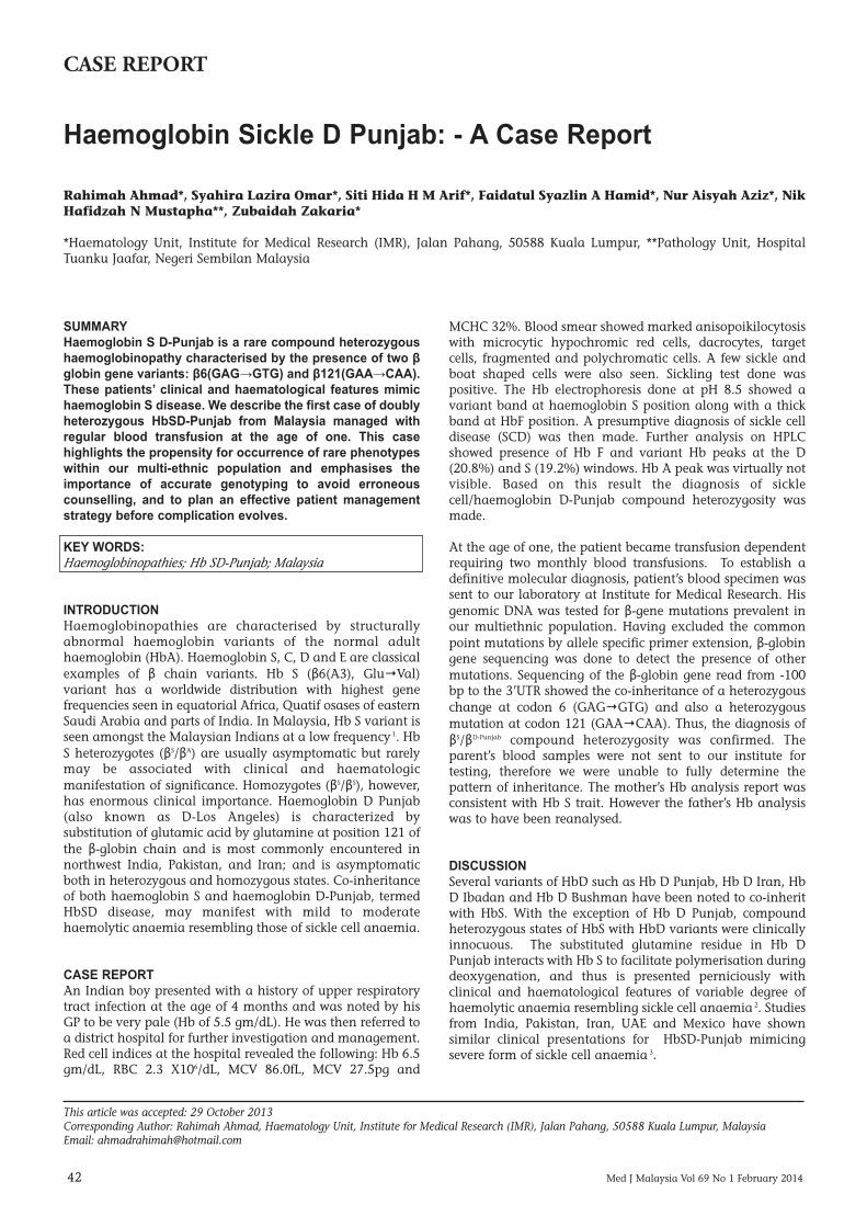
At the age of one, the patient became transfusion dependentrequiring two monthly blood transfusions. To establish adefinitive molecular diagnosis, patient’s blood specimen wassent to our laboratory at Institute for Medical Research. Hisgenomic DNA was tested for β-gene mutations prevalent inour multiethnic population. Having excluded the commonpoint mutations by allele specific primer extension, β-globingene sequencing was done to detect the presence of othermutations. Sequencing of the β-globin gene read from -100bp to the 3’UTR showed the co-inheritance of a heterozygouschange at codon 6 (GAG→GTG) and also a heterozygousmutation at codon 121 (GAA→CAA). Thus, the diagnosis ofβS/βD-Punjab compound heterozygosity was confirmed. Theparent’s blood samples were not sent to our institute fortesting, therefore we were unable to fully determine thepattern of inheritance. The mother’s Hb analysis report wasconsistent with Hb S trait. However the father’s Hb analysiswas to have been reanalysed.
DISCUSSIoNSeveral variants of HbD such as Hb D Punjab, Hb D Iran, HbD Ibadan and Hb D Bushman have been noted to co-inheritwith HbS. With the exception of Hb D Punjab, compoundheterozygous states of HbS with HbD variants were clinicallyinnocuous. The substituted glutamine residue in Hb DPunjab interacts with Hb S to facilitate polymerisation duringdeoxygenation, and thus is presented perniciously withclinical and haematological features of variable degree ofhaemolytic anaemia resembling sickle cell anaemia 2. Studiesfrom India, Pakistan, Iran, UAE and Mexico have shownsimilar clinical presentations for HbSD-Punjab mimicingsevere form of sickle cell anaemia 3.
Haemoglobin Sickle D Punjab: - A Case Report
Rahimah Ahmad*, Syahira Lazira Omar*, Siti Hida H M Arif*, Faidatul Syazlin A Hamid*, Nur Aisyah Aziz*, NikHafidzah N Mustapha**, Zubaidah Zakaria*
*Haematology Unit, Institute for Medical Research (IMR), Jalan Pahang, 50588 Kuala Lumpur, **Pathology Unit, HospitalTuanku Jaafar, Negeri Sembilan Malaysia
CASE REPORT
This article was accepted: 29 October 2013Corresponding Author: Rahimah Ahmad, Haematology Unit, Institute for Medical Research (IMR), Jalan Pahang, 50588 Kuala Lumpur, Malaysia Email: [email protected]
Haemoglobin Sickle D Punjab
Med J Malaysia Vol 69 No 1 February 2014 43
Accurate delineation of these variants is very important tofacilitate an effective response to life threateningcomplication and to avoid erroneous counselling of theserare clinically important Hb S compound heterozygotepatients. As a rule of thumb, all samples showing a singleband at the haemoglobin S position on conservativeelectrophoresis at alkaline pH should be retested by analternative technique either by citrate agar/acid gel method,or by IEF. This step is crucial to exclude the possibility of acompound heterozygote. Alternatively, HPLC should bepreferred if available. Care must be taken if considering adiagnosis on HPLC chromatograph as it is possible for morethan one variant to overlap and co-elute within a givenretention time window. Haemoglobin eluting in the HbSwindow should have a sickle solubility test performed toconfirm the presence of sickling haemoglobin. It should benoted, however, that positive sickling test alone indicates thepresence of a sickling haemoglobin and does not providedefinitive diagnostic information on the identity of thehaemoglobin or which other haemoglobins may be present.Erythrocytes containing other variant haemoglobins inwhich β6(Glu→Val) is present, such as HbC-Geogtown and Hb S-Memphis also exhibit sickling. The sickle cell solubility testmay not be reliable at HbS level below 15-20%, reflecting thatthe test is unreliable in neonates, infants and in recenttransfusion with HbAA blood.
Hb D-Punjab can be readily distinguished from Hb S by itsnormal solubility, difference on electrophoretic mobility onagar gel at acidic pH and its failure to produce sickling.
Some factors affecting variability in clinical manifestation ofHb SD-Punjab may include co-inheritance of α-thalassaemia,enhanced HbF levels and the type of haplotype framework onwhich βS is inherited. The coexistence of α thalassaemia andsickle cell anaemia does have a proven affect on thephenotype reflected by less haemolysis. Moreover, it is knownthat Bantu and Benin haplotypes of βS are associated with theclinically severe sickle cell disease. However, unlike in HbSSsyndrome, elevated levels of HbF concentration do not seemto ameliorate the clinical phenotype of HbS D Punjab 4.
As the pathophysiology HbS D-Punjab is similar to HbSdisease, adopting the guidelines proposed for sickle cellanaemia to manage this clinically significant phenotype willbe critical to reduce morbidity and mortality 5.
CoNCLUSIoNAccording to our extensive literature review and to the best ofour knowledge, this is the first case of HbSD-Punjab reportedfrom Malaysia. Haemoglobin variant such as HbS and HbDare extremely rare in the multi-ethnic Malaysian population.However, demographic changes such as populationmigration, miscegenation causes new spectrum of inheritedhaemoglobin disorders to emerge. Therefore, it is importantto make a precise genotype diagnosis to facilitate error freecounselling and proper management HbS D disease. Acombined data from HPLC, electrophoresis at alkaline andacid pH, and the sickle solubility test enable definitiveidentification of HbA, HbF, HbS, HbC, and several others rarevariants.
ReFeReNCeS1. Lie-Injo LE, Hassan K, Joishy SK, Lim ML. Sickle cell anaemia associated
with alpha-thalassemia in Malaysian Indians. Am J Hematol 1986; 22:265-74.
2. Adachi K, Kim J, Ballas S, Surrey S, Asakura T. Facilitation of Hb Spolymerization by the substitution of Glu for Gln at beta 121. J Biol Chem1988; 263: 5607-10.
3. Mukherjee MB, Surve RR, Gangakhedhar RR, Mohanty D, Colah RB.Hemoglobin sickle D Punjab - a case report. Indian J Hum Genet 2005; 11:154-5.
4. Adekile A, Mullah AA, Akar NA. Does elevated hemoglobin F modulatethe phenotype in Hb SD-Los Angleles? Acta Haematol 2010; 123: 135-9.
5. Report by the Secretariat: Sickle cell anaemia.In Fifty-ninth world healthassembly World Health Organisation, 2006; A59/9: 1-5.
Fig. 1 : The sequence trace of the β globin gene above shows the codon 6 (GAG→GTG) and codon 121 (GAA →CAA) mutation inheritedby the patient.