1/26/09 VIRUS STRUCTURE - Center for Sickle Cell Disease - Howard
PREVALENCE OF THE ΒETA-S GENE AND SICKLE CELL DISEASE IN INDIA
by
Aishwarya Arjunan
BS Biology, Case Western Reserve University, 2010
Submitted to the Graduate Faculty of
the Graduate School of Public Health in partial fulfillment
of the requirements for the degree of
Master of Science
University of Pittsburgh
2013
ii
UNIVERSITY OF PITTSBURGH
GRADUATE SCHOOL OF PUBLIC HEALTH
This thesis was presented
by
Aishwarya Arjunan
It was defended on
February 19, 2013
and approved by
Thesis Advisor:
Lakshmanan Krishnamurti, MD Associate Professor of Pediatric Medicine, Department of Pediatric Medicine
Program Director, Hemoglobinopathy Program Children’s Hospital of Pittsburgh, University of Pittsburgh Medical Center
Committee Members:
Elizabeth Gettig, MS, CGC Associate Professor of Human Genetics
Co-Director, Genetic Counseling Program Graduate School of Public Health
University of Pittsburgh
Martha Ann Terry, PhD Assistant Professor and Director, MPH Program
Department of Behavioral and Community Health Sciences Graduate School of Public Health
University of Pittsburgh

iii
Copyright © by Aishwarya Arjunan
2013
iv
Introduction: Sickle Cell Disease (SCD), an inherited disorder of the red blood cells, is a major
public health problem. India, with a population of 1.2 billion individuals, is estimated to be home
to over 50% of the world’s patients with SCD. While SCD is common among all ethnic groups
in India, high prevalence has been reported in three socio-economically disadvantaged ethnic
categories: the Scheduled Castes (SC), the Scheduled Tribes (ST), and Other Backward Class
(OBC) groups.
Both the prevalence of the βs gene and the clinical phenotype of SCD have not been well
described in these three population groups.
Objective: Our objective was two-fold: to determine the prevalence of the βs gene and to
describe the clinical phenotype of SCD in the district of Nagpur, Maharashtra located in Central
India.
Method: To determine the prevalence of the βs gene, community screening of target populations
was conducted over an eight-year time span. To determine the clinical phenotype of SCD, a
cohort of 726 patients was followed over a four and half year time period during which all
clinical events presented to the hospital were recorded.
Results
Population Screening: Community wide screening of 35,636 individuals identified
5,437 individuals with sickle cell trait and 1,010 with SCD. The trait prevalence was 13%, 12%,
and 3.4% for the SC, ST, and OBC groups respectively.
PREVALENCE OF THE ΒETA-S GENE AND SICKLE CELL DISEASE IN INDIA
Aishwarya Arjunan, MS
University of Pittsburgh, 2013
v
Clinical Phenotype: A total of 726 patients were followed in the comprehensive clinic
over a four and a half year time period. Painful crisis and fever were the most common initial
presentations and the leading causes of hospitalizations. These rates were also higher when
compared to the published data from the Cooperative Study of Sickle Cell Disease.
Implications for Public Health: The population screening program uncovered previously
undiagnosed cases, and provided detailed information for population based disease counseling,
prevention programs and comprehensive care programs. Additionally, we present evidence
suggesting that SCD may not have milder manifestations in India and underscore the need for
detailed studies of phenotype that can form the basis of public health interventions and
mechanistic studies of genetic modifiers of clinical phenotype.
vi
TABLE OF CONTENTS
ACKNOWLEDGEMENTS ........................................................................................................ X
1.0 INTRODUCTION ........................................................................................................ 1
2.0 BACKGROUND & SIGNIFICANCE ....................................................................... 4
2.1 SICKLE CELL DISEASE .................................................................................. 6
2.2 INHERITANCE ................................................................................................... 8
2.3 CLINICAL SYMPTOMS ................................................................................... 8
2.3.1 Blood............................................................................................................... 8
2.3.2 Spleen ............................................................................................................. 9
2.3.3 Neurological ................................................................................................. 10
2.3.4 Pulmonary ................................................................................................... 10
2.3.5 Infection ....................................................................................................... 10
2.3.6 Renal/Kidney ............................................................................................... 11
2.3.7 Ophthalmologic ........................................................................................... 11
2.3.8 Growth and Development .......................................................................... 12
2.3.9 Bone .............................................................................................................. 12
3.0 SPECIFIC AIMS ........................................................................................................ 13
4.0 METHODS ................................................................................................................. 14
4.1 POPULATION SCREENING .......................................................................... 14
vii
4.1.1 Screening Procedure ................................................................................... 15
4.1.2 Genotype Results & Counseling ................................................................ 16
4.1.3 Social Classifications ................................................................................... 16
4.2 CLINICAL PHENOTYPE ............................................................................... 18
4.2.1 Laboratory Diagnosis ................................................................................. 18
4.2.2 Data Collection ............................................................................................ 19
4.2.3 Definition of Events..................................................................................... 20
4.2.4 Statistical Analysis ...................................................................................... 20
5.0 RESULTS ................................................................................................................... 22
5.1 POPULATION SCREENING .......................................................................... 22
5.2 CLINICAL PHENOTYPE ............................................................................... 25
5.2.1 Population Demographics .......................................................................... 25
5.2.2 Clinical Events ............................................................................................. 26
5.2.3 Mortality ...................................................................................................... 27
6.0 DISCUSSION ............................................................................................................. 28
6.1 POPULATION SCREENING .......................................................................... 28
6.2 CLINICAL PHENOTYPE ............................................................................... 30
7.0 CONCLUSION ........................................................................................................... 33
BIBLIOGRAPHY ....................................................................................................................... 36
viii
LIST OF TABLES
Table 1. Different Structural Variants of Hemoglobin ................................................................... 5
Table 2. Totals for all groups (Community wide screenings + High Risk Targeted screenings) . 22
Table 3. Scheduled Caste (SC) Community Wide Screening ....................................................... 23
Table 4. Scheduled Tribe (ST) Community Wide Screening ....................................................... 23
Table 5. Other backward classes (OBC) Community Wide Screening ........................................ 24
Table 6. Ethnic Group Distribution in the Study Population ........................................................ 26
Table 7. Incidence of Clinical Events per 100 person years ......................................................... 27
Table 8. Causes of Mortality by Age Distribution ........................................................................ 27
ix
LIST OF FIGURES
Figure 1. The timeline of human globin gene expression ............................................................... 5
Figure 2. Sickle cell vs. Normal red blood cell ............................................................................... 7
Figure 3. Pathophysiology of SCD ................................................................................................. 7
Figure 4. Districts in the State of Maharashtra, India. .................................................................. 15
x
ACKNOWLEDGEMENTS
I would like to thank the faculty and staff of the Graduate School of Public Health, particularly
those in the Human Genetics Department for all of their guidance and support throughout the
past three years. Candy Kammerer, Betsy Gettig and Robin Grubs have been a big influence and
provided me with the knowledge and support needed to reach my full potential and succeed in
both my public health and genetic counseling programs. And thank you to Dr. Martha Terry for
taking time out of your busy schedule to serve on my committee. A special thank you to Dr. Brad
Therrell for your wisdom and expertise as I started the first phase of my project.
I have much gratitude for Dr. Lakshmanan Krishnamurti who has been a great mentor
and has given me one of the best experiences that I could have ever asked for. Over the past three
years I have been given many unique opportunities from traveling to India, attending
conferences, organizing outreach events, working with the Sickle Cell Foundation and have
developed lifelong relationships for which I am very grateful and appreciative.
None of my work would have been possible without our collaborators in India. Dr. Jain,
Dr. Menon, Dr. Shrikhande, Dr. Italia and all the other team members in Nagpur, Gudalur, and
Valsad: thank you from the bottom of my heart. You were willing to trust me and work with me
and give me a truly wonderful and unforgettable experience in India
I couldn’t have survived graduate school without the Sickle Cell Team at the Children’s
Hospital of Pittsburgh and Andrea and Michael at the Foundation. You all have been my biggest
xi
support system. A special thanks to Beth Kladny, Pixie Talarico, and Mary Campbell for their
friendship and guidance as I navigated the world of a graduate student.
To my classmates in the genetic counseling and public health programs and my
phenomenal buddies: we have been each other’s support systems through the good and the bad,
and I am very grateful that I can call each and every one of you a friend.
Lastly to my family, I honestly could not have completed this program without you. From
driving me to my classes for the last three years, to supporting me through all the career changes
that I have made, I am eternally grateful. And a special thank you to my sister, Akshaya, for
accompanying me on my research trip to India and helping me with my work in Nagpur and
Gudalur.
1
1.0 INTRODUCTION
Sickle cell disease (SCD), the most common single-gene blood disorder (hemoglobinopathy) in
the world, represents a significant public health problem in India. In India, SCD is highly
prevalent in the Western, Central, and Eastern regions and in pockets of the South in the states of
Maharashtra, Madhya Pradesh, Orissa, Andhra Pradesh, Gujarat, Chattisgarh, Tamil Nadu, and
Kerala. [1-9] SCD is an inherited disorder of the red blood characterized by vaso-occlusive pain
crises, risk for pneumococcal infections, acute chest syndrome, stroke and organ failure and is
associated with substantial morbidity and premature mortality. [6]
With a population of over 1.2 billion individuals, it is estimated that India is home to over
50% of the world’s SCD patients. [8] While sickle cell is prevalent in many ethnic groups, the
highest prevalence appears to be in three particular socio-economically disadvantaged
communities, identified as the Scheduled Castes (SC), Scheduled Tribes (ST), and Other
Backward Classes (OBC). [8] Each of these categories consists of several different large ethnic
groups that have practiced endogamy, or the practice of marrying within a specific group, for
millennia and thus represent genetic isolates. In order to be useful for the purposes of planning
for genetic counseling services, control programs, clinical service provision or genetic
epidemiological studies, screening should generate prevalence estimates of the βs gene in each
ethnic group. The prevalence of the βs gene has been well described in the ST population;
2
however, such precise estimates are not available for the OBC population or the SC population.
Together these three groups comprise over 75% of the Indian population. [5, 6, 10, 11]
Similarly, the clinical phenotype of SCD in the Indian population has been described
mainly in the ST group and not in the SC or OBC groups. Previous studies describe the milder
manifestations of SCD in the Indian population compared to that observed in the African and
Caribbean populations. [12] Proposed epistatic factors or modifiers contributing to the
amelioration of clinical phenotype include the high prevalence of the Arab-Indian β globin
haplotype, high Hemoglobin F levels, presence of Xmn1 polymorphic site, and high frequency of
alpha Thalassemia in certain population groups. [13-20] However, there has been a lack of
detailed phenotypic studies of SCD in various populations in India, and such studies are
important not only to understand the clinical and public health impact of the disease in the region
but are also essential prior to undertaking mechanistic studies of molecular mechanisms
modulating disease severity. [15-17] The Cooperative Study of Sickle Cell Disease conducted in
the United States over a 20 year time period is the largest study prospective study of natural
history of sickle cell disease conducted to date and has yielded rich data on the clinical course
and natural history of disease and has contributed to a variety of mechanistic studies.[13]
Central India, which is one of the high prevalence regions for the βs gene, has a
prevalence rate of 9.4-22.2% for this mutation in various communities. [3, 7] The state of
Maharashtra in Central India has a population of 112.3 million of which the SC and ST comprise
10.2% and 8.9% of the population respectively.[11] Additionally, the OBC population comprises
52% of the population.

3
The purpose of our study was two-fold: to determine the prevalence of the βs gene and to
describe the clinical phenotype SCD in the district of Nagpur, Maharashtra, located in Central
India.
4
2.0 BACKGROUND & SIGNIFICANCE
Sickle cell disease or sickle cell anemia is a hereditary genetic disease that is characterized by the
presence of abnormal crescent-shaped red blood cells.[21] While the symptoms related to SCD
were known by various names throughout Africa for centuries, it was not until 1910, when James
Herrick, a physician, noted the presence of a “peculiar elongated sickle shaped red blood cells”
in the blood of an anemic African medical student that the Western world and the scientific
community became aware of the disease. [21] A few years after Dr. Herrick’s report, Dr. Emmel
noted that a patient and her father, both asymptomatic, had sickled blood cells.[22] Furthermore,
three of the patient’s siblings had died from severe anemia. These observations made by Dr.
Emmel were the first to suggest a genetic basis for SCD, although the specific pattern of
inheritance was not delineated until the early 1920s. [22] In 1949, Linus Pauling et al. were the
first to suggest that the cause of SCD was the result of defective hemoglobin molecules. [23]
Hemoglobin (Hb) molecules are a set of proteins formed by pairing of two alpha (α) and
two beta (β) globin polypeptide chains into a tetromeric unit. [24-27] This unit, the α2β2
molecule, forms the major adult hemoglobin responsible for the transport of oxygen from the
lungs to tissues. The α-globin gene is located on chromosome 16 and encodes the adult α-globin
and the ζ-globin chain, which is the embryonic form of α-globin.[25] The β-globin gene is
located on chromosome 11 and encodes four different globin molecules: embryonic ε-globin,
fetal γ-globin, adult δ-globin and adult β-globin, all expressed differently during specific times of
5
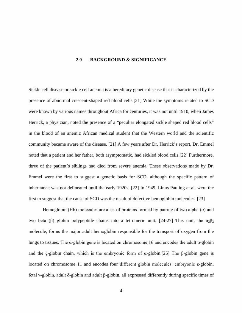
development. [25, 27] Figure 1 illustrates the timeline of the expression of the human globin
genes from early stages of fetal development to the changes that occur at birth and the first year
of life. [25]
Figure 1. The timeline of human globin gene expression Adapted from Schechter[25]
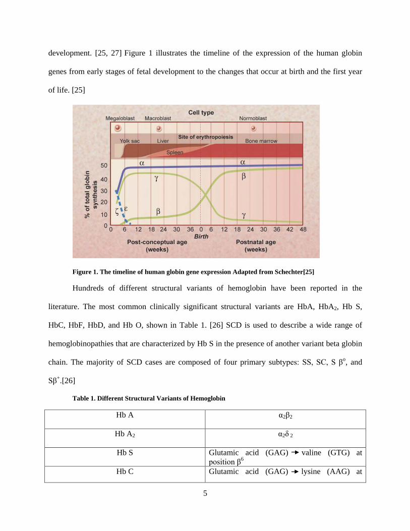
Hundreds of different structural variants of hemoglobin have been reported in the
literature. The most common clinically significant structural variants are HbA, HbA2, Hb S,
HbC, HbF, HbD, and Hb O, shown in Table 1. [26] SCD is used to describe a wide range of
hemoglobinopathies that are characterized by Hb S in the presence of another variant beta globin
chain. The majority of SCD cases are composed of four primary subtypes: SS, SC, S βo, and
Sβ+.[26]
Table 1. Different Structural Variants of Hemoglobin
Hb A α2β2
Hb A2 α2δ 2
Hb S Glutamic acid (GAG) valine (GTG) at position β6
Hb C Glutamic acid (GAG) lysine (AAG) at
6
position β6 Hb F α2γ 2
Hb D Los Angeles or Punjab Glutamic acid (GAG) glutamine (CAG) at position β121
Hb O Arab Glutamic acid (GAG) lysine (AAG) at position β121
Hb E Glutamic acid (GAG) lysine (AAG) at position β26
βo Thalassemia Absence of Hb A due to inability to produce normal β-chain
β + Thalassemia Reduced amount of β-chain production leads to variable amount of Hb A.
2.1 SICKLE CELL DISEASE
Sickle cell disease is an autosomal recessive genetic blood disease that affects individuals of
African, Mediterranean, Indian, Middle-Eastern, Caribbean, South and Central American
ancestry. SCD is characterized by the presence of HbS, caused by a single point mutation
involving GAG GTG transversion at codon 6 of the β-globin gene.[24, 26-28] This results in
an amino acid substitution at position 6 in the β-globin chain from glutamic acid to valine on the
surface of the hemoglobin molecule. This amino acid change results in a change in the
characteristics of the deoxygenated HS which causes HbS to become less soluble compared to
HbA, and when HbS becomes deoxygenated, polymerization of HbS occurs. [24, 26] This
polymerization of HbS results in the formation of elongated rigid fibers that damage the
cytoskeleton of the red blood cell, causing the shape of the cell to change from the smooth,
doughnut-shape to the hallmark sickle-shape. [28-31] (Figure 2) As the red blood cell becomes
re-oxygenated the HbS fibers dissociate and revert back to the doughnut-shape. However, as the
red blood cell passes through the circulatory system repeatedly, it becomes increasingly more
Table 1 continued

7
damaged and the cycles of sickling and unsickling lower the life span of the red blood cell from
120 days to less than 20 days. [24, 26, 29-31]
Figure 2. Hallmark Sickle-Shaped red blood cell shape (L) versus normal red blood cell shape(R)
These sickled cells then form homotypic aggregations and heterotypic adhesion to
endothelial cells and white blood cells and contribute to the obstruction of blood flow and vaso-
occlusion that are hallmark clinical presentations of SCD. [29] This obstruction also results in a
lack of adequate blood supply to organs and tissues resulting in damage. The pathophysiology of
SCD is illustrated in Figure 3.[23]
Figure 3. Pathophysiology of SCD (a) Point mutation in sickle cell disease. (b) Polymerization of hemoglobin S (HbS) under-deoxygenation. (c) Red blood cell shape change in response to HbS polymerization. (d) Reticulocyte adhered to endothelium initiates vaso-occlusion by trapping irreversibly sickled cells and forming aggregates with white blood cells.[23]
8
2.2 INHERITANCE
SCD is inherited in an autosomal recessive pattern, which occurs when an individual inherits one
abnormal hemoglobin gene from each parent. If an individual inherits one abnormal hemoglobin
gene and one normal gene then he/she is considered to have sickle cell trait or a carrier for SCD.
If both parents are carriers for SCD then there is a 25% or 1 in 4 chance in each pregnancy of
having a child who will inherit both abnormal hemoglobin genes and present with SCD. [32, 33]
2.3 CLINICAL SYMPTOMS
Clinical symptoms or manifestations of SCD include hemolysis, anemia, pain crisis, infection,
acute chest syndrome swelling and vascular occlusion potentially leading to ischemic attacks and
organ damage. [34, 35]
2.3.1 Blood
Acute painful crises are the most common type of vaso-occlusive event that occurs in SCD and is
the most common reason for admission to hospital for both adults and children. [36] The exact
mechanism leading to a pain episode is still unclear although it is thought to be a continuous
cycle in which vaso-occlusion results in tissue ischemia and damage, leading to a secondary
inflammatory response that triggers the release of norepinephrine which ultimately leads to
further tissue ischemia. [37] The severity, frequency, location and duration of pain crises are
variable among patients and can be triggered by a number of different events or factors
9
including: extreme temperatures or changes in humidity, dehydration, stress, alcohol
consumption, infection, menses, obstructive sleep apnea and cardiac or pulmonary impairments.
[38-47]
The decreased lifespan of the red blood cells (120 days to <20 days) is also responsible
for patients to experience chronic anemia with varying degrees of severity. [38-47]
2.3.2 Spleen
An organ to be affected early in SCD is the spleen, which is commonly enlarged during the first
decade of life and can undergo progressive atrophy due to repeated attacks of vaso-occlusion and
infarction. [48] Individuals with SCD may experience a wide range of splenic involvement from
minimal change, splenomegaly, asplenia, and splenic sequestration crisis tor hypersplenism.
Splenic complications of SCD are associated with an increased morbidity and in some cases lead
to death. [48]
Splenic sequestration crisis results from the rapid sequestration of red blood cells in the
spleen; it is a serious complication of SCD and considered to be the second leading cause of
death after infection in the first decade of life. [48] Splenic sequestration is characterized by
sudden onset of anemia, splenomegaly, evidence of active bone marrow and a decrease in spleen
size after blood transfusion. Hypersplenism is defined as splenomegaly with anemia,
thrombocytopenia, neutropenia either singly or in combination.[48]
10
2.3.3 Neurological
SCD is one of the most common causes of cerebrovascular accidents (stroke) in children. While
the exact pathophysiology of stroke remains uncertain, contributory factors include anemia,
leukocytosis, hypoxemia, abnormal rheology causing endothelial damage, and functional nitric
oxide deficiency associated with hemolysis. [27, 28, 36] Approximately 11% of patients will
have a stroke by 20 years of age, and once a stroke has occurred, the recurrence risk is greater
than 60%. [36, 49]
2.3.4 Pulmonary
Individuals with SCD may present with a variety of chronic manifestations including restrictive
and obstructive lung disease, hypoxemia, pulmonary hypertension, airway hyper-reactivity,
asthma, or acute chest syndrome. [27, 28, 50-52] Acute chest syndrome is the second most
common cause of hospitalizations in SCD and occurs in 40% of patients. [27, 32, 36, 53] Acute
chest syndrome is characterized by infiltration of the pulmonary vasculature with sickled red
blood cells. Individuals experiencing acute chest syndrome may present with chest pain,
tachypnea, fever, rales and rhonchi, leukocytosis, or lobar consolidations. [28, 54]
2.3.5 Infection
A major cause of morbidity and mortality in patients with sickle cell disease is infection. The
most common infection causing organism is pneumococcus, and children under the age of three
are particularly at an increased risk of infection. [24, 28] Ideally, penicillin prophylaxis is
11
initiated in the first few months of life as a form of treatment for SCD patients to prevent
pneumococcal infection. [24, 28] Impaired splenic functioning, tissue ischemia, micronutrient
deficiencies and defects in the activation of the complement pathway are believed to result in an
increased risk of bacterial and viral infections. [36, 53] Streptococcus, influenza, salmonella,
malaria, hepatitis, parvovirus and chlamydophila are other commonly reported infection in
patients with SCD. [27, 28, 36, 53]
2.3.6 Renal/Kidney
One of the main renal complications seen in individuals with sickle cell disease is occlusion of
the vasarecta capillaries in the renal medulla, leading to a change in the delivery of oxygen to the
kidney, which in turn leads to ischemic damage. [24, 55] Other renal manifestations include
hematuria, proteinuria and hypertension, renal infarction, papillary necrosis, renal colic,
nephrogenic diabetes insipidus leading to polyuria, focal segmental glomerulosclerosis leading to
end-stage renal disease and renal medullary carcinoma.[27, 28, 55]
2.3.7 Ophthalmologic
The blood vessels of the eye may be damaged in patients with SCD due to the physiological
changes that occur in the red blood cells. [28, 56] These changes result in the formation of new
blood vessels that are weaker and more susceptible to rupture. [28, 56] Depending on the tissue
that is involved and the location of the damage, an individual’s vision may or may not be
affected. [24, 28] In patients whose vision is affected the primary complications are proliferative
retinopathy, retinal artery occlusion, or retinal detachment and hemorrhage. [57, 58]
12
2.3.8 Growth and Development
The birth weight and length of infants with SCD are typically along the normal growth curve.
However, as time progresses, the mean height and weight of individuals with SCD may drop
below the 50th percentile, specifically around the age of two. [24, 28] And as the children
become older, the growth deficits can become more pronounced. [24, 28] Both females and
males may experience delays in sexual maturation and progression through the Tanner stages of
development. [24, 28] Females may also experience delays in the onset of menarche while males
may experience lower levels of testosterone, dihydrotestosterone, and androsteneion. [24, 28, 47]
2.3.9 Bone
Due to a limited blood supply to some regions of the body, SCD patients may experience bone
and joint concerns. Bone complications include bone infarction or osteomyelitis. Osteomyelitis,
an acute or chronic bone infection, is most often caused by salmonella. [24, 28] Bone infarctions
include dactylitis, commonly known as hand-foot syndrome, which is characterized by painful
swelling of the hands and feet that generally resolves within a week. [24, 27, 28, 33, 59, 60]
Hand-foot syndrome is typically the first clinical manifestation seen in infants with sickle cell
disease between the ages of six months and two years. [24, 28]
13
3.0 SPECIFIC AIMS
This study has two specific aims:
1) To describe the prevalence of the βs gene in the scheduled caste, scheduled tribe, and
other backward class population in the district of Nagpur, Maharashtra in Central
India.
2) To examine the clinical events in a large cohort of patients with SCD, followed in a
single center in Nagpur, Maharashtra in Central India to determine the clinical
phenotype of SCD in this population.
14
4.0 METHODS
4.1 POPULATION SCREENING
The Regional Hemoglobinopathy Detection and Management Center (RHDMC) at the Indira
Gandhi Medical College located in Nagpur, Maharashtra, has mass community-based and
hospital-based screening, management and counseling for sickle cell disease. The institution
serves as a referral center with a population of over 10 million individuals in the region and
where the high βs prevalence communities of Scheduled Castes, Scheduled Tribes and Other
Backward Classes comprise 55% of the population.
The present study was performed over a span of eight years (2003-2012) and includes the
screening of target populations in all age groups (newborns, school age children, young adults,
and pregnant mothers) as well as screening in hospital for individuals considered to be at high
risk of carrying the βs gene. Informed consent was obtained from all adults and parents of
children once the individuals were educated on the reasoning behind screening. The screening
teams were comprised of a clinician, multiple lab technicians and counselors, and volunteers
from the villages surrounding the screening camp. Screening camps were conducted in villages
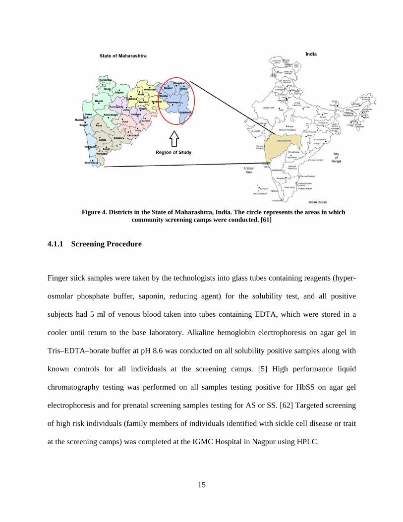
wihin a 300km radius around Nagpur that included the areas of Durg, Raipur, Chandrapur,
Bhandara, Gondia, Gadhchiroli, Amravati, and Wardha (Figure 4).
15
Figure 4. Districts in the State of Maharashtra, India. The circle represents the areas in which
community screening camps were conducted. [61]
4.1.1 Screening Procedure
Finger stick samples were taken by the technologists into glass tubes containing reagents (hyper-
osmolar phosphate buffer, saponin, reducing agent) for the solubility test, and all positive
subjects had 5 ml of venous blood taken into tubes containing EDTA, which were stored in a
cooler until return to the base laboratory. Alkaline hemoglobin electrophoresis on agar gel in
Tris–EDTA–borate buffer at pH 8.6 was conducted on all solubility positive samples along with
known controls for all individuals at the screening camps. [5] High performance liquid
chromatography testing was performed on all samples testing positive for HbSS on agar gel
electrophoresis and for prenatal screening samples testing for AS or SS. [62] Targeted screening
of high risk individuals (family members of individuals identified with sickle cell disease or trait
at the screening camps) was completed at the IGMC Hospital in Nagpur using HPLC.

16
4.1.2 Genotype Results & Counseling
Individuals who were identified through screening to have an abnormal genotype were given
identification cards with their name, age, date of birth, village, and genotype (AS/SS).
Informational booklets were distributed that contained information about sickle cell disease and
trait, inheritance patterns, disease-related issues and premarital counseling. The abnormal
genotype cards were distributed by the counselor, who provided genetic counseling and gave
additional information and answered questions. Subjects with SS disease were also referred to
the onsite clinician, who collected relevant medical history and provided a medical examination.
Patients were referred to comprehensive sickle cell clinic for follow up.
4.1.3 Social Classifications
Articles 341 and 342 of the Constitution of India provide statewide lists of communities
classified as Scheduled Castes and Scheduled Tribes. Scheduled castes (SC) were historically
known as the Untouchables and in the past occupied the lowest status in Indian society. The
Scheduled Tribe (ST) populations are aboriginal people known as Adivasis. [63, 64] Other
Backward Classes (OBC) are defined in the Indian Constitution as “socially and educationally
backward classes” that fall outside the definition of ST and SC. [65] The relative proportion of
these social groups in the State of Maharashtra are 10% (SC), 9% (ST) and 19% (OBC).[11]
Within the SC, ST, and OBC there are different ethnic groups that have been identified
and classified. The Scheduled Castes and Scheduled Tribes Orders (Amendment) Act, of 1976
classifies the various sub-groups in the SC and ST populations. [65] From this list, the SC and
ST populations screened were separated into six and 10 groups respectively, as listed below:

17
Scheduled Caste-(150 sub-groups within 59 groups)
Group 1: Bhangi, Chambhar, Mehtar Group 2: Basori, Burad Group 3: Mang, Matang Group 4: Bauddha (Mahar) Group 5: Balai Group 6: Khatik
Scheduled Tribe(184 sub-groups within 47 groups)
Group 1: Gond, Gowari, Mannewar, Madiya, Mana Group 2: Kawar, Ratha Group 3: Adivasi Group 4: Bhilala, Bhill Group 5: Halba
Group 6: Kolam Group 7: Korku Group 8: Thakur Group 9: Pardhan Group 10: Dhangar
The National Commission for Backward Classes Act of 1993 identifies all the different
sub-groups within the OBC population in every state of India. [66]
Other Backward Classes(261 groups) Group 1: Bari Group 2: Jangum Group 3: Koli Group 4: Koshti, Padmashali Group 5: Kumbhar Group 6: Kunbi Group 7: Kasar Group 8: Navi Group 9: Dhobi Group 10: Bhawsar Group 11: Devang, Sali
Group 12: Shimpi Group 13: Sonar Group 14: Sutar Group 15: Warti Group 16: Wanjari Group 17: Sahu, Shahu, Teli Group 18: Mali, Vanmali Group 19: Yadav Group 20: Kochi Group 21: Gondhali Group 22: Thakur (Thakkar)
Group 23: Kahar, Kirat Group 24: Pawar, Powar, Bhoyar Group 25: Kalar Group 26: Lingayat Group 27: Rangari Group 28: Raut Group 29: Wani Group 30: Puri Group 31: Pahadi
18
4.2 CLINICAL PHENOTYPE
Subjects for the study were recruited from the pediatric comprehensive sickle cell program at the
Government Medical College (GMC) in Nagpur, Maharashtra. Patients have been followed at
this center by a single physician (DJ) for over 20 years. They were enrolled in this prospective
study of sickle cell phenotype from January 1, 2008, to May 31, 2012. Newborn screening
(NBS) was instituted in 2010, and patients detected through NBS were excluded from this
analysis. At the time of registration, the diagnosis of SCD was confirmed by standard procedures
(cellulose acetate hemoglobin electrophoresis/high performance liquid chromatography) in all
subjects. The study was approved by the institutional review board, GMC Nagpur, and written
informed consent was obtained from all the patients and/or their parents at enrollment.
Individuals lost to follow-up were excluded from analysis. Patients were started on Hydroxyurea
(HU) therapy, if they were considered to have a severe phenotype as described in the MSH
study. [67] Individuals already on HU therapy at study initiation were excluded, and data were
excluded at initiation of HU therapy for patients who started HU during the study and only
events before initiation of HU were included. Only events for which the patient was treated in
the GMC hospital for medical care were used in the analysis.
4.2.1 Laboratory Diagnosis
The GMC of Nagpur determined the hemoglobin genotype by hemoglobin electrophoresis (Bio-
Rad)/ High Performance Liquid Chromatography (Hercules, CA, USA). [62] For those
individuals who had received a blood transfusion prior to diagnosis a repeat confirmatory testing
19
was performed after three months. Complete blood count was performed using an auto-analyzer
and reticulocyte count was performed with brilliant cresyl blue staining at the GMC.
4.2.2 Data Collection
These patients were followed up with regularly at the comprehensive sickle cell clinic with all
relevant clinical and laboratory data documented in detailed case report forms. Patients were
advised to follow up on a monthly basis or as symptoms presented. Patients included in this
study received treatment and care, including for acute events, at the GMC, Nagpur. All children
age less than five years were offered penicillin prophylaxis. Starting in 2009, the State
Government of Maharashtra offered polysaccharide pneumococcal vaccination and Haemophilus
influenzae type B vaccination free of charge to all patients. Since 2009 all patients between the
ages of two and five were offered pneumococcal vaccination. Haemophilus influenzae type B,
Typhoid, and Hepatitis B vaccines were given as per recommendations of Indian Academy of
Pediatrics. [68]
Demographic information about the family was recorded at the initial visit after
diagnosis. Clinical records of these patients are available from the time of registration at the
sickle cell clinic till the last available follow-up. Data for this study evaluated clinical events
from January 1, 2008 to May 30, 2012, or until the last available follow-up within this time
frame.
20
4.2.3 Definition of Events
Only events for which the patient presented to the hospital for medical care were recorded and
used for analysis. The event definitions were as follows: a painful event/crisis was defined as
pain in the extremities, back, abdomen, chest, or head for which no other explanation could be
found. Severe anemia was defined as a hemoglobin level less than 5g/dl. Sequestration crisis was
defined as a decrease in the hemoglobin or hematocrit level of at least 20% from baseline
accompanied by an increase in palpated spleen size of at least 2 cm from baseline. A febrile
episode was defined as a hospitalization with fever (≥ 101º Fahrenheit). Confirmatory malarial
diagnosis was defined as an identification of malaria parasite in a peripheral blood smear.
Meningitis was defined as abnormal cerebrospinal fluid (CSF) findings and a positive CSF
culture. Acute chest syndrome was defined by the following three symptoms: fever, tachypnea,
and observation of new pulmonary infiltrates on x-ray. Stroke or a cerebrovascular accident
(CVA) was defined as an acute neurologic syndrome secondary to the occlusion of an artery or
to hemorrhage with resultant neurologic symptoms and signs.
4.2.4 Statistical Analysis
For assessing demographic information, frequency and percentage were computed for
categorical data. For continuous data the median was computed, data categorized into
meaningful groups and the frequencies computed within each group. For clinical data each visit
with a certain condition was counted to assess its overall frequency. Furthermore the number of
patients who had the specific condition at least once during the study period as well as the range
21
of occurrences of the condition within a person across the study population was compiled.
Descriptive statistical analysis was carried out using SAS software 9.3 (Cary, NC, USA).
22
5.0 RESULTS
5.1 POPULATION SCREENING
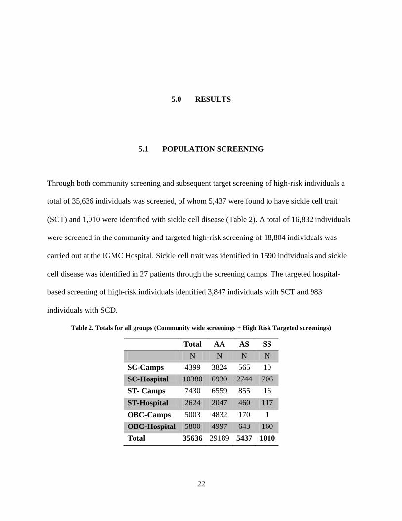
Through both community screening and subsequent target screening of high-risk individuals a
total of 35,636 individuals was screened, of whom 5,437 were found to have sickle cell trait
(SCT) and 1,010 were identified with sickle cell disease (Table 2). A total of 16,832 individuals
were screened in the community and targeted high-risk screening of 18,804 individuals was
carried out at the IGMC Hospital. Sickle cell trait was identified in 1590 individuals and sickle
cell disease was identified in 27 patients through the screening camps. The targeted hospital-
based screening of high-risk individuals identified 3,847 individuals with SCT and 983
individuals with SCD.
Table 2. Totals for all groups (Community wide screenings + High Risk Targeted screenings)
Total AA AS SS N N N N
SC-Camps 4399 3824 565 10 SC-Hospital 10380 6930 2744 706 ST- Camps 7430 6559 855 16 ST-Hospital 2624 2047 460 117 OBC-Camps 5003 4832 170 1 OBC-Hospital 5800 4997 643 160 Total 35636 29189 5437 1010
23
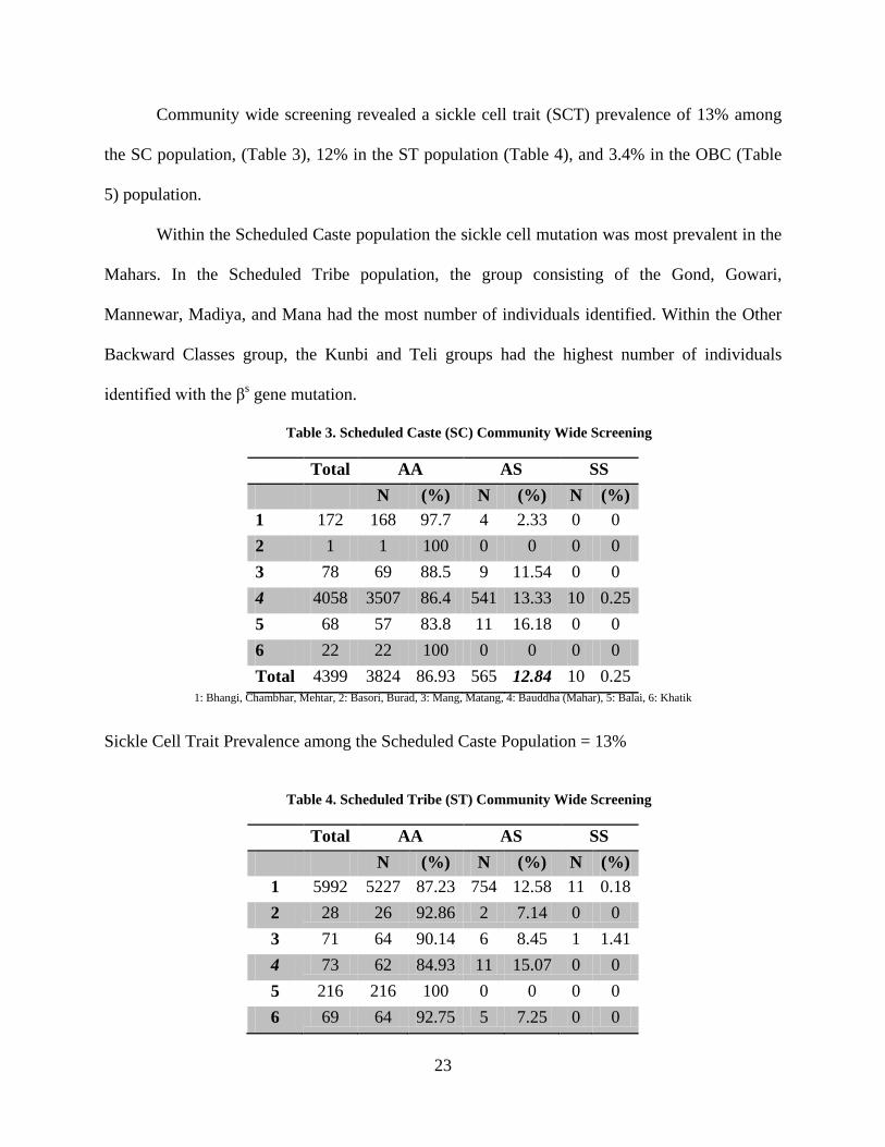
Community wide screening revealed a sickle cell trait (SCT) prevalence of 13% among
the SC population, (Table 3), 12% in the ST population (Table 4), and 3.4% in the OBC (Table
5) population.
Within the Scheduled Caste population the sickle cell mutation was most prevalent in the
Mahars. In the Scheduled Tribe population, the group consisting of the Gond, Gowari,
Mannewar, Madiya, and Mana had the most number of individuals identified. Within the Other
Backward Classes group, the Kunbi and Teli groups had the highest number of individuals
identified with the βs gene mutation.
Table 3. Scheduled Caste (SC) Community Wide Screening
Total AA AS SS N (%) N (%) N (%)
1 172 168 97.7 4 2.33 0 0 2 1 1 100 0 0 0 0 3 78 69 88.5 9 11.54 0 0 4 4058 3507 86.4 541 13.33 10 0.25 5 68 57 83.8 11 16.18 0 0 6 22 22 100 0 0 0 0 Total 4399 3824 86.93 565 12.84 10 0.25
1: Bhangi, Chambhar, Mehtar, 2: Basori, Burad, 3: Mang, Matang, 4: Bauddha (Mahar), 5: Balai, 6: Khatik
Sickle Cell Trait Prevalence among the Scheduled Caste Population = 13%
Table 4. Scheduled Tribe (ST) Community Wide Screening
Total AA AS SS N (%) N (%) N (%) 1 5992 5227 87.23 754 12.58 11 0.18 2 28 26 92.86 2 7.14 0 0 3 71 64 90.14 6 8.45 1 1.41 4 73 62 84.93 11 15.07 0 0 5 216 216 100 0 0 0 0 6 69 64 92.75 5 7.25 0 0
24
7 883 803 90.94 76 8.61 4 0.45 8 2 2 100 0 0 0 0 9 0 0 0 0 0 0 0 10 96 95 98.96 1 1.04 0 0
Total 7430 6559 88.28 855 11.51 16 0.22 1: Gond, Gowari, Mannewar, Madiya, Mana, 2: Kawar, Ratha, 3: Adivasi, 4: Bhilala, Bhill, 5: Halba, 6: Kolam, 7: Korku, 8: Thakur, 9: Pardhan,
10: Dhangar
Sickle Cell Trait Prevalence among the Scheduled Tribe Population = 12%
Table 5. Other backward classes (OBC) Community Wide Screening
Total AA AS SS N (%) N (%) N (%)
1 14 13 92.86 1 7.14 0 0 2 2 2 100 0 0 0 0 3 23 23 100 0 0 0 0 4 96 92 95.83 4 4.17 0 0 5 45 43 95.56 2 4.44 0 0 6 2043 1953 95.59 89 4.36 1 0.05 7 12 12 100 0 0 0 0 8 105 103 98.10 2 1.90 0 0 9 129 126 97.67 3 2.33 0 0 10 14 14 100 0 0 0 0 11 17 17 100 0 0 0 0 12 67 67 100 0 0 0 0 13 174 172 98.85 2 1.15 0 0 14 119 114 95.80 5 4.20 0 0 15 14 14 100 0 0 0 0 16 20 20 100 0 0 0 0 17 1105 1073 97.10 32 2.90 0 0 18 502 486 96.81 16 3.19 0 0 19 60 60 100 0 0 0 0 20 6 6 100 0 0 0 0 21 7 7 100 0 0 0 0 22 100 92 92.00 8 8.00 0 0
Table 4 continued
25
23 11 10 90.91 1 9.09 0 0 24 156 155 99.36 1 0.64 0 0 25 158 154 97.47 4 2.53 0 0 26 4 4 100 0 0 0 0 Total 5003 4832 96.58 170 3.40 1 0.02
1: Bari, 2: Jangum, 3: Koli, 4: Koshti, Padmashali, 5: Kumbhar, 6: Kunbi, 7: Kasar, 8: Navi, 9: Dhobi, 10: Bhawsar, 11: Devang, Sali, 12:
Shimpi, 13: Sonar, 14: Sutar, 15: Warti, 16: Wanjari, 17: Sahu, Shahu, Teli, 18: Mali, Vanmali, 19: Yadav, 20: Kochi, 21: Gondhali, 22: Thakur
(Thakkar), 23: Kahar, Kirat, 24: Pawar, Powar, Bhoyar, 25: Kalar, 26: Lingayat
Sickle Cell Trait Prevalence among the Other Backward Classes = 3.4%
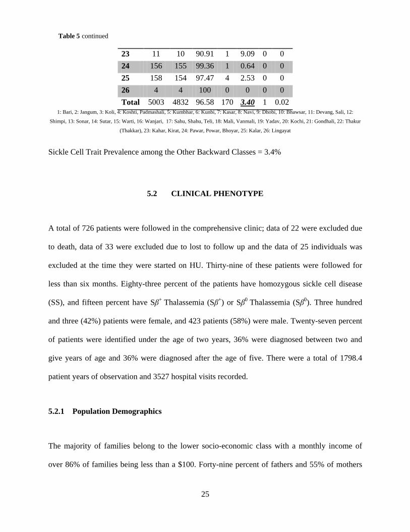
5.2 CLINICAL PHENOTYPE
A total of 726 patients were followed in the comprehensive clinic; data of 22 were excluded due
to death, data of 33 were excluded due to lost to follow up and the data of 25 individuals was
excluded at the time they were started on HU. Thirty-nine of these patients were followed for
less than six months. Eighty-three percent of the patients have homozygous sickle cell disease
(SS), and fifteen percent have Sβ+ Thalassemia (Sβ+) or Sβ0 Thalassemia (Sβ0). Three hundred
and three (42%) patients were female, and 423 patients (58%) were male. Twenty-seven percent
of patients were identified under the age of two years, 36% were diagnosed between two and
give years of age and 36% were diagnosed after the age of five. There were a total of 1798.4
patient years of observation and 3527 hospital visits recorded.
5.2.1 Population Demographics
The majority of families belong to the lower socio-economic class with a monthly income of
over 86% of families being less than a $100. Forty-nine percent of fathers and 55% of mothers
Table 5 continued
26
have an education level that is less than or equivalent to a middle school level. Table 6 illustrates
the caste distribution within the patient cohort. In this cohort, the majority of patients are part of
the SC or "non-tribal” population.
Table 6. Ethnic Group Distribution in the Study Population
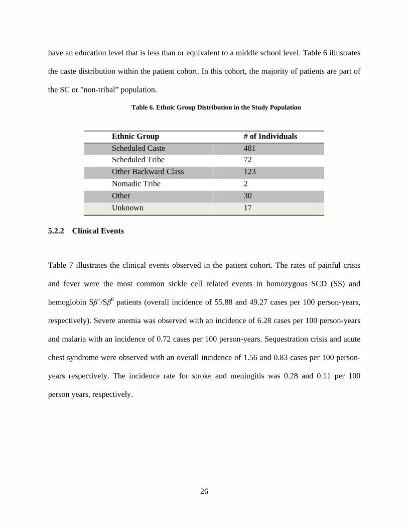
5.2.2 Clinical Events
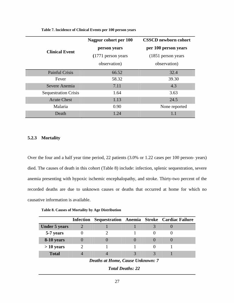
Table 7 illustrates the clinical events observed in the patient cohort. The rates of painful crisis
and fever were the most common sickle cell related events in homozygous SCD (SS) and
hemoglobin Sβ+/Sβ0 patients (overall incidence of 55.88 and 49.27 cases per 100 person-years,
respectively). Severe anemia was observed with an incidence of 6.28 cases per 100 person-years
and malaria with an incidence of 0.72 cases per 100 person-years. Sequestration crisis and acute
chest syndrome were observed with an overall incidence of 1.56 and 0.83 cases per 100 person-
years respectively. The incidence rate for stroke and meningitis was 0.28 and 0.11 per 100
person years, respectively.
Ethnic Group # of Individuals Scheduled Caste 481 Scheduled Tribe 72 Other Backward Class 123 Nomadic Tribe 2 Other
30
Unknown 17
27
Table 7. Incidence of Clinical Events per 100 person years
Clinical Event
Nagpur cohort per 100
person years
(1771 person years
observation)
CSSCD newborn cohort
per 100 person years
(1851 person years
observation)
Painful Crisis 66.52 32.4 Fever 58.32 39.30
Severe Anemia 7.11 4.3 Sequestration Crisis 1.64 3.63
Acute Chest
1.13 24.5 Malaria 0.90 None reported Death 1.24 1.1
5.2.3 Mortality
Over the four and a half year time period, 22 patients (3.0% or 1.22 cases per 100 person- years)
died. The causes of death in this cohort (Table 8) include: infection, splenic sequestration, severe
anemia presenting with hypoxic ischemic encephalopathy, and stroke. Thirty-two percent of the
recorded deaths are due to unknown causes or deaths that occurred at home for which no
causative information is available.
Table 8. Causes of Mortality by Age Distribution
Infection Sequestration Anemia Stroke Cardiac Failure Under 5 years 2 1 1 3 0
5-7 years 0 2 1 0 0 8-10 years 0 0 0 0 0 > 10 years 2 1 1 0 1
Total 4 4 3 3 1 Deaths at Home, Cause Unknown: 7
Total Deaths: 22
28
6.0 DISCUSSION
6.1 POPULATION SCREENING
Our study for the first time describes the results of community based screening for the
prevalence of the βs gene in different SC groups within Central India. The practice of endogamy
in India provides the rationale for the screening of individual populations to better understand the
distribution of the βs gene and guide counseling and awareness programs.
Large scale screening as described in this study has been the foundation for regional
disease control programs. [5] While mass screening has provided detailed descriptions for the ST
population in previous studies, such detailed descriptions are as yet unavailable for the SC and
OBC populations. [1, 3, 4, 6-8, 10, 69-71]
Published studies to date have not distinguished between community-based screening of
the general population and secondary screening of family members as a result of detection of a
sickle cell mutation in an index case. Secondary screening is likely to reveal a higher prevalence
of the βs gene than in the general population. The multiplicity of ethnic groups and overlapping
terminology restricts the utility of prevalence studies. This study addresses these limitations by
using standardized terminology as defined by the Census of India [11] and government
regulations as well as by grouping ethnic groups that are closely related, have social interactions,
and practice inter-marriage to determine the prevalence rates of the βs gene.
29
Description of the prevalence of SCT in individual caste groups has immediate practical
implications. As seen in the study, using community based screening data to target high-risk
individuals has identified a large number of individuals with both sickle cell trait and disease. In
lower resource countries, population screening and secondary target screening can identify
individuals who may not have otherwise sought care. Identifying these individuals can lead to
referrals to comprehensive care clinic to receive the appropriate medicine and follow-up .
The implications for these mass screenings extend beyond identifying carriers of the βs
gene mutation. Emerging data on the potential contribution of sickle cell trait to catastrophic
clinical events following extreme physical exertion, such as in military training or professional
sporting events, suggest the need for individual awareness of sickle cell trait status. Premarital
counseling can be targeted and tailored to the specific groups in which there is a high βs mutation
prevalence. Population-wide screenings can help target specific groups and populations in need
of education to raise awareness levels about screening, health implications, treatment,
management and pre-marital counseling.
Population screening studies help better define the magnitude of a public health problem.
The Mahars, the largest most widespread Scheduled Caste group in India, live in large numbers
in an area extending from the Arabian Sea coast in the west to the Bastar plateau in the east, and
are a varied group considered to have originated by admixture between the autochthons of the
eastern region of Central India and the Proto-Australoid element from the south and constitute
around 9% of the total population of Maharashtra. [71, 72] The SC and ST groups with a
combined population of 18 million and a trait prevalence of 13% and 12% respectively account
for 2.5 million carriers (1.4 million SC & 1.1 million ST) of sickle cell trait in the state of
Maharashtra alone. [11] Additionally, the Other Backward Classes comprising 52% of the state’s
30
population account for 1.9 million carries of sickle cell trait in the state. These groups also spill
over to neighboring states of Gujarat, Madhya Pradesh and Chattisgarh representing an even
larger burden of βs gene. In comparison, the African American population accounts for 40
million individuals in the United States and with a SCT prevalence of 8%, represent over three
million carriers of the βs gene. [73, 74]
6.2 CLINICAL PHENOTYPE
These findings represent the first report of a large prospectively phenotyped cohort of patients
with SCD in India. The clinical picture in the ethnic groups that are present in large numbers in
these areas are described and as such must be interpreted as being descriptive of phenotype in
these groups only. While various ethnic groups are represented in the patient population, the SC
population accounts for a large majority of patients. Vaso-occlusive crisis (VOC) and fever
followed by severe anemia are the most common initial presentations and were the leading
causes of hospitalizations. The rates of pain crisis and fever in this cohort are higher when
compared to the data published from the Cooperative Study of Sickle Cell Disease (CSSCD).
[13]
Severe anemia was also more commonly observed than what was reported in CSSCD.
[13] The cause of a high rate of severe anemia in patients with SCD in this cohort is unknown.
[13] Malaria is a significant co-morbidity in this Indian population. It is possible that
malnutrition, infections and malaria may have contributed to the presentation of severe anemia.
Further investigation, however, is indicated to better understand this common co-morbidity. It is
intriguing that the rate of acute chest syndrome is strikingly lower in this population. This is
31
particularly surprising in light of the high incidence of pneumonia in the country, with India
alone being responsible for over a quarter of the deaths in the world from pneumonia in children
under the age of five years. [75, 76] The lower frequency of splenic sequestration is also
intriguing. It is possible that this may be reflective of lack of access to care and that splenic
sequestration may represent some of the deaths at home with no known cause.
The mortality rate observed in this Indian population (1.24 cases per 100 person-years) is
comparable to the observed rate seen in the CSSCD (1.1 cases per 100 person-years). [13] The
leading causes of death in this cohort include: infection, splenic sequestration, severe anemia
presenting with hypoxic ischemic encephalopathy, stroke and cardiac failure. That nearly one-
third of the recorded deaths occurred at home, due to unknown causes may also be reflective of
barriers to care for this population.
The high rate of VOC, fever, septic arthritis and unique co-morbidities such as severe
anemia and malaria and the high rates of mortality, all suggest that the phenotype of SCD as
observed in Nagpur is not milder than that observed in CSSCD. This is in contrast with previous
published reports suggesting a milder phenotype of SCD in India. [12] However; these reports
were based on cross-sectional studies of a relatively small number of patients. The current
analysis underscores the need for careful study of the clinical course of large cohorts of patients
prospectively to accurately describe phenotype. That the majority of patients are from low
socioeconomic populations with barriers to care is supportive of this possibility.
Environmental and nutritional factors may also contribute to high rate of febrile episodes
especially in a low resource country. These along with endemicity of malaria may further
contribute to high morbidity and possibly premature mortality in patients with SCD in this
region. These findings are also in keeping with our prior retrospective observations. [77] They
32
are also concordant with cross-sectional observations suggesting high rates of VOC events,
infections, organ damage and premature mortality from eastern India. [69] These data provide a
compelling rationale for large prospective studies in different ethnic groups in India to determine
the contribution of environmental, biological and health systems influences to variation in
clinical phenotype.
Median age of diagnosis was four years. This has the potential effect of excluding both
those who succumbed to pneumococcal sepsis in early childhood as well as those with mild
manifestations who may never present for clinical care at a tertiary care hospital. Further, we
excluded patients who were already on HU at the commencement of the study and censored data
at initiation of HU on those enrolled in the study in order to describe phenotype modified by HU.
Thus, patients with severe phenotype who were started on HU therapy are likely to be under
represented in this analysis. These data do, however, indicate that the clinical manifestations of
sickle cell disease in India are no less severe than that observed among those who seek care for
this disease in the United States.
33
7.0 CONCLUSION
We provide for the first time detailed prevalence data of the βs gene among SC and OBC ethnic
groups in Maharashtra in Central India. These population screening programs have uncovered
previously undiagnosed cases, provided detailed information for population based disease
counseling, prevention programs and comprehensive care programs. Over 5,000 individuals were
identified with SCT and more than 1,000 individuals were identified with SCD through
community-based and target high-risk screening. The population screening study shows how
community-based screening can identify individuals who may not have otherwise sought care
and guide individuals to available comprehensive care centers. The screening also helped
identify specific sub-groups within the SC, ST, and OBC population that have higher prevalence
rates of the βs gene. With a SCT prevalence of 13%, 12% and 3.4% in the SC, ST, and OBC
populations respectively, an estimated 4.4 million individuals in the state of Maharashtra alone
are expected to have SCT. Similar community-based screening programs need to be initiated
throughout regions of India that have a high prevalence of SC, ST, and OBC populations to
determine the true prevalence of the βs gene in India.
Additionally, we present the first evidence suggesting that SCD may not have milder
manifestations in India and underscore the need for detailed studies of phenotype that can form
the basis of public health interventions as well as mechanistic studies of genetic modifiers of
clinical phenotype. Our data shows that the rates of painful crises and fever the two most
34
common clinical manifestations were observed at higher rates than individuals in the African
American population in the United States. Other unique co-morbidities such as severe anemia
and malaria were identified that can help direct future research studies to determine exactly how
this co-morbidities affect the course of disease. A limitation of this study is that patients were
diagnosed when they presented with clinical manifestations of SCD rather on newborn
screening. Thus this cohort is not strictly comparable with the CSSCD newborn cohort.
The observation of a severe of phenotype of SCD in India however, has many
implications. It makes the case for interventions such as newborn screening and comprehensive
care and disease modifying therapy such as HU. The magnitudes of both the at-risk population
and the burden of morbidity also suggest the need for public policy to address this major public
health problem. This study, by clearly delineating phenotype can serve as the basis for studies of
genetic modifiers of phenotype in this population.
The burden of SCD in India has not been well documented or studied thus far. These two
studies are of high public health significance and important for the planning of health care
delivery services. The study has implications for the need for comprehensive clinical care and
further population based prevalence studies in other regions of India to truly understand the
impact and burden of SCD in underserved populations.
This thesis reflects the work performed and the author wishes to add a comment.
Comment: Conducting the research for these projects was one of the most unique and greatest learning experiences that I have ever had. I had an opportunity that most students never have the chance to experience and had the honor of meeting and working with four different research groups throughout India on my journey to learn about how Sickle Cell Disease affects individuals in India. I had the chance to attend screening camps and meet individuals from the various villages and every single individual I met was willing to help me learn and teach me all that they knew. Growing up I have made numerous trips to India to visit family members but this project was an eye opening experience and I learned more in my two trips to India for my thesis compared to all the previous trips that I have made before. The sickle cell patients and families

35
were so friendly, open and willing to talk to me so that I can learn about their experiences. While I hope this work truly provides a framework for future research pertaining to SCD in India it has helped me not only learn about the topic but also about myself and was a phenomenal experience.
36
BIBLIOGRAPHY
1. Chandrashekar, V. and M. Soni, Hemoglobin disorders in South India. ISRN Hematol,
2011. 2011: p. 748939.
2. Italia, K., et al., Hydroxyurea in sickle cell disease--a study of clinico-pharmacological
efficacy in the Indian haplotype. Blood Cells Mol Dis, 2009. 42(1): p. 25-31.
3. Kamble, M. and P. Chatruvedi, Epidemiology of sickle cell disease in a rural hospital of
central India. Indian Pediatr, 2000. 37(4): p. 391-6.
4. Kaur, M., G.P. Das, and I.C. Verma, Sickle cell trait & disease among tribal communities
in Orissa, Madhya Pradesh & Kerala. Indian J Med Res, 1997. 105: p. 111-6.
5. Patra, P.K., et al., Screening for the sickle cell gene in Chhattisgarh state, India: an
approach to a major public health problem. J Community Genet, 2011. 2(3): p. 147-51.
6. Balgir, R., Epidemiology, Population Health Genetics and Phenotypic Diversity of Sickle
Cell Disease in India. The Internet Journal of Biological Anthropology, 2007. 1(2).
7. Shukla, R.N. and B.R. Solanki, Sickle Cell Trait in Central India. Lancet, 1985. 1: p.
297-298.
8. Kate, S.L. and D.P. Lingojwar, Epidemiology of Sickle Cell Disorder in the State of
Maharashtra. . International Journal of Human Genetics, 2002. 2(3): p. 161-167.
9. Weatherall, D.J., The inherited diseases of hemoglobin are an emerging global health
burden. Blood, 2010. 115(22): p. 4331-6.
10. Balgir, R.S., Spectrum of hemoglobinopathies in the state of Orissa, India: a ten years
cohort study. J Assoc Physicians India. , 2005. 53: p. 1021-1026.
11. Government-of-India, State Primary Census Abstract for Individual Scheduled Caste and
State Primary Census Abstract for Individual Scheduled Tribe: Census of India M.o.H.
Affairs, Editor 2001: Office of the Registrar General and Census Commissioner.
37
12. Mohanty, D. and M.B. Mukherjee, Sickle cell disease in India. Curr Opin Hematol, 2002.
9(2): p. 117-22.
13. Gill, F.M., et al., Clinical events in the first decade in a cohort of infants with sickle cell
disease. Cooperative Study of Sickle Cell Disease. Blood, 1995. 86(2): p. 776-83.
14. Mashon, R.S., et al., Higher fetal hemoglobin concentration in patients with sickle cell
disease in eastern India reduces frequency of painful crisis. Eur J Haematol, 2009. 83(4):
p. 383-4.
15. Mukherjee, M.B., et al., Effect of alpha-thalassemia on sickle-cell anemia linked to the
Arab-Indian haplotype in India. Am J Hematol, 1997. 55(2): p. 104-9.
16. Mukherjee, M.B., et al., Beta-globin gene cluster haplotypes linked to the betaS gene in
western India. Hemoglobin, 2004. 28(2): p. 157-61.
17. Mukherjee, M.B., et al., Clinical diversity of sickle cell disease in western india -
influence of genetic factors. Acta Haematol, 2000. 103(2): p. 122-3.
18. Patel, D.K., et al., Clinical and molecular characterization of beta(S) and
(G)gamma((A)gammadeltabeta)(0)-thalassemia in eastern India. Hemoglobin, 2010.
34(6): p. 604-9.
19. Ramana, G.V., G.R. Chandak, and L. Singh, Sickle cell gene haplotypes in Relli and
Thurpu Kapu populations of Andhra Pradesh. Hum Biol, 2000. 72(3): p. 535-40.
20. Singh, H., et al., Effective control of sickle cell disease with hydroxyurea therapy. Indian
J Pharmacol, 2010. 42(1): p. 32-5.
21. Herrick, J.B., Peculiar elongated and sickle-shaped red blood corpuscles in a case of
severe anemia. 1910. Yale J Biol Med, 2001. 74(3): p. 179-84.
22. D.V., D. and D. H., Sickle Cell Disease: History and Origin. The Internet Journal of
Hematology, 2004. 1(2).
23. Barabino, G.A., M.O. Platt, and D.K. Kaul, Sickle cell biomechanics. Annu Rev Biomed
Eng, 2010. 12: p. 345-67.
24. Embury, S.H., et al., Sickle Cell Disease: Basic Principles and Clinical Practice. 1994.
25. Schechter, A.N., Hemoglobin research and the origins of molecular medicine. Blood,
2008. 112(10): p. 3927-38.
26. Serjeant, G.R., Sickle Cell Disease. Vol. 2 edition. 1992: Oxford University Press.
38
27. Smith, M.E., Web-based monitoring of pain management in adolescent and young adult
sickle cell patients through daily self-assessment, in Human Genetics2012, University of
Pittsburgh.
28. Costanzo, V.L., Sickle Cell Trait Counseling for Student Athletes, in Human
Genetics2011, University of Pittsburgh.
29. Sickle cell anemia, P.H.S. U.S. Department of Health and Services, National Institutes of
Health Editor 1990: National Institutes of Health (U.S.).
30. Eaton, W.A. and J. Hofrichter, Sickle cell hemoglobin polymerization. Adv Protein
Chem, 1990. 40: p. 63-279.
31. Ferrone, F. and R.L. Nagel, Sickle hemoglobin polymerization, in Disorders of
hemoglobin: Genetics, pathophysiology, clinical management., F.B. Steinberg MH,
Higgs D, Nagel RL. , Editor. 2000, Cambridge University Press.
32. Bender, M. and W. Hobbs, Sickle Cell Disease- Gene Reviews: NCBI Bookshelf.
33. Serjeant, G.R. and B.E. Serjeant, Sickle Cell Disease. 2001, Oxford; New York: Oxford
University Press.
34. Long, K.A., et al., Attitudes and beliefs of African-Americans toward genetics, genetic
testing, and sickle cell disease education and awareness. J Genet Couns, 2011. 20(6): p.
572-92.
35. Wilson, R.E., L. Krishnamurti, and D. Kamat, Management of sickle cell disease in
primary care. Clin Pediatr (Phila), 2003. 42(9): p. 753-61.
36. Rees, D.C., T.N. Williams, and M.T. Gladwin, Sickle-cell disease. Lancet, 2010.
376(9757): p. 2018-31.
37. Ballas, S.K., Pain management of sickle cell disease. Hematol Oncol Clin North Am,
2005. 19(5): p. 785-802, v.
38. Amjad, H., R.M. Bannerman, and J.M. Judisch, Letter: Sickling pain and season. Br Med
J, 1974. 2(5909): p. 54.
39. Ibrahim, A.S., Relationship between meteorological changes and occurrence of painful
sickle cell crises in Kuwait. Trans R Soc Trop Med Hyg, 1980. 74(2): p. 159-61.
40. Jones, S., et al., Windy weather and low humidity are associated with an increased
number of hospital admissions for acute pain and sickle cell disease in an urban
environment with a maritime temperate climate. Br J Haematol, 2005. 131(4): p. 530-3.
39
41. Niscola, P., et al., Pain syndromes in sickle cell disease: an update. Pain Med, 2009.
10(3): p. 470-80.
42. Nolan, V.G., et al., Association between wind speed and the occurrence of sickle cell
acute painful episodes: results of a case-crossover study. Br J Haematol, 2008. 143(3): p.
433-8.
43. Redwood, A.M., et al., Climate and painful crisis of sickle-cell disease in Jamaica.
British Medical Journal, 1976. 1: p. 66-68.
44. Resar, L.M. and F.A. Oski, Cold water exposure and vaso-occlusive crises in sickle cell
anemia. . Journal of Pediatrics, 1991. 118: p. 407-409.
45. Westerman, M.P., et al., Assessment of painful episode frequency in sickle-cell disease.
Am J Hematol, 1997. 54(3): p. 183-8.
46. Yallop, D., et al., The associations between air quality and the number of hospital
admissions for acute pain and sickle-cell disease in an urban environment. Br J
Haematol, 2007. 136(6): p. 844-8.
47. Yoong, W.C. and S.M. Tuck, Menstrual pattern in women with sickle cell anaemia and
its association with sickling crises. J Obstet Gynaecol, 2002. 22(4): p. 399-401.
48. Al-Salem, A.H., Splenic complications of sickle cell anemia and the role of splenectomy.
ISRN Hematol, 2011. 2011: p. 864257.
49. Roseff, S.D., Sickle cell disease: a review. Immunohematology, 2009. 25(2): p. 67-74.
50. D., P., W. J.A., and e.a. Odom-Maryon T., Sickle cell chronic lung disease: prior
morbidity and the risk of pulmonary failure., in Medicine. 1988: Baltimore.
51. Gray, A., et al., Patterns of mortality in sickle cell disease in the United Kingdom. J Clin
Pathol, 1991. 44(6): p. 459-63.
52. Platt, O.S., et al., Mortality in sickle cell disease. Life expectancy and risk factors for
early death. N Engl J Med, 1994. 330(23): p. 1639-44.
53. Booth, C., B. Inusa, and S.K. Obaro, Infection in sickle cell disease: a review. Int J Infect
Dis, 2010. 14(1): p. e2-e12.
54. Taber’s Cyclopedic Medical Dictionary, F.A.D. Company, Editor 2009.
55. Pham, P.T., et al., Renal abnormalities in sickle cell disease. Kidney Int, 2000. 57(1): p.
1-8.
40
56. Fox, P.D., et al., Risk factors for proliferative sickle retinopathy. Br J Ophthalmol, 1990.
74(3): p. 172-6.
57. Hayes, R.J., P.I. Condon, and G.R. Serjeant, Haematological factors associated with
proliferative retinopathy in sickle cell-haemoglobin C disease. Br J Ophthalmol, 1981.
65(10): p. 712-7.
58. Nagpal, K.C., M.F. Goldberg, and M.F. Rabb, Ocular manifestations of sickle
hemoglobinopathies. Surv Ophthalmol, 1977. 21(5): p. 391-411.
59. Foucan, L., et al., Early onset dactylitis associated with the occurrence of severe events
in children with sickle cell anaemia. The Paediatric Cohort of Guadeloupe (1984-99).
Paediatr Perinat Epidemiol, 2006. 20(1): p. 59-66.
60. Platt, O.S., The acute chest syndrome of sickle cell disease. N Engl J Med, 2000. 342(25):
p. 1904-7.
61. Board, M.S.A.M. Agriculture Produce Market Committee 2008; Available from:
http://www.msamb.com/english/apmc/profile.htm.
62. Eastman, J.W., et al., Automated HPLC screening of newborns for sickle cell anemia and
other hemoglobinopathies. Clin Chem, 1996. 42(5): p. 704-10.
63. Constitution of India: Article 341, 1950. http://lawmin.nic.in/ld/subord/rule3a.htm
64. Consitution of India Article 342, 1950. http://lawmin.nic.in/ld/subord/rule9a.htm
65. The Scheduled Castes and Scheduled Tribes Orders Amendment Act. Constitution of
India, 1976.
66. National Commission for Backward Classes Act of 1993 J.A.C.A. MINISTRY OF LAW,
Editor 1993.
67. Charache, S., et al., Design of the multicenter study of hydroxyurea in sickle cell anemia.
Investigators of the Multicenter Study of Hydroxyurea. Control Clin Trials, 1995. 16(6):
p. 432-46.
68. Indian Academy of Pediatrics Committee on, I., Consensus recommendations on
immunization and IAP immunization timetable 2012. Indian Pediatr, 2012. 49(7): p. 549-
64.
69. Kar, B.C., Sickle cell disease in India. J Assoc Physicians India, 1991. 39(12): p. 954-60.
70. Lehmann, H. and M. Cutbush, Sickle-cell trait in southern India. Br Med J, 1952.
1(4755): p. 404-5.
41
71. Negi, R.S., The Incidence of Sickle-Cell Trait in Two Bastar Tribes, I., in Man. 1962,
Royal Anthropological Institute of Great Britain and Ireland. p. 84-86.
72. Mahar. Encyclopædia Britannica. Encyclopædia Britannica Online.
Encyclopædia Britannica Inc., 2013. Web. 03 Apr. 2013
<http://www.britannica.com/EBchecked/topic/357931/Mahar>.
73. United States Census, U.S.C. Bureau, Editor 2011.
74. Sickle Cell Disease (SCD), 2011: Centers for Disease Control and Prevention.
75. Liu, L., et al., Global, regional, and national causes of child mortality: an updated
systematic analysis for 2010 with time trends since 2000. Lancet, 2012. 379(9832): p.
2151-61.
76. Million Death Study, C., et al., Causes of neonatal and child mortality in India: a
nationally representative mortality survey. Lancet, 2010. 376(9755): p. 1853-60.
77. Jain, D., et al., Sickle cell anemia from central India: a retrospective analysis. Indian
Pediatr, 2012. 49(11): p. 911-3.