Expression of spoIIIJ in the Prespore Is Sufficient for Activation of G
13
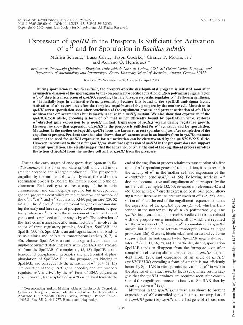
JOURNAL OF BACTERIOLOGY, July 2003, p. 3905–3917 Vol. 185, No. 13 0021-9193/03/$08.000 DOI: 10.1128/JB.185.13.3905–3917.2003 Copyright © 2003, American Society for Microbiology. All Rights Reserved. Expression of spoIIIJ in the Prespore Is Sufficient for Activation of G and for Sporulation in Bacillus subtilis Mo ´nica Serrano, 1 Luísa Co ˆrte, 1 Jason Opdyke, 2 Charles P. Moran, Jr., 2 and Adriano O. Henriques 1 * Instituto de Tecnologia Química e Biolo ´gica, Universidade Nova de Lisboa, 2781-901 Oeiras Codex, Portugal, 1 and Department of Microbiology and Immunology, Emory University School of Medicine, Atlanta, Georgia 30322 2 Received 25 November 2002/Accepted 9 April 2003 During sporulation in Bacillus subtilis, the prespore-specific developmental program is initiated soon after asymmetric division of the sporangium by the compartment-specific activation of RNA polymerase sigma factor F . F directs transcription of spoIIIG, encoding the late forespore-specific regulator G . Following synthesis, G is initially kept in an inactive form, presumably because it is bound to the SpoIIAB anti-sigma factor. Activation of G occurs only after the complete engulfment of the prespore by the mother cell. Mutations in spoIIIJ arrest sporulation soon after conclusion of the engulfment process and prevent activation of G . Here we show that G accumulates but is mostly inactive in a spoIIIJ mutant. We also show that expression of the spoIIIGE155K allele, encoding a form of G that is not efficiently bound by SpoIIAB in vitro, restores G -directed gene expression to a spoIIIJ mutant. Expression of spoIIIJ occurs during vegetative growth. However, we show that expression of spoIIIJ in the prespore is sufficient for G activation and for sporulation. Mutations in the mother cell-specific spoIIIA locus are known to arrest sporulation just after completion of the engulfment process. Previous work has also shown that G accumulates in an inactive form in spoIIIA mutants and that the need for spoIIIA expression for G activation can be circumvented by the spoIIIGE155K allele. However, in contrast to the case for spoIIIJ, we show that expression of spoIIIA in the prespore does not support efficient sporulation. The results suggest that the activation of G at the end of the engulfment process involves the action of spoIIIA from the mother cell and of spoIIIJ from the prespore. During the early stages of endospore development in Ba- cillus subtilis, the rod-shaped bacterial cell is divided into a smaller prespore and a larger mother cell. The prespore is engulfed by the mother cell, which lyses at the end of the sporulation process to liberate the mature spore into the en- vironment. Each cell type receives a copy of the bacterial chromosome, and each deploys specific but interdependent genetic programs controlled by the successive appearance of the F , E , G , and K subunits of RNA polymerase (29, 32, 42, 46). The F and G regulators control gene expression dur- ing the early and late stages of prespore development, respec- tively, whereas E controls the expression of early mother cell genes and is replaced at later stages by K . The activation of the first compartment-specific sigma factor, F , involves the action of three regulatory proteins, SpoIIAA, SpoIIAB, and SpoIIE (33, 48). SpoIIAB is an anti-sigma factor that binds to F as a dimer and inhibits its transcriptional activity (6, 7, 14, 36), whereas SpoIIAA is an anti-anti-sigma factor that in an unphosphorylated state interacts with SpoIIAB and releases F from the SpoIIAB- F complex (1, 12, 13). SpoIIE, a sep- tum-bound phosphatase, promotes the preferential dephos- phorylation of SpoIIAA-P in the prespore, its binding to SpoIIAB, and consequently the activation of F (3, 4, 12, 15). Transcription of the spoIIIG gene, encoding the late prespore regulator G , is driven by the F form of RNA polymerase (55). However, transcription of spoIIIG is delayed towards the end of the engulfment process relative to transcription of a first class of F -dependent genes (41). In addition, it requires both the activity of E in the mother cell and expression of the F -controlled gene spoIIQ (41, 56). Following synthesis, G does not become active until engulfment of the prespore by the mother cell is complete (32, 53; reviewed in references 42 and 46). Once active, G directs expression of its own gene, allow- ing a rapid increase in the cellular levels of G (25, 55). Acti- vation of G at the end of the engulfment sequence demands the expression of the spoIIIA operon (26, 43), which is tran- scribed in the mother cell by E RNA polymerase (23). The spoIIIA locus encodes eight proteins predicted to be associated with the prespore outer membrane, all of which are required for the activation of G (23, 53). G accumulates in a spoIIIA mutant but is unable to activate transcription from its target promoters (26). Genetic, biochemical, and structural evidence suggests that the anti-sigma factor SpoIIAB negatively regu- lates G (7, 8, 17, 26, 28, 44). In particular, during sporulation SpoIIAB tends to disappear from the forespore soon after completion of the engulfment sequence in a spoIIIA-depen- dent mode (28), and expression of an allele of spoIIIG (spoIIIGE155K) encoding a form of G that is not efficiently bound by SpoIIAB in vitro permits activation of G in vivo in the absence of an intact spoIIIA locus (26). These results sug- gest that the spoIIIA products are required soon after conclu- sion of the engulfment process to inactivate SpoIIAB, thereby releasing active G (26). Mutations in the spoIIIJ locus were also shown to prevent expression of G -controlled genes but not transcription of the spoIIIG gene (16). spoIIIJ is the first gene of a bicistronic * Corresponding author. Mailing address: Instituto de Tecnologia Química e Biolo ´gica, Universidade Nova de Lisboa, Av. da Repu ´blica, Apartado 127, 2781-901 Oeiras Codex, Portugal. Phone: 351-21- 4469521. Fax: 351-21-4411277. E-mail: [email protected]. 3905 Downloaded from https://journals.asm.org/journal/jb on 15 February 2022 by 77.45.84.7.
Transcript of Expression of spoIIIJ in the Prespore Is Sufficient for Activation of G
JOURNAL OF BACTERIOLOGY, July 2003, p. 3905–3917 Vol. 185, No. 130021-9193/03/$08.00�0 DOI: 10.1128/JB.185.13.3905–3917.2003Copyright © 2003, American Society for Microbiology. All Rights Reserved.
Expression of spoIIIJ in the Prespore Is Sufficient for Activationof �G and for Sporulation in Bacillus subtilis
Monica Serrano,1 Luísa Corte,1 Jason Opdyke,2 Charles P. Moran, Jr.,2and Adriano O. Henriques1*
Instituto de Tecnologia Química e Biologica, Universidade Nova de Lisboa, 2781-901 Oeiras Codex, Portugal,1 andDepartment of Microbiology and Immunology, Emory University School of Medicine, Atlanta, Georgia 303222
Received 25 November 2002/Accepted 9 April 2003
During sporulation in Bacillus subtilis, the prespore-specific developmental program is initiated soon afterasymmetric division of the sporangium by the compartment-specific activation of RNA polymerase sigma factor�F. �F directs transcription of spoIIIG, encoding the late forespore-specific regulator �G. Following synthesis,�G is initially kept in an inactive form, presumably because it is bound to the SpoIIAB anti-sigma factor.Activation of �G occurs only after the complete engulfment of the prespore by the mother cell. Mutations inspoIIIJ arrest sporulation soon after conclusion of the engulfment process and prevent activation of �G. Herewe show that �G accumulates but is mostly inactive in a spoIIIJ mutant. We also show that expression of thespoIIIGE155K allele, encoding a form of �G that is not efficiently bound by SpoIIAB in vitro, restores�G-directed gene expression to a spoIIIJ mutant. Expression of spoIIIJ occurs during vegetative growth.However, we show that expression of spoIIIJ in the prespore is sufficient for �G activation and for sporulation.Mutations in the mother cell-specific spoIIIA locus are known to arrest sporulation just after completion of theengulfment process. Previous work has also shown that �G accumulates in an inactive form in spoIIIA mutantsand that the need for spoIIIA expression for �G activation can be circumvented by the spoIIIGE155K allele.However, in contrast to the case for spoIIIJ, we show that expression of spoIIIA in the prespore does not supportefficient sporulation. The results suggest that the activation of �G at the end of the engulfment process involvesthe action of spoIIIA from the mother cell and of spoIIIJ from the prespore.
During the early stages of endospore development in Ba-cillus subtilis, the rod-shaped bacterial cell is divided into asmaller prespore and a larger mother cell. The prespore isengulfed by the mother cell, which lyses at the end of thesporulation process to liberate the mature spore into the en-vironment. Each cell type receives a copy of the bacterialchromosome, and each deploys specific but interdependentgenetic programs controlled by the successive appearance ofthe �F, �E, �G, and �K subunits of RNA polymerase (29, 32,42, 46). The �F and �G regulators control gene expression dur-ing the early and late stages of prespore development, respec-tively, whereas �E controls the expression of early mother cellgenes and is replaced at later stages by �K. The activation ofthe first compartment-specific sigma factor, �F, involves theaction of three regulatory proteins, SpoIIAA, SpoIIAB, andSpoIIE (33, 48). SpoIIAB is an anti-sigma factor that binds to�F as a dimer and inhibits its transcriptional activity (6, 7, 14,36), whereas SpoIIAA is an anti-anti-sigma factor that in anunphosphorylated state interacts with SpoIIAB and releases�F from the SpoIIAB-�F complex (1, 12, 13). SpoIIE, a sep-tum-bound phosphatase, promotes the preferential dephos-phorylation of SpoIIAA-P in the prespore, its binding toSpoIIAB, and consequently the activation of �F (3, 4, 12, 15).Transcription of the spoIIIG gene, encoding the late presporeregulator �G, is driven by the �F form of RNA polymerase(55). However, transcription of spoIIIG is delayed towards the
end of the engulfment process relative to transcription of a firstclass of �F-dependent genes (41). In addition, it requires boththe activity of �E in the mother cell and expression of the�F-controlled gene spoIIQ (41, 56). Following synthesis, �G
does not become active until engulfment of the prespore by themother cell is complete (32, 53; reviewed in references 42 and46). Once active, �G directs expression of its own gene, allow-ing a rapid increase in the cellular levels of �G (25, 55). Acti-vation of �G at the end of the engulfment sequence demandsthe expression of the spoIIIA operon (26, 43), which is tran-scribed in the mother cell by �E RNA polymerase (23). ThespoIIIA locus encodes eight proteins predicted to be associatedwith the prespore outer membrane, all of which are requiredfor the activation of �G (23, 53). �G accumulates in a spoIIIAmutant but is unable to activate transcription from its targetpromoters (26). Genetic, biochemical, and structural evidencesuggests that the anti-sigma factor SpoIIAB negatively regu-lates �G (7, 8, 17, 26, 28, 44). In particular, during sporulationSpoIIAB tends to disappear from the forespore soon aftercompletion of the engulfment sequence in a spoIIIA-depen-dent mode (28), and expression of an allele of spoIIIG(spoIIIGE155K) encoding a form of �G that is not efficientlybound by SpoIIAB in vitro permits activation of �G in vivo inthe absence of an intact spoIIIA locus (26). These results sug-gest that the spoIIIA products are required soon after conclu-sion of the engulfment process to inactivate SpoIIAB, therebyreleasing active �G (26).
Mutations in the spoIIIJ locus were also shown to preventexpression of �G-controlled genes but not transcription ofthe spoIIIG gene (16). spoIIIJ is the first gene of a bicistronic
* Corresponding author. Mailing address: Instituto de TecnologiaQuímica e Biologica, Universidade Nova de Lisboa, Av. da Republica,Apartado 127, 2781-901 Oeiras Codex, Portugal. Phone: 351-21-4469521. Fax: 351-21-4411277. E-mail: [email protected].
3905
Dow
nloa
ded
from
http
s://j
ourn
als.
asm
.org
/jour
nal/j
b on
15
Febr
uary
202
2 by
77.
45.8
4.7.
operon transcribed during vegetative growth (16, 39). It en-codes a putative lipoprotein that localizes to the cell mem-brane and septal regions during vegetative growth and sporu-lation but also to the membranes that surround the developingspore (39). SpoIIIJ is related to the YidC Sec-independent andSec-dependent membrane protein translocase of Escherichiacoli (about 33% identity and 51% conserved residues for aregion of overlap between the two proteins of 226 amino acids)(47, 49, 57). SpoIIIJ is also related to the CcfA protein ofEnterococcus faecalis, with about 65% similarity and 39% iden-tity between the two proteins (2). Like spoIIIJ, ccfA codes fora putative lipoprotein precursor, but it also encodes the cCF10peptide pheromone within the signal peptide (2). The cCF10peptide pheromone is required for the conjugative transfer ofthe E. faecalis plasmid pCF10. CcfA, like YidC in E. coli, seemsto be required for normal growth of E. faecalis (2).
Here we have analyzed the role of spoIIIJ in the activationof �G. We show that �G accumulates in a spoIIIJ mutantbut is mostly inactive. We also show that expression of thespoIIIGE155K allele restores transcription of the �G-depen-dent gene sspE to spoIIIJ mutant cells. Expression of spoIIIJfrom a prespore-specific promoter produces a sporulation-pro-ficient strain, whereas its expression from a mother cell-specificpromoter results in a block soon after completion of the en-gulfment process. Conversely, expression of spoIIIA in theprespore does not support efficient sporulation. These resultssuggest that SpoIIIJ from the prespore and the spoIIIA-en-coded products from the mother cell compartment are celltype-specific elements of a signaling pathway that antagonizes
the inhibitory action of SpoIIAB and promotes the activationof �G soon after conclusion of the engulfment process.
MATERIALS AND METHODS
Bacterial strains and general methods. The B. subtilis strains used in this work,most of which are congenic derivatives of the wild-type strain MB24 (trpC2metC3), are listed in Table 1. Luria-Bertani medium was used for routine growthof E. coli DH5� (BRL) and of B. subtilis strains. Sporulation was induced byexhaustion in Difco sporulation medium (DSM), and the extent of sporulation(expressed as the percentage of heat resistant CFU relative to the total cellcount) was assessed as described previously (20, 21). Antibiotics were used aspreviously described (20, 21).
Construction of a spoIIIJ nonpolar null mutant. To create a nonpolar inser-tion into the spoIIIJ gene, we used PCR with Taq DNA polymerase and primersRNPA-75 and JAGD-235R (Table 2) to produce a DNA fragment extendingfrom nucleotide 421 upstream of the spoIIIJ start codon to nucleotide 240downstream of the jag stop codon. This DNA fragment was cloned into thepCR2.1 TOPO vector (Invitrogen) to create plasmid pJO34. An EcoRI fragmentcontaining the spoIIIJ operon was released from pJO34 and inserted into theEcoRI site of pLitmus38 (New England Biolabs) to create pJO57. Inverse PCRwith primers JXMA-1 and JXMA-2 (Table 2) was performed on pJO57 to createa linear fragment that deleted nucleotides �41 to �707 of the spoIIIJ gene (withrespect to the transcription start site). This PCR product was ligated with anXmaI restriction fragment containing a promoterless kanamycin resistance(Kmr) determinant resulting from the digestion of pUC18-K (35). The DNAresulting from this ligation was a recircularized plasmid in which the spoIIIJ genewas replaced with the km gene, which was now under control of the spoIIIJpromoter. This plasmid was named pJO60. Strains JOB34 and JOB44 werecreated by transformation of AH1042 (sspE-lacZ) and MB24 (wild type) (Table1), respectively, with ScaI-linearized pJO60, selecting for Kmr. Disruption of thespoIIIJ gene by a double-crossover event (marker replacement) was verified byPCR. The resulting strains, JOB34 and JOB44 (Table 1), were tested by reversetranscription-PCR analysis to confirm that the insertion was nonpolar on jag (notshown). Strains JOB34 and AH1042 were transformed with chromosomal DNA
TABLE 1. B. subtilis strains
Strain Relevant genotype Origin (reference)
MB24 trpC2 metC3 Laboratory stockAH45 trpC2 metC3 spoIIIG�1 Laboratory stockAH62 trpC2 metC3 spoIIIA::Tn917�HU24 Laboratory stock (26)EUE9537 trpC2 metC3 SP�::sspE-lacZ spoIIIG::pEK15 (wild-type spoIIIG) Laboratory stock (26)EUE9538 trpC2 metC3 SP�::sspE-lacZ spoIIIG::pEK16 (spoIIIGE155K) Laboratory stock (26)AH1042 trpC2 metC3 �sspE::sspE-lacZ Laboratory stockAH2350 trpC2 metC3(pMK3) This workAH5008 trpC2 metC3 �amyE::spoIIIG-lacZ This workAH5009 trpC2 metC3 �spoIIIJ::km �amyE::spoIIIG-lacZ This workAH5011 trpC2 metC3 �spoIIIJ::km �amyE::PspoIIIJspoIIIJ This workAH5012 trpC2 metC3 �spoIIIJ::km �amyE::PspoIIJQ-spoIIIJ This workAH5013 trpC2 metC3 �spoIIIJ::km �amyE::PspoIID-spoIIIJ This workAH5014 trpC2 metC3 �spoIIIJ::km �amyE::PspoIIIJ-spoIIIJ �sspE::sspE-lacZ This workAH5015 trpC2 metC3 �spoIIIJ::km �amyE::PspoIIQ-spoIIIJ �sspE::sspE-lacZ This workAH5016 trpC2 metC3 �spoIIIJ::km �amyE::PspoIID-spoIIIJ �sspE::sspE-lacZ This workAH5017 trpC2 metC3 �spoIIIJ::km �amyE::PspoIIIJ-spoIIIJ-gfp This workAH5018 trpC2 metC3 �spoIIIJ::km �amyE::PspoIIQ-spoIIIJ-gfp This workAH5019 trpC2 metC3 �spoIIIJ::km �amyE::PspoIID-spoIIIJ-gfp This workAH5020 trpC2 metC3 spoIIIA::PspoIIQ-spoIIIA This workAH5024 trpC2 metC3 �spoIIIJ::km �sspE::sspE-lacZ spoIIIG::pEK15 This workAH5025 trpC2 metC3 �spoIIIJ::km �sspE::sspE-lacZ spoIIIG::pEK16 This workAH5029 trpC2 metC3 spoIIIA::PspoIID-spoIIIA This workAH5030 trpC2 metC3 �sspE::sspE-lacZ spoIIIG::pEK15 This workAH5031 trpC2 metC3 �sspE::sspE-lacZ spoIIIG::pEK16 This workAH5035 trpC2 metC3 spoIIIJ::sp(pMK3) This workAH5036 trpC2 metC3 spoIIIJ::sp(pMS210) This workAH5037 trpC2 metC3(pMS210) This workJOB20 trpC2 metC3 �spoIIIJ::sp This workJOB34 trpC2 metC3 �spoIIIJ::km �sspE::sspE-lacZ This workJOB44 trpC2 metC3 �spoIIIJ::km This work
3906 SERRANO ET AL. J. BACTERIOL.
Dow
nloa
ded
from
http
s://j
ourn
als.
asm
.org
/jour
nal/j
b on
15
Febr
uary
202
2 by
77.
45.8
4.7.
from EUE9538 (spoIIIGE155K allele) or EUE9537 (wild-type spoIIIG allele)with selection for spectinomycin resistance (Spr), and appropriate transformantswere identified as described previously (Table 1) (26). During the course of thiswork we also used a spoIIIJ mutant created by insertion of an Spr cassette. pJO34(see above) was digested with EcoRI, and the resulting fragment was cloned intothe EcoRI site of pUC19 to create pJO36. An Spr cassette was then liberatedfrom pAH250 (20) with SmaI and XhoI and cloned between the EcoRV andXhoI sites of pJO36 to create pJO41. ScaI-digested pJO41 was used to transformMB24 to Spr, yielding strain JOB20 (Table 1; Fig. 1).
Construction of a spoIIIG-lacZ fusion. To construct a spoIIIG-lacZ transla-tional fusion, we first isolated a 448-bp EcoRI-to-PvuII fragment from pTK4encompassing the spoIIGB-spoIIIG intergenic region and including the first 30codons of spoIIIG (27). The 448-bp fragment was introduced between the EcoRIand SmaI sites of pNM480 (37), yielding pMS140. pMS140 was digested withEcoRI and SacI, and the 2,448-bp fragment encompassing the spoIIGB-spoIIIGintergenic region and 2,000 bp of the lacZ gene was cloned between the samesites of the amyE integrational vector pSN32 (38), yielding pMS141. MB24 andJOB44 were both transformed with pMS141 to chloramphenicol resistance
(Cmr), and Cmr AmyE� transformants were identified and named AH5008 andAH5009, respectively (Table 1).
Fusions of spoIIIJ to different sporulation promoters. To insert a copy of thespoIIIJ gene under the control of its promoter at the nonessential amyE locus, wefirst used PCR amplification with Taq DNA polymerase and primers RNPA-75and spoIIIJ-150R (Table 2) to produce a DNA fragment extending from nucle-otide 421 upstream of the spoIIIJ translational start codon to nucleotide 144downstream of the end of the spoIIIJ gene. This fragment was cloned intopCR2.1 TOPO (Invitrogen), creating plasmid pJO46. An EcoRI restriction frag-ment containing the spoIIIJ gene from pJO46 was subcloned into the EcoRIrestriction site of pDG364 (10) to create pJO49. Fusions of spoIIIJ to the spoIIQand spoIID promoters were constructed by the method of splicing by overlayextension (SOE) (22). The spoIIQ and spoIID promoters included regions from�322 to �14 and �464 to �31 relative to the transcriptional start site, respec-tively. Initially, the spoIIIJ and promoter sequences were amplified separatelyfrom chromosomal DNA of MB24 (Table 1). The following primers were used(Table 2): for spoIIIJ, spoIIIJ-spoIIQ or spoIIIJ-spoIID and spoIIIJ-1039R; forspoIIQ, spoIIQ-152D and spoIIQ-500R; and for spoIID, spoIID-1D and spoIID-
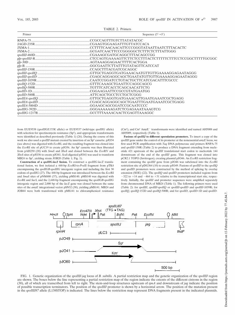
FIG. 1. Genetic organization of the spoIIIJ-jag locus of B. subtilis. A partial restriction map and the genetic organization of the spoIIIJ regionare shown. The boxes below the line representing a partial restriction map of the region indicate the extents of the different cistrons in the region(30), all of which are transcribed from left to right. The stem-and-loop structures upstream of rpnA and downstream of jag indicate the positionof possible transcription terminators. The position of the spoIIIJ promoter is shown by a horizontal arrow. The position of the mutation presentin the spoIIIJ87 allele (L138STOP) is indicated. The lines below the restriction map represent DNA fragments present in the indicated plasmids.
TABLE 2. Primers
Primer Sequence (5�33�)
RNPA-75.........................................................................CCGCCAGTTTGTCTTATATACGCJAGD-235R ....................................................................CGAAGTGGAAGATTTGTTATCCACAJXMA-1...........................................................................CCTTTTCAACAACATTCCCGGGTATAATTAATCTTTACACTCJXMA-2...........................................................................GCGATCAACTTCCCGGGGGCTCTTTCTCTTTATTGGGspoIIIJ-460D....................................................................CGAAAGCGATGCAGGCTTTACAGCCGGgfp-spoIIIJ-R....................................................................CTCCAGTGAAAAGTTCTTCTCCTTTACTCTTTTTCTTTCCTCCGGCTTTTTGCGGCgfp-30D ............................................................................AGTAAAGGAGAACTTTTCACTGGAgfp-R ................................................................................GGCGAATTCTTATTTGTATAGTTCATCCATspoIIIJ-150R....................................................................CCAGCTTTACGATCGCAGGCspoIIIJ-spoIIQ .................................................................GTTGCTGAGGTGATGAAACAATGTTGTTGAAAAGGAGAATAGGGspoIIIJ-spoIID .................................................................CGAGCAGGAGGCAGCTGAATATGTTGTTGAAAAGGAGAATAGGGspoIIIJ-1039R..................................................................CAATCCGGATCCTGTACTGCTTCATCGACATTTCGCCCspoIIQ-152D....................................................................GTTTCAAAGCTGAATTCCAGGCAGCGspoIIQ-500R....................................................................TGTTTCATCACCTCAGCAACATTCTGspoIID-1D........................................................................CGGAAGAATTCCGCCGTATGAATGGspoIID-500R....................................................................ATTCAGCTGCCTCCTGCTCGGGspoIIIA-spoIIQ ................................................................GTTGCTGAGGTGATGAAACATTGAATGAAATCGCTGAGGspoIIIA-spoIID ................................................................CGAGCAGGAGGCAGCTGAATTTGAATGAAATCGCTGAGGspoIIIA-5004D ................................................................GGAAGCAGCGGATCCGCAATCCCCspoIIIG-392D..................................................................GGGAAAAAAGATCTCGAGAAATAAAGTCGspoIIIG-1217R ................................................................GCCTTTAAAACAACTCGAGTTAAAGGC
VOL. 185, 2003 ROLE OF spoIIIJ IN ACTIVATION OF �G 3907
Dow
nloa
ded
from
http
s://j
ourn
als.
asm
.org
/jour
nal/j
b on
15
Febr
uary
202
2 by
77.
45.8
4.7.
500R. The 839-bp spoIIIJ fragment was mixed with the 350-bp spoIIQ fragmentor with the 500-bp spoIID fragment, and the resulting fragments of 1,189 or 1,339bp were amplified with primers spoIIQ-152D and spoIIIJ-1039R, to create aPspoIIQ-spoIIIJ fusion, or with primers spoIID-1D and spoIIIJ-1039R, to create aPspoIID-spoIIIJ fusion. The PspoIIQ-spoIIIJ and PspoIID-spoIIIJ fragments weredigested with EcoRI and BamHI and ligated to pDG364 (10) digested with thesame enzymes, to yield pMS189 and pMS190, respectively. JOB44 was trans-formed with pJO49, pMS189, and pMS190 to produce the Cmr AmyE� strainsAH5011, AH5012, and AH5013, respectively (Table 1). Derivatives of thesestrains bearing an sspE-lacZ fusion were obtained by transformation with chro-mosomal DNA from AH1042 (Table 1). To construct a PspoIID-spoIIIJ fusion ina multicopy vector, we isolated a 1,339-bp EcoRI-to-BamHI fragment frompMS190 encompassing the PspoIID-spoIIIJ fusion. The 1,339-bp fragment wasintroduced between the same sites of pMK3 (54), yielding pMS210. PlasmidspMK3 and pMS210 were introduced in both MB24 and JOB20 (Table 1).
Construction of a spoIIIJ-gfp fusion. We used the SOE technique (see above)to construct a fusion of spoIIIJ to gfp. Initially, the spoIIIJ and gfp sequences wereamplified separately, from chromosomal DNA of a wild-type B. subtilis strain andfrom pEA18 (a gift from Alan Grossman), respectively. The following primerpairs were used (Table 2): for spoIIIJ, spoIIIJ-460D and gfp-spoIIIJ-R; and forgfp, gfp-30D and gfp-R. The 551-bp spoIIIJ fragment and the 772-bp gfp fragmentwere purified, mixed together, and amplified with primers spoIIIJ-460D andgfp-R (see above). This produced a 1,245-bp spoIIIJ-gfp fragment which wascloned into pCR2.1 TOPO (Invitrogen) to yield pLC1. The presence and orien-tation of the insert were confirmed by restriction analysis. An Spr cassette wasreleased from pAH256 (20) with EcoRV and XhoI and cloned in pLC1 that hadbeen cut with the same restriction enzymes. The resulting plasmid, pLC3, wasused to transform strains AH5011, AH5012, and AH5013; Spr transformants thatwere the result of a single reciprocal crossover event (Campbell-type mechanism)were identified and designated AH5017, AH5018, and AH5019, respectively(Table 1).
Fusions of spoIIIA to different sporulation promoters. Fusions of spoIIIA tothe spoIIQ and spoIID promoters were constructed by SOE (see above). Initially,the spoIIIA and promoter sequences were amplified separately from chromo-somal DNA of a wild-type B. subtilis strain. The following primers were used(Table 2): for spoIIIA, spoIIIA-spoIIQ, spoIIIA-spoIID, and spoIIIA-5004D; forspoIIQ, spoIIQ-152D and spoIIQ-500R; and for spoIID, spoIID-1D, and spoIID-500R. The 386-bp spoIIIA fragment was mixed with the 350-bp spoIIQ fragmentor with the 500-bp spoIID fragment, and the mixture was subjected to PCRamplification with primers spoIIQ-152D and spoIIIA-5004D for spoIIQ or prim-ers spoIID-1D and spoIIIA-5004D for spoIID. The resulting PspoIIQ-spoIIIAand PspoIID-spoIIIA fragments were digested with BamHI and introduced intoBamHI- and SmaI-digested pUS19 (5), to yield pMS155 and pMS198. Transfor-mation of MB24 with pMS155 or pMS198 (with selection for Spr cells) producedstrains AH5020 and AH5029, respectively (Table 1). In both strains the Camp-bell-type integration of pMS155 or pMS198 separated spoIIIAA from its nativepromoter and placed the spoIIIA operon under the control of the spoIIQ orspoIID promoter, respectively. Correct integration was confirmed by PCR.
Overproduction and purification of �G. The spoIIIG coding region was PCRamplified with primers spoIIIG-392D and spoIIIG-1217R (Table 2). The PCRproduct was digested with BglII and XhoI and inserted between the BamHI andXhoI sites of pET30c(�) (Novagen), creating pMS112, in which a His6 tag wasintroduced between the first and second codons of spoIIIG. Plasmid pMS112 wasintroduced into competent cells of BL21(DE3)/pLysS (Novagen). Growth, in-duction, and lysate preparation were essentially as described previously (50). TheHis6-�
G fusion protein was partially purified on Hi-Trap chelating columns in thepresence of 8 M urea as described by the manufacturer (Amersham PharmaciaBiotech) and used to raise a polyclonal anti-�G antibody in rabbits (Eurogentec,Herstal, Belgium).
Immunoblotting. Preparation of B. subtilis whole-cell lysates and Western blotanalysis were essentially as described previously (50). The anti-�G polyclonalantiserum was incubated with the membranes at a dilution of 1:1,000 in phos-phate-buffered saline (8 mM sodium phosphate [pH 7.5], 150 mM NaCl) con-taining 0.5% low-fat milk. Incubation with an anti-rabbit secondary antibodyconjugated to horseradish peroxidase (from Sigma) was for 30 min at a 1:5,000dilution. A rabbit anti-green fluorescent protein (anti-GFP) antiserum (Clon-tech) was used according to the manufacturer’s protocol.
�-Galactosidase assays. �-Galactosidase activity was determined with thesubstrate o-nitrophenol-�-D-galactopyranoside, and enzyme activity was ex-pressed in Miller units as described previously (20, 21).
Transmission electron microscopy. Samples (5 ml) of DSM cultures of variousstrains were collected 12 h after the onset of sporulation. The cells were fixed for2 h with 2.5% glutaraldehyde in 0.1 M cocadylate buffer (pH 7.4) on ice. Samples
were washed twice with 0.1 M cocadylate buffer (pH 7.4) and then were treatedwith 1% osmium tetroxide solution [1.5% K4Fe(CN)6, 1% OsO4, 0.1 M cocady-late buffer]. The cells were dehydrated at room temperature by washing withincreasing concentrations of ethanol (up to 100%), followed by a wash withacetone. The cells were embedded in EM bed-812 resin (Electron MicroscopySciences). Ultrathin sections were processed for observation as described previ-ously (21). Transmission electron microscopy was performed on a Hitachi H7500transmission electron microscope operated at 75 keV.
Light microscopy and image processing. Samples (0.5 ml) of DSM cultureswere collected throughout growth and sporulation and resuspended in the samevolume of phosphate-buffered saline supplemented with DAPI (4�,6�-diamidino-2-phenylindole) (10 g/ml). Microscope slides were prepared by using an adap-tation of the method developed by van Helvoort and Woldringh (58). Imageswere acquired on a scientific-grade cooled charge-coupled device (Cooke Co.)on a multiwavelength wide-field three-dimensional microscopy system (63x/1.4OIL Plan Apochromat objective, Zeiss 100 M; Intelligent Imaging Innovation).Samples were imaged in successive 0.2-m focal planes, and out-of-focus lightwas removed with a constrained iterative deconvolution algorithm. Standardfilters for fluorescein isothiocyanate (for GFP) and DAPI were used. The patternof nucleoid staining with DAPI was used to as an indication of the sporulationstage. Just after formation of the asymmetric septum, the prespore nucleoidappears highly condensed, whereas soon after conclusion of the engulfmentprocess, the mother cell and prespore nucleoids appear equally condensed (19,51).
RESULTS
�G accumulates but is mostly inactive in a spoIIIJ mutant.spoIIIJ is the first gene of a bicistronic operon (16) (Fig. 1).Mutations in the spoIIIJ locus block sporulation after comple-tion of prespore engulfment and abolish �G activity (16). Err-ington et al. have examined the effect on sporulation of thespoIIIJ87 allele (16), which we found to be a nonsense muta-tion at codon 138 of SpoIIIJ (Fig. 1). Those authors have alsostudied the effect of a spoIIIJ insertional mutation on sporu-lation-specific gene expression and found that neither alleleimpaired transcription of the �E-dependent phoAIII gene or ofthe �F-dependent spoIIIG gene (16). However, both spoIIIJ87and the insertional mutation prevented expression of the �G-dependent spoVA operon (16). Disruption of the second geneof the spoIIIJ operon, jag, did not affect either growth orsporulation (16). However, because both types of spoIIIJ mu-tants could have a polar effect on the expression of the jaggene, the sporulation phenotype could be caused by the lack ofexpression of the two genes. Here, we have used a nonpolarmutation in spoIIIJ (�spoIIIJ::km; strain JOB44) (Table 1),which permits expression of the jag gene, as verified by reversetranscription-PCR (not shown), to reexamine the individualcontribution of this gene to sporulation. In agreement with theresults of Errington and coauthors (16), we found that inacti-vation of spoIIIJ has little effect on the transcription of genesexpressed in the mother cell (such as spoIID) or in the pre-spore (such as spoIIQ) prior to the completion of engulfment(not shown). In contrast, and as also found by Errington andcoauthors (16), the �spoIIIJ::km mutation reduced the rate ofspoIIIG-lacZ expression (Fig. 2A [Fig. 2A is included to allowdirect comparison with the immunoblot analysis of �G accu-mulation; see below]). The �spoIIIJ::km mutation also greatlyreduced transcription of the �G-dependent gene sspE (34)(data not shown). The �spoIIIJ::sp mutant JOB20 (Table 1)behaved like JOB44 (�spoIIIJ::km) with respect to spoIIIG-and sspE-lacZ expression (not shown).
Since substantial expression of a spoIIIG-lacZ fusion occursin a spoIIIJ mutant (16) (Fig. 2A), it is likely that SpoIIIJ acts
3908 SERRANO ET AL. J. BACTERIOL.
Dow
nloa
ded
from
http
s://j
ourn
als.
asm
.org
/jour
nal/j
b on
15
Febr
uary
202
2 by
77.
45.8
4.7.
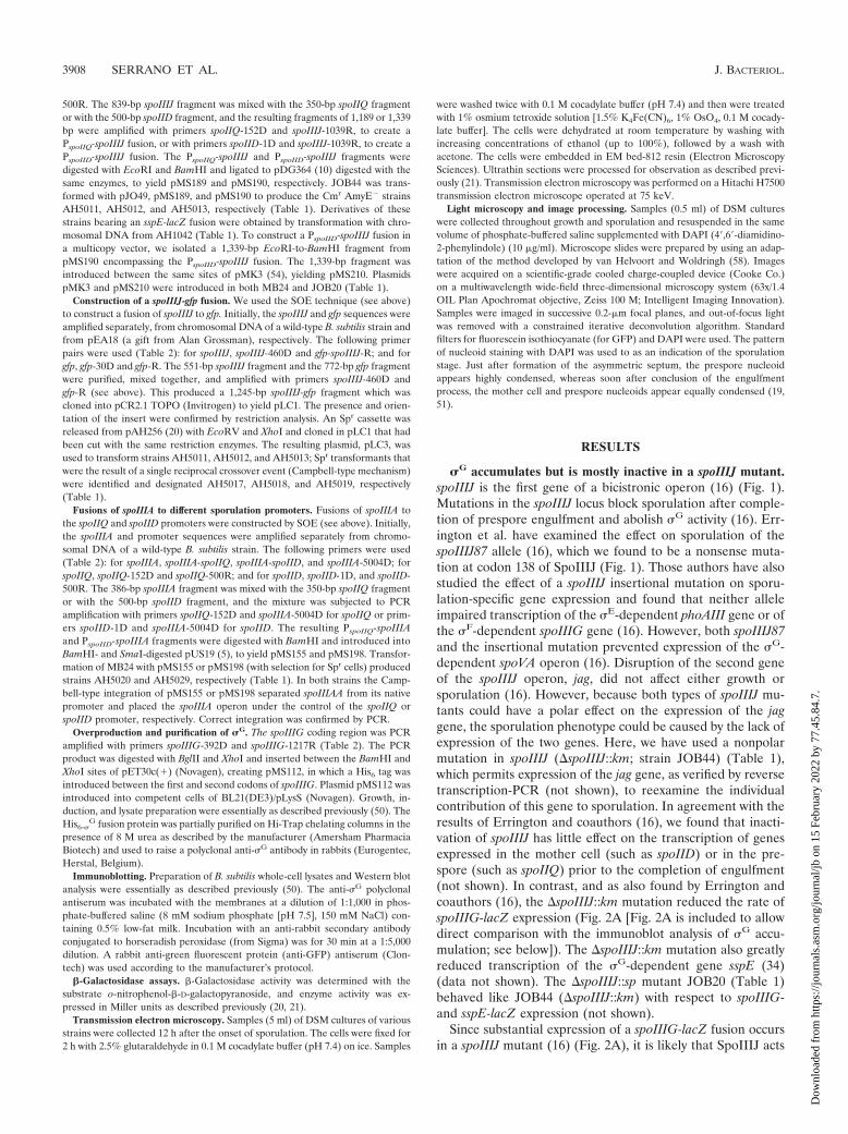
at the level of �G accumulation or activity. To distinguishbetween these two possibilities, we used an antiserum directedagainst �G to monitor its accumulation during sporulation inthe wild type and in the �spoIIIJ::km mutant (JOB44) (Table1). This �G antiserum detected a species of 35 kDa in immu-noblot analysis of extracts prepared from sporulating wild-typecultures of B. subtilis but not from cultures of a spoIIIG dele-tion mutant (Fig. 2B and C, lanes 7) (26). In the wild-typestrain, MB24, �G was first detected at 2 h after the onset ofsporulation, and its levels increased during h 3 and 4 (Fig. 2B).In the spoIIIJ mutant, �G accumulated to nearly wild-typelevels, although at h 3 the level of �G was reduced aboutthreefold compared to that in the wild type (Fig. 2C). Thepattern of �G accumulation in the spoIIIJ mutant probablyreflects the decreased rate of spoIIIG transcription (Fig. 2).
The somewhat reduced levels of spoIIIG transcription and �G
accumulation in the spoIIIJ mutant are probably a result of theautoregulatory effect that �G exerts on transcription of itsencoding gene (55). We conclude that the failure to detectexpression of �G-dependent genes in the spoIIIJ mutant (16;this work) results from the inactivity of �G.
The E155K form of �G no longer requires SpoIIIJ for acti-vation. In a spoIIIA mutant, �G also accumulates in an inactiveform (26). Activity of �G can be restored to spoIIIA mutantcells by the production of a form of �G (�GE155K) that is notefficiently bound by SpoIIAB in vitro (26). To test whether�GE155K could also bypass the need for spoIIIJ expressionfor �G activity, we introduced the wild-type or spoIIIGE155Kallele of spoIIIG (the later encoding �GE155K) linked to a Spr
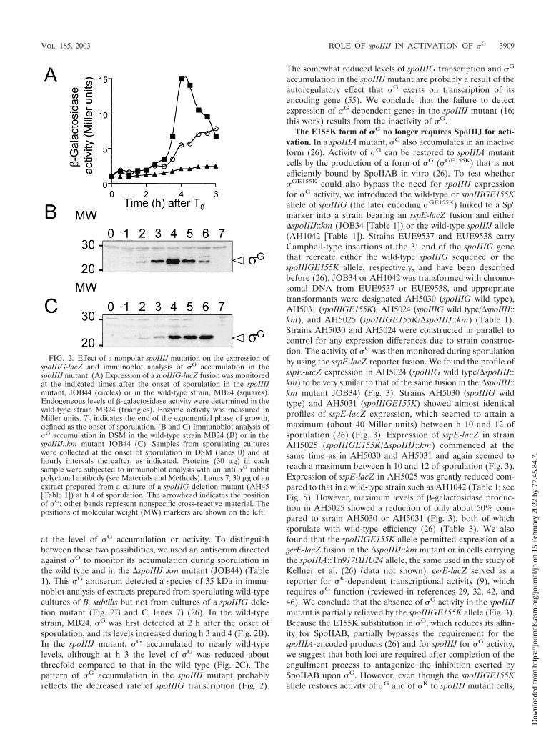
marker into a strain bearing an sspE-lacZ fusion and either�spoIIIJ::km (JOB34 [Table 1]) or the wild-type spoIIIJ allele(AH1042 [Table 1]). Strains EUE9537 and EUE9538 carryCampbell-type insertions at the 3� end of the spoIIIG genethat recreate either the wild-type spoIIIG sequence or thespoIIIGE155K allele, respectively, and have been describedbefore (26). JOB34 or AH1042 was transformed with chromo-somal DNA from EUE9537 or EUE9538, and appropriatetransformants were designated AH5030 (spoIIIG wild type),AH5031 (spoIIIGE155K), AH5024 (spoIIIG wild type/�spoIIIJ::km), and AH5025 (spoIIIGE155K/�spoIIIJ::km) (Table 1).Strains AH5030 and AH5024 were constructed in parallel tocontrol for any expression differences due to strain construc-tion. The activity of �G was then monitored during sporulationby using the sspE-lacZ reporter fusion. We found the profile ofsspE-lacZ expression in AH5024 (spoIIIG wild type/�spoIIIJ::km) to be very similar to that of the same fusion in the �spoIIIJ::km mutant JOB34) (Fig. 3). Strains AH5030 (spoIIIG wildtype) and AH5031 (spoIIIGE155K) showed almost identicalprofiles of sspE-lacZ expression, which seemed to attain amaximum (about 40 Miller units) between h 10 and 12 ofsporulation (26) (Fig. 3). Expression of sspE-lacZ in strainAH5025 (spoIIIGE155K/�spoIIIJ::km) commenced at thesame time as in AH5030 and AH5031 and again seemed toreach a maximum between h 10 and 12 of sporulation (Fig. 3).Expression of sspE-lacZ in AH5025 was greatly reduced com-pared to that in a wild-type strain such as AH1042 (Table 1; seeFig. 5). However, maximum levels of �-galactosidase produc-tion in AH5025 showed a reduction of only about 50% com-pared to strain AH5030 or AH5031 (Fig. 3), both of whichsporulate with wild-type efficiency (26) (Table 3). We alsofound that the spoIIIGE155K allele permitted expression of agerE-lacZ fusion in the �spoIIIJ::km mutant or in cells carryingthe spoIIIA::Tn917�HU24 allele, the same used in the study ofKellner et al. (26) (data not shown). gerE-lacZ served as areporter for �K-dependent transcriptional activity (9), whichrequires �G function (reviewed in references 29, 32, 42, and46). We conclude that the absence of �G activity in the spoIIIJmutant is partially relieved by the spoIIIGE155K allele (Fig. 3).Because the E155K substitution in �G, which reduces its affin-ity for SpoIIAB, partially bypasses the requirement for thespoIIIA-encoded products (26) and for spoIIIJ for �G activity,we suggest that both loci are required after completion of theengulfment process to antagonize the inhibition exerted bySpoIIAB upon �G. However, even though the spoIIIGE155Kallele restores activity of �G and of �K to spoIIIJ mutant cells,
FIG. 2. Effect of a nonpolar spoIIIJ mutation on the expression ofspoIIIG-lacZ and immunoblot analysis of �G accumulation in thespoIIIJ mutant. (A) Expression of a spoIIIG-lacZ fusion was monitoredat the indicated times after the onset of sporulation in the spoIIIJmutant, JOB44 (circles) or in the wild-type strain, MB24 (squares).Endogeneous levels of �-galactosidase activity were determined in thewild-type strain MB24 (triangles). Enzyme activity was measured inMiller units. T0 indicates the end of the exponential phase of growth,defined as the onset of sporulation. (B and C) Immunoblot analysis of�G accumulation in DSM in the wild-type strain MB24 (B) or in thespoIIIJ::km mutant JOB44 (C). Samples from sporulating cultureswere collected at the onset of sporulation in DSM (lanes 0) and athourly intervals thereafter, as indicated. Proteins (30 g) in eachsample were subjected to immunoblot analysis with an anti-�G rabbitpolyclonal antibody (see Materials and Methods). Lanes 7, 30 g of anextract prepared from a culture of a spoIIIG deletion mutant (AH45[Table 1]) at h 4 of sporulation. The arrowhead indicates the positionof �G; other bands represent nonspecific cross-reactive material. Thepositions of molecular weight (MW) markers are shown on the left.
VOL. 185, 2003 ROLE OF spoIIIJ IN ACTIVATION OF �G 3909
Dow
nloa
ded
from
http
s://j
ourn
als.
asm
.org
/jour
nal/j
b on
15
Febr
uary
202
2 by
77.
45.8
4.7.
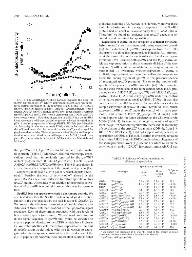
the spoIIIGE155K/�spoIIIJ::km double mutant is still unableto sporulate (Table 3). Moreover, electron microscopy obser-vations reveal that, as previously reported for the spoIIIJ87mutant (16), in both JOB44 (�spoIIIJ::km) (Table 1) andAH5025 (spoIIIGE155K/�spoIIIJ::km) (Table 1) sporulation isarrested soon after completion of the engulfment process (Fig.4, compare panels B and C with panel A, which depicts a Spo�
strain). Possibly, the level of activity of �G allowed by thespoIIIGE155K allele is not sufficient to restore sporulation to aspoIIIJ mutant. Alternatively, in addition to promoting activa-tion of �G, SpoIIIJ is required in some other way for sporula-tion.
SpoIIIJ does not appear to encode a pheromone peptide. Wealso tested whether the SpoIIIJ protein could yield a peptidesimilar to the one encoded by the ccfA locus of E. faecalis (2).We assayed the effects on sporulation of double alanine sub-stitutions at three different locations of the lipoprotein signalsequence. Each of these strains produced wild-type levels ofheat-resistant spores (not shown). We also made substitutionsin the signal sequence of spoIIIJ that would be expected tocreate a peptide identical to the cCF10 peptide from E. faeca-lis. We tested whether cell-free culture supernatants from thisB. subtilis strain would induce wild-type E. faecalis to aggre-gate, which is a response consistent with the production of thecCF10 peptide (2); however, these supernatant solutions failed
to induce clumping of E. faecalis (not shown). Moreover thesemultiple substitutions in the signal sequence of the SpoIIIJprotein had no effect on sporulation by this B. subtilis strain.Therefore, we found no evidence that spoIIIJ encodes a se-creted peptide required for sporulation.
Expression of spoIIIJ in the prespore is sufficient for sporu-lation. spoIIIJ is normally expressed during vegetative growth(16), but induction of spoIIIJ transcription from the IPTG(isopropyl-�-D-thiogalactopyranoside)-inducible Pspac promot-er at the onset of sporulation is sufficient for efficient sporeformation (39). Because both spoIIIJ and the Pspac-spoIIIJ al-lele are expressed prior to the asymmetric division of the spo-rangium, SpoIIIJ could accumulate in the prespore and in themother cell. To investigate whether expression of spoIIIJ isexplicitly required in either the mother cell or the prespore, wefused the coding region of spoIIIJ to the prespore-specific�F-recognized spoIIQ promoter (31) or to the mother cell-specific �E-dependent spoIID promoter (45). The promoterfusions were introduced at the nonessential amyE locus, pro-ducing strains AH5012 (PspoIIQ-spoIIIJ) and AH5013 (PspoIID-spoIIIJ) (Table 1). A strain carrying spoIIIJ under the controlof its native promoter at amyE (AH5011 [Table 1]) was alsoconstructed in parallel to control for any differences due toectopic expression of spoIIIJ at amyE. Strain AH5011, whichexpresses spoIIIJ at amyE under the control of its native pro-moter, and strain AH5012 (PspoIIQ-spoIIIJ at amyE) bothformed spores with the same efficiency as the wild-type strainMB24 (Table 3). In contrast, although expression of spoIIIJfrom the spoIID promoter significantly increased the frequencyof sporulation of the �spoIIIJ::km mutant (JOB44), from 1 102 to 5.9 106 (Table 3), it did not support wild-type levels ofsporulation (AH5013) (Table 3). Electron microscopy revealedthat strains AH5011 and AH5012 completed morphogenesis ofthe spore protective layers (Fig. 4A and D), which relies on theactivities of �G and �K (29, 32). In contrast, strain AH5013 was
FIG. 3. The spoIIIGE155K allele partially bypasses the need forspoIIIJ expression for �G activity. Expression of sspE-lacZ was moni-tored during sporulation in the following strains (Table 1): AH5030(spoIIIG::pEK15) (closed squares), AH5031 (spoIIIG::pEK16) (opencircles), AH5024 (spoIIIG::pEK15 spoIIIJ::km) (open squares), AH5025(spoIIIG::pEK16 spoIIIJ::km) (open diamonds), and JOB34 (spoIIIJ::km) (closed circles). Note that integration of pEK15 into the spoIIIGlocus recreates a wild-type spoIIIG sequence, whereas integration ofpEK16 results in expression of the spoIIIGE155K allele (see Materialsand Methods). Strains were grown in DSM, and samples were taken atthe indicated times after the onset of sporulation (T0) and assayed for�-galactosidase activity. The endogenous levels of �-galactosidase pro-duction were determined in the wild-type strain MB24 (closed trian-gles). Enzyme activity is indicated in Miller units (see Materials andMethods).
TABLE 3. Influence of various mutations onefficiency of sporulation
Strain Genotype
Sporulation (CFU/ml)a
% Sporu-lationViable
cells
Heat-resistant
cells
MB24 Wild type 5.9 108 3.5 108 59.0JOB44 spoIIIJ::km 1.8 108 1.0 102 �0.001JOB20 spoIIIJ::sp 3.0 108 1.9 103 �0.001AH5030 spoIIIGwtb 5.4 108 1.6 108 30.0AH5031 spoIIIGE155K 9.0 108 2.9 108 32.0AH5024 spoIIIJ spoIIIGwt 3.5 108 1.8 103 �0.001AH5025 spoIIIJ spoIIIGE155K 3.4 108 2.9 103 �0.001AH5011 spoIIIJ PspoIIIJ-spoIIIJ 5.5 108 2.2 108 40.0AH5012 spoIIIJ PspoIIQ-spoIIIJ 4.3 108 2.6 108 61.0AH5013 spoIIIJ PspoIID-spoIIIJ 1.2 108 5.9 106 5.0AH2550 Wild type(pMK3) 7.9 108 4.5 108 57.0AH5037 Wild type(pMS210) 1.5 108 6.0 106 4.0AH5035 spoIIIJ(pMK3) 1.8 108 5.0 103 0.003AH5036 spoIIIJ(pMS210) 1.5 108 5.8 106 3.9AH62 spoIIIA�HU24 2.3 107 9.0 102 �0.001AH5020 PspoIIQ-spoIIIA 3.2 108 9.0 103 0.003AH5029 PspoIID-spoIIIA 5.5 108 3.1 108 56.0
a Sporulation was measured 24 h after the onset of the process in liquidsporulation medium as described in Materials and Methods.
b spoIIIGwt, wild-type spoIIIG.
3910 SERRANO ET AL. J. BACTERIOL.
Dow
nloa
ded
from
http
s://j
ourn
als.
asm
.org
/jour
nal/j
b on
15
Febr
uary
202
2 by
77.
45.8
4.7.
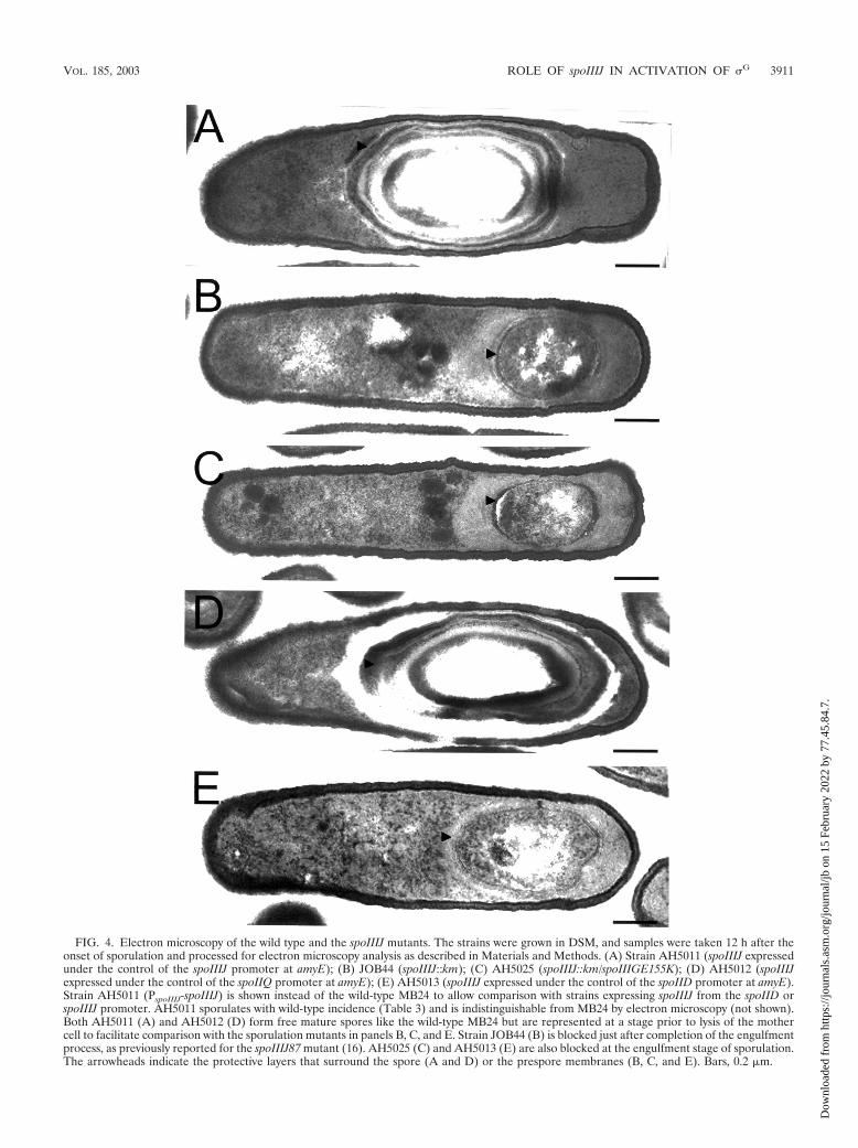
FIG. 4. Electron microscopy of the wild type and the spoIIIJ mutants. The strains were grown in DSM, and samples were taken 12 h after theonset of sporulation and processed for electron microscopy analysis as described in Materials and Methods. (A) Strain AH5011 (spoIIIJ expressedunder the control of the spoIIIJ promoter at amyE); (B) JOB44 (spoIIIJ::km); (C) AH5025 (spoIIIJ::km/spoIIIGE155K); (D) AH5012 (spoIIIJexpressed under the control of the spoIIQ promoter at amyE); (E) AH5013 (spoIIIJ expressed under the control of the spoIID promoter at amyE).Strain AH5011 (PspoIIIJ-spoIIIJ) is shown instead of the wild-type MB24 to allow comparison with strains expressing spoIIIJ from the spoIID orspoIIIJ promoter. AH5011 sporulates with wild-type incidence (Table 3) and is indistinguishable from MB24 by electron microscopy (not shown).Both AH5011 (A) and AH5012 (D) form free mature spores like the wild-type MB24 but are represented at a stage prior to lysis of the mothercell to facilitate comparison with the sporulation mutants in panels B, C, and E. Strain JOB44 (B) is blocked just after completion of the engulfmentprocess, as previously reported for the spoIIIJ87 mutant (16). AH5025 (C) and AH5013 (E) are also blocked at the engulfment stage of sporulation.The arrowheads indicate the protective layers that surround the spore (A and D) or the prespore membranes (B, C, and E). Bars, 0.2 m.
VOL. 185, 2003 ROLE OF spoIIIJ IN ACTIVATION OF �G 3911
Dow
nloa
ded
from
http
s://j
ourn
als.
asm
.org
/jour
nal/j
b on
15
Febr
uary
202
2 by
77.
45.8
4.7.
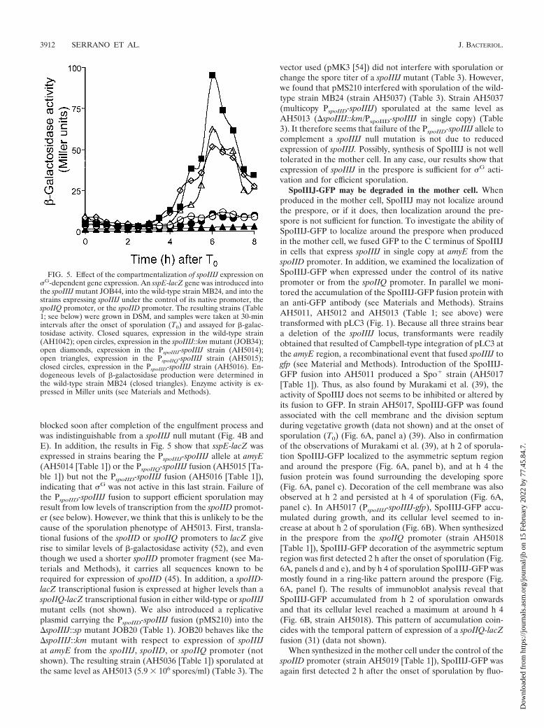
blocked soon after completion of the engulfment process andwas indistinguishable from a spoIIIJ null mutant (Fig. 4B andE). In addition, the results in Fig. 5 show that sspE-lacZ wasexpressed in strains bearing the PspoIIIJ-spoIIIJ allele at amyE(AH5014 [Table 1]) or the PspoIIQ-spoIIIJ fusion (AH5015 [Ta-ble 1]) but not the PspoIID-spoIIIJ fusion (AH5016 [Table 1]),indicating that �G was not active in this last strain. Failure ofthe PspoIID-spoIIIJ fusion to support efficient sporulation mayresult from low levels of transcription from the spoIID promot-er (see below). However, we think that this is unlikely to be thecause of the sporulation phenotype of AH5013. First, transla-tional fusions of the spoIID or spoIIQ promoters to lacZ giverise to similar levels of �-galactosidase activity (52), and eventhough we used a shorter spoIID promoter fragment (see Ma-terials and Methods), it carries all sequences known to berequired for expression of spoIID (45). In addition, a spoIID-lacZ transcriptional fusion is expressed at higher levels than aspoIIQ-lacZ transcriptional fusion in either wild-type or spoIIIJmutant cells (not shown). We also introduced a replicativeplasmid carrying the PspoIID-spoIIIJ fusion (pMS210) into the�spoIIIJ::sp mutant JOB20 (Table 1). JOB20 behaves like the�spoIIIJ::km mutant with respect to expression of spoIIIJat amyE from the spoIIIJ, spoIID, or spoIIQ promoter (notshown). The resulting strain (AH5036 [Table 1]) sporulated atthe same level as AH5013 (5.9 106 spores/ml) (Table 3). The
vector used (pMK3 [54]) did not interfere with sporulation orchange the spore titer of a spoIIIJ mutant (Table 3). However,we found that pMS210 interfered with sporulation of the wild-type strain MB24 (strain AH5037) (Table 3). Strain AH5037(multicopy PspoIID-spoIIIJ) sporulated at the same level asAH5013 (�spoIIIJ::km/PspoIID-spoIIIJ in single copy) (Table3). It therefore seems that failure of the PspoIID-spoIIIJ allele tocomplement a spoIIIJ null mutation is not due to reducedexpression of spoIIIJ. Possibly, synthesis of SpoIIIJ is not welltolerated in the mother cell. In any case, our results show thatexpression of spoIIIJ in the prespore is sufficient for �G acti-vation and for efficient sporulation.
SpoIIIJ-GFP may be degraded in the mother cell. Whenproduced in the mother cell, SpoIIIJ may not localize aroundthe prespore, or if it does, then localization around the pre-spore is not sufficient for function. To investigate the ability ofSpoIIIJ-GFP to localize around the prespore when producedin the mother cell, we fused GFP to the C terminus of SpoIIIJin cells that express spoIIIJ in single copy at amyE from thespoIID promoter. In addition, we examined the localization ofSpoIIIJ-GFP when expressed under the control of its nativepromoter or from the spoIIQ promoter. In parallel we moni-tored the accumulation of the SpoIIIJ-GFP fusion protein withan anti-GFP antibody (see Materials and Methods). StrainsAH5011, AH5012 and AH5013 (Table 1; see above) weretransformed with pLC3 (Fig. 1). Because all three strains beara deletion of the spoIIIJ locus, transformants were readilyobtained that resulted of Campbell-type integration of pLC3 atthe amyE region, a recombinational event that fused spoIIIJ togfp (see Material and Methods). Introduction of the SpoIIIJ-GFP fusion into AH5011 produced a Spo� strain (AH5017[Table 1]). Thus, as also found by Murakami et al. (39), theactivity of SpoIIIJ does not seems to be inhibited or altered byits fusion to GFP. In strain AH5017, SpoIIIJ-GFP was foundassociated with the cell membrane and the division septumduring vegetative growth (data not shown) and at the onset ofsporulation (T0) (Fig. 6A, panel a) (39). Also in confirmationof the observations of Murakami et al. (39), at h 2 of sporula-tion SpoIIIJ-GFP localized to the asymmetric septum regionand around the prespore (Fig. 6A, panel b), and at h 4 thefusion protein was found surrounding the developing spore(Fig. 6A, panel c). Decoration of the cell membrane was alsoobserved at h 2 and persisted at h 4 of sporulation (Fig. 6A,panel c). In AH5017 (PspoIIIJ-spoIIIJ-gfp), SpoIIIJ-GFP accu-mulated during growth, and its cellular level seemed to in-crease at about h 2 of sporulation (Fig. 6B). When synthesizedin the prespore from the spoIIQ promoter (strain AH5018[Table 1]), SpoIIIJ-GFP decoration of the asymmetric septumregion was first detected 2 h after the onset of sporulation (Fig.6A, panels d and e), and by h 4 of sporulation SpoIIIJ-GFP wasmostly found in a ring-like pattern around the prespore (Fig.6A, panel f). The results of immunoblot analysis reveal thatSpoIIIJ-GFP accumulated from h 2 of sporulation onwardsand that its cellular level reached a maximum at around h 4(Fig. 6B, strain AH5018). This pattern of accumulation coin-cides with the temporal pattern of expression of a spoIIQ-lacZfusion (31) (data not shown).
When synthesized in the mother cell under the control of thespoIID promoter (strain AH5019 [Table 1]), SpoIIIJ-GFP wasagain first detected 2 h after the onset of sporulation by fluo-
FIG. 5. Effect of the compartmentalization of spoIIIJ expression on�G-dependent gene expression. An sspE-lacZ gene was introduced intothe spoIIIJ mutant JOB44, into the wild-type strain MB24, and into thestrains expressing spoIIIJ under the control of its native promoter, thespoIIQ promoter, or the spoIID promoter. The resulting strains (Table1; see below) were grown in DSM, and samples were taken at 30-minintervals after the onset of sporulation (T0) and assayed for �-galac-tosidase activity. Closed squares, expression in the wild-type strain(AH1042); open circles, expression in the spoIIIJ::km mutant (JOB34);open diamonds, expression in the PspoIIIJ-spoIIIJ strain (AH5014);open triangles, expression in the PspoIIQ-spoIIIJ strain (AH5015);closed circles, expression in the PspoIID-spoIIIJ strain (AH5016). En-dogeneous levels of �-galactosidase production were determined inthe wild-type strain MB24 (closed triangles). Enzyme activity is ex-pressed in Miller units (see Materials and Methods).
3912 SERRANO ET AL. J. BACTERIOL.
Dow
nloa
ded
from
http
s://j
ourn
als.
asm
.org
/jour
nal/j
b on
15
Febr
uary
202
2 by
77.
45.8
4.7.
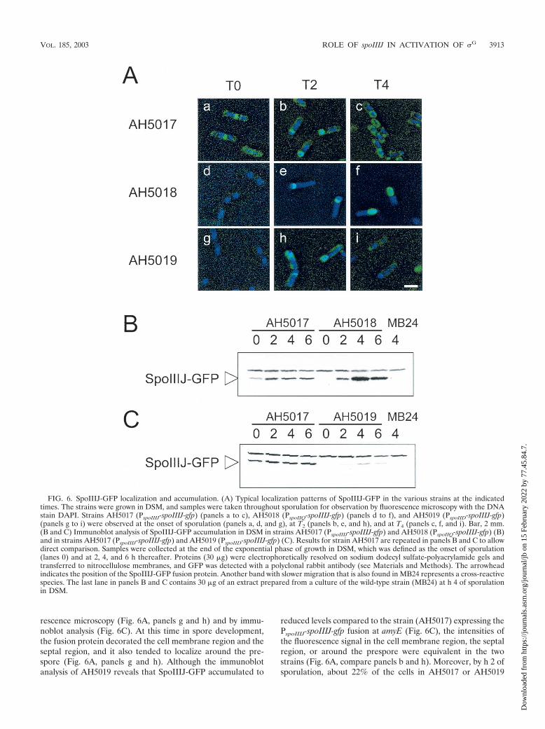
rescence microscopy (Fig. 6A, panels g and h) and by immu-noblot analysis (Fig. 6C). At this time in spore development,the fusion protein decorated the cell membrane region and theseptal region, and it also tended to localize around the pre-spore (Fig. 6A, panels g and h). Although the immunoblotanalysis of AH5019 reveals that SpoIIIJ-GFP accumulated to
reduced levels compared to the strain (AH5017) expressing thePspoIIIJ-spoIIIJ-gfp fusion at amyE (Fig. 6C), the intensities ofthe fluorescence signal in the cell membrane region, the septalregion, or around the prespore were equivalent in the twostrains (Fig. 6A, compare panels b and h). Moreover, by h 2 ofsporulation, about 22% of the cells in AH5017 or AH5019
FIG. 6. SpoIIIJ-GFP localization and accumulation. (A) Typical localization patterns of SpoIIIJ-GFP in the various strains at the indicatedtimes. The strains were grown in DSM, and samples were taken throughout sporulation for observation by fluorescence microscopy with the DNAstain DAPI. Strains AH5017 (PspoIIIJ-spoIIIJ-gfp) (panels a to c), AH5018 (PspoIIQ-spoIIIJ-gfp) (panels d to f), and AH5019 (PspoIID-spoIIIJ-gfp)(panels g to i) were observed at the onset of sporulation (panels a, d, and g), at T2 (panels b, e, and h), and at T4 (panels c, f, and i). Bar, 2 mm.(B and C) Immunoblot analysis of SpoIIIJ-GFP accumulation in DSM in strains AH5017 (PspoIIIJ-spoIIIJ-gfp) and AH5018 (PspoIIQ-spoIIIJ-gfp) (B)and in strains AH5017 (PspoIIIJ-spoIIIJ-gfp) and AH5019 (PspoIID-spoIIIJ-gfp) (C). Results for strain AH5017 are repeated in panels B and C to allowdirect comparison. Samples were collected at the end of the exponential phase of growth in DSM, which was defined as the onset of sporulation(lanes 0) and at 2, 4, and 6 h thereafter. Proteins (30 g) were electrophoretically resolved on sodium dodecyl sulfate-polyacrylamide gels andtransferred to nitrocellulose membranes, and GFP was detected with a polyclonal rabbit antibody (see Materials and Methods). The arrowheadindicates the position of the SpoIIIJ-GFP fusion protein. Another band with slower migration that is also found in MB24 represents a cross-reactivespecies. The last lane in panels B and C contains 30 g of an extract prepared from a culture of the wild-type strain (MB24) at h 4 of sporulationin DSM.
VOL. 185, 2003 ROLE OF spoIIIJ IN ACTIVATION OF �G 3913
Dow
nloa
ded
from
http
s://j
ourn
als.
asm
.org
/jour
nal/j
b on
15
Febr
uary
202
2 by
77.
45.8
4.7.
showed condensed prespore chromosomes (�100 cells count-ed), as judged from the analysis of DAPI-stained images(see Materials and Methods). These cells are presumed tohave completed the asymmetric division of the sporangium.Of these, about 80% in either AH5017 (PspoIIIJ-spoIIIJ-gfp)or AH5019 (PspoIID-spoIIIJ-gfp) showed decoration of the sep-tum or prespore regions by SpoIIIJ-GFP (see also below). Itappears that the PspoIID-spoIIIJ-gfp allele results in sufficientproduct to decorate the prespore region in most of the sporu-lating cells at h 2 of sporulation. About 17% of the cells (over100 cells counted) of both AH5017 or AH5019 appeared tohave completed the engulfment process by h 4 of sporulation,based on the analysis of DAPI images (see Materials andMethods). However, at this time in sporulation, SpoIIIJ-GFPdecoration of both the cell membrane and the prespore regionwas considerably reduced in AH5019 compared to AH5017(Fig. 6A, compare panels c and i). This reduction in the dec-oration of the prespore was not accompanied by an increase incytoplasmic fluorescence, even though the accumulation ofSpoIIIJ-GFP as monitored by immunoblot analysis appearedto increase (Fig. 6C). The immunoblot analysis did not revealsigns of proteolysis of SpoIIIJ-GFP (Fig. 6C). Nevertheless,even though a spoIID-lacZ fusion appears to be expressed athigher levels in both wild-type and spoIIIJ mutant cells than isa spoIIQ-lacZ fusion (see above), our PspoIID-spoIIIJ-gfp fusionresulted in reduced cellular levels of SpoIIIJ-GFP. The obser-vation that a PspoIID-spoIIIJ multicopy allele interfered withsporulation of a wild-type strain (see above) suggests that ac-cumulation of SpoIIIJ in the mother cell is not well tolerated.Together, the results suggest that when produced in themother cell, the SpoIIIJ-GFP protein may be subjected todegradation.
Expression of spoIIIA in the prespore does not permit ac-tivation of �G. The results described above suggest thatSpoIIIJ and the spoIIIA-encoded products may function at theend of the engulfment process to antagonize the action exertedby SpoIIAB upon �G. However, while expression of spoIIIJ issufficient in the prespore (this work), earlier studies haveshown that the spoIIIA operon, which is transcribed from apromoter recognized by the �E form of RNA polymerase, isspecifically required in the mother cell (23, 24). To determinewhether expression of spoIIIA in the mother cell is an absoluterequirement or whether the spoIIIA-encoded products couldpromote �G activation and sporulation if produced only in theprespore, we fused the spoIIIA operon to the prespore-specificspoIIQ promoter (strain AH5020 [Table 1]). The results inTable 3 indicate that expression of the spoIIIA operon from thespoIIQ promoter resulted in very poor sporulation (about 104
spores/ml). We also found that expression of the spoIIIA op-eron solely from the spoIIQ promoter (in strain AH5020, bear-ing the PspoIIQ-spoIIIA allele) did not support expression of the�G-dependent sspE-lacZ fusion (not shown). Moreover, elec-tron microscopy observations revealed that strain AH5020 isblocked soon after completion of the engulfment sequence(data not shown). In contrast, expression of the spoIIIA operonfrom the similarly expressed mother cell-specific spoIID pro-moter (45, 52) resulted in wild-type levels of sporulation (Table3). We suggest that at the end of the engulfment process, thespoIIIA-encoded products from the mother cell and SpoIIIJfrom the prespore are both required to antagonize the in-
hibitory action imposed by SpoIIAB upon �G in the pre-spore (Fig. 7).
DISCUSSION
Here we have analyzed the role of spoIIIJ in the activation of�G. Mutations in spoIIIJ arrest development soon after engulf-ment of the prespore by the mother cell, and greatly diminishthe activity of �G (16). However, mutations in spoIIIJ do notprevent transcription of the spoIIIG gene (16; this work), andas shown here they allow the accumulation of �G to nearlywild-type levels (Fig. 2). Therefore, most of the �G that accu-mulates in spoIIIJ mutant cells is inactive. This is reminiscentof the situation in a spoIIIA mutant, in which �G also accumu-lates but is inactive (26). Activation of �G at the end of theengulfment process then relies on spoIIIA- and spoIIIJ-depen-dent events. The available evidence suggests that the inhibitoryaction of SpoIIAB is responsible for the absence of �G activityin a spoIIIA mutant. SpoIIAB tends to disappear from theprespore compartment after conclusion of the engulfment pro-cess in a spoIIIA-dependent manner (28). More importantly, asingle amino acid substitution in �G that results in inefficientbinding by SpoIIAB in vitro bypasses the requirement forspoIIIA expression for �G activity (26). This implies thatSpoIIIA acts after engulfment of the prespore by the mothercell to antagonize the inhibition of �G activity by SpoIIAB(26). We found that expression of the spoIIIGE155K allele(encoding the �GE155K form of �G) also allows �G activity inthe absence of SpoIIIJ (Fig. 3). Therefore, our results suggestthat both SpoIIIJ and the spoIIIA-encoded products are in-volved in relieving the inhibition imposed by SpoIIAB upon �G
in the prespore. The spoIIIGE155K mutation does not causespremature expression of sspE-lacZ and in that sense does not
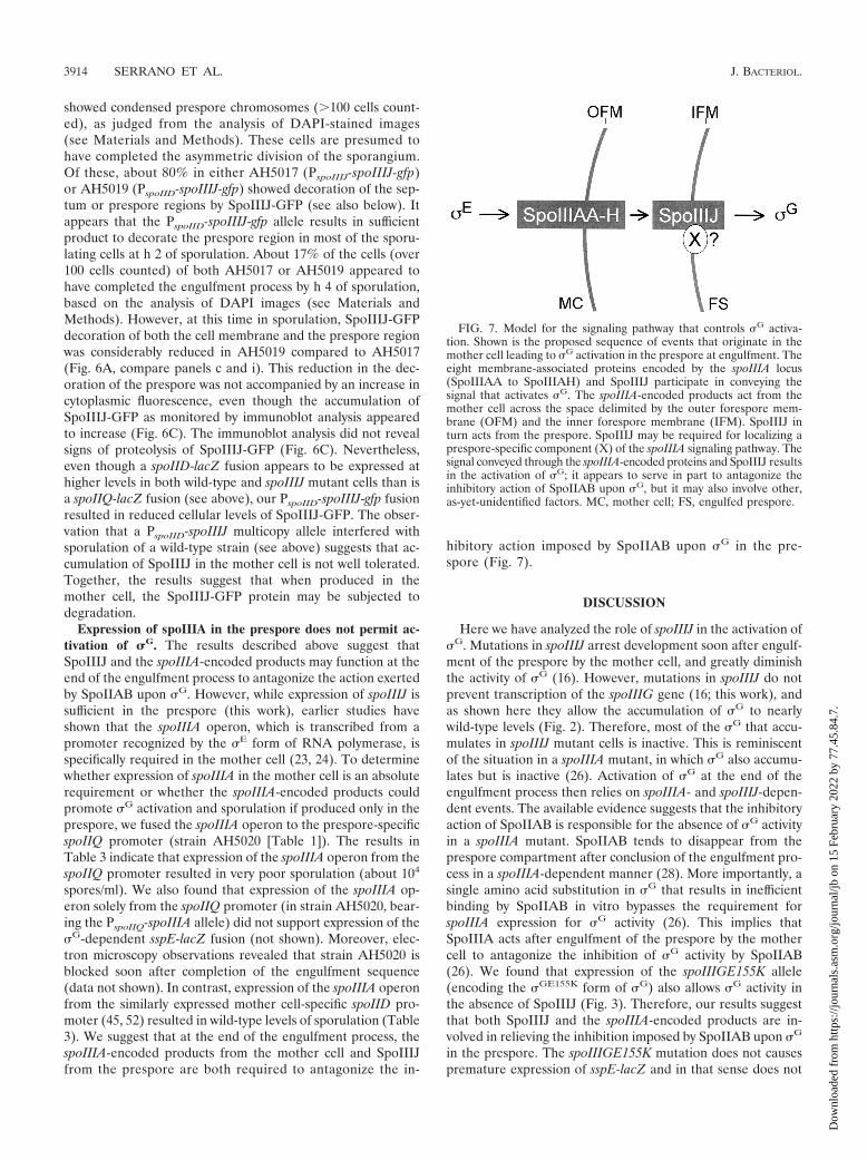
FIG. 7. Model for the signaling pathway that controls �G activa-tion. Shown is the proposed sequence of events that originate in themother cell leading to �G activation in the prespore at engulfment. Theeight membrane-associated proteins encoded by the spoIIIA locus(SpoIIIAA to SpoIIIAH) and SpoIIIJ participate in conveying thesignal that activates �G. The spoIIIA-encoded products act from themother cell across the space delimited by the outer forespore mem-brane (OFM) and the inner forespore membrane (IFM). SpoIIIJ inturn acts from the prespore. SpoIIIJ may be required for localizing aprespore-specific component (X) of the spoIIIA signaling pathway. Thesignal conveyed through the spoIIIA-encoded proteins and SpoIIIJ resultsin the activation of �G; it appears to serve in part to antagonize theinhibitory action of SpoIIAB upon �G, but it may also involve other,as-yet-unidentified factors. MC, mother cell; FS, engulfed prespore.
3914 SERRANO ET AL. J. BACTERIOL.
Dow
nloa
ded
from
http
s://j
ourn
als.
asm
.org
/jour
nal/j
b on
15
Febr
uary
202
2 by
77.
45.8
4.7.
uncouple the activity of �G from the conclusion of the engulf-ment sequence (Fig. 3) (26). One possible explanation is thatSpoIIAB is important to antagonize �G only towards the endof the engulfment sequence and that another, as-yet-unidenti-fied factor contributes to the negative regulation of �G activityduring the process of engulfment.
We also note that even though the spoIIIGE155K allelebypasses the requirement for SpoIIIJ for both �G and �K
activities, it did not restore formation of heat-resistant spores(Fig. 4C; Table 3). Interestingly, the spoIIIGE155K allele doesnot restore sporulation to a spoIIIA mutant either (26). Thereare several possible explanations for these observations. First,because �G is autoregulatory (25, 55), a mutation that wouldmake it less susceptible to SpoIIAB could in principle result inincreased levels of �G activity in the mother cell. However,ectopic activation of �G in the mother cell is unlikely to bethe cause of the sporulation phenotype of the spoIIIGE155K/�spoIIIJ::km double mutant, since a strain carrying thespoIIIGE155K allele in an otherwise wild-type backgroundproduces heat-resistant spores with wild-type efficiency (26)(not shown). The block in sporulation of the spoIIIGE155K/�spoIIIJ::km double mutant could also result from insufficientactivity of �G, even though peak levels of sspE-lacZ expressionin the spoIIIGE155K/�spoIIIJ::km double mutant are only halfof what is found in a strain carrying only the spoIIIGE155Kallele and which is Spo� (Fig. 3). The E155K substitution wasintroduced in �G (26) on the basis of the finding that a glu-tamic acid-to-lysine substitution was found at an equivalentposition (E149K) in a screen for �F mutants with reducedaffinity for SpoIIAB (11). The structure of a complex betweena dimer of the Bacillus stearothermophilus SpoIIAB proteinand �F helps to explain the specificity of SpoIIAB for �F andfor �G (7). In the B. stearothermophilus �F protein, the residueequivalent to E149 of B. subtilis �F, as well as three otherresidues found in genetic screens for mutants resistant to in-hibition by SpoIIAB (11), is located within a region that con-tains 17 amino acids found to interact with SpoIIAB in thecrystal structure (7). Of those residues, 15 are either identicalor homologous in �G and 3 are uniquely conserved between �F
and �G (7). However, the structure also suggests that eventhough �G appears to contact SpoIIAB through the same re-gion that mediates the interaction of �F with SpoIIAB, thedetails of the interaction of �G with SpoIIAB may differ (7). Itis possible that the E155K substitution makes �G less refrac-tory to SpoIIAB than the corresponding mutation (E149K) in�F. Lastly, another possibility for the incapacity of spoIIIGE155K/spoIIIA or spoIIIGE155K/�spoIIIJ::km double mutants to sporu-late is that in addition to the activation of �G, SpoIIIJ and theeight products of the spoIIIA operon play other roles in sporu-lation (26).
In any case, SpoIIIJ and the products of the spoIIIA locusfunction at least in part in the same signaling pathway that isinvolved in the activation of �G in the prespore after comple-tion of the engulfment sequence. This signaling pathway ap-pears to originate in the mother cell with the �E-dependenttranscription of the spoIIIA operon (23). Because their synthe-sis is restricted to the mother cell compartment, it seems un-likely that the spoIIIA-encoded proteins interact directly withSpoIIAB in the prespore (26). However, other than SpoIIABand �G, no prespore-specific components of the �E-to-�G sig-
naling pathway have been identified. We tested whether thefunction of SpoIIIJ is specifically required in either the pre-spore or the mother cell by expressing spoIIIJ under the controlof a promoter utilized by the �F (spoIIQ) or �E (spoIID) formof RNA polymerase. We found that expression of spoIIIJ issufficient in the prespore both for the activation of �G and forsporulation (Fig. 6; Table 3). Nevertheless, expression of aPspoIID-spoIIIJ fusion in single copy significantly increases thesporulation efficiency of a spoIIIJ null mutant, and thus itappears that SpoIIIJ retains some functionality when ex-pressed in the mother cell (Table 3). The lack of full comple-mentation of a spoIIIJ null mutant by PspoIID-spoIIIJ does notseem to be due to reduced expression of spoIIIJ, because thepresence of the same fusion in a multicopy plasmid does notfurther increase the efficiency of sporulation. Since the multi-copy PspoIID-spoIIIJ allele interferes with sporulation of thewild-type strain, we speculate that SpoIIIJ may promote theincorporation into the mother cell side of the prespore mem-branes of a protein that interferes with the signaling events thatlead to �G activation and that the mother cell has mechanismsto prevent SpoIIIJ accumulation to high levels. Preferentialdegradation of SpoIIIJ in the mother cell could be anotherexample of proteolysis playing a role in the reinforcement ofthe correct compartmentalization on � factor activity duringsporulation (see, for example, references 18 and 40).
In contrast to the case for spoIIIJ, we found that expressionof spoIIIA from the prespore-specific spoIIQ promoter doesnot support efficient sporulation (Fig. 6; Table 3). Like SpoIIIJ(39; this work), the spoIIIA-encoded products also localizearound the developing spore (46; W. Blaylock and C. P. Mo-ran, Jr., unpublished results). Together, our results suggest thatthe spoIIIA-encoded products from the mother cell andSpoIIIJ from the prespore act together to convey a signal thatresults in the activation of �G in the prespore after completionof the engulfment process (Fig. 7). We favor the idea that thespoIIIA-encoded proteins and SpoIIIJ function in the samepathway as suggested in Fig. 7, because mutations in either locirespond similarly to the spoIIIGE155K allele. However, wecannot presently exclude that spoIIIA and spoIIIJ act indepen-dently of each other. SpoIIIJ is related to the YidC membraneprotein translocase of E. coli (47, 49) and may have a similarfunction in B. subtilis (57). Since its function is sufficient in theprespore, SpoIIIJ may be required for the membrane localiza-tion of an as-yet-unknown forespore-specific component in the�E to �G signaling pathway (Fig. 7). It remains unclear whetherSpoIIAB is the only factor responsible for keeping �G inactiveprior to engulfment and, if this is the case, how �F can escapefrom SpoIIAB while �G is held inactive. It is also unknownwhether SpoIIAA and SpoIIE, which are required for �F ac-tivation (1, 3, 4, 12, 13, 15), play a role in the activation of �G.
ACKNOWLEDGMENTS
We thank Bill Blaylock and Goncalo Real for helpful discussionsand for critically reading the manuscript, Patrick Piggot and AlanGrossman for the gift of strains, Daniel Kalman for help with thefluorescence microscope and for helpful discussions, and Hong Hy(Emory Microscopy Core Facility) and Jan Pohl (Emory Microchem-ical Facility) for technical assistance.
This work was supported by grants Praxis XXI/PCNA/C/BIO/13201/98 and PRAXIS/BIO/35109/99 from the Fundacao para a Ciencia e aTecnologia to A.O.H. and by grant GM54395 from the National In-
VOL. 185, 2003 ROLE OF spoIIIJ IN ACTIVATION OF �G 3915
Dow
nloa
ded
from
http
s://j
ourn
als.
asm
.org
/jour
nal/j
b on
15
Febr
uary
202
2 by
77.
45.8
4.7.
stitutes of Health to C.P.M. M.S. is the recipient of a Ph.D. fellowship(PRAXIS XXI/BD 18 251/98) from the Fundacao para a Ciencia e aTecnologia.
REFERENCES
1. Alper, S., L. Duncan, and R. Losick. 1994. An adenosine nucleotide switchcontrolling the activity of a cell type-specific transcription factor in B. subtilis.Cell 77:195–205.
2. Antiporta, M. H., and G. M. Dunny. 2002. ccfA, the genetic determinant forthe cCF10 peptide pheromone in Enterococcus faecalis OG1RF. J. Bacteriol.184:1155–1162.
3. Arigoni, F., K. Pogliano, C. D. Webb, P. Stragier, and R. Losick. 1995.Localization of protein implicated in establishment of cell type to sites ofasymmetric division. Science 270:637–640.
4. Arigoni, F., L. Duncan, S. Alper, R. Losick, and P. Stragier. 1996. SpoIIEgoverns the phosphorylation state of a protein regulating transcription factorsigma F during sporulation in Bacillus subtilis. Proc. Natl. Acad. Sci. USA93:3238–3242.
5. Benson, A. K., and W. G. Haldenwang. 1993. Regulation of �B levels andactivity in Bacillus subtilis. J. Bacteriol. 175:2347–2356.
6. Campbell, E. A., and S. A. Darst. 2000. The anti-� factor SpoIIAB forms a2:1 complex with �F, contacting multiple conserved regions of the � factor.J. Mol. Biol. 300:17–28.
7. Campbell, E. A., S. Masuda, J. L. Sun, O. Muzzin, C. A. Olson, S. Wang, andS. A. Darst. 2002. Crystal structure of the Bacillus stearothermophilus anti-�factor SpoIIAB with the sporulation factor �F. Cell 108:795–807.
8. Coppolechia, R., H. DeGrazia, and C. P. Moran, Jr. 1991. Deletion ofspoIIAB blocks endospore formation in Bacillus subtilis at an early stage.J. Bacteriol. 173:6678–6685.
9. Cutting, S., S. Panzer, and R. Losick. 1989. Regulatory studies on thepromoter for a gene governing synthesis and assembly of the spore coat inBacillus subtilis. J. Mol. Biol. 207:393–404.
10. Cutting, S. M., and P. B. V. Horn. 1990. Genetics analysis, p. 27–74. In C. R.Harwood and S. M. Cutting (ed.), Molecular biological methods for Bacillus.John Wiley & Sons Ltd., Chichester, England.
11. Decatur, A., and R. Losick. 1996. Multiple sites of contact between theBacillus subtilis developmental transcription factor �F and its anti-sigmafactor SpoIIAB. Genes Dev. 10:2348–2358.
12. Diederich, B., J. F. Wilkinson, T. Magnin, M. Najafi, J. Errington, and M. D.Yudkin. 1994. Role of interactions between SpoIIAA and SpoIIAB in reg-ulating cell-specific transcription factor sigma F of Bacillus subtilis. GenesDev. 8:2653–2663.
13. Duncan, L., A. Alper, and R. Losick. 1996. SpoIIAA governs the release ofthe cell-type specific transcription factor �F from its anti-sigma factorSpoIIAB. J. Mol. Biol. 260:147–164.
14. Duncan, L., and R. Losick. 1993. SpoIIAB is an anti-sigma factor that bindsto and inhibits transcription by regulatory protein sigma F from Bacillussubtilis. Proc. Natl. Acad. Sci. USA 90:2325–2329.
15. Duncan, L., S. Alper, F. Arigoni, R. Losick, and P. Stragier. 1995. Activationof cell-specific transcription by a serine phosphatase at the site of asymmetricdivision. Science 270:641–644.
16. Errington, J., L. Appleby, R. A. Daniel, H. Goodfellow, S. R. Partridge, andM. D. Yudkin. 1992. Structure and function of the spoIIIJ gene of Bacillussubtilis: a vegetatively expressed gene that is essential for sigma G activity atan intermediate stage of sporulation. J. Gen. Microbiol. 138:2609–2618.
17. Foulger, D., and J. Errington. 1993. Effects of new mutations in the spoIIABgene of Bacillus subtilis on the regulation of �F and �G activities. J. Gen.Microbiol. 139:3197–3203.
18. Fujita, M., and R. Losick. 2002. An investigation into the compartmental-ization of the sporulation transcription factor sigma E in Bacillus subtilis.Mol. Microbiol. 43:27–38.
19. Harry, E. J., K. Pogliano, and R. Losick. 1995. Use of immunofluorescenceto visualize cell-specific gene expression during sporulation in Bacillus sub-tilis. J. Bacteriol. 177:3386–3393.
20. Henriques, A. O., B. W. Beall, and C. P. Moran, Jr. 1997. CotM of Bacillussubtilis, a member of the alpha-crystallin family of stress proteins, is inducedduring development and participates in spore outer coat formation. J. Bac-teriol. 179:1887–1897.
21. Henriques, A. O., L. R. Melsen, and C. P. Moran, Jr. 1998. Involvement ofsuperoxide dismutase in spore coat assembly in Bacillus subtilis. J. Bacteriol.180:2285–2291.
22. Horton, R. M., H. D. Hunt, S. N. Ho, J. K. Pullen, and L. R. Pease. 1989.Engineering hybrid genes without the use of restriction enzymes: gene splic-ing by overlap extension. Gene 77:61–68.
23. Illing, N., and J. Errington. 1991. The spoIIIA operon of Bacillus subtilisdefines a new temporal class of mother-cell-specific sporulation genes underthe control of the sigma E form of RNA polymerase. Mol. Microbiol. 5:1927–1940.
24. Illing, N., M. Young, and J. Errington. 1990. Use of integrational plasmidexcision to identify cellular localization of gene expression during sporula-tion in Bacillus subtilis. J. Bacteriol. 172:6937–6941.
25. Karmazyn-Campelli, C., C. Bonamy, B. Savelli, and P. Stragier. 1989. Tan-
dem genes encoding �-factors for consecutive steps of development in Ba-cillus subtilis. Genes Dev. 3:150–157.
26. Kellner, E. M., A. Decatur, and C. P. Moran, Jr. 1996. Two-stage regulationof an anti-sigma factor determines developmental fate during bacterial en-dospore formation. Mol. Microbiol. 21:913–924.
27. Kenney, T. J., and C. P. Moran, Jr. 1987. Organization and regulation of anoperon that encodes a sporulation-essential sigma factor in Bacillus subtilis.J. Bacteriol. 169:3329–3339.
28. Kirchman, P. A., H. DeGrazia, E. M. Kellner, and C. P. Moran, Jr. 1993.Forespore-specific disappearance of the sigma-factor antagonist spoIIAB:implications for its role in determination of cell fate in Bacillus subtilis. Mol.Microbiol. 8:663–671.
29. Kroos, L., B. Zhang, H. Ichikawa, and Y.-T. N. Yu. 1999. Control of � factoractivity during Bacillus subtilis sporulation. Mol. Microbiol. 31:1285–1294.
30. Kunst, F., et al. 1997. The complete genome sequence of the Gram-positivebacterium Bacillus subtilis. Nature 390:249–256.
31. Londono-Vallejo, J.-A., C. Frehel, and P. Stragier. 1997. spoIIQ, a forespore-expressed gene required for engulfment in Bacillus subtilis. Mol. Microbiol.24:29–39.
32. Losick, R., and P. Stragier. 1992. Crisscross regulation of cell-type-specificgene expression during development in Bacillus subtilis. Nature 355:601–604.
33. Margolis, P., A. Driks, and R. Losick. 1991. Establishment of cell type bycompartmentalized activation of a transcription factor. Science 254:562–565.
34. Mason, J. M., R. H. Hackett, and P. Setlow. 1988. Regulation of transcrip-tion of genes coding for small, acid-soluble proteins of Bacillus subtilisspores: studies using lacZ fusions. J. Bacteriol. 170:239–244.
35. Menard, R., P. J. Sansonetti, and C. Parsot. 1993. Nonpolar mutagenesis ofthe ipa genes defines IpaB, IpaC, and IpaD as effectors of Shigella flexnerientry into epithelial cells. J. Bacteriol. 175:5899–5906.
36. Min, K.-T., C. M. Hilditch, B. Diederich, J. Errington, and M. D. Yudkin.1993. �F, the first compartment-specific transcription factor of B. subtilis, isregulated by an anti-� factor that is also a protein kinase. Cell 74:735–742.
37. Minton, N. P. 1984. Improved plasmid vectors for the isolation of transla-tional lac gene fusions. Gene 31:269–273.
38. Mota, L. J., P. Tavares, and I. Sa-Nogueira. 1999. Mode of action of AraR,the key regulator of L-arabinose metabolism in Bacillus subtilis. Mol. Micro-biol. 33:476–489.
39. Murakami, T., K. Haga, M. Takeuchi, and T. Sato. 2002. Analysis of theBacillus subtilis spoIIIJ gene and its paralogue gene, yqjG. J. Bacteriol.184:1998–2004.
40. Pan, Q., D. A. Garsin, and R. Losick. 2001. Self-reinforcing activation of acell-specific transcription factor by proteolysis of an anti-� factor in B. sub-tilis. Mol. Cell 8:873–883.
41. Partridge, S., and J. Errington. 1993. The importance of morphologicalevents and intercellular interactions in the regulation of prespore-specificgene expression during sporulation in Bacillus subtilis. Mol. Microbiol.8:945–955.
42. Piggot, P. J., and R. Losick. 2001. Sporulation genes and intercompartmentalregulation, p. 483–517. In A. L. Sonenshein, J. A. Hoch, and R. Losick (ed.),Bacillus subtilis and its closest relatives from genes to cells. ASM Press,Washington, D.C.
43. Rather, P. N., and C. P. Moran, Jr. 1988. Compartment-specific transcrip-tion in Bacillus subtilis: identification of the promoter gdh. J. Bacteriol.170:5086–5092.
44. Rather, P. N., R. Coppolechia, H. DeGrazia, and C. P. Moran, Jr. 1990.Negative regulator of �G-controlled gene expression in stationary-phaseBacillus subtilis. J. Bacteriol. 172:709–715.
45. Rong, S., M. S. Rosenkrantz, and A. L. Sonenshein. 1986. Transcriptionalcontrol of the Bacillus subtilis spoIID gene. J. Bacteriol. 165:771–779.
46. Rudner, D. Z., and R. Losick. 2001. Morphological coupling in development:lessons from prokaryotes. Dev. Cell 1:733–742.
47. Samuelson, J. C., M. Chen, F. Jiang, I. Moller, M. Wiedmann, A. Kuhn, G. J.Phillips, and R. E. Dalbey. 2000. YidC mediates membrane protein insertionin bacteria. Nature 406:637–641.
48. Schmidt, R., P. Margolis, L. Duncan, R. Coppolecchia, C. P. Moran, Jr., andR. Losick. 1990. Control of developmental transcription factor sigma F bysporulation regulatory proteins SpoIIAA and SpoIIAB in Bacillus subtilis.Proc. Natl. Acad. Sci. USA 87:9221–9225.
49. Scotti, P. A., M. L. Urbanus, J. Brunner, J. W. de Gier, G. von Heijne, C. vander Does, A. J. Driessen, B. Oudega, and J. Luirink. 2000. YidC, the Esch-erichia coli homologue of mitochondrial Oxa1p, is a component of the Sectranslocase. EMBO J. 19:542–549.
50. Serrano, M., R. Zilhao, A. J. Ozin, E. Ricca, C. P. Moran, Jr., and A. O.Henriques. 1999. A Bacillus subtilis secreted protein with a role in endosporecoat assembly and function. J. Bacteriol. 181:3632–3643.
51. Setlow, B., N. Magill, P. Febbroriello, L. Nakhimovsky, D. E. Koppel, and P.Setlow. 1991. Condensation of the forespore nucleoid early in sporulation ofBacillus species. J. Bacteriol. 173:6270–6278.
52. Sharp, M. D., and K. Pogliano. 2002. Role of cell-specific SpoIIIE assemblyin polarity of DNA transfer. Science 295:137–139.
53. Stragier, P. 1992. Establishment of forespore-specific gene expression during
3916 SERRANO ET AL. J. BACTERIOL.
Dow
nloa
ded
from
http
s://j
ourn
als.
asm
.org
/jour
nal/j
b on
15
Febr
uary
202
2 by
77.
45.8
4.7.
sporulation of Bacillus subtilis, p. 297–310. In J. A. Cole, F. Mohan, and C.Dow (ed.), Prokaryotic structure and function. Society for General Micro-biology, Cambridge, United Kingdom.
54. Sullivan, M. A., R. E. Yasbin, and F. E. Young. 1984. New shuttle vectors forBacillus subtilis and E. coli which allow rapid detection of inserted fragments.Gene 29:21–26.
55. Sun, D., R. M. Cabrera-Martinez, and P. Setlow. 1991. Control of transcrip-tion of the Bacillus subtilis spoIIIG gene, which codes for the forespore-specific transcription factor �G. J. Bacteriol. 173:2977–2984.
56. Sun, Y.-L., M. D. Sharp, and K. Pogliano. 2000. A dispensable role forforespore-specific gene expression in engulfment of the forespore duringsporulation of Bacillus subtilis. J. Bacteriol. 182:2919–2927.
57. Tjalsma, H., S. Bron, and J. M. van Dijl. 2003. Complementary impact ofparalogous Oxa1-like proteins of Bacillus subtilis on post-translocationalstages in protein secretion. J. Biol. Chem., 278:15622–15632.
58. van Helvoort, J. M., and C. L. Woldringh. 1994. Nucleoid partitioning inEscherichia coli during steady-state growth and upon recovery from chlor-amphenicol treatment. Mol. Microbiol. 13:577–583.
VOL. 185, 2003 ROLE OF spoIIIJ IN ACTIVATION OF �G 3917
Dow
nloa
ded
from
http
s://j
ourn
als.
asm
.org
/jour
nal/j
b on
15
Febr
uary
202
2 by
77.
45.8
4.7.