NF-κB and androgen receptor variant expression correlate ... · pathway is linked to both...
21
NF-kB and Androgen Receptor Variant Expression Correlate With Human BPH Progression David C. Austin, 1 Douglas W. Strand, 2 * Harold L. Love, 2 Omar E. Franco, 3 Alex Jang, 2 Magdalena M. Grabowska, 2 Nicole L. Miller, 2 Omar Hameed, 4 Peter E. Clark, 2,5 Jay H. Fowke, 2,6 Robert J. Matusik, 1,5 Ren J. Jin, 2 and Simon W. Hayward 2,3 * 1 Department of Cancer Biology, Vanderbilt University Medical Center, Nashville, Tennessee 2 Department of Urologic Surgery, Vanderbilt University Medical Center, Nashville, Tennessee 3 Department of Surgery, NorthShore University HealthSystem Research Institute, Evanston, Illinois 4 Department of Pathology, Vanderbilt University Medical Center, Nashville, Tennessee 5 Vanderbilt-Ingram Cancer Center, Vanderbilt University Medical Center, Nashville, Tennessee 6 Department of Medicine, Vanderbilt University Medical Center, Nashville, Tennessee BACKGROUND. Benign prostatic hyperplasia (BPH) is a common, chronic progressive disease. Inflammation is associated with prostatic enlargement and resistance to 5a-reductase inhibitor (5ARI) therapy. Activation of the nuclear factor-kappa B (NF-kB) pathway is linked to both inflammation and ligand-independent prostate cancer progression. METHODS. NF-kB activation and androgen receptor variant (AR-V) expression were quantified in transition zone tissue samples from patients with a wide range of AUASS from incidental BPH in patients treated for low grade, localized peripheral zone prostate cancer to advanced disease requiring surgical intervention. To further investigate these pathways, human prostatic stromal and epithelial cell lines were transduced with constitutively active or kinase dead forms of IKK2 to regulate canonical NF-kB activity. The effects on AR full length (AR-FL) and androgen-independent AR-V expression as well as cellular growth and differentiation were assessed. RESULTS. Canonical NF-kB signaling was found to be upregulated in late versus early stage BPH, and to be strongly associated with non-insulin dependent diabetes mellitus. Elevated expression of AR-variant 7 (AR-V7), but not other AR variants, was found in advanced BPH samples. Expression of AR-V7 significantly correlated with the patient AUASS and TRUS volume. Forced activation of canonical NF-kB in human prostatic epithelial and stromal cells resulted in elevated expression of both AR-FL and AR-V7, with concomitant ligand-independent activation of AR reporters. Activation of NF-kB and over expression of AR-V7 in human prostatic epithelial cells maintained cell viability in the face of 5ARI treatment. Grant sponsor: National Institutes of Health; Grant numbers: DK098277; R01-CA076142; R01-DK055748; DK103483; DK067049; DK097782; DK104280; Grant sponsor: National Cancer Institute; Grant number: P30 CA068485. Present address of Douglas W. Strand is Department of Urology, University of Texas Southwestern Medical Center, Dallas, TX. Disclosure Statement: The authors have nothing to disclose. Correspondence to: Simon W. Hayward, Department of Surgery, NorthShore University HealthSystem Research Institute, 1001 University Place, Evanston, IL 60201. E-mail: [email protected] Received 26 June 2015; Accepted 1 December 2015 DOI 10.1002/pros.23140 Published online in Wiley Online Library (wileyonlinelibrary.com). The Prostate ß 2015 Wiley Periodicals, Inc.
Transcript of NF-κB and androgen receptor variant expression correlate ... · pathway is linked to both...
NF-kB and Androgen Receptor Variant ExpressionCorrelate With Human BPH Progression
David C. Austin,1 Douglas W. Strand,2* Harold L. Love,2 Omar E. Franco,3 Alex Jang,2
Magdalena M. Grabowska,2 Nicole L. Miller,2 Omar Hameed,4 Peter E. Clark,2,5
Jay H. Fowke,2,6 Robert J. Matusik,1,5 Ren J. Jin,2 and Simon W. Hayward2,3*1Department of Cancer Biology, Vanderbilt University Medical Center, Nashville, Tennessee2Department of Urologic Surgery, Vanderbilt University Medical Center, Nashville, Tennessee
3Department of Surgery, NorthShore University HealthSystem Research Institute, Evanston, Illinois4Department of Pathology, Vanderbilt University Medical Center, Nashville, Tennessee
5Vanderbilt-Ingram Cancer Center, Vanderbilt University Medical Center, Nashville, Tennessee6Department of Medicine, Vanderbilt University Medical Center, Nashville, Tennessee
BACKGROUND. Benign prostatic hyperplasia (BPH) is a common, chronic progressivedisease. Inflammation is associated with prostatic enlargement and resistance to5a-reductase inhibitor (5ARI) therapy. Activation of the nuclear factor-kappa B (NF-kB)pathway is linked to both inflammation and ligand-independent prostate cancerprogression.METHODS. NF-kB activation and androgen receptor variant (AR-V) expression werequantified in transition zone tissue samples from patients with a wide range of AUASS fromincidental BPH in patients treated for low grade, localized peripheral zone prostate cancer toadvanced disease requiring surgical intervention. To further investigate these pathways,human prostatic stromal and epithelial cell lines were transduced with constitutively activeor kinase dead forms of IKK2 to regulate canonical NF-kB activity. The effects on AR fulllength (AR-FL) and androgen-independent AR-V expression as well as cellular growth anddifferentiation were assessed.RESULTS. Canonical NF-kB signaling was found to be upregulated in late versus earlystage BPH, and to be strongly associated with non-insulin dependent diabetes mellitus.Elevated expression of AR-variant 7 (AR-V7), but not other AR variants, was found inadvanced BPH samples. Expression of AR-V7 significantly correlated with the patientAUASS and TRUS volume. Forced activation of canonical NF-kB in human prostaticepithelial and stromal cells resulted in elevated expression of both AR-FL and AR-V7, withconcomitant ligand-independent activation of AR reporters. Activation of NF-kB and overexpression of AR-V7 in human prostatic epithelial cells maintained cell viability in the faceof 5ARI treatment.
Grant sponsor: National Institutes of Health; Grant numbers: DK098277; R01-CA076142; R01-DK055748; DK103483; DK067049; DK097782;DK104280; Grant sponsor: National Cancer Institute; Grant number: P30 CA068485.Present address of Douglas W. Strand is Department of Urology, University of Texas Southwestern Medical Center, Dallas, TX.Disclosure Statement: The authors have nothing to disclose.�Correspondence to: Simon W. Hayward, Department of Surgery, NorthShore University HealthSystem Research Institute, 1001 UniversityPlace, Evanston, IL 60201. E-mail: [email protected] 26 June 2015; Accepted 1 December 2015DOI 10.1002/pros.23140Published online in Wiley Online Library(wileyonlinelibrary.com).
The Prostate
� 2015 Wiley Periodicals, Inc.
CONCLUSION. Activation of NF-kB and AR-V7 in the prostate is associated with increaseddisease severity. AR-V7 expression is inducible in human prostate cells by forced activationof NF-kB resulting in resistance to 5ARI treatment, suggesting a potential mechanism bywhich patients may become resistant to 5ARI therapy. Prostate # 2015 Wiley Periodicals, Inc.
KEY WORDS: NF-kB; Inflammation; BPH; androgen receptor; androgen receptorvariant 7
INTRODUCTION
Benign prostatic hyperplasia (BPH) is the mostcommon urologic disease in men over the age of 50 [1].Comorbidities of BPH include age, systemic inflamma-tion, autoimmune and inflammatory disease, andindividual components of metabolic syndrome [2–5].There are many potential etiological factors contribut-ing to BPH pathogenesis such as disruption of growthfactor and hormone signaling, inflammation, fibrosis,and sympathetic nerve activity [6–9]. Consistent withconcepts discussed by other investigators [10,11] wehave recently demonstrated that gene expression pro-files of advanced BPH show marked similarities withconditions such as rheumatoid arthritis, psoriasis, andinflammatory bowel disease suggesting the possibilityof an autoimmune/inflammatory component to thedisease process [12].
There are two major medical approaches forpatients presenting with symptoms suggestive ofBPH: alpha-adrenergic receptor antagonists (a-blockers) that decrease smooth muscle tone [13] and5a-reductase inhibitors (5ARI) that reduce the enzy-matic conversion of testosterone to dihydrotestoster-one, resulting in apoptosis and a decrease ofaround 25% in total prostate volume [14]. Combina-tion treatment with these therapies was shown toprovide a significant reduction in the risk ofsymptomatic progression. However, nearly 20% ofpatients display serious adverse complications tothese medications and many patients either fail torespond or become resistant over time, with 5–7%progressing to surgical intervention [15]. Given theage and comorbidity profile of this population,many of these patients are not good candidates forsurgery [16]. The variability in clinical responses toexisting BPH therapies highlights the need to betterunderstand the molecular basis of BPH progressionwith a view to developing new therapies appropri-ately targeted to specific patient groups [17].
The eukaryotic nuclear factor-kappa B (NF-kB)transcription factor family regulates the expression ofa large variety of genes involved in inflammatory andimmune responses as well as cellular growth anddevelopment [18]. NF-kB transcription factors areactivated as a response to a variety of stress signals,
including cytokines and pathogens. Activation ofNF-kB proteins is tightly regulated, and inappropriateactivation of these signaling pathways has beenlinked to autoimmunity, chronic inflammation, andvarious cancers [19,20]. NF-kB signaling occursthrough canonical (p50/RelA) and non-canonical(p52/RelB) pathways resulting in activation of over-lapping sets of downstream genes.
The androgen receptor (AR) has been shown tobe expressed in the form of C-terminal truncatedvariants (AR-V) in prostate cancer [21–27]. TheseAR-Vs lack the ligand-binding domain of full-lengthAR. AR-V can be constitutively active, driving AR-regulated transcription and promoting tumor pro-gression, even under castrate conditions [23,28–30].Expression of AR variant 7 (AR-V7) in circulatingprostate tumor cells has been shown to be predic-tive of resistance to enzalutamide (which interactswith the ligand binding domain of the AR) andabiraterone (which depletes androgen levels) [28].Gao et al. have reported that non-canonical NF-kBsignaling induces the expression of AR-V in pros-tate cancer [31]. We have reported that AR-Vs arealso induced by canonical NF-kB signaling resultingin castrate-resistant prostate cancer (CRPC) [32].Inhibition of NF-kB expression results in AR-Vdown-regulation and restores sensitivity of CRPC toanti-androgens [32].
Baseline prostate volume is the most reliablepredictor of therapeutic failure of BPH and lowerurinary tract symptom (LUTS) progression [33] and ismost commonly targeted by 5ARI therapy; therefore,our goal is to understand the potential mechanisms of5ARI resistance. Currently, there is not an establishedlink between AR-V expression and resistance to 5ARItherapy in BPH.
In this study, we investigated the activation ofNF-kB and AR-V7 in BPH. We compared humantissue samples from patients who underwent surgeryfor moderate-to-severe BPH/LUTS to a cohort ofpatients with, mildly symptomatic BPH incidental toradical prostatectomy for prostate cancer. We utilizedbenign human prostate epithelial and stromal cells, totest the presence and consequences of NF-kB activa-tion on AR and AR-V7. To our knowledge, this isthe first study to link chronic activation of NF-kB
2 Austin et al.
The Prostate
signaling in BPH to increased AR-V7 expression. Thisprovides the basis for a mechanism that could explainwhy certain patients with BPH fail 5ARI therapy. Ourprevious study with CRPC demonstrated that inhibi-tion of NF-kB and AR-V7 expression restores respon-siveness to medical therapy [34], suggesting thattargeting both NF-kB and AR could have an impact inreducing failure of treatment for BPH.
MATERIALS AND METHODS
Cells, Reagents, and Antibodies
BHPrS1 human prostate stromal cells [35] werecultured in RPMI 1640 (Gibco, Grand Island, NY), 5%fetal bovine serum (FBS; Atlanta Biologicals, Lawrence-ville, GA), and 1% penicillin/streptomycin (Gibco).NHPrE1 prostate epithelial cells [36] were cultured inDMEM/F12 (Gibco) containing either 5% completefetal bovine serum (FBS) or 5% dextran-coated charcoal-stripped FBS (CS-FBS) (Atlanta Biologicals), 1% penicil-lin/streptomycin (Gibco), 10ng/ml epidermal growthfactor (Sigma–Aldrich, St. Louis, MO), 1% insulin-transferrin-selenium (ITS) (Gibco), and 0.4% bovinepituitary extract (Atlanta Biologicals). Primary antibod-ies against p65, acetyl-p65 (K310) were purchased fromCell Signaling (Beverly, MA), AR (N-20 and C-19) fromSanta Cruz Biotechnology (Santa Cruz, CA), AR-V7from Precision Antibody (Western blotting) (Columbia,MD) and Abcam (IHC) (Cambridge, MA) and, andphospho-p65(S276) from Abcam. Secondary antibodieswere purchased from GE Healthcare (Pittsburg, PA).
Matrigel Culture
Single cell suspensions of NHPrE1 cells in mono-layer culture medium containing 2% growth factorreduced Matrigel (BD Bioscience, Oxford, UK) wereseeded at 2,000 cells/well in 48 well plate containing200ml of medium containing 4% growth factor reducedMatrigel, 5% charcoal- stripped FBS (CS-FBS inDMEM/F12 medium with or without DHT (10�8M).Cells were incubated at 37°C with replacement of thegrowth medium containing 4% growth factor reducedMatrigel with/without DHT at day 3. Medium wasthen changed every 2 days. NHPrE1 cells wereextracted from the Matrigel using cell recovery solution(BD Biosciences). Extracted NHPrE1 cells were thendissociated to a single cell suspension using enzymaticdisaggregation (0.25% Trypsin EDTA, Sigma).
Viral Transduction
The pCFG5-IEGZ retroviral vector, gift fromMartin Leverkus (Heidelberg, Germany) containing
complementary DNA inserts of IKK2-Empty Vector(EV), IKK2-constitutively active (EE), IKK2-kinase dead(KD) (gifts from Dr. Timothy Blackwell, Vanderbilt),AR-FL and V7 (gift from Dr. Ganesh Raj, University ofTexas Southwestern) were used for infection of NHPrE1and BHPrS1 prostatic epithelial and stromal cells aspreviously described [37–39]. Briefly, the amphotropicproducer cell line fNX was transfected with 10mg ofthe retroviral vectors by calcium phosphate precipita-tion. To select transfected producer cells, 0.5mg/mlzeocin (IKK2) (Life Technologies, Carlsbad, CA) orblasticidin (AR-FL, V7) (Life Technologies) was added tothe culture medium for 3 days to obtain >95% greenfluorescent protein-positive producer cells. Cell culturesupernatants containing viral particles were generatedby incubation of producer cells with DMEM containing10% FBS overnight. Following filtration at 45mm (Milli-pore Billerica, MA), culture supernatant was addedto NHPrE1 or BHPrS1 cells seeded in six-well plates24hr earlier in the presence of 1mg/ml polybrene.After 5 days recovery of bulk-infected cultures, FACSanalysis for green fluorescent protein expression andwestern blot analysis was performed on expandedpolyclonal cells to confirm ectopic expression of therespective molecules. Cell lines used were theIKK2-empty vector (NHPrE1-EV and BHPrS1-EV),IKK2-kinase dead (NHPrE1-KD andBHPrS1-KD),or IKK2-constitutively active, (NHPrE1-EE andBHPrS1-EE).
Transient Transfection Assay
The NGL vector (a NF-kB responsive reporter vectorthat has luciferase and green fluorescent protein (GFP)reporter genes [40]) was a gift from Dr. TimothyBlackwell (Vanderbilt), ARR2PB-Luc vector (anAR-responsive reporter vector that does not responddirectly to NF-kB [41]), and pGL3-PSA-PSAE1-Lucreporters [42] along with p65 and AR siRNA pur-chased from Cell Signaling, Life Technologies, andSanta Cruz Biotechnology, respectively. The transfec-tion efficiency was determined by co-transfectingpRL-CMV containing the Renilla luciferase reportergene (Promega, Madison, WI). Luciferase activity wasdetermined using the Promega Corp luciferase assaysystem 24hr after transfection. The values plottedrepresent the mean of at least three individualsamples� SD.
Cell Viability Assay
Cell viability assay was performed using CellTiter-Glo (Promega) according to manufacturer’s instruc-tions. NHPrE1 cells were seeded at 2,000 cells/well ina 96-well plate, grown over 5 and 7 days in the
NK-kB Stimulates Androgen Receptor Variant Expression 3
The Prostate
presence and absence of testosterone and Finasteride.CellTiter-Glo measurements were taken at severaltime points to track cell survival.
Determination of Androgen Synthesis by Cells
NHPrE1-AR-FL cells where grown in CS media for5 days and cell pellet and media samples werecollected in triplicates and analyzed in one analyticalrun for five androgens (testosterone [T], dihydrotestos-terone [DHT], dehydroepiandrosterone [DHEA],androstenedione [ASD], and androsterone [AND])using a modification of a validated high pressure liquidchromatographic (HPLC) assay with tandem massspectrometric detection (LC/MS/MS) performedBioanalytics, Metabolomics, and Pharmacokinetics(BMPK) Shared Resource as previously described [43].
Prostate Tissue
As part of an Institutional Review Board (IRB)-approved protocol, fresh prostate specimens werecollected from 46 consented patients undergoing
holmium laser enucleation of the transition zone ofthe prostate (HoLEP) for symptomatic BPH withLUTS, referred to here as “Surgical BPH.” In themajority of cases, these patients had failed medicaltherapy with an a-blocker, a 5ARI, or both (seeTable I). As a control, a pathologist resected transitionzone nodules from 53 patients undergoing radicalprostatectomy for small volume, low risk, clinicallylocalized peripheral zone prostate cancer (referred tohere as “Incidental BPH”) at Vanderbilt UniversityMedical Center (Nashville, TN) as previouslydescribed [12]. Low risk prostate cancer was definedas clinical stage T1c, pathologic stage T2a, pre-opera-tive PSA <10 ng/ml, Gleason Score 3þ 3¼ 6 or lower,and cancer present in less than 5% of the specimenisolated to the peripheral zone. For all patients,demographic and clinical data were collected includ-ing medication history, past medical history (includ-ing hypertension, hypercholesterolemia, and diabetesmellitus), demographic and anthropomorphic meas-urements such as height and weight, and LUTS usingthe self-reported American Urological AssociationSymptom Score (AUASS).
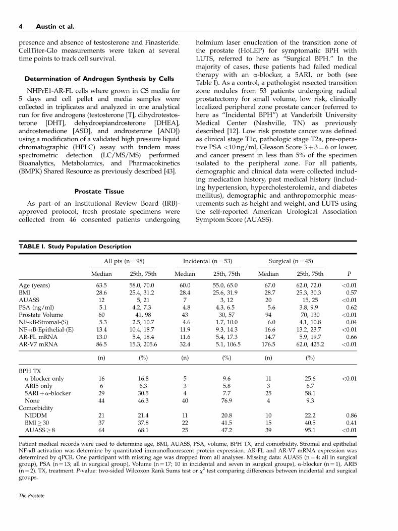
TABLE I. Study Population Description
All pts (n¼ 98) Incidental (n¼ 53) Surgical (n¼ 45)
Median 25th, 75th Median 25th, 75th Median 25th, 75th P
Age (years) 63.5 58.0, 70.0 60.0 55.0, 65.0 67.0 62.0, 72.0 <0.01BMI 28.6 25.4, 31.2 28.4 25.6, 31.9 28.7 25.3, 30.3 0.57AUASS 12 5, 21 7 3, 12 20 15, 25 <0.01PSA (ng/ml) 5.1 4.2, 7.3 4.8 4.3, 6.5 5.6 3.8, 9.9 0.62Prostate Volume 60 41, 98 43 30, 57 94 70, 130 <0.01NF-kB-Stromal-(S) 5.3 2.5, 10.7 4.6 1.7, 10.0 6.0 4.1, 10.8 0.04NF-kB-Epithelial-(E) 13.4 10.4, 18.7 11.9 9.3, 14.3 16.6 13.2, 23.7 <0.01AR-FL mRNA 13.0 5.4, 18.4 11.6 5.4, 17.3 14.7 5.9, 19.7 0.66AR-V7 mRNA 86.5 15.3, 205.6 32.4 5.1, 106.5 176.5 62.0, 425.2 <0.01
(n) (%) (n) (%) (n) (%)
BPH TXa blocker only 16 16.8 5 9.6 11 25.6 <0.01ARI5 only 6 6.3 3 5.8 3 6.75ARIþa-blocker 29 30.5 4 7.7 25 58.1None 44 46.3 40 76.9 4 9.3
ComorbidityNIDDM 21 21.4 11 20.8 10 22.2 0.86BMI� 30 37 37.8 22 41.5 15 40.5 0.41AUASS� 8 64 68.1 25 47.2 39 95.1 <0.01
Patient medical records were used to determine age, BMI, AUASS, PSA, volume, BPH TX, and comorbidity. Stromal and epithelialNF-kB activation was determine by quantitated immunofluorescent protein expression. AR-FL and AR-V7 mRNA expression wasdetermined by qPCR. One participant with missing age was dropped from all analyses. Missing data: AUASS (n¼ 4; all in surgicalgroup), PSA (n¼ 13; all in surgical group), Volume (n¼ 17; 10 in incidental and seven in surgical groups), a-blocker (n¼ 1), ARI5(n¼ 2). TX, treatment. P-value: two-sided Wilcoxon Rank Sums test or x2 test comparing differences between incidental and surgicalgroups.
4 Austin et al.
The Prostate
Tissue Processing and Pathology
After gross pathological examination, all prostatesamples used for this study were stored at 4°C andprocessed within 24 hr. Processing of samplesinvolved flash freezing in liquid nitrogen followed bystorage at �80°C until use, as well formalin fixationfor paraffin embedding. Samples were reviewed by apathologist (O.H.) to confirm histologic findings andto exclude those with any foci of cancer.
Quantitative-Real-Time PCR, Western Blot andIHC
For quantitative real-time PCR (qPCR) 50mg flash-frozen tissue was ground using a mortar and pestle inliquid nitrogen and RNA was extracted with Trizol(Ambion) from 46 Surgical BPH and 53 IncidentalBPH specimens. Subsequently, 500 ng RNA wasreverse transcribed into cDNA using RT2 First StrandKit (Qiagen, Valencia, CA). qPCR was performedusing IQ SYBR Green Supermix (BioRad, Hercules,CA) and results were analyzed using BioRad CFXmanager software. All results were calculated usingDDCt analysis and normalized to GAPDH expression.Primer sequences are listed in Table II.
For Western blotting, approximately 50mg of flashfrozen human prostate tissue was ground in liquidnitrogen using a mortar and pestle. Protein wasextracted with 2% SDS buffer and 30mg protein wasrun on pre-made 10% polyacrylamide gels (LifeTechnologies). Primary antibodies were incubated in
5% BSA in TBST overnight at 4°C followed byincubation in secondary antibodies and developmentusing ECL. Membranes were stripped and re-probedwith an antibody against b-actin (Sigma).
Immunohistochemistry was performed as previ-ously described [44]. Briefly, 5mm sections were de-waxed, rehydrated and endogenous peroxidases wereblocked with hydrogen peroxide. Sections were thenboiled in citrate and blocked in 5% serum for 1 hr.Primary antibodies were incubated overnight at4°C at the following concentrations: p-p65-S276(1:200), AR (N-20 1:200) (C-19 1:50), AR-V7 (1:100)(Abcam). Biotinylated anti-mouse or -rabbit second-ary antibodies (DAKO Carpentaria, CA) were incu-bated for 60min at room temperature after slideswere washed for 1 hr in PBS. Slides where incubatedin ABC-HRP complex (Vector Laboratories Burlin-game, CA) for 30min. Bound antibodies where thenvisualized by incubation with 3,30 diaminobenzidinetetrahydrochloride (liquid DAB, DAKO). Slides wherethen rinsed in tap water, counterstained with hema-toxylin, and mounted.
Immunofluorescence
Tissues were fixed with 4% paraformaldehyde,permeablized with 0.5% Triton X-100 and incubatedwith 1% BSA to block non-specific binding. Tissueswere then incubated with rabbit anti-phospho-p65(276) (Abcam) and mouse anti-wide-spectrum cyto-keratin (DAKO) and were visualized with anti-rabbit
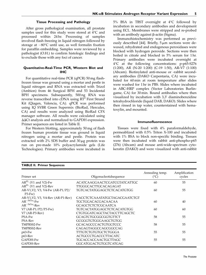
TABLE II. Primer Sequences
Primer set OligonucleotidsequenceAnnealing temp.
(°C)Amplification
cycles
ARFL (V1 and V2)-Fw ACATCAAGGAACTCGATCGTATCATTGC 60 55ARFL (V1 and V2)-Rev TTGGGCACTTGCACAGAGATAR-V1,V2, V3, V4-Fw (AR-P1/P2/P3-Fw)
TGTCACTATGGAGCTCTCACATGTGG 60 55
AR-V1,V2, V3, V4-Rev (AR-P1-Rev) CACCTCTCAAATATGCTAGACGAATCTGTAR�567es-Fw TGCTGGACACGACAACAA 60 40AR�567es-Rev GCAGCTCTCTCGCAATCAV7 (AR-P1/P2/P3-Fw) TGTCACTATGGAGCTCTCACATGTGG 60 60V7 (AR-P3-Rev) CTGTGGATCAGCTACTACCTTCAGCTCPSA-Fw GCAGTCTGCGGCGGTGTTCT 58 55PSA-Rev GCGGGTGTGGGAAGCTGTGGTMPRSS2-Fw GCACAGCCCACTGTGGTCCC 58 55TMPRSS2-Rev CAGAGTAGGCCAGCGGCCAGp63-Fw TTTGTCTGTGTGCTCTGGGA 55 55p63-Rev ACTGCCCTGACCCTTACATCGAPDH-Fw TGCACCACCAACTGCTTAGC 55 55GAPDH-Rev GGCATGGACTGTGGTCATGAG
NK-kB Stimulates Androgen Receptor Variant Expression 5
The Prostate
594-rhodamine-conjugated or anti-mouse FITC-conjugated secondary antibodies (Life Technologies)and nuclei were visualized with DAPI (Vector Labora-tories).
Image Analysis
Immunostained tissue images were captured usinga high throughput Leica SCN400 Slide Scanner auto-mated digital image system from Leica Microsystems.Whole slides were imaged at 20� magnification to aresolution of 0.5mm/pixel. Tissue cores were mappedusing Ariol1 Review software. The numbers of posi-tive (brown) and negative (blue) nuclei were deter-mined by analysis of the high-resolution images in theAriol1 software. Fluorescent, immunostained tissueslides were imaged on an Ariol SL-50 automated slidescanner (Leica Biosystems). Tissue cores were imagedat 20� magnification to a resolution of 0.323mm/pixel. The software used in this work, CellProfiler 2.0,is an open source package for Windows (availablefrom the Broad Institute at www.cellprofiler.org). Thesoftware uses a pipeline (available upon request fromthe authors) of modules designed to automaticallyidentify, quantify, and export the area-shape measure-ments of cells stained for wide spectrum cytokeratin,phosphorylated p65, and DAPI (nucleus). The pipe-line also saves a resultant image with each celldetected (outlined in color). Analysis included totalcell count to determine NF-kB positive epithelial cells(wide spectrum cytokeratin, p65-S276 and DAPI) orNF-kB positive stromal cells (p65-S276 and DAPI).
Statistical Analysis
Chi-square and the Wilcoxon-Rank Sum tests wereused for univariate comparisons of study character-istics between Surgical and Incidental patients. Pri-mary analyses of differences in NF-kB, AR, or AR-V7expression across Incidental and Surgical groups wereperformed within a linear regression model thatallowed us to adjust for differences in age, body massindex (BMI), and Non-insulin dependent diabetesmellitus (NIDDM) between groups. Additionalregression analyses investigated the age- and group-adjusted associations between NF-kB, AR, and AR-V7Levels with BMI, NIDDM, and 5ARI and a-blockeruse. Tissue markers were natural log transformedprior to analysis to normalize these distributions asnecessary to meet model assumptions, and adjustedmean biomarker values were then back transformedsuch that geometric means are reported. A two-sidedP-value of 0.05 or less was considered statisticallysignificant. A One-Way ANOVA test was used forcomparisons of characteristics between IKK2-empty
vector (EV), IKK2-kinase dead (KD), and IKK2-constitutively active (EE) gene expression. A Wil-coxon-Rank Sum test was used when comparinginhibition of NF-kB in EE to EV. Correlation betweenAR-FL, AR-V7, and AUASS and TRUS volume wasevaluated by the determination of the Spearmancorrelation coefficient (GraphPad Prism).
RESULTS
Activation of NF-kB Signaling Is Associated WithClinical Progression of BPH
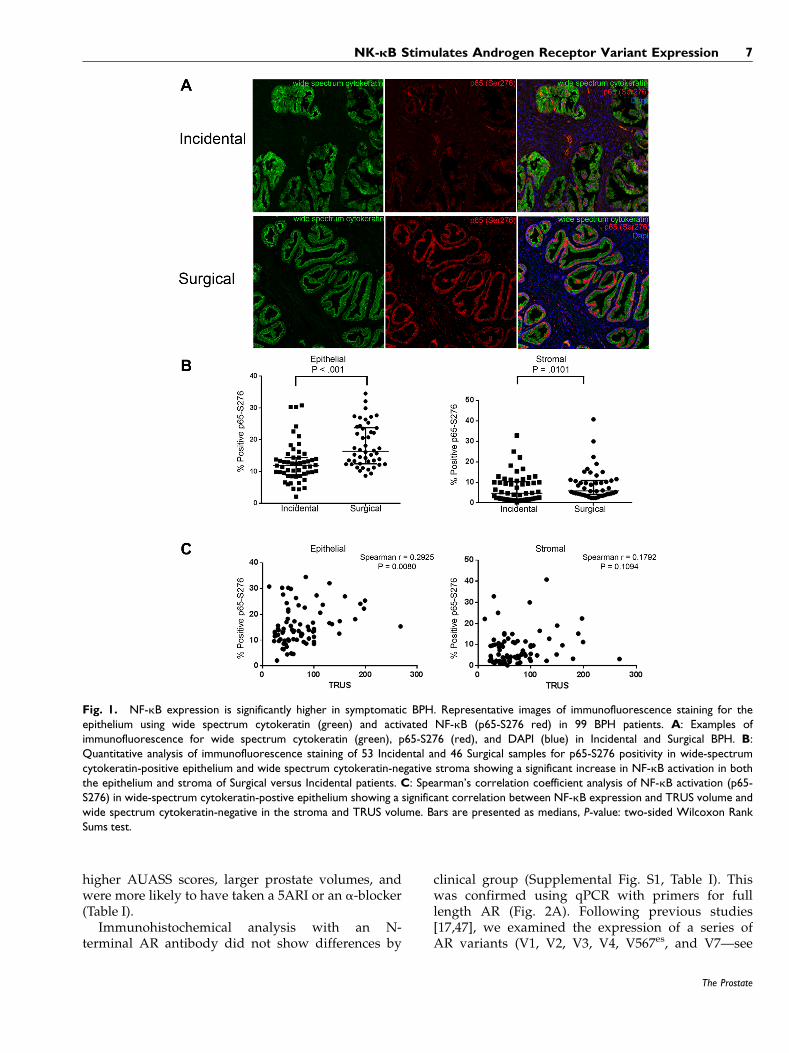
Progressive BPH is commonly associated withinflammation [45], and indeed has molecular similari-ties to other autoimmune inflammatory conditions[12]. The NF-kB pathway is a molecular program thatplays an important role in the regulation of inflamma-tion. An unbiased high resolution scanning analysisof immunofluorescent nuclear phospho-p65 Localiza-tion (indicating canonical NF-kB signaling) was per-formed to compare NF-kB activation in 46 clinicallyadvanced Surgical BPH specimens versus 53 Inciden-tal BPH specimens. As illustrated in a representativeexample in Figure 1A, and quantified in Figure1B there is significantly higher nuclear phospho-p65in the more advanced Surgical samples compared toIncidental BPH. Nuclear p65 Levels were also higherwithin the epithelial versus stromal compartment inboth clinical groups, and the epithelial compartmentin Surgical BPH patients displayed the highest p65Levels (P< 0.01). We next investigated whether acti-vated NF-kB expression was associated withincreased LUTS. We examined all Incidental andSurgical BPH patients (n¼ 99), and analyzed patientswhose TRUS (TransRectal UltraSound) volume meas-urements was included in our data set (n¼ 81). Welooked at NF-kB activation by compartment and sawthat epithelial NF-kB activation significantly corre-lated with TRUS volume while there was no signifi-cant correlation with stromal NF-kB activation andTRUS volume (Fig. 1C).
BPH Progression Is Associated With IncreasedExpression of AR-V7
Recent studies have linked NF-kB activation toandrogen receptor expression, and in particular to theexpression of AR variants, in castrate-resistant pros-tatic cancer [31,34,46]. Given that most advanced BPHpatients are refractory to 5ARI therapy, we examinedthe expression of both full length and variant forms ofthe AR in Surgical and Incidental BPH patientsamples. When compared to Incidental BPH patients,Surgical BPH patients were significantly older, had
6 Austin et al.
The Prostate
higher AUASS scores, larger prostate volumes, andwere more likely to have taken a 5ARI or an a-blocker(Table I).
Immunohistochemical analysis with an N-terminal AR antibody did not show differences by
clinical group (Supplemental Fig. S1, Table I). Thiswas confirmed using qPCR with primers for fulllength AR (Fig. 2A). Following previous studies[17,47], we examined the expression of a series ofAR variants (V1, V2, V3, V4, V567es, and V7—see
Fig. 1. NF-kB expression is significantly higher in symptomatic BPH. Representative images of immunofluorescence staining for theepithelium using wide spectrum cytokeratin (green) and activated NF-kB (p65-S276 red) in 99 BPH patients. A: Examples ofimmunofluorescence for wide spectrum cytokeratin (green), p65-S276 (red), and DAPI (blue) in Incidental and Surgical BPH. B:Quantitative analysis of immunofluorescence staining of 53 Incidental and 46 Surgical samples for p65-S276 positivity in wide-spectrumcytokeratin-positive epithelium and wide spectrum cytokeratin-negative stroma showing a significant increase in NF-kB activation in boththe epithelium and stroma of Surgical versus Incidental patients. C: Spearman’s correlation coefficient analysis of NF-kB activation (p65-S276) in wide-spectrum cytokeratin-postive epithelium showing a significant correlation between NF-kB expression and TRUS volume andwide spectrum cytokeratin-negative in the stroma and TRUS volume. Bars are presented as medians, P-value: two-sided Wilcoxon RankSums test.
NK-kB Stimulates Androgen Receptor Variant Expression 7
The Prostate
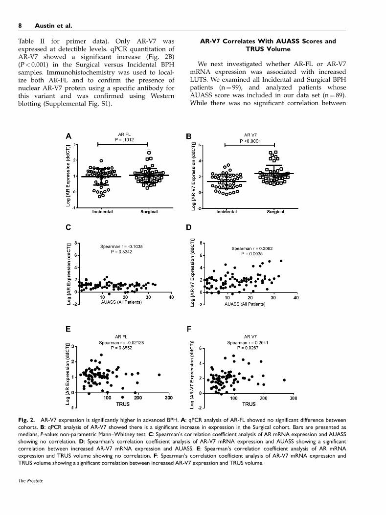
Table II for primer data). Only AR-V7 wasexpressed at detectible levels. qPCR quantitation ofAR-V7 showed a significant increase (Fig. 2B)(P< 0.001) in the Surgical versus Incidental BPHsamples. Immunohistochemistry was used to local-ize both AR-FL and to confirm the presence ofnuclear AR-V7 protein using a specific antibody forthis variant and was confirmed using Westernblotting (Supplemental Fig. S1).
AR-V7 Correlates With AUASS Scores andTRUS Volume
We next investigated whether AR-FL or AR-V7mRNA expression was associated with increasedLUTS. We examined all Incidental and Surgical BPHpatients (n¼ 99), and analyzed patients whoseAUASS score was included in our data set (n¼ 89).While there was no significant correlation between
Fig. 2. AR-V7 expression is significantly higher in advanced BPH. A: qPCR analysis of AR-FL showed no significant difference betweencohorts. B: qPCR analysis of AR-V7 showed there is a significant increase in expression in the Surgical cohort. Bars are presented asmedians, P-value: non-parametric Mann–Whitney test. C: Spearman’s correlation coefficient analysis of AR mRNA expression and AUASSshowing no correlation. D: Spearman’s correlation coefficient analysis of AR-V7 mRNA expression and AUASS showing a significantcorrelation between increased AR-V7 mRNA expression and AUASS. E: Spearman’s correlation coefficient analysis of AR mRNAexpression and TRUS volume showing no correlation. F: Spearman’s correlation coefficient analysis of AR-V7 mRNA expression andTRUS volume showing a significant correlation between increased AR-V7 expression and TRUS volume.
8 Austin et al.
The Prostate
AR-FL expression and AUASS (Fig. 2C), there was asignificant correlation between increased AUASS andAR-V7 expression (P< 0.0035) (Fig. 2D). To use anon-subjective measurement to determine the associa-tion between AR-FL and AR-V7 mRNA expressionwith increased LUTS, we used TRUS volumes thatwere included in our data set (n¼ 81). While therewas no significant correlation between AR-FL expres-sion and TRUS volume (Fig. 2E), there was a signifi-cant correlation between increased TRUS volume andAR-V7 expression (P¼ 0.0267).
Inflammation Is Associated With Increased ARExpression
To determine whether adjacent inflammation isassociated with increased AR and NF-kB expressionwe performed immunohistochemistry for inflamma-tory cells, AR, and activated NF-kB (p-p65). Inflam-matory markers such as CD3e and CD4 were used todetermine na€ıve T-lymphocytes and T-helper lympho-cytes respectively. As shown in representative images
in Supplemental Figure S2, epithelial cells adjacent toareas with low inflammation (Supplemental Fig. S2A)had lower expression levels of AR and activatedNF-kB. In areas with high areas of inflammation(Supplemental Fig. S2B) correlated with increasedexpression of AR and NF-kB. Incidental diseasesamples displayed relatively low levels of AR expres-sion and NF-kB activation while Surgical BPHpatients displayed increased expression of AR andNF-kB in inflamed areas.
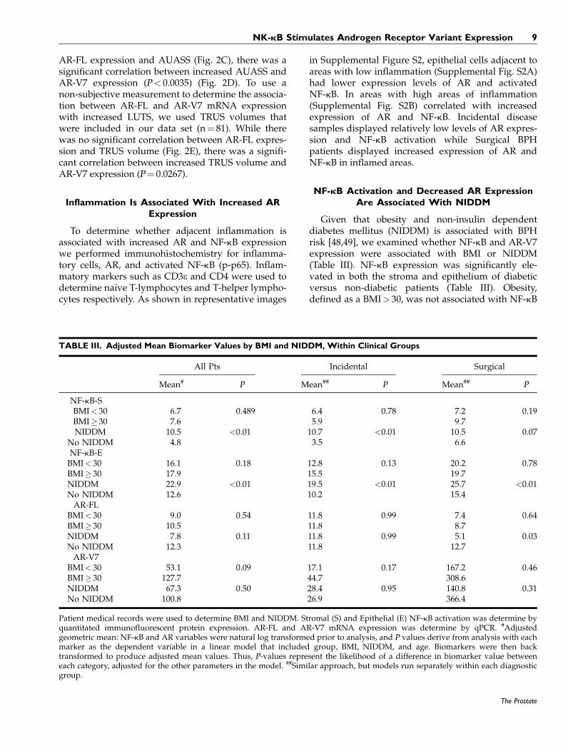
NF-kB Activation and Decreased AR ExpressionAre Associated With NIDDM
Given that obesity and non-insulin dependentdiabetes mellitus (NIDDM) is associated with BPHrisk [48,49], we examined whether NF-kB and AR-V7expression were associated with BMI or NIDDM(Table III). NF-kB expression was significantly ele-vated in both the stroma and epithelium of diabeticversus non-diabetic patients (Table III). Obesity,defined as a BMI> 30, was not associated with NF-kB
TABLE III. Adjusted Mean Biomarker Values by BMI and NIDDM, Within Clinical Groups
All Pts Incidental Surgical
Mean# P Mean## P Mean## P
NF-kB-SBMI< 30 6.7 0.489 6.4 0.78 7.2 0.19BMI� 30 7.6 5.9 9.7NIDDM 10.5 <0.01 10.7 <0.01 10.5 0.07
No NIDDM 4.8 3.5 6.6NF-kB-EBMI< 30 16.1 0.18 12.8 0.13 20.2 0.78BMI� 30 17.9 15.5 19.7NIDDM 22.9 <0.01 19.5 <0.01 25.7 <0.01No NIDDM 12.6 10.2 15.4AR-FL
BMI< 30 9.0 0.54 11.8 0.99 7.4 0.64BMI� 30 10.5 11.8 8.7NIDDM 7.8 0.11 11.8 0.99 5.1 0.03No NIDDM 12.3 11.8 12.7AR-V7
BMI< 30 53.1 0.09 17.1 0.17 167.2 0.46BMI� 30 127.7 44.7 308.6NIDDM 67.3 0.50 28.4 0.95 140.8 0.31No NIDDM 100.8 26.9 366.4
Patient medical records were used to determine BMI and NIDDM. Stromal (S) and Epithelial (E) NF-kB activation was determine byquantitated immunofluorescent protein expression. AR-FL and AR-V7 mRNA expression was determine by qPCR. #Adjustedgeometric mean: NF-kB and AR variables were natural log transformed prior to analysis, and P values derive from analysis with eachmarker as the dependent variable in a linear model that included group, BMI, NIDDM, and age. Biomarkers were then backtransformed to produce adjusted mean values. Thus, P-values represent the likelihood of a difference in biomarker value betweeneach category, adjusted for the other parameters in the model. ##Similar approach, but models run separately within each diagnosticgroup.
NK-kB Stimulates Androgen Receptor Variant Expression 9
The Prostate

activation after controlling for NIDDM. Patterns weresimilar within Incidental and Surgical BPH groups,with no significant difference in epithelial or stromalcell NF-kB activity with either high or low BMI. Incontrast, AR-FL expression was significantly lower inSurgical BPH patients with NIDDM, while differencesin AR-V7 expression levels were not significantlyassociated with BMI or NIDDM.
Since androgen ablation can lead to the expressionof AR-Vs in prostate cancer [30], we also examinedwhether medication used to treat BPH was associatedwith NF-kB activation, AR-FL or AR-V7 expression(Table IV). There was a significant increase in AR-V7in patients with a history of a-blocker use (Table IV),whether in Incidental or Surgical BPH groups. Whilethis result certainly warrants future investigation, wenote that a-blockers are the front line therapy for BPHsymptoms and this may simply reflect an underlyingbiology of the disease where patients with high levelsof AR-V7 may be more likely to show symptoms thatare treatable with a-blockers.
Activation of NF-kB Upregulates AR-FL andAR-V7 Expression in Benign Prostate Epithelial
and Stromal Cells
Activation of AR-Vs by NF-kB has previouslybeen described in prostate cancer but not in BPH.To determine whether chronic activation of NF-kBresults in increased expression of AR and influ-enced cell growth and function, we retrovirallytransduced benign human prostatic NHPrE1 (epi-thelial) and BHPrS1 (stromal) cells [35,36], usingempty vector (EV), kinase dead IKK2 (KD), orconstitutively active IKK2 (EE) retroviral constructs.KD will block NF-kB activity while EE will increaseNF-kB activity. Cells were grown in charcoal-stripped FBS (CS) in the presence/absence of10�8M DHT. Activation of NF-kB signaling wasassessed using the NGL reporter, a plasmid with anNF-kB responsive element coupled to GFP/Lucifer-ase. We confirmed the successful transduction andincreased expression of NF-kB signaling in NHPrE1cells (Fig. 3A) and BHPrS1 cells (Fig. 3B). Asexpected, the background level of NF-kB activitywas suppressed by the KD construct and enhancedby the constitutively active EE form of IKK2. Thepresence or absence of DHT had no significanteffect on the activation of NF-kB.
Having established that the transductions wereeffective, we used the ARR2PB-Luc reporter thatresponds to activation of the AR but has no bindingsites for NF-kB. Both NHPrE1 and BHPrS1 showedsignificant activation of the reporter construct whenNF-kB signaling was constitutively activated even in
the absence of androgens (Fig. 3C and D). Stromalcells have a stronger basal reporter activity and asmaller induction of the reporter with DHT withoutthe expression of NF-kB, likely reflecting basal expres-sion of low levels of AR in culture.
NHPrE1 epithelial cells, which do not normallyexpress AR in vitro, showed a clearer induction ofsignal. Addition of DHT increased the activity of theandrogen-driven reporter in both epithelial and stro-mal cells (Fig. 3C and D). We repeated this experimentusing a PSA promoter driven luciferase reporter. Thisconstruct contains AR responsive elements and alsoan NF-kB responsive element, previously identifiedin the PSA gene [50]. Activation of NF-kB droveincreased PSA promoter activity that was onlyslightly increased by the addition of androgens(Fig. 3E). These data demonstrate that cells in which
TABLE IV. Adjusted Mean Biomarker Values by BPHMedication History
Mean
Marker Drug(s) All Inci Surg
NF-kB-S 5ARI only 4.9 5.3 3.9a blocker only 5.3 2.0 9.75ARIþa-blocker 5.2 3.1 7.1None 6.2 5.1 3.4P 0.90 0.26 0.04
NF-kB-E 5ARI only 12.4 12.7 12.2a blocker only 15.3 11.8 19.45ARIþa-blocker 13.2 7.1 17.5None 14.5 11.9 14.9P 0.64 0.30 0.24
AR-FL 5ARI only 14.1 4.4 31.2a blocker only 13.7 21.3 10.25ARIþa-blocker 11.5 43.7 8.1None 9.1 9.9 16.2P 0.69 0.01 0.24
AR-V7 5ARI only 22.9 1.8 150.9a blocker only 450.8 142.5 1290.55ARIþa-blocker 92.4 60.4 212.2None 45.9 20.0 51.9P 0.01 0.06 0.11
Patient medical records were used to determine medicationhistory. Stromal and Epithelial NF-kB activation was determineby quantitated immunofluorescent protein expression. AR-FLand AR-V7 mRNA expression was determined by qPCR.Control for group and age; Group specific analyses control forage; P-values—omnibus test of significant variability within thegroup being tested, simply indicating whether or not there is asignificant association between drug use and biomarker. Spe-cific mean values may be based on only a handful of subjects.Check n values from Table I. Early associations between 5ARIand NF-kB were due to confounding by differences in 5ARI usebetween groups. Once group differences are controlled for,5ARI is no longer associated.
10 Austin et al.
The Prostate
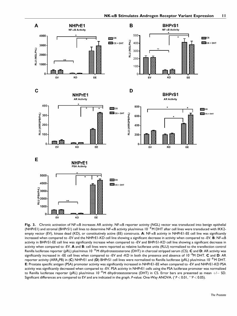
Fig. 3. Chronic activation of NF-kB increases AR activity. NF-kB reporter activity (NGL) vector was transduced into benign epithelial(NHPrE1) and stromal (BHPrS1) cell lines to determine NF-kB activity plus/minus 10�8M DHT after cell lines were transduced with IKK2-empty vector (EV), kinase dead (KD), or constitutively active (EE) constructs. A: NF-kB activity in NHPrE1-EE cell line was significantlyincreased when compared to -EV and the NHPrE1-KD cell line showing a significant decrease in activity when compared to -EV. B: NF-kBactivity in BHPrS1-EE cell line was significantly increase when compared to -EV and BHPrS1-KD cell line showing a significant decrease inactivity when compared to -EV. A and B: cell lines were reported as relative luciferase units (RLU) normalized to the transfection controlRenilla luciferase reporter (pRL) plus/minus 10�8M dihydrotestosterone (DHT) in charcoal stripped serum (CS). C and D: AR activity wassignificantly increased in -EE cell lines when compared to -EV and -KD in both the presence and absence of 10�8M DHT. C and D: ARreporter activity (ARR2PB) in (C) NHPrE1 and (D) BHPrS1 cell lines were normalized to Renilla luciferase (pRL) plus/minus 10�8M DHT.E: Prostate specific antigen (PSA) promoter activity was significantly increased in NHPrE1-EE when compared to -EV and NHPrE1-KD PSAactivity was significantly decreased when compared to -EV. PSA activity in NHPrE1 cells using the PSA luciferase promoter was normalizedto Renilla luciferase reporter (pRL) plus/minus 10�8M dihydrotestosterone (DHT) in CS. Error bars are presented as mean þ/� SD.Significant differences are compared to EV and are indicated in the graph. P-value: One-Way ANOVA. (�P< 0.01, ��P< 0.05).
NK-kB Stimulates Androgen Receptor Variant Expression 11
The Prostate
NF-kB is active are able to activate AR reporters in theabsence of ligand, but retain an additional and greaterligand-driven response.
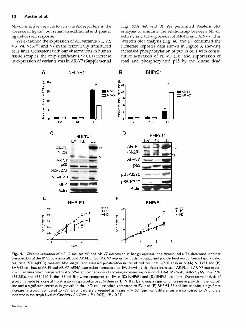
We examined the expression of AR variants V1, V2,V3, V4, V567es, and V7 in the retrovirally transducedcells lines. Consistent with our observations in humantissue samples, the only significant (P< 0.01) increasein expression of variants was in AR-V7 (Supplemental
Figs. S3A, 4A and B). We performed Western blotanalysis to examine the relationship between NF-kBactivity and the expression of AR-FL and AR-V7. ThisWestern blot analysis (Fig. 4C and D) confirmed theluciferase reporter data shown in Figure 3, showingincreased phosphorylation of p65 in cells with consti-tutive activation of NF-kB (EE) and suppression oftotal and phosphorylated p65 by the kinase dead
Fig. 4. Chronic activation of NF-kB induces AR and AR-V7 expression in benign epithelial and stromal cells. To determine whethertransduction of the IKK2 construct affected AR-FL and/or AR-V7 expression at the message and protein level we performed quantitativereal time PCR (qPCR), western blot analysis and assessed proliferation in transduced cell lines. qPCR analysis of (A) NHPrE1 and (B)BHPrS1 cell lines of AR-FL and AR-V7 mRNA expression normalized to -EV, showing a significant increase in AR-FL and AR-V7 expressionin -EE cell lines when compared to -EV. Western blot analysis of showing increased expression of AR/ARV (N-20), AR-V7, p65, p65-S276,p65-S536, and p65K310 in the -EE cell line when compared to -EV in (C) NHPrE1 and (D) BHPrS1 cell lines. Quantitative analysis ofgrowth is made by a crystal violet assay using absorbance at 570 nm in (E) NHPrE1, showing a significant increase in growth in the -EE cellline and a significant decrease in growth in the -KD cell line when compared to EV, and (F) BHPrS1-EE cell line showing a significantincrease in growth compared to -EV. Error bars are presented as means þ/� SD. Significant differences are compared to EV and areindicated in the graph P-value: One-Way ANOVA. (�P< 0.05), ��P< 0.01).
12 Austin et al.
The Prostate
(KD) construct. Both AR-FL and AR-V7 proteins wereupregulated in cells in which NF-kB signaling wasconstitutively activated. To evaluate the biologicaleffects of NF-kB on proliferation, we performedgrowth assays. Constitutive activation of NF-kB inprostate epithelial and stromal cells was associatedwith an increase in proliferation starting by 3 days inculture in the absence of androgens, compared to theempty vector and kinase dead NF-kB (Fig. 4E and F).
To determine whether the increased proliferationwas due to NF-kB or AR-FL we used a cell viabilityassay based on quantitation of ATP, using cellularmetabolism as a surrogate for cell number. AR expres-sion was knocked down in NHPrE1-EV and NHPrE1-EE cell lines. Cells were grown in charcoal-stripped(CS) serum in vehicle or in the presence of10�7M Testosterone (T). There was a significantincrease in proliferation in EE (P< 0.001) and EE-ARknockdown cells (P< 0.003) when compared to EVand EV-AR knockdown respectively (SupplementalFig. S3B). This highlights that the increased prolifera-tion is due to both the expression of NF-kB andexpression of AR and increased proliferation whenNF-kB and AR are coexpressed.
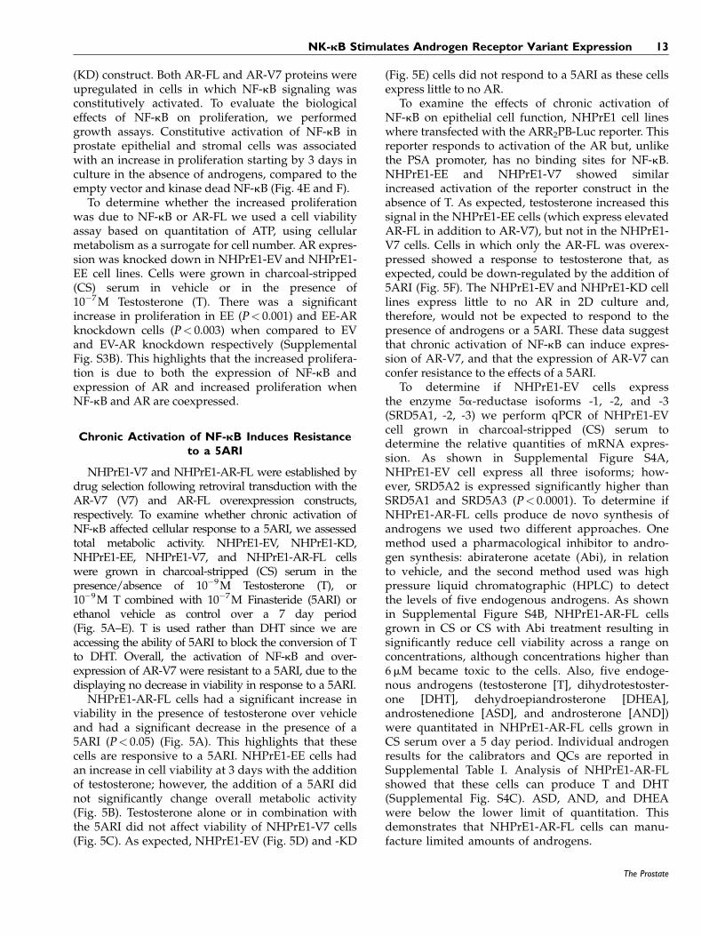
Chronic Activation of NF-kB Induces Resistanceto a 5ARI
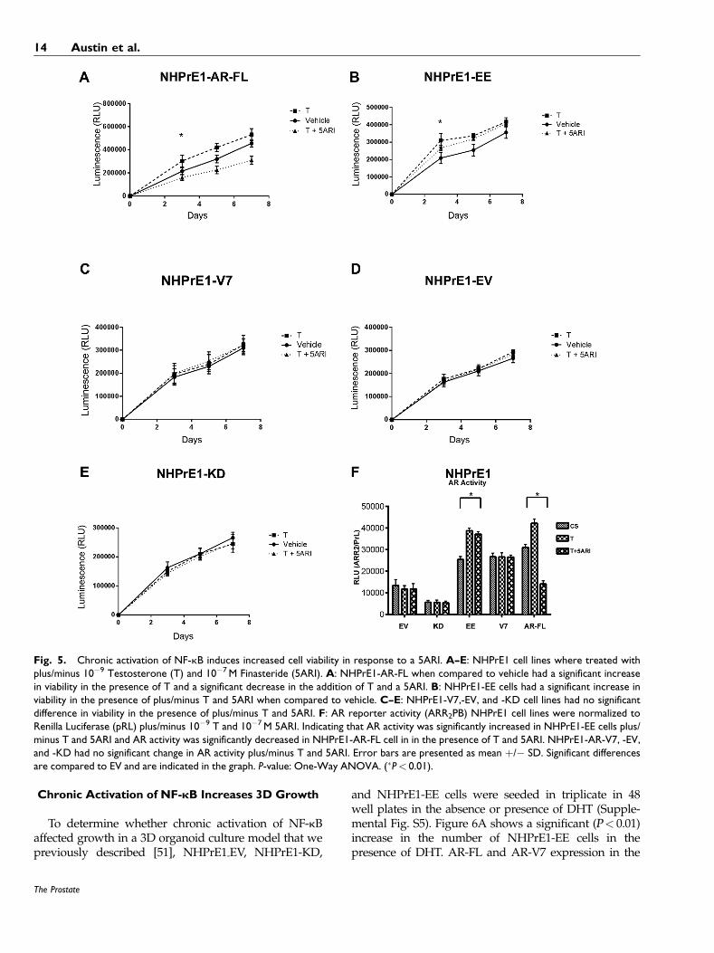
NHPrE1-V7 and NHPrE1-AR-FL were established bydrug selection following retroviral transduction with theAR-V7 (V7) and AR-FL overexpression constructs,respectively. To examine whether chronic activation ofNF-kB affected cellular response to a 5ARI, we assessedtotal metabolic activity. NHPrE1-EV, NHPrE1-KD,NHPrE1-EE, NHPrE1-V7, and NHPrE1-AR-FL cellswere grown in charcoal-stripped (CS) serum in thepresence/absence of 10�9M Testosterone (T), or10�9M T combined with 10�7M Finasteride (5ARI) orethanol vehicle as control over a 7 day period(Fig. 5A–E). T is used rather than DHT since we areaccessing the ability of 5ARI to block the conversion of Tto DHT. Overall, the activation of NF-kB and over-expression of AR-V7 were resistant to a 5ARI, due to thedisplaying no decrease in viability in response to a 5ARI.
NHPrE1-AR-FL cells had a significant increase inviability in the presence of testosterone over vehicleand had a significant decrease in the presence of a5ARI (P< 0.05) (Fig. 5A). This highlights that thesecells are responsive to a 5ARI. NHPrE1-EE cells hadan increase in cell viability at 3 days with the additionof testosterone; however, the addition of a 5ARI didnot significantly change overall metabolic activity(Fig. 5B). Testosterone alone or in combination withthe 5ARI did not affect viability of NHPrE1-V7 cells(Fig. 5C). As expected, NHPrE1-EV (Fig. 5D) and -KD
(Fig. 5E) cells did not respond to a 5ARI as these cellsexpress little to no AR.
To examine the effects of chronic activation ofNF-kB on epithelial cell function, NHPrE1 cell lineswhere transfected with the ARR2PB-Luc reporter. Thisreporter responds to activation of the AR but, unlikethe PSA promoter, has no binding sites for NF-kB.NHPrE1-EE and NHPrE1-V7 showed similarincreased activation of the reporter construct in theabsence of T. As expected, testosterone increased thissignal in the NHPrE1-EE cells (which express elevatedAR-FL in addition to AR-V7), but not in the NHPrE1-V7 cells. Cells in which only the AR-FL was overex-pressed showed a response to testosterone that, asexpected, could be down-regulated by the addition of5ARI (Fig. 5F). The NHPrE1-EV and NHPrE1-KD celllines express little to no AR in 2D culture and,therefore, would not be expected to respond to thepresence of androgens or a 5ARI. These data suggestthat chronic activation of NF-kB can induce expres-sion of AR-V7, and that the expression of AR-V7 canconfer resistance to the effects of a 5ARI.
To determine if NHPrE1-EV cells expressthe enzyme 5a-reductase isoforms -1, -2, and -3(SRD5A1, -2, -3) we perform qPCR of NHPrE1-EVcell grown in charcoal-stripped (CS) serum todetermine the relative quantities of mRNA expres-sion. As shown in Supplemental Figure S4A,NHPrE1-EV cell express all three isoforms; how-ever, SRD5A2 is expressed significantly higher thanSRD5A1 and SRD5A3 (P< 0.0001). To determine ifNHPrE1-AR-FL cells produce de novo synthesis ofandrogens we used two different approaches. Onemethod used a pharmacological inhibitor to andro-gen synthesis: abiraterone acetate (Abi), in relationto vehicle, and the second method used was highpressure liquid chromatographic (HPLC) to detectthe levels of five endogenous androgens. As shownin Supplemental Figure S4B, NHPrE1-AR-FL cellsgrown in CS or CS with Abi treatment resulting insignificantly reduce cell viability across a range onconcentrations, although concentrations higher than6mM became toxic to the cells. Also, five endoge-nous androgens (testosterone [T], dihydrotestoster-one [DHT], dehydroepiandrosterone [DHEA],androstenedione [ASD], and androsterone [AND])were quantitated in NHPrE1-AR-FL cells grown inCS serum over a 5 day period. Individual androgenresults for the calibrators and QCs are reported inSupplemental Table I. Analysis of NHPrE1-AR-FLshowed that these cells can produce T and DHT(Supplemental Fig. S4C). ASD, AND, and DHEAwere below the lower limit of quantitation. Thisdemonstrates that NHPrE1-AR-FL cells can manu-facture limited amounts of androgens.
NK-kB Stimulates Androgen Receptor Variant Expression 13
The Prostate
Chronic Activation of NF-kB Increases 3D Growth
To determine whether chronic activation of NF-kBaffected growth in a 3D organoid culture model that wepreviously described [51], NHPrE1-EV, NHPrE1-KD,
and NHPrE1-EE cells were seeded in triplicate in 48well plates in the absence or presence of DHT (Supple-mental Fig. S5). Figure 6A shows a significant (P< 0.01)increase in the number of NHPrE1-EE cells in thepresence of DHT. AR-FL and AR-V7 expression in the
Fig. 5. Chronic activation of NF-kB induces increased cell viability in response to a 5ARI. A–E: NHPrE1 cell lines where treated withplus/minus 10�9 Testosterone (T) and 10�7M Finasteride (5ARI). A: NHPrE1-AR-FL when compared to vehicle had a significant increasein viability in the presence of T and a significant decrease in the addition of T and a 5ARI. B: NHPrE1-EE cells had a significant increase inviability in the presence of plus/minus T and 5ARI when compared to vehicle. C–E: NHPrE1-V7,-EV, and -KD cell lines had no significantdifference in viability in the presence of plus/minus T and 5ARI. F: AR reporter activity (ARR2PB) NHPrE1 cell lines were normalized toRenilla Luciferase (pRL) plus/minus 10�9 T and 10�7M 5ARI. Indicating that AR activity was significantly increased in NHPrE1-EE cells plus/minus T and 5ARI and AR activity was significantly decreased in NHPrE1-AR-FL cell in in the presence of T and 5ARI. NHPrE1-AR-V7, -EV,and -KD had no significant change in AR activity plus/minus T and 5ARI. Error bars are presented as mean þ/� SD. Significant differencesare compared to EV and are indicated in the graph. P-value: One-Way ANOVA. (�P< 0.01).
14 Austin et al.
The Prostate
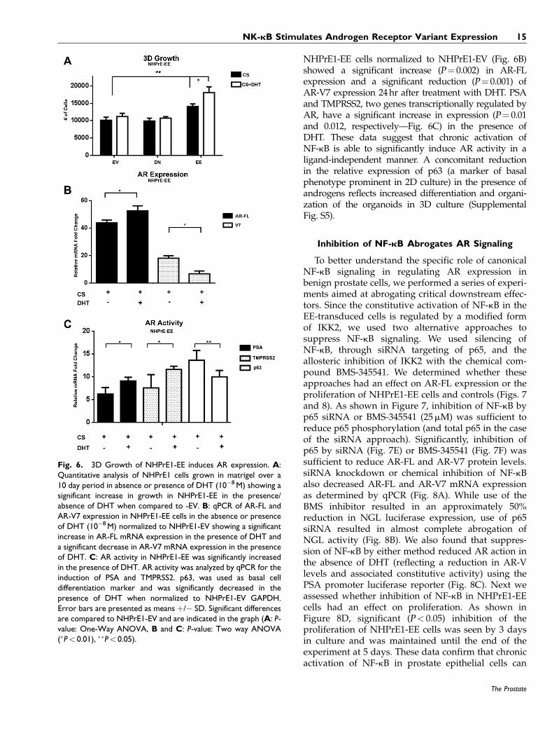
NHPrE1-EE cells normalized to NHPrE1-EV (Fig. 6B)showed a significant increase (P¼ 0.002) in AR-FLexpression and a significant reduction (P¼ 0.001) ofAR-V7 expression 24hr after treatment with DHT. PSAand TMPRSS2, two genes transcriptionally regulated byAR, have a significant increase in expression (P¼ 0.01and 0.012, respectively—Fig. 6C) in the presence ofDHT. These data suggest that chronic activation ofNF-kB is able to significantly induce AR activity in aligand-independent manner. A concomitant reductionin the relative expression of p63 (a marker of basalphenotype prominent in 2D culture) in the presence ofandrogens reflects increased differentiation and organi-zation of the organoids in 3D culture (SupplementalFig. S5).
Inhibition of NF-kB Abrogates AR Signaling
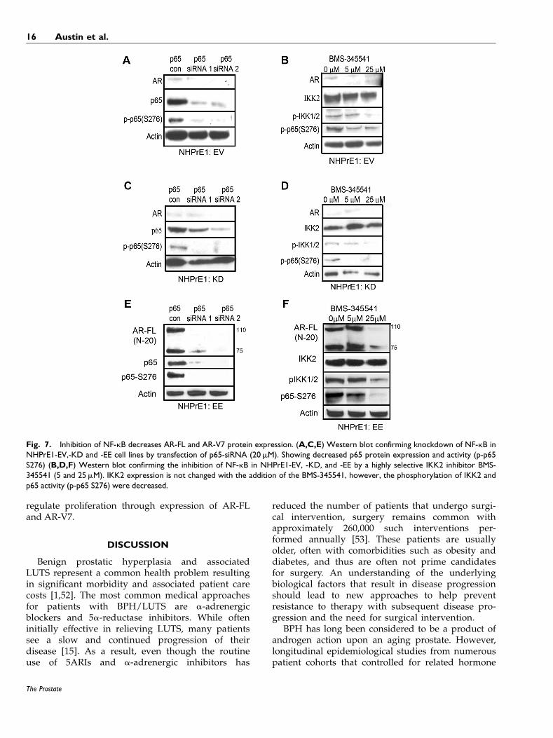
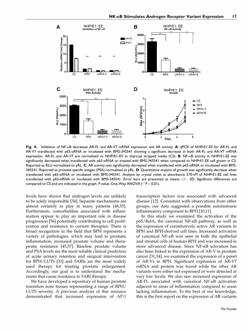
To better understand the specific role of canonicalNF-kB signaling in regulating AR expression inbenign prostate cells, we performed a series of experi-ments aimed at abrogating critical downstream effec-tors. Since the constitutive activation of NF-kB in theEE-transduced cells is regulated by a modified formof IKK2, we used two alternative approaches tosuppress NF-kB signaling. We used silencing ofNF-kB, through siRNA targeting of p65, and theallosteric inhibition of IKK2 with the chemical com-pound BMS-345541. We determined whether theseapproaches had an effect on AR-FL expression or theproliferation of NHPrE1-EE cells and controls (Figs. 7and 8). As shown in Figure 7, inhibition of NF-kB byp65 siRNA or BMS-345541 (25mM) was sufficient toreduce p65 phosphorylation (and total p65 in the caseof the siRNA approach). Significantly, inhibition ofp65 by siRNA (Fig. 7E) or BMS-345541 (Fig. 7F) wassufficient to reduce AR-FL and AR-V7 protein levels.siRNA knockdown or chemical inhibition of NF-kBalso decreased AR-FL and AR-V7 mRNA expressionas determined by qPCR (Fig. 8A). While use of theBMS inhibitor resulted in an approximately 50%reduction in NGL luciferase expression, use of p65siRNA resulted in almost complete abrogation ofNGL activity (Fig. 8B). We also found that suppres-sion of NF-kB by either method reduced AR action inthe absence of DHT (reflecting a reduction in AR-Vlevels and associated constitutive activity) using thePSA promoter luciferase reporter (Fig. 8C). Next weassessed whether inhibition of NF-kB in NHPrE1-EEcells had an effect on proliferation. As shown inFigure 8D, significant (P< 0.05) inhibition of theproliferation of NHPrE1-EE cells was seen by 3 daysin culture and was maintained until the end of theexperiment at 5 days. These data confirm that chronicactivation of NF-kB in prostate epithelial cells can
Fig. 6. 3D Growth of NHPrE1-EE induces AR expression. A:Quantitative analysis of NHPrE1 cells grown in matrigel over a10 day period in absence or presence of DHT (10�8M) showing asignificant increase in growth in NHPrE1-EE in the presence/absence of DHT when compared to -EV. B: qPCR of AR-FL andAR-V7 expression in NHPrE1-EE cells in the absence or presenceof DHT (10�8M) normalized to NHPrE1-EV showing a significantincrease in AR-FL mRNA expression in the presence of DHT anda significant decrease in AR-V7 mRNA expression in the presenceof DHT. C: AR activity in NHPrE1-EE was significantly increasedin the presence of DHT. AR activity was analyzed by qPCR for theinduction of PSA and TMPRSS2. p63, was used as basal celldifferentiation marker and was significantly decreased in thepresence of DHT when normalized to NHPrE1-EV GAPDH.Error bars are presented as means þ/� SD. Significant differencesare compared to NHPrE1-EV and are indicated in the graph (A: P-value: One-Way ANOVA, B and C: P-value: Two way ANOVA(�P< 0.01), ��P< 0.05).
NK-kB Stimulates Androgen Receptor Variant Expression 15
The Prostate
regulate proliferation through expression of AR-FLand AR-V7.
DISCUSSION
Benign prostatic hyperplasia and associatedLUTS represent a common health problem resultingin significant morbidity and associated patient carecosts [1,52]. The most common medical approachesfor patients with BPH/LUTS are a-adrenergicblockers and 5a-reductase inhibitors. While ofteninitially effective in relieving LUTS, many patientssee a slow and continued progression of theirdisease [15]. As a result, even though the routineuse of 5ARIs and a-adrenergic inhibitors has
reduced the number of patients that undergo surgi-cal intervention, surgery remains common withapproximately 260,000 such interventions per-formed annually [53]. These patients are usuallyolder, often with comorbidities such as obesity anddiabetes, and thus are often not prime candidatesfor surgery. An understanding of the underlyingbiological factors that result in disease progressionshould lead to new approaches to help preventresistance to therapy with subsequent disease pro-gression and the need for surgical intervention.
BPH has long been considered to be a product ofandrogen action upon an aging prostate. However,longitudinal epidemiological studies from numerouspatient cohorts that controlled for related hormone
Fig. 7. Inhibition of NF-kB decreases AR-FL and AR-V7 protein expression. (A,C,E) Western blot confirming knockdown of NF-kB inNHPrE1-EV,-KD and -EE cell lines by transfection of p65-siRNA (20mM). Showing decreased p65 protein expression and activity (p-p65S276) (B,D,F) Western blot confirming the inhibition of NF-kB in NHPrE1-EV, -KD, and -EE by a highly selective IKK2 inhibitor BMS-345541 (5 and 25mM). IKK2 expression is not changed with the addition of the BMS-345541, however, the phosphorylation of IKK2 andp65 activity (p-p65 S276) were decreased.
16 Austin et al.
The Prostate
levels have shown that androgen levels are unlikelyto be solely responsible [54]. Separate mechanisms arealmost certainly in play in many patients [48,55].Furthermore, comorbidities associated with inflam-mation appear to play an important role in diseaseprogression [56] potentially contributing to cell prolif-eration and resistance to current therapies. There isbroad recognition in the field that BPH represents avariety of pathologies, which may lead to prostaticinflammation, increased prostate volume and thera-peutic resistance [45,57]. Baseline prostate volumeand PSA levels are the most reliable clinical predictorsof acute urinary retention and surgical interventionfor BPH/LUTS [33] and 5ARIs are the most widelyused therapy for treating prostatic enlargement.Accordingly, our goal is to understand the mecha-nisms that cause resistance to 5ARI therapy.
We have developed a repository of human prostatetransition zone tissues representing a range of BPH/LUTS severity. A previous analysis of this resourcedemonstrated that increased expression of AP-1
transcription factors was associated with advanceddisease [12]. Consistent with observations from othergroups, our data suggested a possible autoimmuneinflammatory component to BPH [10,11].
In this study we examined the activation of thep65/RelA, the canonical NF-kB pathway, as well asthe expression of constitutively active AR variants inBPH and BPH-derived cell lines. Increased activationof canonical NF-kB was seen in both the epithelialand stromal cells of human BPH and was increased inmore advanced disease. Since NF-kB activation hasalso been linked to the expression of AR-V in prostatecancer [31,34], we examined the expression of a panelof AR-Vs in BPH. Significant expression of AR-V7mRNA and protein was found while the other ARvariants were either not expressed or were detected atvery low levels. We also saw increased expression ofAR-FL associated with canonical NF-kB activationadjacent to areas of inflammation compared to areaslacking inflammation. To the best of our knowledge,this is the first report on the expression of AR variants
Fig. 8. Inhibition of NF-kB decreases AR-FL and AR-V7 mRNA expression and AR activity. A: qPCR of NHPrE1-EE for AR-FL andAR-V7 transfected with p65-siRNA or incubated with BMS-345541 showing a significant decrease in both AR-FL and AR-V7 mRNAexpression. AR-FL and AR-V7 are normalized to NHPrE1-EV in charcoal stripped media (CS). B: NF-kB activity in NHPrE1-EE wassignificantly decreased when transfected with p65-siRNA or treated with BMS-345541 when compared to NHPrE1-EE cell grown in CS.Reported as RLU normalized to pRL. C: AR activity was significantly decreased when transfected with p65-siRNA or incubated with BMS-345541. Reported as prostate specific antigen (PSA) normalized to pRL. D: Quantitative analysis of growth was significantly decrease whentransfected with p65-siRNA or incubated with BMS-345541. Analysis by crystal violet at absorbance 570 nM of NHPrE1-EE cell linestransfected with p65-siRNA or incubated with BMS-345541. Error bars are presented as means þ/� SD. Significant differences arecompared to CS and are indicated in the graph. P-value: One-Way ANOVA (��P< 0.01).
NK-kB Stimulates Androgen Receptor Variant Expression 17
The Prostate
in human BPH and suggests a mechanism for escapefrom growth regulation by 5ARI therapy.
Analysis of the clinical samples revealed a numberof interesting correlations in detection of NF-kB andAR-V7. We determined that patients with moreadvanced disease showed higher levels of both epithe-lial and stromal nuclear p-p65 (NF-kB activation) andalso increased AR-V7 transcript levels. We determinedthat increased AR-V7 Levels positively correlate withAUASS and TRUS volume. We also determined thatincreased activation of epithelial cell NF-kB correlateswith increased TRUS volume. This suggests a linkagebetween disease progression, activation of NF-kB andexpression of AR-V7. We hypothesized that obesitywould also be linked to these same outcomes, butbased on a binary analysis (BMI< 30 vs. BMI� 30)there was no significant relationship between obesityand either NF-kB or AR-V7 expression after adjustingfor differences in NIDDM prevalence between groups.In contrast, NIDDM was positively associated withNF-kB expression. Past studies have found directassociations between diabetes and BPH progres-sion [58]. This might suggest that specific forms ofsystemic stress exert distinct influences in the patho-genesis of BPH, and reinforces the idea that, althoughprostatic hyperplasia is associated with both obesityand diabetes, these conditions may act through sepa-rate or overlapping mechanisms [2]. We also saw anassociation between a-blocker treated patients as hav-ing increased NF-kB activation and AR-V7 expression.Conflicting reports in literature suggest that a-adrener-gic receptors are both pro-inflammatory [59–61] andanti-inflammatory [62–64]. Our data would be consis-tent with the latter scenario, that a-adrenergic receptorsare anti-inflammatory and that patients on an a-blockerwould have NF-kB activation increased. This couldalso support our observations of increased AR-V7Levels in this patient population. The role of a-blockerto alter inflammation needs to be elucidated further.
We performed a series of 2D and 3D in vitroexperiments to investigate whether activation ofNF-kB can result in the expression of both AR fulllength (AR-FL) and AR-V7 in benign epithelial andstromal cell lines. The findings were consistent inepithelial and stromal cell lines in that the constitutiveactivation of NF-kB resulted in the coordinate expres-sion of both AR-FL and AR-V7. This is consistent withobservations in prostate cancer where both forms ofthe androgen receptor are regulated together by theNF-kB pathway [34].We next performed a series of invitro experiments to determine if NF-kB and specifi-cally AR-V7 can induce resistance to a 5ARI. Thesefindings demonstrated that forced activation of NF-kBand over expression of AR-V7 are able to induceresistance to a 5ARI. We also showed by HLPC
LC/MS/MS analysis that NHPrE1-AR-FL cells canundertake androgen synthesis. This could suggestanother mechanism by which BPH patients could fail5ARI therapy. Stromal cells are an important compo-nent of BPH, which is often considered to be a diseaseof the stromal tissue with epithelial growth as asecondary consequence. This follows the early obser-vations of McNeal who suggested the reawakening offetal mesenchymal potential contributed to the devel-opment of BPH, a concept subsequently validated byCunha and colleagues in rodent and tissue recombi-nation models [65,66]. The activation of NF-kBpresented here is consistent with the idea that inflam-mation plays a role in BPH progression as well asproviding a potential molecular mechanism of resis-tance to 5ARIs via activation of AR-V7 expression.AR-V7 has been shown to modulate expression of anumber of tumor-promoting autocrine/paracrinegrowth factors in prostate cancer [47]. However, it islikely that this is a multifactorial process in whichunderlying comorbidities play a complex role. Theremay well be a number of alternative and possiblycomplementary pathways involved in this process.For example, our previous observations that AP-1transcription factors (molecules known to be relatedto several immune/inflammatory conditions) arehighly enriched in symptomatic BPH [12]. AP-1transcription factors are post-transcriptionally regu-lated by upstream factors such as NF-kB, JNK, ERK,and p38 [67] and therefore activation of NF-kB canupregulate not only AR-FL and AR-V7 but also AP-1factors which can serve as AR co-factors to regulatetranscription, which could lead to the developmentand progression of therapy-resistant BPH. These path-ways might be interrelated, with no individual change(e.g., androgen synthesis, activation of stress factors,expression of AR-V7) being sufficient to allow escapefrom therapy but that these individual changes maybe additive and allow for eventual regrowth of theprostate in the face of therapy.
In summary, the observations presented hereprovide a potential mechanism to explain thepreviously observed links between prostatic inflam-mation and 5ARI resistance [45], whereby aberrantactivation of NF-kB drives AR-V7 expression result-ing in ligand independent activation of AR result-ing in 5ARI resistance. BPH is a complex conditionand there are likely multiple pathways in play inindividual patients relating to common comorbid-ities including diabetes, obesity, and metabolicsyndrome. An understanding of the stress pathwaysactive in individual patients may provide a route toappropriately tailor therapy and avoid the profile ofresistance and subsequent progression to surgerythat is seen in many patients.
18 Austin et al.
The Prostate
ACKNOWLEDGMENTS
The authors thank Dr. Timothy Blackwell (Vander-bilt) and Dr. Ganesh Raj (UT Southwestern) their kindgifts of reagents. Prostate tissue was provided by theCooperative Human Tissue Network (CHTN) - WesternDivision. We thank the NorthShore Histology andImaging Core for help with the immunohistochemicallocalization of AR and AR-V. The LC-MS/MS analysiswas conducted by the Bioanalytics, Metabolomics andPharmacokinetics (BMPK) Shared Resource of RoswellPark Cancer Institute, and partially supported byNational Cancer Institute (NCI) grant P30CA016056.
REFERENCES
1. Platz EA, Joshu CE, Mondul AM, Peskoe SB, Willett WC,Giovannucci E. Incidence and progression of lower urinarytract symptoms in a large prospective cohort of United Statesmen. J Urol 2012;2:496–501.
2. Hammarsten J, Peeker R. Urological aspects of the metabolicsyndrome. Nat Rev Urol 2011;9:483–494.
3. McNeal J. Pathology of benign prostatic hyperplasia. Insightinto etiology. Urol Clin North Am 1990;3:477–486.
4. Rohrmann S, De Marzo AM, Smit E, Giovannucci E, Platz EA.Serum C-reactive protein concentration and lower urinary tractsymptoms in older men in the Third National Health andNutrition Examination Survey (NHANES III). Prostate2005;1:27–33.
5. Schenk JM, Calip GS, Tangen CM, Goodman P, Parsons JK,Thompson IM, Kristal AR. Indications for and use of nonsteroi-dal antiinflammatory drugs and the risk of incident, symptom-atic benign prostatic hyperplasia: Results from the prostatecancer prevention trial. Am J Epidemiol 2012;2:156–163.
6. Fibbi B, Penna G, Morelli A, Adorini L, Maggi M. Chronicinflammation in the pathogenesis of benign prostatic hyperpla-sia. Int J Androl 2010;3:475–488.
7. McVary KT, Rademaker A, Lloyd GL, Gann P. Autonomicnervous system overactivity in men with lower urinary tractsymptoms secondary to benign prostatic hyperplasia. J Urol2005;4(Pt 1):1327–1433.
8. Ho CK, Habib FK. Estrogen and androgen signaling in thepathogenesis of BPH. Nat Rev Urol 2011;1:29–41.
9. Rodriguez-Nieves JA, Macoska JA. Prostatic fibrosis, lowerurinary tract symptoms, and BPH. Nat Rev Urol 2013;9:546–550.
10. Kramer G, Mitteregger D, Marberger M. Is benign prostatichyperplasia (BPH) an immune inflammatory disease? Eur Urol2007;5:1202–1216.
11. Madigan AA, Sobek KM, Cummings JL, Green WR, Bacich DJ,O’Keefe DS. Activation of innate anti-viral immune responsegenes in symptomatic benign prostatic hyperplasia. GenesImmun 2012;7:566–572.
12. Lin-Tsai O, Clark PE, Miller NL, Fowke JH, Hameed O, HaywardSW, Strand DW. Surgical intervention for symptomatic benignprostatic hyperplasia is correlated with expression of the AP-1transcription factor network. Prostate 2014;6:669–679.
13. Lepor H, Kazzazi A, Djavan. B. alpha-Blockers for benignprostatic hyperplasia: The new era. Curr Opin Urol 2012;1:7–15.
14. Roehrborn CG, Boyle P, Nickel JC, Hoefner K, Andriole G, AriaA, Investigators AS. Efficacy and safety of a dual inhibitor of5-alpha-reductase types 1 and 2 (dutasteride) in men withbenign prostatic hyperplasia. Urology 2002;3:434–441.
15. McConnell JD, Roehrborn CG, Bautista OM, Andriole GL Jr.,Dixon CM, Kusek JW, Lepor H, McVary KT, Nyberg LM Jr.,Clarke HS, Crawford ED, Diokno A, Foley JP, Foster HE, JacobsSC, Kaplan SA, Kreder KJ, Lieber MM, Lucia MS, Miller GJ,Menon M, Milam DF, Ramsdell JW, Schenkman NS, SlawinKM, Smith JA, G. the Medical Therapy of Prostatic SymptomsResearch. The long-term effect of doxazosin, finasteride, andcombination therapy on the clinical progression of benignprostatic hyperplasia. N Engl J Med 2003;25:2387–2398.
16. Lu-Yao GL, Barry MJ, Chang CH, Wasson JH, Wennberg JE.Transurethral resection of the prostate among Medicare benefi-ciaries in the United States: Time trends and outcomes. Prostatepatient outcomes research team (PORT). Urology 1994;5:692–698; discussion 698–699.
17. Hu DG, Hickey TE, Irvine C, Wijayakumara DD, Lu L, TilleyWD, Selth LA, Mackenzie PI. Identification of androgen recep-tor splice variant transcripts in breast cancer cell lines andhuman tissues. Horm Cancer 2014;2:61–71.
18. Ghosh S, Hayden MS. New regulators of NF-kappaB ininflammation. Nat Rev Immunol 2008;11:837–848.
19. Basseres DS, Baldwin AS. Nuclear factor-kappaB and inhibitorof kappaB kinase pathways in oncogenic initiation and progres-sion. Oncogene 2006;51:6817–6830.
20. Toubi E, Shoenfeld Y. Toll-like receptors and their role in thedevelopment of autoimmune diseases. Autoimmunity2004;3:183–188.
21. Dehm SM, Schmidt LJ, Heemers HV, Vessella RL, Tindall DJ.Splicing of a novel androgen receptor exon generates aconstitutively active androgen receptor that mediates prostatecancer therapy resistance. Cancer Res 2008;13:5469–5477.
22. Guo Z, Yang X, Sun F, Jiang R, Linn DE, Chen H, Chen H, KongX, Melamed J, Tepper CG, Kung HJ, Brodie AM, Edwards J,Qiu Y. A novel androgen receptor splice variant is up-regulatedduring prostate cancer progression and promotes androgendepletion-resistant growth. Cancer Res 2009;6:2305–2313.
23. Hu R, Dunn TA, Wei S, Isharwal S, Veltri RW, Humphreys E,Han M, Partin AW, Vessella RL, Isaacs WB, Bova GS, Luo J.Ligand-independent androgen receptor variants derived fromsplicing of cryptic exons signify hormone-refractory prostatecancer. Cancer Res 2009;1:16–22.
24. Hu R, Isaacs WB, Luo J. A snapshot of the expression signatureof androgen receptor splicing variants and their distinctivetranscriptional activities. Prostate 2011;15:1656–1667.
25. Sun S, Sprenger CC, Vessella RL, Haugk K, Soriano K,Mostaghel EA, Page ST, Coleman IM, Nguyen HM, Sun H,Nelson PS, Plymate SR. Castration resistance in human prostatecancer is conferred by a frequently occurring androgen receptorsplice variant. J Clin Invest 2010;8:2715–2730.
26. Watson PA, Chen YF, Balbas MD, Wongvipat J, Socci ND, VialeA, Kim K, Sawyers CL. Constitutively active androgen receptorsplice variants expressed in castration-resistant prostate cancerrequire full-length androgen receptor. Proc Natl Acad Sci USA2010;39:16759–16765.
27. Zhang X, Morrissey C, Sun S, Ketchandji M, Nelson PS, TrueLD, Vakar-Lopez F, Vessella RL, Plymate SR. Androgen receptorvariants occur frequently in castration resistant prostate cancermetastases. PLoS ONE 2011;11:e27970.
NK-kB Stimulates Androgen Receptor Variant Expression 19
The Prostate
28. Antonarakis ES, Lu C, Wang H, Luber B, Nakazawa M, RoeserJC, Chen Y, Mohammad TA, Chen Y, Fedor HL, Lotan TL,Zheng Q, De Marzo AM, Isaacs JT, Isaacs WB, Nadal R, PallerCJ, Denmeade SR, Carducci MA, Eisenberger MA, Luo J. AR-V7 and resistance to enzalutamide and abiraterone in prostatecancer. N Engl J Med 2014;11:1028–1038.
29. Li Y, Chan SC, Brand LJ, Hwang TH, Silverstein KA, Dehm SM.Androgen receptor splice variants mediate enzalutamide resis-tance in castration-resistant prostate cancer cell lines. CancerRes 2013;2:483–489.
30. Yu Z, Chen S, Sowalsky AG, Voznesensky OS, Mostaghel EA,Nelson PS, Cai C, Balk SP. Rapid induction of androgenreceptor splice variants by androgen deprivation in prostatecancer. Clin Cancer Res 2014;20:1590–1600.
31. Nadiminty N, Tummala R, Liu C, Yang J, Lou W, Evans CP, GaoAC. NF-kappaB2/p52 induces resistance to enzalutamide inprostate cancer: Role of androgen receptor and its variants. MolCancer Ther 2013;8:1629–1637.
32. Jin R, Yamashita H, Yu X, Wang J, Franco OE, Wang Y, HaywardSW, Matusik RJ. Inhibition of NF-kappa B signaling restoresresponsiveness of castrate-resistant prostate cancer cells to anti-androgen treatment by decreasing androgen receptor-variantexpression. Oncogene 2015;28:3700–3710.
33. Roehrborn CG. Definition of at-risk patients: Baseline variables.BJU Int 2006;97(Suppl 2):7–11.
34. Jin R, Yamashita H, Yu X, Wang J, Franco OE, Wang Y, HaywardSW, Matusik RJ. Inhibition of NF-kappa B signaling restoresresponsiveness of castrate-resistant prostate cancer cells to anti-androgen treatment by decreasing androgen receptor-variantexpression. Oncogene 2014.
35. Franco OE, Jiang M, Strand DW, Peacock J, Fernandez S,Jackson RS 2nd, Revelo MP, Bhowmick NA, Hayward SW.Altered TGF-beta signaling in a subpopulation of humanstromal cells promotes prostatic carcinogenesis. Cancer Res2011;4:1272–1281.
36. Jiang M, Strand DW, Fernandez S, He Y, Yi Y, Birbach A, Qiu Q,Schmid J, Tang DG, Hayward SW. Functional remodeling ofbenign human prostatic tissues in vivo by spontaneouslyimmortalized progenitor and intermediate cells. Stem Cells2010;344–356.
37. Diessenbacher P, Hupe M, Sprick MR, Kerstan A, Geserick P,Haas TL, Wachter T, Neumann M, Walczak H, Silke J, LeverkusM. NF-kappaB inhibition reveals differential mechanisms ofTNF versus TRAIL-induced apoptosis upstream or at the levelof caspase-8 activation independent of cI AP2. J Invest Derma-tol 2008;5:1134–1147.
38. Leverkus M, Sprick MR, Wachter T, Denk A, Brocker EB,Walczak H, Neumann M. TRAIL-induced apoptosis and geneinduction in HaCaT keratinocytes: Differential contribution ofTRAIL receptors 1 and 2. J Invest Dermatol 2003;1:149–155.
39. Marienfeld R, Berberich-Siebelt F, Berberich I, Denk A, SerflingE, Neumann M. Signal-specific and phosphorylation-depen-dent RelB degradation: A potential mechanism of NF-kappaBcontrol. Oncogene 2001;56:8142–8147.
40. Everhart MB, Han W, Sherrill TP, Arutiunov M, Polosukhin VV,Burke JR, Sadikot RT, Christman JW, Yull FE, Blackwell TS.Duration and intensity of NF-kappaB activity determine theseverity of endotoxin-induced acute lung injury. J Immunol2006;8:4995–5005.
41. Zhang J, Thomas TZ, Kasper S, Matusik RJ. A small compositeprobasin promoter confers high levels of prostate-specific gene
expression through regulation by androgens and glucocorti-coids in vitro and in vivo. Endocrinology 2000;12:4698–4710.
42. Gao N, Zhang J, Rao MA, Case TC, Mirosevich J, Wang Y, Jin R,Gupta A, Rennie PS, Matusik RJ. The role of hepatocyte nuclearfactor-3 alpha (Forkhead Box A1) and androgen receptor intranscriptional regulation of prostatic genes. Mol Endocrinol2003;8:1484–1507.
43. Wilton JH, Titus MA, Efstathiou E, Fetterly GJ, Mohler JL.Androgenic biomarker profiling in human matrices and cellculture samples using high throughput, electrospray tandemmass spectrometry. Prostate 2014;74(7):722–731.
44. Strand DW, Jiang M, Murphy TA, Yi Y, Konvinse KC, FrancoOE, Wang Y, Young JD, Hayward SW. PPARgamma isoformsdifferentially regulate metabolic networks to mediate mouseprostatic epithelial differentiation. Cell Death Dis 2012;e361.
45. Nickel JC, Roehrborn CG, O’Leary MP, Bostwick DG, Somer-ville MC, Rittmaster RS. The relationship between prostateinflammation and lower urinary tract symptoms: Examinationof baseline data from the REDUCE trial. Eur Urol 2008;6:1379–1384.
46. Zhang L, Altuwaijri S, Deng F, Chen L, Lal P, Bhanot UK,Korets R, Wenske S, Lilja HG, Chang C, Scher HI, Gerald WL.NF-kappaB regulates androgen receptor expression and pros-tate cancer growth. Am J Pathol 2009;2:489–499.
47. Sun F, Chen HG, Li W, Yang X, Wang X, Jiang R, Guo Z, ChenH, Huang J, Borowsky AD, Qiu Y. Androgen receptor splicevariant AR3 promotes prostate cancer via modulating expres-sion of autocrine/paracrine factors. J Biol Chem 2014;3:1529–1539.
48. Parsons JK. Modifiable risk factors for benign prostatic hyper-plasia and lower urinary tract symptoms: New approaches toold problems. J Urol 2007;2:395–401.
49. Parsons JK. Benign prostatic hyperplasia and male lowerurinary tract symptoms: Epidemiology and risk factors. CurrBladder Dysfunct Rep 2010;4:212–218.
50. Chen CD, Sawyers CL. NF-kappa B activates prostate-specificantigen expression and is upregulated in androgen-indepen-dent prostate cancer. Mol Cell Biol 2002;8:2862–2870.
51. Strand DW, Degraff DJ, Jiang M, Sameni M, Franco OE, LoveHD, Hayward WJ, Lin-Tsai O, Wang AY, Cates JM, Sloane BF,Matusik RJ, Hayward SW. Deficiency in metabolic regulatorsPPARgamma and PTEN cooperates to drive keratinizingsquamous metaplasia in novel models of human tissue regener-ation. Am J Pathol 2013;2:449–459.
52. Saigal CS, Joyce G. Economic costs of benign prostatic hyper-plasia in the private sector. J Urol 2005;4:1309–1313.
53. Wei JT, Calhoun E, Jacobsen SJ. Urologic diseases in Americaproject: Benign prostatic hyperplasia. J Urol 2005;4:1256–1261.
54. Arrighi HM, Metter EJ, Guess HA, Fozzard JL. Natural history ofbenign prostatic hyperplasia and risk of prostatectomy. TheBaltimore longitudinal study of aging. Urology 1991; 38(Suppl 1):4–8.
55. Parsons JK, Carter HB, Partin AW, Windham BG, Metter EJ,Ferrucci L, Landis P, Platz EA. Metabolic factors associatedwith benign prostatic hyperplasia. J Clin Endocrinol Metab2006;7:2562–2568.
56. Schenk JM, Kristal AR, Arnold KB, Tangen CM, NeuhouserML, Lin DW, White E, Thompson IM. Association of symptom-atic benign prostatic hyperplasia and prostate cancer: Resultsfrom the prostate cancer prevention trial. Am J Epidemiol2011;12:1419–1428.
20 Austin et al.
The Prostate
57. Roehrborn CG. Combination medical therapy for lower urinarytract symptoms and benign prostatic hyperplasia. Rev Urol2005;S43–S51.
58. Breyer BN, Sarma AV. Hyperglycemia and insulin resistanceand the risk of BPH/LUTS: An update of recent literature. CurrUrol Rep 2014;12:462.
59. Maestroni GJ. Dendritic cell migration controlled by alpha1b-adrenergic receptors. J Immunol 2000;12:6743–6747.
60. Heijnen CJ, Rouppe van der Voort C, van de Pol M, KavelaarsA. Cytokines regulate alpha(1)-adrenergic receptor mRNAexpression in human monocytic cells and endothelial cells.J Neuroimmunol 2002;1-2:66–72.
61. Perez DM, Papay RS, Shi T. Alpha1-Adrenergic receptorstimulates interleukin-6 expression and secretion through bothmRNA stability and transcriptional regulation: Involvement ofp38 mitogen-activated protein kinase and nuclear factor-kappaB. Mol Pharmacol 2009;1:144–152.
62. Farmer P, Pugin J. beta-adrenergic agonists exert their “anti-inflammatory” effects in monocytic cells through the IkappaB/NF-kappaB pathway. Am J Physiol Lung Cell Mol Physiol2000;4:L675–L682.
63. Romero-Sandoval EA, McCall C, Eisenach JC. Alpha2-adreno-ceptor stimulation transforms immune responses in neuritisand blocks neuritis-induced pain. J Neurosci 2005;39:8988–8994.
64. Elenkov IJ, Wilder RL, Chrousos GP, Vizi ES. The sympatheticnerve-an integrative interface between two supersystems: Thebrain and the immune system. Pharmacol Rev 2000;4:595–638.
65. McNeal JE. Prostate anatomy and BPH morphogenisis. ProgClin Biol Res 1984;27–54.
66. Hayward SW, Haughney PC, Rosen MA, Greulich KM, WeierHU, Dahiya R, Cunha GR. Interactions between adult humanprostatic epithelium and rat urogenital sinus mesenchyme in atissue recombination model. Differentiation 1998;3:131–140.
67. Eferl R, Wagner EF. AP-1: A double-edged sword in tumorigen-esis. Nat Rev Cancer 2003;11:859–868.
SUPPORTING INFORMATION
Additional supporting information may be found inthe online version of this article at the publisher’sweb-site.
NK-kB Stimulates Androgen Receptor Variant Expression 21
The Prostate