Dualmodeantibacterialactivityof .... Antibioticloadedionsubstitutednanoapatites...
10
ORIGINAL RESEARCH published: 01 May 2015 doi: 10.3389/fbioe.2015.00059 Edited by: Malcolm Xing, University of Manitoba, Canada Reviewed by: Bingyun Li, West Virginia University School of Medicine, USA Wen Zhong, University of Manitoba, Canada *Correspondence: T. S. Sampath Kumar, Medical Materials Laboratory, Department of Metallurgical and Materials Engineering, Indian Institute of Technology Madras, Chennai, Tamil Nadu 600036, India [email protected] Specialty section: This article was submitted to Biomaterials, a section of the journal Frontiers in Bioengineering and Biotechnology Received: 30 December 2014 Accepted: 17 April 2015 Published: 01 May 2015 Citation: Sampath Kumar TS, Madhumathi K, Rubaiya Y and Doble M (2015) Dual mode antibacterial activity of ion substituted calcium phosphate nanocarriers for bone infections. Front. Bioeng. Biotechnol. 3:59. doi: 10.3389/fbioe.2015.00059 Dual mode antibacterial activity of ion substituted calcium phosphate nanocarriers for bone infections T. S. Sampath Kumar 1 *, K. Madhumathi 1 , Y. Rubaiya 2 and Mukesh Doble 2 1 Medical Materials Laboratory, Department of Metallurgical and Materials Engineering, Indian Institute of Technology Madras, Chennai, India, 2 Department of Biotechnology, Indian Institute of Technology Madras, Chennai, India Nanotechnology has tremendous potential for the management of infectious diseases caused by multi-drug resistant bacteria, through the development of newer antibacterial materials and efficient modes of antibiotic delivery. Calcium phosphate (CaP) bioceramics are commonly used as bone substitutes due to their similarity to bone mineral and are widely researched upon for the treatment of bone infections associated with bone loss. CaPs can be used as local antibiotic delivery agents for bone infections and can be substituted with antibacterial ions in their crystal structure to have a wide spectrum, sustained antibacterial activity even against drug resistant bacteria. In the present work, a dual mode antibiotic delivery system with antibacterial ion substituted calcium deficient hydroxyapatite (CDHA) nanoparticles has been developed. Antibacterial ions such as zinc, silver, and strontium have been incorporated into CDHA at concentrations of 6, 0.25–0.75, and 2.5–7.5 at. %, respectively. The samples were found to be phase pure, acicular nanoparticles of length 40–50 nm and width 5–6 nm approximately. The loading and release profile of doxycycline, a commonly used antibiotic, was studied from the nanocarriers. The drug release was studied for 5 days and the release profile was influenced by the ion concentrations. The release of antibacterial ions was studied over a period of 21 days. The ion substituted CDHA samples were tested for antibacterial efficacy on Staphylococcus aureus and Escherichia coli by MIC/MBC studies and time-kill assay. AgCDHA and ZnCDHA showed high antibacterial activity against both bacteria, while SrCDHA was weakly active against S. aureus. Present study shows that the antibiotic release can provide the initial high antibacterial activity, and the sustained ion release can provide a long-term antibacterial activity. Such dual mode antibiotic and antibacterial ion release offers an efficient and potent way to treat an incumbent drug resistant infection. Keywords: doxycycline, calcium phosphate bioceramics, antibacterial ion substitutions, bone infections, silver, zinc, strontium, calcium deficient hydroxyapatite Introduction Bone infections are becoming a frequent occurrence due to human longevity, increasing usage of prosthetic implants, and prevalence of drug resistant bacteria. These antibiotic resistant infections place immense burden on the health care system due to the medical complications, long duration of hospitalization, and associated costs. Bone and joint infections mostly fall into three types namely, osteomyelitis (infection of bones), septic arthritis (infection of joints), and prosthetic joint infections Frontiers in Bioengineering and Biotechnology | www.frontiersin.org May 2015 | Volume 3 | Article 59 1
Transcript of Dualmodeantibacterialactivityof .... Antibioticloadedionsubstitutednanoapatites...
ORIGINAL RESEARCHpublished: 01 May 2015
doi: 10.3389/fbioe.2015.00059
Edited by:Malcolm Xing,
University of Manitoba, Canada
Reviewed by:Bingyun Li,
West Virginia University School ofMedicine, USAWen Zhong,
University of Manitoba, Canada
*Correspondence:T. S. Sampath Kumar,
Medical Materials Laboratory,Department of Metallurgical and
Materials Engineering, Indian Instituteof Technology Madras, Chennai, Tamil
Nadu 600036, [email protected]
Specialty section:This article was submitted to
Biomaterials, a section of the journalFrontiers in Bioengineering and
Biotechnology
Received: 30 December 2014Accepted: 17 April 2015Published: 01 May 2015
Citation:Sampath Kumar TS, Madhumathi K,Rubaiya Y and Doble M (2015) Dual
mode antibacterial activity of ionsubstituted calcium phosphate
nanocarriers for bone infections.Front. Bioeng. Biotechnol. 3:59.doi: 10.3389/fbioe.2015.00059
Dual mode antibacterial activity ofion substituted calcium phosphatenanocarriers for bone infectionsT. S. Sampath Kumar 1*, K. Madhumathi 1, Y. Rubaiya 2 and Mukesh Doble 2
1Medical Materials Laboratory, Department of Metallurgical and Materials Engineering, Indian Institute of Technology Madras,Chennai, India, 2 Department of Biotechnology, Indian Institute of Technology Madras, Chennai, India
Nanotechnology has tremendous potential for the management of infectious diseasescaused by multi-drug resistant bacteria, through the development of newer antibacterialmaterials and efficient modes of antibiotic delivery. Calcium phosphate (CaP) bioceramicsare commonly used as bone substitutes due to their similarity to bone mineral and arewidely researched upon for the treatment of bone infections associated with bone loss.CaPs can be used as local antibiotic delivery agents for bone infections and can besubstituted with antibacterial ions in their crystal structure to have a wide spectrum,sustained antibacterial activity even against drug resistant bacteria. In the present work,a dual mode antibiotic delivery system with antibacterial ion substituted calcium deficienthydroxyapatite (CDHA) nanoparticles has been developed. Antibacterial ions such aszinc, silver, and strontium have been incorporated into CDHA at concentrations of6, 0.25–0.75, and 2.5–7.5 at. %, respectively. The samples were found to be phasepure, acicular nanoparticles of length 40–50 nm and width 5–6 nm approximately. Theloading and release profile of doxycycline, a commonly used antibiotic, was studied fromthe nanocarriers. The drug release was studied for 5 days and the release profile wasinfluenced by the ion concentrations. The release of antibacterial ions was studied over aperiod of 21 days. The ion substituted CDHA samples were tested for antibacterial efficacyon Staphylococcus aureus and Escherichia coli by MIC/MBC studies and time-kill assay.AgCDHA and ZnCDHA showed high antibacterial activity against both bacteria, whileSrCDHA was weakly active against S. aureus. Present study shows that the antibioticrelease can provide the initial high antibacterial activity, and the sustained ion release canprovide a long-term antibacterial activity. Such dual mode antibiotic and antibacterial ionrelease offers an efficient and potent way to treat an incumbent drug resistant infection.
Keywords: doxycycline, calcium phosphate bioceramics, antibacterial ion substitutions, bone infections, silver,zinc, strontium, calcium deficient hydroxyapatite
Introduction
Bone infections are becoming a frequent occurrence due to human longevity, increasing usage ofprosthetic implants, and prevalence of drug resistant bacteria. These antibiotic resistant infectionsplace immense burden on the health care system due to the medical complications, long duration ofhospitalization, and associated costs. Bone and joint infections mostly fall into three types namely,osteomyelitis (infection of bones), septic arthritis (infection of joints), and prosthetic joint infections
Frontiers in Bioengineering and Biotechnology | www.frontiersin.org May 2015 | Volume 3 | Article 591
Sampath Kumar et al. Antibiotic loaded ion substituted nanoapatites
(Bejon and Robinson, 2013). Gram-positive bacteria like Staphy-lococcus aureus, β-hemolytic Streptococci, Enterococci, Coagulase-negative Staphylococci, Haemophilus influenza, Escherichia coli,and Enterobacteriaceae are commonly implicated in these infec-tions (Bejon and Robinson, 2013). Osteomyelitis is caused bybacteria introduced through trauma, surgery, direct colonizationfrom a proximal infection, or through systemic circulation. Pros-thetic joint infections are most often hospital acquired or noso-comial in origin. Contamination of implant site can occur fromsources like the air of the operating room and resident bacteria onthe patient’s skin and body. Bacterial adhesion on the prosthesisresults in biofilm formation, which renders bacteria resistant tomost first-line antibiotics (Gristina, 1987). It is estimated thatevery year more than 2.2 million people are treated surgically formusculoskeletal disorders (Gentleman and Polak, 2006). Anotherstudy indicates the incidence of bone infections as approximately5% of surgeries for fracture fixation devices, 2% of primary jointreplacements, and 1.4–4% of total hip and knee replacements(Verron et al., 2012).
Treatment of bone infection involves systemic (intravenous)antibiotic administration in acute conditions and surgical excisionof necrosed bone in chronic infections. Systemic administration ofantibiotics suffers from side effects and is influenced by factors likethe dosage interval and duration (Wu and Grainger, 2006). Boneinfections associated with poorly vascularized or necrotic areasreceive fluctuating and inadequate dose not capable of destroyingthe bacterial biofilm. Due to these problems, local drug deliveryof antibiotics is favored. The advantages of local delivery includelowered dose, greater control over drug bioavailability and releaseprofile, maintenance of therapeutic concentration at the infectionsite, avoidance of side effects, and lowered cost (Huh and Kwon,2011). Currently, the gold standard in local delivery for boneinfections is poly methyl methacrylate (PMMA) containing gen-tamycin available as beads and cement (McLaren, 2004). Variousother biopolymers have been tried as drug carriers. However,mostpolymers are not bioactive, mostly non-biodegradable, and thedegradation products produce inflammatory reactions at the localsite (Madhumathi and Sampath Kumar, 2014). Bone destructionand defects commonly occur because of bone infections or postsurgery. In such cases, osteoconductive or osteoinductive bioma-terials promoting bone repair, and regeneration is preferable overpolymers as delivery vehicles of antibiotics.
Bioactive ceramics such as calcium phosphate ceramics (CPCs)are attractive candidates for local drug delivery. Hydroxyapatite(HA), calcium deficient hydroxyapatite (CDHA), and tricalciumphosphate (TCP) are some of the CPCs of interest in bonetherapeutics. Their degradation products are calcium and phos-phate ions, which are commonly present in the human body(Bose and Tarafder, 2012). Human bone is considered as a com-posite of collagen biopolymer (~20%), reinforced with carbon-ated non-stoichiometric apatite (~70%) nanocrystals (Dorozhkin,2010). These apatite nanocrystals have plate like morphologywith width 15–30 nm and length 30–50 nm. Among the CPCs,CDHA [Ca10-x(HPO4)x(PO4)6-x(OH)2-x; Ca/P= 1.33–1.66] withtailorable Ca/P ratio and degradability is preferred for drug deliv-ery applications (Victor and Kumar, 2008; Madhumathi and Sam-pathKumar, 2014). CDHA is structurally similar to stoichiometric
HA (non-biodegradable) and compositionally can be varied evento that of TCP (rapidly biodegradable) (Victor and Kumar, 2008).The apatitic structure presents multiple functional groups towhich many biomolecules can bind. Our earlier studies haveshown that CDHA nanoparticles of Ca/P ratio 1.61 exhibit themaximum uptake and release of drugs like doxycycline and tetra-cycline (Victor and Kumar, 2008; Madhumathi and SampathKumar, 2014).
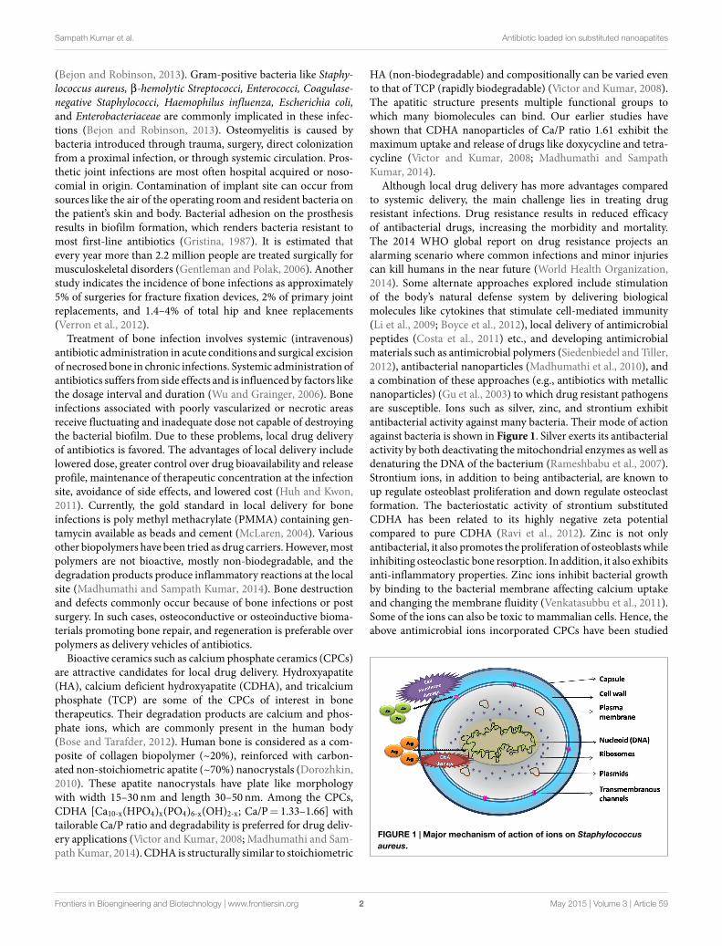
Although local drug delivery has more advantages comparedto systemic delivery, the main challenge lies in treating drugresistant infections. Drug resistance results in reduced efficacyof antibacterial drugs, increasing the morbidity and mortality.The 2014 WHO global report on drug resistance projects analarming scenario where common infections and minor injuriescan kill humans in the near future (World Health Organization,2014). Some alternate approaches explored include stimulationof the body’s natural defense system by delivering biologicalmolecules like cytokines that stimulate cell-mediated immunity(Li et al., 2009; Boyce et al., 2012), local delivery of antimicrobialpeptides (Costa et al., 2011) etc., and developing antimicrobialmaterials such as antimicrobial polymers (Siedenbiedel and Tiller,2012), antibacterial nanoparticles (Madhumathi et al., 2010), anda combination of these approaches (e.g., antibiotics with metallicnanoparticles) (Gu et al., 2003) to which drug resistant pathogensare susceptible. Ions such as silver, zinc, and strontium exhibitantibacterial activity against many bacteria. Their mode of actionagainst bacteria is shown in Figure 1. Silver exerts its antibacterialactivity by both deactivating themitochondrial enzymes as well asdenaturing the DNA of the bacterium (Rameshbabu et al., 2007).Strontium ions, in addition to being antibacterial, are known toup regulate osteoblast proliferation and down regulate osteoclastformation. The bacteriostatic activity of strontium substitutedCDHA has been related to its highly negative zeta potentialcompared to pure CDHA (Ravi et al., 2012). Zinc is not onlyantibacterial, it also promotes the proliferation of osteoblastswhileinhibiting osteoclastic bone resorption. In addition, it also exhibitsanti-inflammatory properties. Zinc ions inhibit bacterial growthby binding to the bacterial membrane affecting calcium uptakeand changing the membrane fluidity (Venkatasubbu et al., 2011).Some of the ions can also be toxic to mammalian cells. Hence, theabove antimicrobial ions incorporated CPCs have been studied
FIGURE 1 | Major mechanism of action of ions on Staphylococcusaureus.
Frontiers in Bioengineering and Biotechnology | www.frontiersin.org May 2015 | Volume 3 | Article 592
Sampath Kumar et al. Antibiotic loaded ion substituted nanoapatites
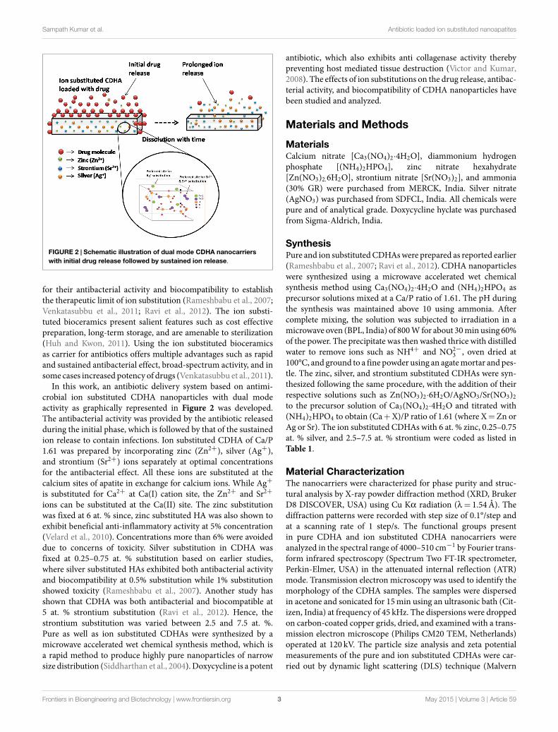
FIGURE 2 | Schematic illustration of dual mode CDHA nanocarrierswith initial drug release followed by sustained ion release.
for their antibacterial activity and biocompatibility to establishthe therapeutic limit of ion substitution (Rameshbabu et al., 2007;Venkatasubbu et al., 2011; Ravi et al., 2012). The ion substi-tuted bioceramics present salient features such as cost effectivepreparation, long-term storage, and are amenable to sterilization(Huh and Kwon, 2011). Using the ion substituted bioceramicsas carrier for antibiotics offers multiple advantages such as rapidand sustained antibacterial effect, broad-spectrum activity, and insome cases increased potency of drugs (Venkatasubbu et al., 2011).
In this work, an antibiotic delivery system based on antimi-crobial ion substituted CDHA nanoparticles with dual modeactivity as graphically represented in Figure 2 was developed.The antibacterial activity was provided by the antibiotic releasedduring the initial phase, which is followed by that of the sustainedion release to contain infections. Ion substituted CDHA of Ca/P1.61 was prepared by incorporating zinc (Zn2+), silver (Ag+),and strontium (Sr2+) ions separately at optimal concentrationsfor the antibacterial effect. All these ions are substituted at thecalcium sites of apatite in exchange for calcium ions. While Ag+is substituted for Ca2+ at Ca(I) cation site, the Zn2+ and Sr2+ions can be substituted at the Ca(II) site. The zinc substitutionwas fixed at 6 at. % since, zinc substituted HA was also shown toexhibit beneficial anti-inflammatory activity at 5% concentration(Velard et al., 2010). Concentrations more than 6% were avoideddue to concerns of toxicity. Silver substitution in CDHA wasfixed at 0.25–0.75 at. % substitution based on earlier studies,where silver substituted HAs exhibited both antibacterial activityand biocompatibility at 0.5% substitution while 1% substitutionshowed toxicity (Rameshbabu et al., 2007). Another study hasshown that CDHA was both antibacterial and biocompatible at5 at. % strontium substitution (Ravi et al., 2012). Hence, thestrontium substitution was varied between 2.5 and 7.5 at. %.Pure as well as ion substituted CDHAs were synthesized by amicrowave accelerated wet chemical synthesis method, which isa rapid method to produce highly pure nanoparticles of narrowsize distribution (Siddharthan et al., 2004). Doxycycline is a potent
antibiotic, which also exhibits anti collagenase activity therebypreventing host mediated tissue destruction (Victor and Kumar,2008). The effects of ion substitutions on the drug release, antibac-terial activity, and biocompatibility of CDHA nanoparticles havebeen studied and analyzed.
Materials and Methods
MaterialsCalcium nitrate [Ca3(NO4)2·4H2O], diammonium hydrogenphosphate [(NH4)2HPO4], zinc nitrate hexahydrate[Zn(NO3)2.6H2O], strontium nitrate [Sr(NO3)2], and ammonia(30% GR) were purchased from MERCK, India. Silver nitrate(AgNO3) was purchased from SDFCL, India. All chemicals werepure and of analytical grade. Doxycycline hyclate was purchasedfrom Sigma-Aldrich, India.
SynthesisPure and ion substitutedCDHAswere prepared as reported earlier(Rameshbabu et al., 2007; Ravi et al., 2012). CDHA nanoparticleswere synthesized using a microwave accelerated wet chemicalsynthesis method using Ca3(NO4)2·4H2O and (NH4)2HPO4 asprecursor solutions mixed at a Ca/P ratio of 1.61. The pH duringthe synthesis was maintained above 10 using ammonia. Aftercomplete mixing, the solution was subjected to irradiation in amicrowave oven (BPL, India) of 800W for about 30min using 60%of the power. The precipitate was then washed thrice with distilledwater to remove ions such as NH4+ and NO2−
3 , oven dried at100°C, and ground to a fine powder using an agatemortar andpes-tle. The zinc, silver, and strontium substituted CDHAs were syn-thesized following the same procedure, with the addition of theirrespective solutions such as Zn(NO3)2·6H2O/AgNO3/Sr(NO3)2to the precursor solution of Ca3(NO4)2·4H2O and titrated with(NH4)2HPO4 to obtain (Ca+X)/P ratio of 1.61 (where X=Zn orAg or Sr). The ion substituted CDHAs with 6 at. % zinc, 0.25–0.75at. % silver, and 2.5–7.5 at. % strontium were coded as listed inTable 1.
Material CharacterizationThe nanocarriers were characterized for phase purity and struc-tural analysis by X-ray powder diffraction method (XRD, BrukerD8 DISCOVER, USA) using Cu Kα radiation (λ = 1.54Å). Thediffraction patterns were recorded with step size of 0.1°/step andat a scanning rate of 1 step/s. The functional groups presentin pure CDHA and ion substituted CDHA nanocarriers wereanalyzed in the spectral range of 4000–510 cm−1 by Fourier trans-form infrared spectroscopy (Spectrum Two FT-IR spectrometer,Perkin-Elmer, USA) in the attenuated internal reflection (ATR)mode. Transmission electron microscopy was used to identify themorphology of the CDHA samples. The samples were dispersedin acetone and sonicated for 15min using an ultrasonic bath (Cit-izen, India) at frequency of 45 kHz. The dispersions were droppedon carbon-coated copper grids, dried, and examined with a trans-mission electron microscope (Philips CM20 TEM, Netherlands)operated at 120 kV. The particle size analysis and zeta potentialmeasurements of the pure and ion substituted CDHAs were car-ried out by dynamic light scattering (DLS) technique (Malvern
Frontiers in Bioengineering and Biotechnology | www.frontiersin.org May 2015 | Volume 3 | Article 593
Sampath Kumar et al. Antibiotic loaded ion substituted nanoapatites
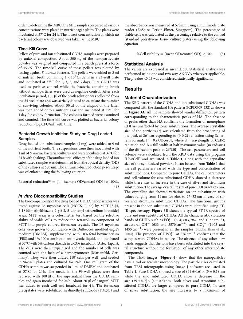
TABLE 1 | List of cell parameters, crystallite size, loading and release profiles for ion substituted CDHAs.
Sample code % ionicsubstitution
Cellparameters (Å)
Cellvolume (Å)3
Average crystallitesize (nm)
Doxycycline loadingpercentage (%)(mean±SD)
Doxycycline releasepercentage (%)(mean±SD)
a c XRD TEM (mean±SD)
CDHA – 9.110 6.80 519 25 (41±0.6)× (5±0.1) 68±9 61±1.0ZnCDHA 6 9.014 6.72 509 19 (39±0.7)× (4±0.3) 47±8 57±1.20.25AgCDHA 0.25 9.190 6.82 524 27 (41±0.5)×6±0.3) 37±4 49±1.60.5AgCDHA 0.5 9.195 6.81 527 28 (42±0.9)× (6±0.4) 30±3 52±0.80.75AgCDHA 0.75 9.198 6.83 529 29 (44±0.5)× (6±0.4) 27±5 55±0.82.5SrCDHA 2.5 9.410 6.80 541 34 (47±0.5)× (6±0.3) 31±6 51±1.15SrCDHA 5 9.440 6.82 547 38 (49±0.4)× (6±0.2) 26±5 54±0.77.5SrCDHA 7.5 9.460 6.83 549 38 (51±0.2)× (6±0.2) 21±6 56±0.9
Zetasizer Nano ZS-90, UK). One milligram of the samples weredispersed in 10ml distilled water and sonicated for 15min. Onemilliliter of the supernatant was then removed and used for DLSmeasurements.
In vitro Loading and Release Studies ofDoxycyclineLoading and release studies of doxycycline from pure CDHAand ion substituted CDHA were performed as described earlier(Madhumathi and Sampath Kumar, 2014). About 10mg of thedrug was dispersed in 10ml of phosphate buffer solution (PBS)of pH 7.4. To this, 10mg of CDHA nanocarriers was added. Thesamples were placed in water bath at 37°C for 24 h. After 24 h, 2mlof supernatantwas removed for estimation of doxycycline concen-tration at 274 nm using UV-Vis spectrophotometer (Lambda 35,Perkin-Elmer, USA). The samples were centrifuged and dried atroom temperature for 24 h. The amount of drug loaded onto thenanocarriers was determined by the following equation:
%Drug loading = Ic − Fc/Ic × 100 (1)
where Ic and Fc are initial and final concentration of doxycyclinein PBS. The release study was performed by dispersing 10mg ofdoxycycline loaded CDHAnanocarriers in PBS solution of pH 7.4kept in a constant temperature water bath at 37°C. About 2ml ofsupernatant was removed for doxycycline estimation and replacedby fresh PBS at periodic intervals over a period of 7 days. The drugrelease profile was determined by measuring the absorbance val-ues at different time intervals (Fc) from the initial concentration(Ic). All the experiments were performed in triplicates.
In vitro Dissolution and Ion Release StudiesThe in vitro dissolution studies were carried out in PBS of pH7.4 kept in a constant temperature water bath and maintained at37°C over a period of 21 days. Ten milligrams of the samples inpowder form were dispersed in 10ml of PBS at a concentrationof 1mg/ml. The weight loss of the samples, pH variations aswell as the ionic concentration in the supernatant solution, wasregularly monitored. At the end of each experiment, the sampleswere filtered, dried at 100°C, and weighed to calculate the per-centage weight loss. The ion release studies were performed byremoving 2ml of the supernatant from each sample and dilutingwith 23ml of distilled water to obtain a total volume of 25ml.
The concentration of the released Zn2+, Ag+, and Sr2+ ions weredetermined using an inductively coupled plasma optical emissionspectrometer (ICP-OES) (PerkinElmer Optima 5300 DV, USA).These studies were carried out in triplicates for 1st, 3rd, 7th, 14th,and 21st day.
In vitro Antibacterial StudiesVarious antibacterial studies such as minimum inhibitory con-centration (MIC), minimum bactericidal concentration (MBC),time-kill assays were performed on ion substituted CDHAsagainst theE. coli (NCIM2931) and S. aureus (NCIM5021), whichwere purchased from the National Chemical Laboratory, Pune,India. The bacteria were stored in glycerol stock at -20°C andused after revival as and when required. The stage of bacteria wasdetermined by plotting a growth curve. They were found to be instationary phase for MIC/MBC and bacterial growth inhibitionstudies. However, they were in log phase for time kill assay in viewof the longer duration of the bacterial study. Pure CDHAwas usedas the control. All tests were performed in triplicates.
Minimum Inhibitory ConcentrationMinimum inhibitory concentration is the lowest concentrationof an antimicrobial agent that inhibited the visible growth ofmicroorganism after incubation of 24 h. Ion substituted CDHAnanoparticles were suspended in nutrient broth at concentrationsof 300, 200, 100, 75, 50, 25, 10, and 5mg/ml and ultrasonicatedto ensure optimal dispersion. After 24 h, 10 µl of the inoculumof each microorganism was added to the nanoparticulate suspen-sion. The suspension was then incubated at 37°C for 24 h in ashaking incubator (180 rpm). Nanoparticle-free broths containingbacterial inoculum were used as negative controls. Resazurin dyewas used to assess the viability of bacteria with presence of bluecolor indicating bacterial growth inhibition and a change to redcolor indicating viable bacteria. About 10 µl of 0.01% resazurinsolution was added to the suspension and incubated for 2 h. TheMIC of the ion substituted CDHAs was calculated as the lowestconcentration of the nanoparticles that did not permit any visiblegrowth of bacteria (blue color) during 24 h of incubation.
Minimum Bactericidal ConcentrationMinimum bactericidal concentration refers to the lowest concen-tration of an antibacterial agent required to kill the bacteria. In
Frontiers in Bioengineering and Biotechnology | www.frontiersin.org May 2015 | Volume 3 | Article 594
Sampath Kumar et al. Antibiotic loaded ion substituted nanoapatites
order to determine theMBC, theMIC samples prepared at variousconcentrations were plated in nutrient agar plates. The plates wereincubated at 37°C for 24 h. The lowest concentration at which nobacterial colony was observed was taken as the MBC.
Time-Kill CurvePellets of pure and ion substituted CDHA samples were preparedby uniaxial compaction. About 300mg of the nanoparticulatepowder was weighed and compacted in a bench press at a forceof 15 kN. The time-kill curve of these pellets was plotted bytesting against S. aureus bacteria. The pellets were added to 2mlof nutrient broth containing 1× 10⁸ CFU/ml in a 24-well plateand incubated at 37°C for 1, 3, 5, and 7 days. Pure CDHA wasused as positive control while the bacteria containing brothwithout nanoparticles were used as negative control. After eachincubation period, 100 µl of the broth solution was collected fromthe 24-well plate and was serially diluted to calculate the numberof surviving colonies. About 50 µl of the aliquot of the latterwas then added onto a nutrient agar and incubated at 37°C for1 day for colony formation. The colonies formed were examinedand counted. The time-kill curve was plotted as bacterial colonyreduction (log CFU/ml) with time.
Bacterial Growth Inhibition Study on Drug LoadedSamplesDrug loaded ion substituted samples (1mg) were added to 9mlof the nutrient broth. The suspensions were then inoculated with1ml of S. aureus bacterial cultures and were incubated at 37°C for24 hwith shaking. The antibacterial efficacy of the drug loaded ionsubstituted sampleswas determined from the optical density (OD)of the cultures at 600 nm. The antimicrobial reduction percentagewas calculated using the following equation
Bacterial reduction% = {1− (sample OD/control OD)}×100%.
(2)
In vitro Biocompatibility StudiesThe biocompatibility of the drug loaded CDHAnanoparticles wastested against L6 myoblast cells (NCCS, Pune) by MTT [3-(4,5-181dimethylthiazole-2-yl)-2, 5-diphenyl tetrazolium bromide]assay. MTT assay is a colorimetric test based on the selectiveability of viable cells to reduce the tetrazolium component ofMTT into purple colored formazan crystals. The L6 myoblastcells were grown to confluence with Dulbecco’s modifed eagle’smedium (DMEM), supplemented with 10% fetal bovine serum(FBS) and 1% 100× antibiotic-antimycotic liquid, and incubatedat 37°Cwith 5% carbon dioxide in a CO2 incubator (Astec, Japan).The cells were then trypsinized and the number of cells wascounted with the help of a hemocytometer (Marienfeld, Ger-many). They were then diluted (104 cells per well) and seededin 96-well plates and cultured for 24 h. One milligram of theCDHA samples was suspended in 1ml of DMEM and incubatedat 37°C for 24 h. The media in the 96-well plates were thenreplaced with 100 µl of the supernatant from the CDHA sam-ples and again incubated for 24 h. About 20 µl of 5mg/ml MTTwas added to each well and incubated for 4 h. The formazanprecipitates were solubilized in dimethyl sulfoxide (DMSO) and
the absorbance was measured at 570 nm using a multimode platereader (EnSpire, Perkin-Elmer, Singapore). The percentage ofviable cells was calculated as the percentage relative to the control(standard polystyrene tissue culture plates) using the followingequation
%Cell viability = (mean OD/control OD)× 100. (3)
Statistical AnalysisThe values are expressed as mean± SD. Statistical analysis wasperformed using one and two way ANOVA wherever applicable.The p-value <0.05 was considered statistically significant.
Results
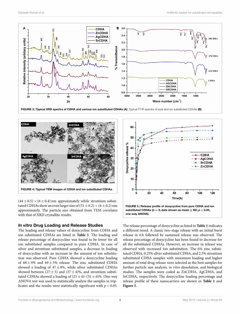
Material CharacterizationThe XRD pattern of the CDHA and ion substituted CDHAs wascomparedwith the standardHApattern (JCPDS 09-432) as shownin Figure 3A. All the samples showed similar diffraction patterncorresponding to the characteristic peaks of HA. The absenceof peaks other than HA confirms the formation of monophaseCDHAs unaffected by ionic substitutions. The average crystallitesize of the particles (t) was calculated from the broadening ofthe peak at 26° corresponding to (0 0 2) reflection using Scher-rer’s formula [t= 0.9λ/Bcosθ], where λ =wavelength of CuKαradiation and B= full width at half maximum value (in radians)of the diffraction peak at 26°(2θ). The cell parameters and cellvolume were calculated from the XRD data using the program“UnitCell” and are listed in Table 1, along with the crystallitesize of the synthesized powders. It can be seen from Table 1 thatthe cell parameters varied with the type and concentration ofsubstituted ions. Compared to pure CDHAs, the cell parametersand cell volume for zinc substituted CDHA showed a decreasewhile there was an increase in the case of silver and strontiumsubstitution. The average crystallite size of pureCDHAwas 25 nm.The crystallite size showed variations on ion substitution withvalues ranging from 19 nm for zinc to 27–32 nm in case of sil-ver and strontium substituted CDHAs. The functional groupspresent in the ion substituted CDHAs were identified using FT-IR spectroscopy. Figure 3B shows the typical FT-IR spectra ofpure and ions substituted CDHAs. All the characteristic vibrationbands of CDHA such as PO3−
4 (564, 603, 962, and 1032 cm−1),structural OH− (633 and 3570 cm−1), and CO2−
3 (1403 and1455 cm−1) were present in all the samples (Siddharthan et al.,2004). The presence of HPO2−
4 at 876 cm−1 confirms that thesamples were CDHAs in nature. The absence of any other newbands suggests that the ions have been substituted into the crys-tal structure without the formation of any other intermediatecompounds.
The TEM images (Figure 4) show that the nanoparticleshave a rod or acicular morphology. The particle sizes calculatedfrom TEM micrographs using Image J software are listed inTable 1. Pure CDHA showed a size of (41± 0.6)× (5± 0.1) nmwhile the zinc substituted CDHA show a decrease in thesize (39± 0.7)× (4± 0.3) nm. Both silver and strontium sub-stituted CDHAs are larger compared to pure CDHA. In caseof silver substitution, the size increases to a maximum of
Frontiers in Bioengineering and Biotechnology | www.frontiersin.org May 2015 | Volume 3 | Article 595
Sampath Kumar et al. Antibiotic loaded ion substituted nanoapatites
FIGURE 3 | Typical XRD spectra of CDHA and various ion substituted CDHAs (A). Typical FT-IR spectra of pure and ion substituted CDHAs (B).
FIGURE 4 | Typical TEM images of CDHA and ion substituted CDHAs.
(44± 0.5)× (6± 0.4) nm approximately while strontium substi-tutedCDHAs show an even larger size of (51± 0.2)× (6± 0.2) nmapproximately. The particle size obtained from TEM correlateswith that of XRD crystallite results.
In vitro Drug Loading and Release StudiesThe loading and release values of doxycycline from CDHA andion substituted CDHAs are listed in Table 1. The loading andrelease percentage of doxycycline was found to be lower for allion substituted samples compared to pure CDHA. In case ofsilver and strontium substituted samples, a decrease in loadingof doxycycline with an increase in the amount of ion substitu-tion was observed. Pure CDHA showed a doxycycline loadingof 68± 9% and 69± 5% release. The zinc substituted CDHAshowed a loading of 47± 8%, while silver substituted CDHAsshowed between (27± 5) and (37± 4)%, and strontium substi-tuted CDHAs showed a loading of (21± 6)–(31± 6)%. One-wayANOVA test was used to statistically analyze the samples in trip-licates and the results were statistically significant with p< 0.05.
FIGURE 5 | Release profile of doxycycline from pure CDHA and ionsubstituted CDHAs (n=3; data shown as mean±SD; p< 0.05,one-way ANOVA).
The release percentage of doxycycline as listed inTable 1 indicatesa different trend. A classic two-stage release with an initial burstrelease in 6 h followed by sustained release was observed. Therelease percentage of doxycycline has been found to decrease forall the substituted CDHAs. However, an increase in release wasobserved with increased ion substitution. The 6% zinc substi-tuted CDHA, 0.25% silver substituted CDHA, and 2.5% strontiumsubstituted CDHA samples with maximum loading and higheramount of total drug release were selected as the best samples forfurther particle size analysis, in vitro dissolution, and biologicalstudies. The samples were coded as ZnCDHA, AgCDHA, andSrCDHA, respectively. The doxycycline loading percentage andrelease profile of these nanocarriers are shown in Table 1 andFigure 5.
Frontiers in Bioengineering and Biotechnology | www.frontiersin.org May 2015 | Volume 3 | Article 596
Sampath Kumar et al. Antibiotic loaded ion substituted nanoapatites
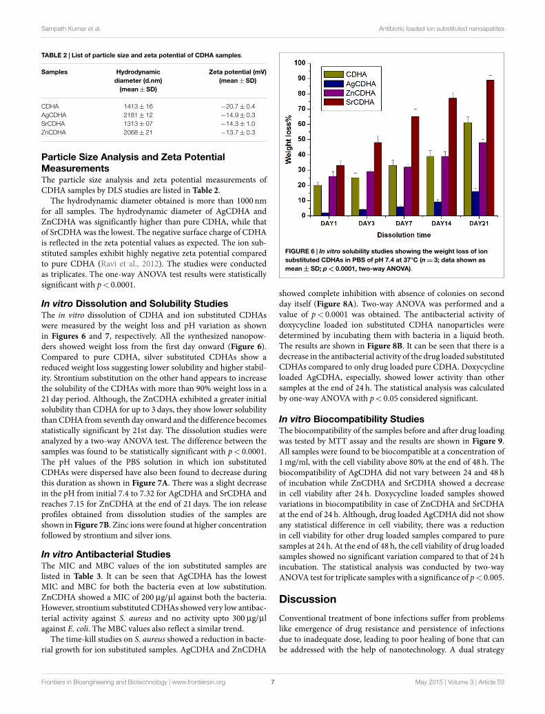
TABLE 2 | List of particle size and zeta potential of CDHA samples.
Samples Hydrodynamicdiameter (d.nm)(mean±SD)
Zeta potential (mV)(mean±SD)
CDHA 1413±16 −20.7±0.4AgCDHA 2181±12 −14.9±0.3SrCDHA 1313±07 −14.3±1.0ZnCDHA 2068±21 −13.7±0.3
Particle Size Analysis and Zeta PotentialMeasurementsThe particle size analysis and zeta potential measurements ofCDHA samples by DLS studies are listed in Table 2.
The hydrodynamic diameter obtained is more than 1000 nmfor all samples. The hydrodynamic diameter of AgCDHA andZnCDHA was significantly higher than pure CDHA, while thatof SrCDHA was the lowest. The negative surface charge of CDHAis reflected in the zeta potential values as expected. The ion sub-stituted samples exhibit highly negative zeta potential comparedto pure CDHA (Ravi et al., 2012). The studies were conductedas triplicates. The one-way ANOVA test results were statisticallysignificant with p< 0.0001.
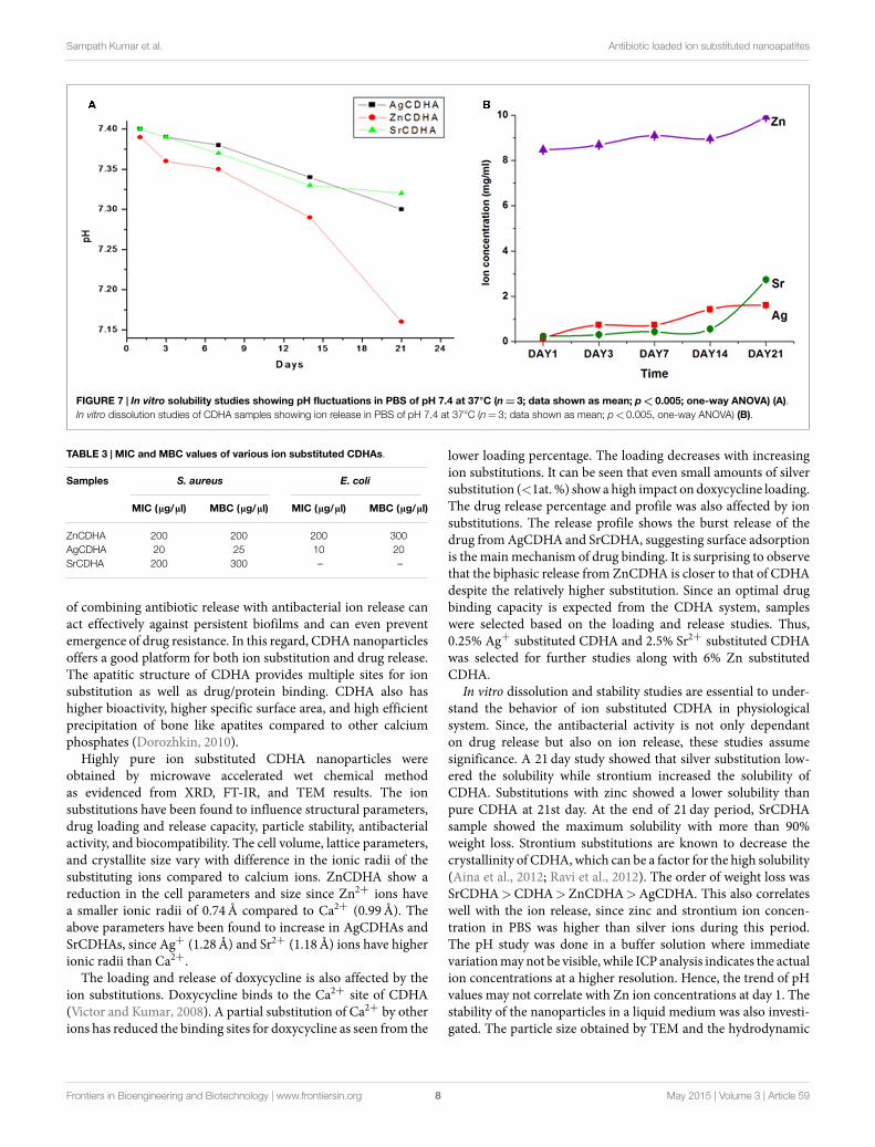
In vitro Dissolution and Solubility StudiesThe in vitro dissolution of CDHA and ion substituted CDHAswere measured by the weight loss and pH variation as shownin Figures 6 and 7, respectively. All the synthesized nanopow-ders showed weight loss from the first day onward (Figure 6).Compared to pure CDHA, silver substituted CDHAs show areduced weight loss suggesting lower solubility and higher stabil-ity. Strontium substitution on the other hand appears to increasethe solubility of the CDHAs with more than 90% weight loss in a21 day period. Although, the ZnCDHA exhibited a greater initialsolubility than CDHA for up to 3 days, they show lower solubilitythan CDHA from seventh day onward and the difference becomesstatistically significant by 21st day. The dissolution studies wereanalyzed by a two-way ANOVA test. The difference between thesamples was found to be statistically significant with p< 0.0001.The pH values of the PBS solution in which ion substitutedCDHAs were dispersed have also been found to decrease duringthis duration as shown in Figure 7A. There was a slight decreasein the pH from initial 7.4 to 7.32 for AgCDHA and SrCDHA andreaches 7.15 for ZnCDHA at the end of 21 days. The ion releaseprofiles obtained from dissolution studies of the samples areshown in Figure 7B. Zinc ions were found at higher concentrationfollowed by strontium and silver ions.
In vitro Antibacterial StudiesThe MIC and MBC values of the ion substituted samples arelisted in Table 3. It can be seen that AgCDHA has the lowestMIC and MBC for both the bacteria even at low substitution.ZnCDHA showed a MIC of 200 µg/µl against both the bacteria.However, strontium substituted CDHAs showed very low antibac-terial activity against S. aureus and no activity upto 300 µg/µlagainst E. coli. The MBC values also reflect a similar trend.
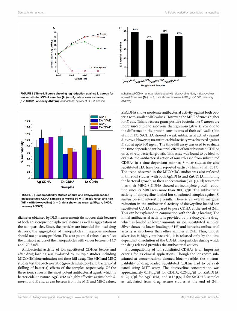
The time-kill studies on S. aureus showed a reduction in bacte-rial growth for ion substituted samples. AgCDHA and ZnCDHA
FIGURE 6 | In vitro solubility studies showing the weight loss of ionsubstituted CDHAs in PBS of pH 7.4 at 37°C (n= 3; data shown asmean±SD; p< 0.0001, two-way ANOVA).
showed complete inhibition with absence of colonies on secondday itself (Figure 8A). Two-way ANOVA was performed and avalue of p< 0.0001 was obtained. The antibacterial activity ofdoxycycline loaded ion substituted CDHA nanoparticles weredetermined by incubating them with bacteria in a liquid broth.The results are shown in Figure 8B. It can be seen that there is adecrease in the antibacterial activity of the drug loaded substitutedCDHAs compared to only drug loaded pure CDHA. Doxycyclineloaded AgCDHA, especially, showed lower activity than othersamples at the end of 24 h. The statistical analysis was calculatedby one-way ANOVA with p< 0.05 considered significant.
In vitro Biocompatibility StudiesThe biocompatibility of the samples before and after drug loadingwas tested by MTT assay and the results are shown in Figure 9.All samples were found to be biocompatible at a concentration of1mg/ml, with the cell viability above 80% at the end of 48 h. Thebiocompatibility of AgCDHA did not vary between 24 and 48 hof incubation while ZnCDHA and SrCDHA showed a decreasein cell viability after 24 h. Doxycycline loaded samples showedvariations in biocompatibility in case of ZnCDHA and SrCDHAat the end of 24 h. Although, drug loaded AgCDHA did not showany statistical difference in cell viability, there was a reductionin cell viability for other drug loaded samples compared to puresamples at 24 h. At the end of 48 h, the cell viability of drug loadedsamples showed no significant variation compared to that of 24 hincubation. The statistical analysis was conducted by two-wayANOVA test for triplicate samples with a significance of p< 0.005.
Discussion
Conventional treatment of bone infections suffer from problemslike emergence of drug resistance and persistence of infectionsdue to inadequate dose, leading to poor healing of bone that canbe addressed with the help of nanotechnology. A dual strategy
Frontiers in Bioengineering and Biotechnology | www.frontiersin.org May 2015 | Volume 3 | Article 597
Sampath Kumar et al. Antibiotic loaded ion substituted nanoapatites
FIGURE 7 | In vitro solubility studies showing pH fluctuations in PBS of pH 7.4 at 37°C (n=3; data shown as mean; p<0.005; one-way ANOVA) (A).In vitro dissolution studies of CDHA samples showing ion release in PBS of pH 7.4 at 37°C (n= 3; data shown as mean; p<0.005, one-way ANOVA) (B).
TABLE 3 | MIC and MBC values of various ion substituted CDHAs.
Samples S. aureus E. coli
MIC (μg/μl) MBC (μg/μl) MIC (μg/μl) MBC (μg/μl)
ZnCDHA 200 200 200 300AgCDHA 20 25 10 20SrCDHA 200 300 – –
of combining antibiotic release with antibacterial ion release canact effectively against persistent biofilms and can even preventemergence of drug resistance. In this regard, CDHAnanoparticlesoffers a good platform for both ion substitution and drug release.The apatitic structure of CDHA provides multiple sites for ionsubstitution as well as drug/protein binding. CDHA also hashigher bioactivity, higher specific surface area, and high efficientprecipitation of bone like apatites compared to other calciumphosphates (Dorozhkin, 2010).
Highly pure ion substituted CDHA nanoparticles wereobtained by microwave accelerated wet chemical methodas evidenced from XRD, FT-IR, and TEM results. The ionsubstitutions have been found to influence structural parameters,drug loading and release capacity, particle stability, antibacterialactivity, and biocompatibility. The cell volume, lattice parameters,and crystallite size vary with difference in the ionic radii of thesubstituting ions compared to calcium ions. ZnCDHA show areduction in the cell parameters and size since Zn2+ ions havea smaller ionic radii of 0.74Å compared to Ca2+ (0.99Å). Theabove parameters have been found to increase in AgCDHAs andSrCDHAs, since Ag+ (1.28Å) and Sr2+ (1.18Å) ions have higherionic radii than Ca2+.
The loading and release of doxycycline is also affected by theion substitutions. Doxycycline binds to the Ca2+ site of CDHA(Victor and Kumar, 2008). A partial substitution of Ca2+ by otherions has reduced the binding sites for doxycycline as seen from the
lower loading percentage. The loading decreases with increasingion substitutions. It can be seen that even small amounts of silversubstitution (<1at. %) show a high impact on doxycycline loading.The drug release percentage and profile was also affected by ionsubstitutions. The release profile shows the burst release of thedrug from AgCDHA and SrCDHA, suggesting surface adsorptionis the main mechanism of drug binding. It is surprising to observethat the biphasic release from ZnCDHA is closer to that of CDHAdespite the relatively higher substitution. Since an optimal drugbinding capacity is expected from the CDHA system, sampleswere selected based on the loading and release studies. Thus,0.25% Ag+ substituted CDHA and 2.5% Sr2+ substituted CDHAwas selected for further studies along with 6% Zn substitutedCDHA.
In vitro dissolution and stability studies are essential to under-stand the behavior of ion substituted CDHA in physiologicalsystem. Since, the antibacterial activity is not only dependanton drug release but also on ion release, these studies assumesignificance. A 21 day study showed that silver substitution low-ered the solubility while strontium increased the solubility ofCDHA. Substitutions with zinc showed a lower solubility thanpure CDHA at 21st day. At the end of 21 day period, SrCDHAsample showed the maximum solubility with more than 90%weight loss. Strontium substitutions are known to decrease thecrystallinity of CDHA, which can be a factor for the high solubility(Aina et al., 2012; Ravi et al., 2012). The order of weight loss wasSrCDHA>CDHA>ZnCDHA>AgCDHA. This also correlateswell with the ion release, since zinc and strontium ion concen-tration in PBS was higher than silver ions during this period.The pH study was done in a buffer solution where immediatevariationmaynot be visible, while ICP analysis indicates the actualion concentrations at a higher resolution. Hence, the trend of pHvalues may not correlate with Zn ion concentrations at day 1. Thestability of the nanoparticles in a liquid medium was also investi-gated. The particle size obtained by TEM and the hydrodynamic
Frontiers in Bioengineering and Biotechnology | www.frontiersin.org May 2015 | Volume 3 | Article 598
Sampath Kumar et al. Antibiotic loaded ion substituted nanoapatites
FIGURE 8 | Time-kill curve showing log reduction against S. aureus forion substituted CDHA samples (A) (n=3; data shown as mean;p< 0.0001, one-way ANOVA). Antibacterial activity of CDHA and ion
substituted CDHA nanoparticles loaded with doxycycline (doxy – doxycycline)against S. aureus (B) (n= 3; data shown as mean±SD; p<0.005, one-wayANOVA).
FIGURE 9 | Biocompatibility studies of pure and doxycycline loadedion substituted CDHA samples (1mg/ml) by MTT assay for 24 and 48h(WD – with doxycycline) (n=3; data shown as mean±SD; p< 0.005,two-way ANOVA).
diameter obtained byDLSmeasurements do not correlate becauseof both anisotropic non-spherical nature as well as aggregation ofthe nanoparticles. Since, the particles are intended for local drugdelivery, the aggregation of nanoparticles in aqueous mediumshould not pose any problem. The zeta potential values also reflectthe unstable nature of the nanoparticles with values between -13.7and -20.7mV.
Antibacterial activity of ion substituted CDHAs before andafter drug loading was evaluated by multiple studies includingMIC/MBC determination and time-kill assay. The MIC and MBCstudies test the bacteriostatic (growth inhibitory) and bactericidal(killing of bacteria) effects of the samples respectively. Of thethree ions, silver is the most potent antibacterial agent, which isbactericidal in nature. AgCDHA is highly effective against both S.aureus and E. coli, as can be seen from the MIC and MBC values.
ZnCDHA shows moderate antibacterial activity against both bac-teria with similar MIC values. However, the MBC of zinc is higherfor E. coli. This is because gram-positive bacteria like S. aureus aremore susceptible to zinc ions than gram-negative E. coli due tothe difference in the protein constituents of their cell walls (Jainet al., 2013). SrCDHA showed a weak antibacterial activity againstS. aureus.However, no antimicrobial activity was observed againstE. coli at upto 300 µg/µl. The time-kill assay was used to evaluatethe time dependant antibacterial effect of ion substituted CDHAson S. aureus bacterial growth. This assay was found to be ideal toevaluate the antibacterial action of ions released from substitutedCDHAs in a time dependant manner. Similar studies for zincsubstituted HA have been reported earlier (Thian et al., 2013).The trend observed in the MIC/MBC studies was also reflectedin time-kill studies, with both AgCDHA and ZnCDHA inhibitingthe bacterial growth, as their concentration (300 µg/µl) was morethan their MBC. SrCDHA showed an incomplete growth reduc-tion since its MBC was more than 300 µg/µl. The antibacterialactivity of doxycycline loaded ion substituted samples against S.aureus present interesting results. There is an overall marginalreduction in the antibacterial activity of doxycycline loaded ionsubstituted CDHAs compared to pure CDHA at the end of 24 h.This can be explained in conjunction with the drug loading. Theinitial antibacterial activity is provided by the doxycycline drug,which is loaded at lower amounts in ion substituted samples.Silver shows the lowest loading (~31%) and hence its antibacterialactivity is also lower than other samples at 24 h. Thus, thoughsilver ion is highly antibacterial, it is released only by the timedependant dissolution of the CDHA nanoparticles during whichthe drug released provides the antibacterial activity.
Biocompatibility of ion substituted CDHAs is an importantcriteria for its clinical applications. Though the ions were sub-stituted at concentrations deemed biocompatible, the biocom-patibility of drug loaded substituted CDHAs had to be eval-uated using MTT assay. The doxycycline concentration wasapproximately 0.18 µg/µl for CDHA, 0.26 µg/µl for ZnCDHA,0.12 µg/µl for AgCDHA, and 0.15 µg/µl for SrCDHA samplesas calculated from drug release studies at the end of 24 h.
Frontiers in Bioengineering and Biotechnology | www.frontiersin.org May 2015 | Volume 3 | Article 599
Sampath Kumar et al. Antibiotic loaded ion substituted nanoapatites
The doxycycline release reduces the cell viability during first 24 hfor ZnCDHA and SrCDHA while no appreciable difference wasobserved for AgCDHA samples with and without drug loading.However, a statistically significant decrease in cell viability wasobserved for ZnCDHA and SrCDHA at the end of 48 h comparedto 24 h. However, all the samples were found to be biocompatibleat the given concentration. The results clearly demonstrate theadvantage of ion substituted CDHAs, as the controlled releaseof ions incorporated into CDHAs plays an important role inimproving the biocompatibility.
Among various ion substituted CDHAs, AgCDHA with 0.25%substitution shows the lowest doxycycline loading. However, itexhibits the highest antibacterial activity and is also biocompat-ible. The SrCDHA sample shows low loading of doxycycline andlowest antibacterial activity but exhibits higher biocompatibility.Although, the ZnCDHA sample exhibits reasonable drug loadingand satisfactory antibacterial activity, it shows lower cell viability
compared to pure CDHA and other ion substituted samples.The CDHA and AgCDHA samples exhibit drug release up to5 days while there was no change in the drug release profileafter first day for SrCDHA and ZnCDHA samples. But bothZnCDHA and AgCDHA show high antibacterial activity at day 2in spite of their different drug release profiles. Although, SrCDHAshow maximum drug release by first day, it exhibits antibacte-rial activity from day 2 onward. A long-term antibacterial andcytotoxic study may clearly bring out the combined role of ionsubstitution and drug release in combating antimicrobial resis-tance.
Thus, the present work demonstrates that by combining antibi-otics and antimicrobial ions, a biocompatible, sustained, highlyefficient antibacterial bioceramic system with additional benefi-cial properties such as anti-inflammatory activity or bone remod-eling activity can be developed to efficiently contain and treat boneinfections.
ReferencesAina, V., Lusvardi, G., Annaz, B., Gibson, I. R., Flora, E., and Malavasi, I. G. (2012).
Magnesium- and strontium-co-substituted hydroxyapatite: the effects of doped-ions on the structure and chemico-physical properties. J. Mater. Sci. Mater. Med.23, 2867–2879. doi:10.1007/s10856-012-4767-3
Bejon, P., and Robinson, E. (2013). Bone and joint infection.Medicine 41, 719–722.doi:10.1016/j.mpmed.2013.09.008
Bose, S., and Tarafder, S. (2012). Calcium phosphate ceramic systems in growthfactor and drug delivery. Acta Biomater. 8, 1401–1421. doi:10.1016/j.actbio.2011.11.017
Boyce, B. M., Lindsey, B. A., Clovis, N. B., Smith, S., Hobbs, G. R., Hubbard, D. F.,et al. (2012). Additive effects of exogenous IL-12 supplementation and antibiotictreatment in infection prophylaxis. J. Orthop. Res. 30, 196–202. doi:10.1002/jor.21520
Costa, F., Carvalho, I. F., Montelaro, R. C., Gomes, P., and Martins, M. C. L. (2011).Covalent immobilization of antimicrobial peptides (AMPs) onto biomaterialsurfaces. Acta Biomater. 7, 1431–1440. doi:10.1016/j.actbio.2010.11.005
Dorozhkin, S. V. (2010). Nanosized and nanocrystalline calcium orthophosphates.Acta Biomater. 6, 715–734. doi:10.1016/j.actbio.2009.10.031
Gentleman, E., and Polak, J. (2006). Historic and current strategies in bone tis-sue engineering: do we have a hope in hench? J. Mater. Sci. Mater. Med. 17,1029–1035. doi:10.1007/s10856-006-0440-z
Gristina, A. G. (1987). Biomaterial-centered infection: microbial adhesion versustissue integration. Science 237, 1588–1595. doi:10.1126/science.3629258
Gu, H., Ho, P. L., Tong, E., Wang, L., and Xu, B. (2003). Presenting vancomycinon nanoparticles to enhance antimicrobial activities. Nano Lett. 3, 1261–1263.doi:10.1021/nl034396z
Huh, A. J., and Kwon, Y. J. (2011). Nanoantibiotics: a new paradigm for treatinginfectious diseases using nanomaterials in the antibiotics resistant era. J. Control.Release 156, 128–145. doi:10.1016/j.jconrel.2011.07.002
Jain, A., Bhargava, R., and Poddar, P. (2013). Probing interaction of Gram-positiveand Gram-negative bacterial cells with ZnO nanorods. Mater. Sci. Eng. C 33,1247–1253. doi:10.1016/j.msec.2012.12.019
Li, B., Jiang, B., Boyce, B. M., and Lindsey, B. A. (2009). Multilayer polypeptidenanoscale coatings incorporating IL-12 for the prevention of biomedical device-associated infections. Biomaterials 30, 2552–2558. doi:10.1016/j.biomaterials.2009.01.042
Madhumathi, K., and Sampath Kumar, T. S. (2014). Regenerative potential and anti-bacterial activity of tetracycline loaded apatitic nanocarriers for the treatment ofperiodontitis. Biomed. Mater. 9, 035002. doi:10.1088/1748-6041/9/3/035002
Madhumathi, K., Sudeesh Kumar, P. T., Abhilash, S., Sreeja, V., Tamura, H.,Manzoor, K., et al. (2010). Development of novel chitin/nanosilver compositescaffolds for wound dressing applications. J. Mater. Sci. Mater. Med. 21, 807–813.doi:10.1007/s10856-009-3877-z
McLaren, A. (2004). Alternative materials to acrylic bone cement for deliveryof depot antibiotics in orthopaedic infections. Clin. Orthop. Relat. Res. 427,101–106. doi:10.1097/01.blo.0000143554.56897.26
Rameshbabu, N., Kumar, T. S., Prabhakar, T., Sastry, V., Murty, K., and Rao, K. P.(2007). Antibacterial nanosized silver substituted hydroxyapatite: synthesis andcharacterization. J. Biomed. Mater. Res. A 80, 581–591. doi:10.1002/jbm.a.30958
Ravi, N. D., Balu, R., and Sampath Kumar, T. S. (2012). Strontium-substitutedcalcium deficient hydroxyapatite nanoparticles: synthesis, characterization, andantibacterial properties. J. Am. Ceram. Soc. 95, 2700–2708. doi:10.1111/j.1551-2916.2012.05262
Siddharthan, A., Seshadri, S. K., and Sampath Kumar, T. S. (2004). Microwaveaccelerated synthesis of nanosized calcium deficient hydroxyapatite. J. Mater.Sci. Mater. Med. 15, 1279–1284. doi:10.1007/s10856-004-5735-3
Siedenbiedel, F., and Tiller, J. C. (2012). Antimicrobial polymers in solution andon surfaces: overview and functional principles. Polymers 4, 46–71. doi:10.3390/polym4010046
Thian, E. S., Konishi, T., Kawanobe, Y., Lim, P. N., Choong, C., Ho, B., et al. (2013).Zinc-substituted hydroxyapatite: a biomaterial with enhanced bioactivity andantibacterial properties. J. Mater. Sci. Mater. Med. 24, 437–445. doi:10.1007/s10856-012-4817-x
Velard, F., Laurent-Maquin, D., Braux, J., Guillaume, C., Bouthors, S., Jallot, E., et al.(2010). The effect of zinc on hydroxyapatite-mediated activation of human poly-morphonuclear neutrophils and bone implant-associated acute inflammation.Biomaterials 31, 2001–2009. doi:10.1016/j.biomaterials.2009.11.066
Venkatasubbu, G. D., Ramasamy, S., Ramakrishnan, V., and Kumar, J. (2011).Nanocrystalline hydroxyapatite and zinc-doped hydroxyapatite as carrier mate-rial for controlled delivery of ciprofloxacin. 3 Biotech 1, 173–186. doi:10.1007/s13205-011-0021-9
Verron, E., Bouler, J. M., and Guicheux, J. (2012). Controlling the biologicalfunction of calcium phosphate bone substitutes with drugs. Acta Biomater. 8,3541–3551. doi:10.1016/j.actbio.2012.06.022
Victor, S. P., and Kumar, T. S. (2008). Tailoring calcium-deficient hydroxyapatitenanocarriers for enhanced release of antibiotics. J. Biomed. Nanotechnol. 4, 1–7.doi:10.1166/jbn.2008.019
World Health Organization. (2014). Antimicrobial Resistance Global Report onSurveillance. Geneva: World Health Organization.
Wu, P., and Grainger, D. W. (2006). Drug/device combinations for local drugtherapies and infection prophylaxis. Biomaterials 27, 2450–2467. doi:10.1016/j.biomaterials.2005.11.031
Conflict of Interest Statement: The authors declare that the research was con-ducted in the absence of any commercial or financial relationships that could beconstrued as a potential conflict of interest.
Copyright © 2015 Sampath Kumar, Madhumathi, Rubaiya and Doble. This is anopen-access article distributed under the terms of the Creative Commons AttributionLicense (CC BY). The use, distribution or reproduction in other forums is permitted,provided the original author(s) or licensor are credited and that the original publica-tion in this journal is cited, in accordance with accepted academic practice. No use,distribution or reproduction is permitted which does not comply with these terms.
Frontiers in Bioengineering and Biotechnology | www.frontiersin.org May 2015 | Volume 3 | Article 5910