Streptococci and Enterococci - Columbia University · Streptococci and Enterococci ... responsible...
9
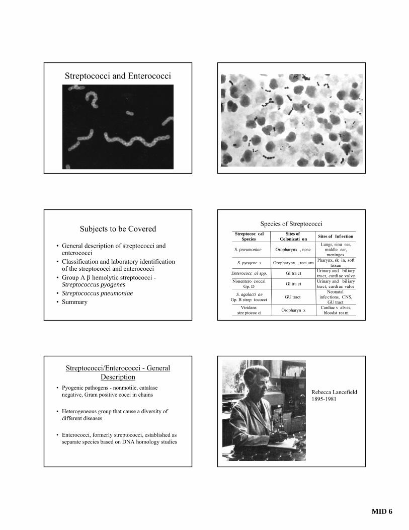
MID 6 Streptococci and Enterococci Subjects to be Covered • General description of streptococci and enterococci • Classification and laboratory identification of the streptococci and enterococci • Group A β hemolytic streptococci - Streptococcus pyogenes • Streptococcus pneumoniae • Summary Streptococci/Enterococci - General Description • Pyogenic pathogens - nonmotile, catalase negative, Gram positive cocci in chains • Heterogeneous group that cause a diversity of different diseases • Enterococci, formerly streptococci, established as separate species based on DNA homology studies Species of Streptococci Streptococ cal Species Sites of Colonizati on Sites of Inf ection S. pneumoniae Oropharynx , nose Lungs, sinu ses, middle ear, meninges S. pyogene s Oropharynx , rect um Pharynx, sk in, soft tissue Enterococc al spp. GI tra ct Urinary and bil iary tract, cardiac valve Nonentero coccal Gp. D GI tra ct Urinary and bil iary tract, cardiac valve S. agalacti ae Gp. B strep tococci GU tract Neonatal infe ctions, CNS, GU tract Viridans stre ptococ ci Oropharyn x Cardiac v alves, bloodst ream Rebecca Lancefield 1895-1981
Transcript of Streptococci and Enterococci - Columbia University · Streptococci and Enterococci ... responsible...
MID 6
Streptococci and Enterococci
Subjects to be Covered
• General description of streptococci and enterococci
• Classification and laboratory identification of the streptococci and enterococci
• Group A β hemolytic streptococci -Streptococcus pyogenes
• Streptococcus pneumoniae• Summary
Streptococci/Enterococci - General Description
• Pyogenic pathogens - nonmotile, catalase negative, Gram positive cocci in chains
• Heterogeneous group that cause a diversity of different diseases
• Enterococci, formerly streptococci, established as separate species based on DNA homology studies
Species of StreptococciStreptococ cal
Species Sites of
Colonizati on Sites of Inf ection
S. pneumoniae Oropharynx , nose Lungs, sinu ses,
middle ear, meninges
S. pyogene s Oropharynx , rect um Pharynx, sk in, soft tissue
Enterococc al spp. GI tra ct Urinary and bil iary tra ct, cardi ac valve
Nonentero coccal Gp. D GI tra ct Urinary and bil iary
tra ct, cardi ac valve
S. agalacti ae Gp. B strep tococci GU tract
Neonatal infe ctions, CNS,
GU tract Viridans
stre ptococ ci Oropharyn x Cardiac v alves, bloodst ream
Rebecca Lancefield1895-1981
MID 6
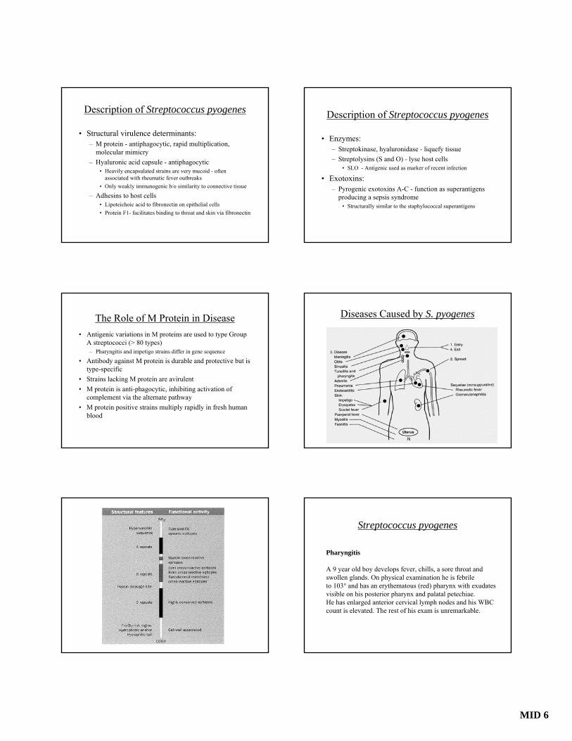
Classification Systems for Streptococci
• Hemolysis on blood agar plates – S. pyogenes is β hemolytic (complete) – Viridans streptococci are α hemolytic (incomplete)– Enterococci are γ hemolytic (no hemolysis)
• Lancefield grouping based on group specific carbohydrate antigens. Most β and some αhemolytic streptococci can be typed by this method
• Biochemical properties– Catalase negative, facultative anaerobes
Beta Hemolysis
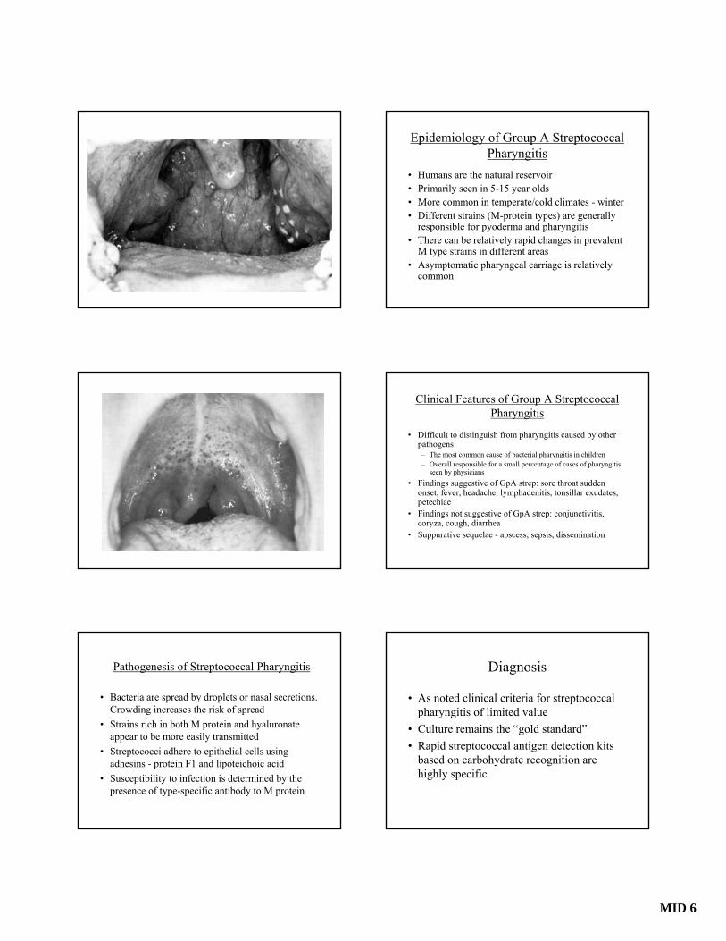
Alpha Hemolysis
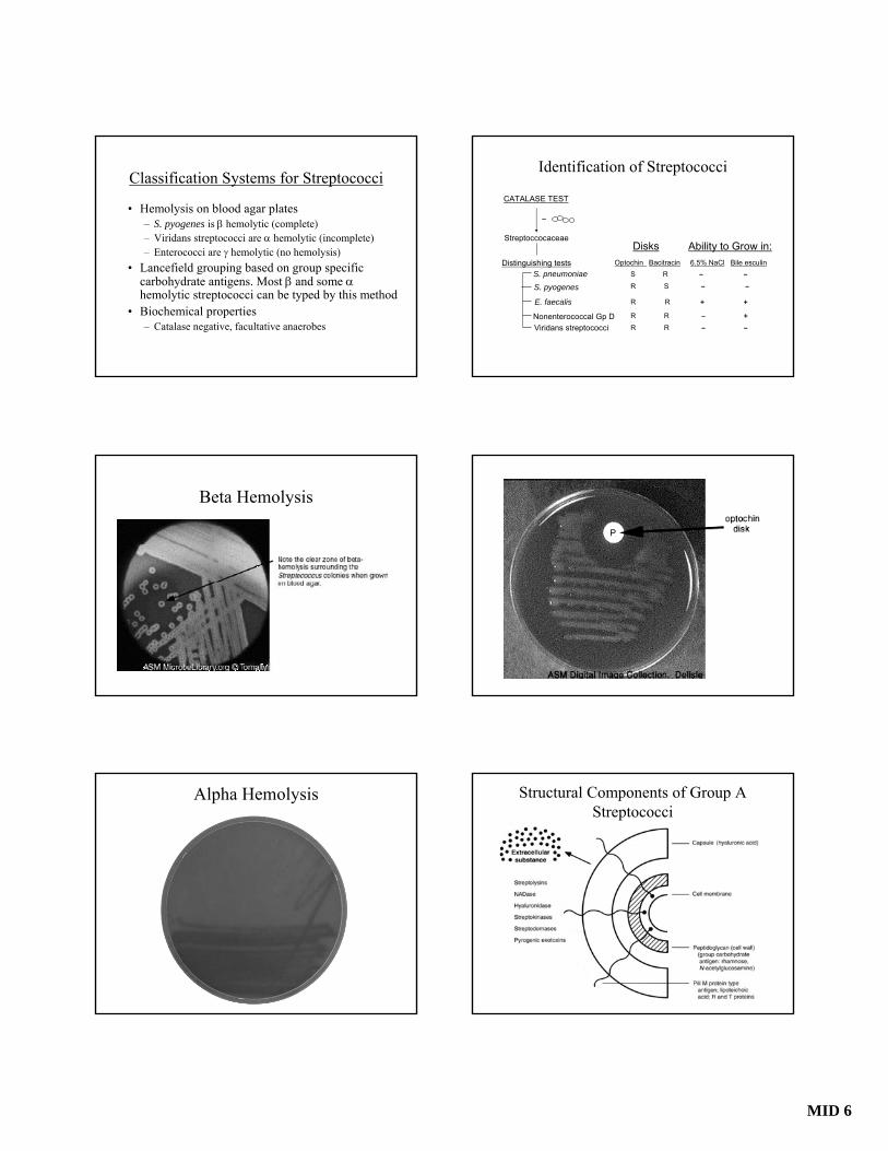
Identification of Streptococci
CATALASE TEST
Nonenterococcal Gp D
–
Streptoccocaceae
Distinguishing tests S. pneumoniaeS. pyogenes
E. faecalis
Viridans streptococci
Optochin Bacitracin 6.5% NaCl Bile esculinS R – –R S – –
R R + +
R R – +R R – –
Disks Ability to Grow in:
P
Structural Components of Group A Streptococci
MID 6
Description of Streptococcus pyogenes
• Structural virulence determinants:– M protein - antiphagocytic, rapid multiplication,
molecular mimicry– Hyaluronic acid capsule - antiphagocytic
• Heavily encapsulated strains are very mucoid - often associated with rheumatic fever outbreaks
• Only weakly immunogenic b/o similarity to connective tissue
– Adhesins to host cells • Lipoteichoic acid to fibronectin on epithelial cells• Protein F1- facilitates binding to throat and skin via fibronectin
The Role of M Protein in Disease• Antigenic variations in M proteins are used to type Group
A streptococci (> 80 types)– Pharyngitis and impetigo strains differ in gene sequence
• Antibody against M protein is durable and protective but is type-specific
• Strains lacking M protein are avirulent• M protein is anti-phagocytic, inhibiting activation of
complement via the alternate pathway• M protein positive strains multiply rapidly in fresh human
blood
Description of Streptococcus pyogenes
• Enzymes:– Streptokinase, hyaluronidase - liquefy tissue– Streptolysins (S and O) - lyse host cells
• SLO - Antigenic used as marker of recent infection
• Exotoxins:– Pyrogenic exotoxins A-C - function as superantigens
producing a sepsis syndrome• Structurally similar to the staphylococcal superantigens
Diseases Caused by S. pyogenes
Streptococcus pyogenes
Pharyngitis
A 9 year old boy develops fever, chills, a sore throat and swollen glands. On physical examination he is febrile to 103° and has an erythematous (red) pharynx with exudatesvisible on his posterior pharynx and palatal petechiae. He has enlarged anterior cervical lymph nodes and his WBC count is elevated. The rest of his exam is unremarkable.
MID 6
Pathogenesis of Streptococcal Pharyngitis
• Bacteria are spread by droplets or nasal secretions. Crowding increases the risk of spread
• Strains rich in both M protein and hyaluronate appear to be more easily transmitted
• Streptococci adhere to epithelial cells using adhesins - protein F1 and lipoteichoic acid
• Susceptibility to infection is determined by the presence of type-specific antibody to M protein
Epidemiology of Group A Streptococcal Pharyngitis
• Humans are the natural reservoir• Primarily seen in 5-15 year olds• More common in temperate/cold climates - winter• Different strains (M-protein types) are generally
responsible for pyoderma and pharyngitis• There can be relatively rapid changes in prevalent
M type strains in different areas• Asymptomatic pharyngeal carriage is relatively
common
Clinical Features of Group A Streptococcal Pharyngitis
• Difficult to distinguish from pharyngitis caused by other pathogens– The most common cause of bacterial pharyngitis in children– Overall responsible for a small percentage of cases of pharyngitis
seen by physicians• Findings suggestive of GpA strep: sore throat sudden
onset, fever, headache, lymphadenitis, tonsillar exudates, petechiae
• Findings not suggestive of GpA strep: conjunctivitis, coryza, cough, diarrhea
• Suppurative sequelae - abscess, sepsis, dissemination
Diagnosis
• As noted clinical criteria for streptococcal pharyngitis of limited value
• Culture remains the “gold standard”• Rapid streptococcal antigen detection kits
based on carbohydrate recognition are highly specific
MID 6
Nonsuppurative Sequelae of Pharyngitis
• Rheumatic fever: syndromic diagnosis made using the Jones criteria– Carditis, polyarthritis, erythema marginatum,
subcutaneous nodules, chorea (+ minor criteria)• Pathogenesis believed to involve “molecular
mimicry”• Cross reactive epitopes with myosin and M protein
• Glomerulonephritis: – Immunologically mediated damage perhaps resulting
from streptococcal antigens that cross react with kidney tissue
Impetigo - Pyoderma
A 3 year old boy presents with a rash on his face.The lesions started as small pustules that progressedto thick “honey”-crusted lesions on his face. There is alarge primary lesion by his nose and several satellite lesions on his face. His mother states that he was scratchinga mosquito bite there just before the rash started.
Pathogenesis and Epidemiology of Streptococcal Pyoderma
• Primarily seen in 2-5 year olds• Pyoderma is most commonly encountered in economically
disadvantaged populations– Influenced by climate and hygiene
• Cutaneous colonization (prior to injury) leads to autoinoculation at sites of injury
• Strains differ from those that cause pharyngitis although pharyngeal carriage of these strains also occurs
• Complications rare: lymphadenitis, immune-complex glomerulonephritis
Erysipelas: GAS infection of the superficial skin and cutaneous lymphatics. Most cases involve the legs and feet. Bacteremia is rare.
Cellulitis
MID 6
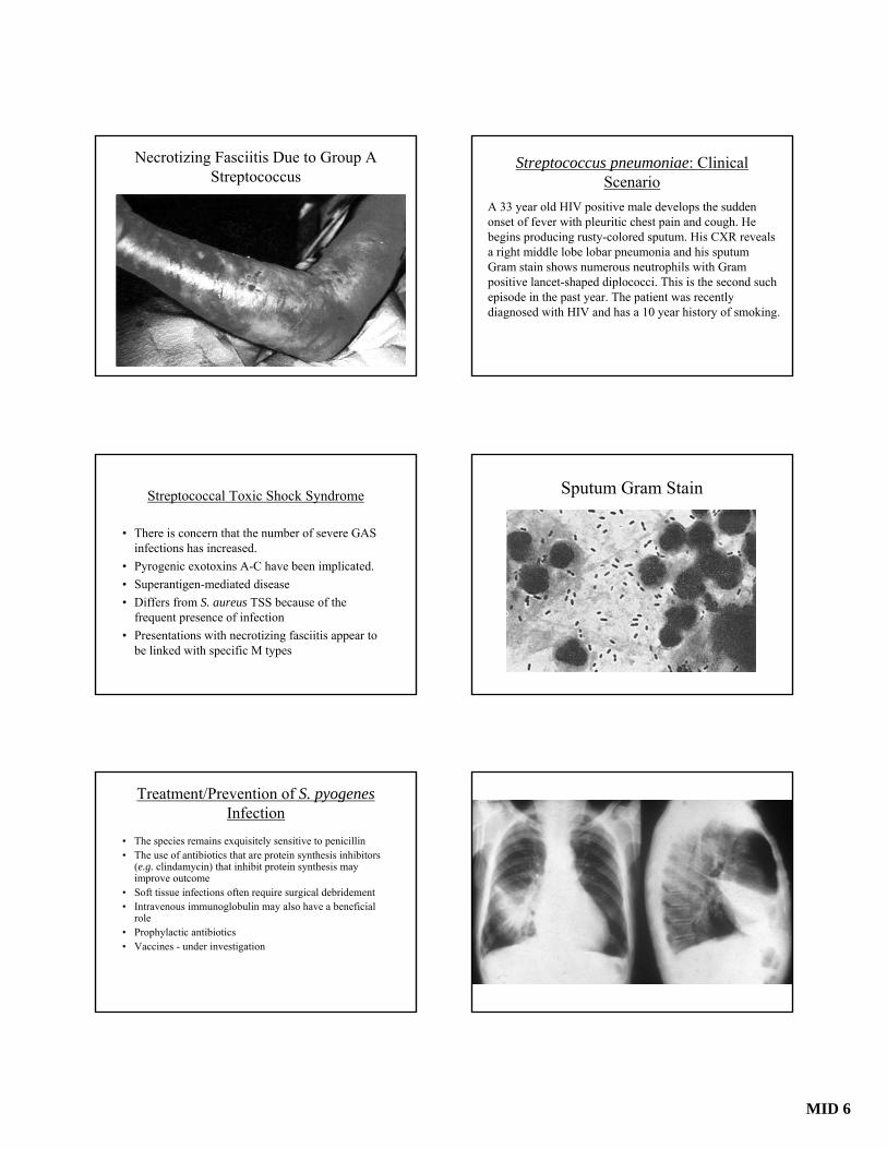
Necrotizing Fasciitis Due to Group A Streptococcus
Streptococcal Toxic Shock Syndrome
• There is concern that the number of severe GAS infections has increased.
• Pyrogenic exotoxins A-C have been implicated. • Superantigen-mediated disease• Differs from S. aureus TSS because of the
frequent presence of infection• Presentations with necrotizing fasciitis appear to
be linked with specific M types
Treatment/Prevention of S. pyogenes Infection
• The species remains exquisitely sensitive to penicillin• The use of antibiotics that are protein synthesis inhibitors
(e.g. clindamycin) that inhibit protein synthesis may improve outcome
• Soft tissue infections often require surgical debridement• Intravenous immunoglobulin may also have a beneficial
role• Prophylactic antibiotics• Vaccines - under investigation
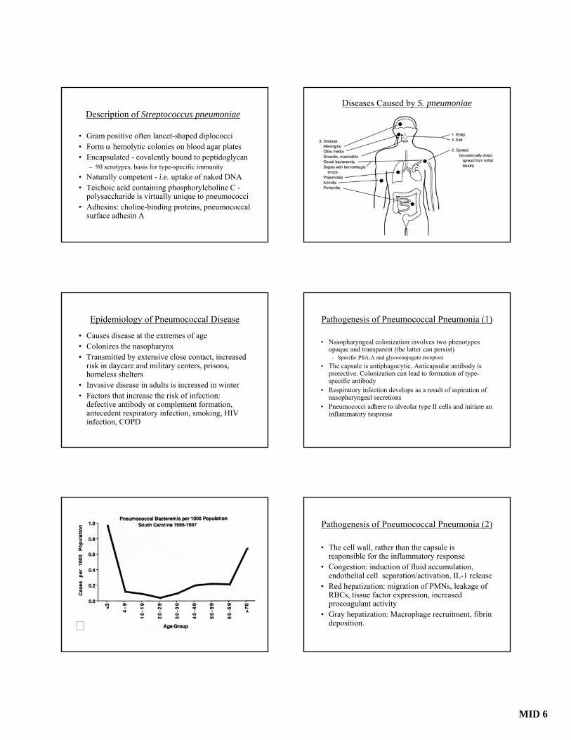
Streptococcus pneumoniae: Clinical Scenario
A 33 year old HIV positive male develops the sudden onset of fever with pleuritic chest pain and cough. He begins producing rusty-colored sputum. His CXR reveals a right middle lobe lobar pneumonia and his sputum Gram stain shows numerous neutrophils with Gram positive lancet-shaped diplococci. This is the second such episode in the past year. The patient was recently diagnosed with HIV and has a 10 year history of smoking.
Sputum Gram Stain
MID 6
Description of Streptococcus pneumoniae
• Gram positive often lancet-shaped diplococci • Form α hemolytic colonies on blood agar plates• Encapsulated - covalently bound to peptidoglycan
– 90 serotypes, basis for type-specific immunity• Naturally competent - i.e. uptake of naked DNA• Teichoic acid containing phosphorylcholine C -
polysaccharide is virtually unique to pneumococci • Adhesins: choline-binding proteins, pneumococcal
surface adhesin A
Epidemiology of Pneumococcal Disease
• Causes disease at the extremes of age• Colonizes the nasopharynx• Transmitted by extensive close contact, increased
risk in daycare and military centers, prisons, homeless shelters
• Invasive disease in adults is increased in winter• Factors that increase the risk of infection:
defective antibody or complement formation, antecedent respiratory infection, smoking, HIV infection, COPD
Diseases Caused by S. pneumoniae
Pathogenesis of Pneumococcal Pneumonia (1)
• Nasopharyngeal colonization involves two phenotypes opaque and transparent (the latter can persist)– Specific PSA-A and glycoconjugate receptors
• The capsule is antiphagocytic. Anticapsular antibody is protective. Colonization can lead to formation of type-specific antibody
• Respiratory infection develops as a result of aspiration of nasopharyngeal secretions
• Pneumococci adhere to alveolar type II cells and initiate an inflammatory response
Pathogenesis of Pneumococcal Pneumonia (2)
• The cell wall, rather than the capsule is responsible for the inflammatory response
• Congestion: induction of fluid accumulation, endothelial cell separation/activation, IL-1 release
• Red hepatization: migration of PMNs, leakage of RBCs, tissue factor expression, increased procoagulant activity
• Gray hepatization: Macrophage recruitment, fibrin deposition.
MID 6
Pathogenesis of Pneumococcal Pneumonia (3)
• Resolution of pneumonia starts with development of anticapsular antibody
• If the infection is not contained, the pneumo-coccus can spread to other sites such as joints or the meninges
• Spread to the meninges may be via an antecedent CSF leak or through the choroid plexus
Treatment of Pneumococcal Infections
• Strains have become increasingly resistant to penicillin as well as to other anti-microbial agents
• Need to test for antimicrobial susceptibility and, in settings where there is a high incidence of penicillin resistance, use other agents as initial empirical therapy.
Impact of Therapy on Survival in Pneumococcal Pneumonia
Austrian and Gold, 1964
Prevention of Pneumococcal Disease
• Rationale: Early South African vaccine studies, Austrian bacteremia data, emerging antimicrobial resistance
• Types of vaccines– Polysaccharide (23 types) - T cell independent– Polysaccharide protein conjugate vaccine (7 types) T
cell dependent, more effective in infants ≤2 years of age
(< 2 Years)
(≥ 2 Years)
Invasive Disease Caused by Penicillin-Susceptibleand NonsusceptiblePneumococci among Children (1996-2004)
Kyaw et al., N Engl J Med 2006
Summary (1)
• Streptococci are a diverse group of species that cause a variety of different diseases
• S. pyogenes are primarily responsible for cutaneous and pharyngeal infections. More severe disease is associated with toxin producing strains and particular M serotypes– The M protein, the hylauronate capsule, and the pyrogenic
exotoxins are the most important virulence determinants
• Development of a vaccine has been hampered by the large number of M serotypes and the concern about epitopes that cross react with human tissue
MID 6
Summary (2)
• S. pneumoniae is among the most common causes of pneumonia, otitis and meningitis
• Capsules are antiphagocytic and capsular antibody induces protection against subsequent infection
• Peptidoglycan is largely responsible for the brisk inflammatory response induced during infection
• Antimicrobial resistance has become a serious concern in the management of these infections