Study of a 2-branched (1→→3)-ββ-D-glucan from ... · solution a fairly rough morphology with...
9
Study of a 2-branched (1→3)-β β β-D-glucan from Lactobacillus suebicus CUPV221 as observed by Tapping mode Atomic Force Microscopy C. Marieta 1,2 and M. T. Dueñas 1 1 Department of Applied Chemistry, University of the Basque Country, UPV/EHU, Pº Manuel Lardiazabal, 3, 20018 Donostia-San Sebastián, Spain 2 Materials and Technologies Group, Department of Chemical Engineering and Environmental, University of the Basque Country, UPV/EHU, Plaza Europa 1, 20018 Donostia-San Sebastián, Spain Tapping Mode Atomic Force Microscopy (TM-AFM) has been used to analyze the supramolecular structure and conformation of the (1→3)(1→2)-β-D-glucan produced by Lactobacillus suebicus CUPV221 isolated from cider. Solutions for TM-AFM observation were prepared by dispersing the solid glucan in distilled water and in alkaline aqueous solutions. It was found that from the distilled water at 10 mg/L the (1→3)(1→2)-β-D-glucan forms networks. From the alkaline aqueous solutions different supramolecular structures were observed depending on pH. From the weakest alkaline solution a fairly rough morphology with a high density of spike-like growth features was revealed. As the ionic force of the medium increased, the size of the spike-like growth features diminished, and even many disaggregated fibers could be found. At 0.4 M NaOH (pH 13.16), the aggregates had disappeared almost totally. At 0.1 M NaOH the aggregates were partially detached, and many free microfibers were found to which a helical conformation could be assigned due to their stiffness and rodlike character. At 0.4 M NaOH (pH 13.16), the denaturation was observed. After neutralizing the alkaline solution with HCl, the formation of short linear, circular, and hairpin structures was observed. Keywords (1→3)-β-glucan; lactic acid bacteria; atomic force microscope; supramolecular structure; conformation; 1. Introduction Polysaccharides are relatively complex carbohydrates. They are biopolymers made up of many monosaccharides joined together by glycosidic bonds. They are important constituents of plants and microorganisms, where they perform a variety of functions [1]. (1→3)-β-D-glucan polysaccharides are chains of D-glucose molecules, with the six-sided D- glucose rings connected at the 1 and 3 positions. They are found in the cell wall of yeast, fruiting bodies of fungi, and as exopolysaccharides (EPSs) in the culture medium of some bacteria. It has been shown that these (1→3)-β-D-glucans have broad biological activities such as antitumor, antibacterial, antiviral, and anticoagulatory effects [2]. The biological activity of these biomacromolecules is strongly dependent upon either chemical or physical properties as well as conformation or structural features, which also depends on the environmental conditions [3]. Besides, these (1→3)-β-D- glucans have a growing interest because of their applications in the dairy industry. They may act both as texturizers and stabilizers. A better understanding of the structure-function relationship of (1→3)-β-D-glucans remains a challenge to further improve applications of these biopolymers to better satisfy the consumer demand for appealing, tasty and even healthier products [4]. It has been reported that some species of lactic bacteria are able to synthesize these polysaccharides, which are secreted into the surrounding environment [5]. Some strains of Pediococcus parvulus have been described as β-glucan- producing bacteria in cider [6] and wine [7]. In our laboratory, an EPS-producing Lactobacillus suebicus CUPV221 strain has been isolated from a ropy cider of the Basque Country (Spain). The objective of our research has been to know the polysaccharide secreted by this strain. FT-IR spectroscopy was used to know at chemical level and TM-AFM for supramolecular structure and conformation. 2. Materials and Methods 2.1 Bacterial strain and media L. suebicus CUPV221 was isolated from a ropy Basque cider. Strain was routinely cultured at 28 ºC in MRS and stored at -80 ºC with glycerol at 20% (vol/vol). For EPS production a SMD broth was used without yeast extract, beef extract or peptone, as these ingredients interfere with the β-glucan purification; it contained (in grams per litre of distilled water): glucose 20, casamino acids (Difco) 10, sodium acetate 5, Bacto yeast nitrogen base (BYNB) (Difco) 6.7, K 2 HPO 4 1, KH 2 PO 4 1, Mg 2 SO 4 ·7H 2 O 0.2, MnSO 4 ·4H 2 O 0.1, KCl 0.45, di-ammonium citrate 3.5, Tween 80 1, adenine, uracil, thymine, and guanine 0.005. Both sugar and BYNB were sterilized by filtering them through a 0.22-μm-pore size Millex-GS filter unit (Millipore, Bedford, MA, USA) and added after autoclaving. The pH of the SMD medium was adjusted to 4.8 prior to sterilization. Microscopy: Science, Technology, Applications and Education A. Méndez-Vilas and J. Díaz (Eds.) ©FORMATEX 2010 537 ______________________________________________
Transcript of Study of a 2-branched (1→→3)-ββ-D-glucan from ... · solution a fairly rough morphology with...
Study of a 2-branched (1→→→→3)-ββββ-D-glucan from Lactobacillus suebicus CUPV221 as observed by Tapping mode Atomic Force Microscopy
C. Marieta1,2 and M. T. Dueñas
1
1Department of Applied Chemistry, University of the Basque Country, UPV/EHU, Pº Manuel Lardiazabal, 3,
20018 Donostia-San Sebastián, Spain 2Materials and Technologies Group, Department of Chemical Engineering and Environmental, University of the Basque
Country, UPV/EHU, Plaza Europa 1, 20018 Donostia-San Sebastián, Spain
Tapping Mode Atomic Force Microscopy (TM-AFM) has been used to analyze the supramolecular structure and
conformation of the (1→3)(1→2)-β-D-glucan produced by Lactobacillus suebicus CUPV221 isolated from cider.
Solutions for TM-AFM observation were prepared by dispersing the solid glucan in distilled water and in alkaline aqueous
solutions. It was found that from the distilled water at 10 mg/L the (1→3)(1→2)-β-D-glucan forms networks. From the
alkaline aqueous solutions different supramolecular structures were observed depending on pH. From the weakest alkaline
solution a fairly rough morphology with a high density of spike-like growth features was revealed. As the ionic force of
the medium increased, the size of the spike-like growth features diminished, and even many disaggregated fibers could be
found. At 0.4 M NaOH (pH 13.16), the aggregates had disappeared almost totally. At 0.1 M NaOH the aggregates were
partially detached, and many free microfibers were found to which a helical conformation could be assigned due to their
stiffness and rodlike character. At 0.4 M NaOH (pH 13.16), the denaturation was observed. After neutralizing the alkaline
solution with HCl, the formation of short linear, circular, and hairpin structures was observed.
Keywords (1→3)-β-glucan; lactic acid bacteria; atomic force microscope; supramolecular structure; conformation;
1. Introduction
Polysaccharides are relatively complex carbohydrates. They are biopolymers made up of many monosaccharides joined
together by glycosidic bonds. They are important constituents of plants and microorganisms, where they perform a
variety of functions [1]. (1→3)-β-D-glucan polysaccharides are chains of D-glucose molecules, with the six-sided D-
glucose rings connected at the 1 and 3 positions. They are found in the cell wall of yeast, fruiting bodies of fungi, and as
exopolysaccharides (EPSs) in the culture medium of some bacteria. It has been shown that these (1→3)-β-D-glucans
have broad biological activities such as antitumor, antibacterial, antiviral, and anticoagulatory effects [2]. The biological
activity of these biomacromolecules is strongly dependent upon either chemical or physical properties as well as
conformation or structural features, which also depends on the environmental conditions [3]. Besides, these (1→3)-β-D-
glucans have a growing interest because of their applications in the dairy industry. They may act both as texturizers and
stabilizers. A better understanding of the structure-function relationship of (1→3)-β-D-glucans remains a challenge to
further improve applications of these biopolymers to better satisfy the consumer demand for appealing, tasty and even
healthier products [4].
It has been reported that some species of lactic bacteria are able to synthesize these polysaccharides, which are
secreted into the surrounding environment [5]. Some strains of Pediococcus parvulus have been described as β-glucan-
producing bacteria in cider [6] and wine [7]. In our laboratory, an EPS-producing Lactobacillus suebicus CUPV221
strain has been isolated from a ropy cider of the Basque Country (Spain). The objective of our research has been to
know the polysaccharide secreted by this strain. FT-IR spectroscopy was used to know at chemical level and TM-AFM
for supramolecular structure and conformation.
2. Materials and Methods
2.1 Bacterial strain and media
L. suebicus CUPV221 was isolated from a ropy Basque cider. Strain was routinely cultured at 28 ºC in MRS and stored
at -80 ºC with glycerol at 20% (vol/vol). For EPS production a SMD broth was used without yeast extract, beef extract
or peptone, as these ingredients interfere with the β-glucan purification; it contained (in grams per litre of distilled
water): glucose 20, casamino acids (Difco) 10, sodium acetate 5, Bacto yeast nitrogen base (BYNB) (Difco) 6.7,
K2HPO4 1, KH2PO4 1, Mg2SO4·7H2O 0.2, MnSO4·4H2O 0.1, KCl 0.45, di-ammonium citrate 3.5, Tween 80 1, adenine,
uracil, thymine, and guanine 0.005. Both sugar and BYNB were sterilized by filtering them through a 0.22-µm-pore
size Millex-GS filter unit (Millipore, Bedford, MA, USA) and added after autoclaving. The pH of the SMD medium
was adjusted to 4.8 prior to sterilization.
Microscopy: Science, Technology, Applications and Education A. Méndez-Vilas and J. Díaz (Eds.)
©FORMATEX 2010 537
______________________________________________
2.2 Isolation and purification of β-glucan
The clear supernatant obtained by centrifugation for 30 min at 16 000 g was collected. Crude EPS was precipitated from
the supernatant by addition of three volumes of cold ethanol, followed by storage overnight at 4 ºC. The polysaccharide
was purified by precipitation with ethanol three times and the final precipitate was resuspended in distilled water,
dialyzed (Mw cut-off 12000-14000 Da) against distilled water for 48 h with water replacement twice a day, and finally
lyophilized.
2.3 FT-IR spectroscopy analysis
The KBr pressed disc technique (approximately 1 mg of sample mixed with 200 mg of KBr) was used for preparing the
samples. FT-IR spectra were recorded on a Nicolet Nexus FT-IR spectrometer in the wavenumber range of 750 to 4000
cm-1
at a resolution of 4 cm-1
. Twenty scans for each specimen were made.
2.4 Samples Preparation for TM-AFM Imaging
AFM has been applied successfully to visualize a range of polysaccharides including curdlan [8], schizophyllan [9],
scleroglucan [10], carragenans [11], oat β-glucan [12], etc. In these mentioned works different AFM techniques and
different methods of preparation of samples have been used. To develop the present work Tapping Mode AFM (TM-
AFM) has been used by the drop deposition method to prepare the samples. Thus, several solutions of the (1→3)(1→2)-
β-D-glucan were prepared in distilled water (100, 10, and 1 mg/L) or in alkaline aqueous solution (1 mg/L) at different
pHs: 11.75 (5 mM NaOH), 12.86 (0.1 M NaOH), and 13.16 (0.4 M NaOH). The 100 mg/L water solutions were
obtained by two different ways: with stirring or without stirring, both at room temperature during 24 hours. Without
stirring the biopolymer was swollen and finally dispersed in the water. The alkaline aqueous solutions of different pHs
were prepared by dissolving the β-glucan with slight stirring during al least 24 hours. The AFM samples were made by
the drop deposition method: 0.5 µL of solutions were pipetted onto cleaved sheets of mica and then the solvent was
naturally evaporated in a dust-free enclosure. During drying of the samples from the alkaline aqueous solutions, a slight
flux of N2 was applied to avoid sodium carbonate production by the reaction between sodium hydroxide and carbon
dioxide from air.
Some authors have documented the ability of the triple helixes to spontaneously reform upon restoration of the
thermodynamic conditions (renaturation) [13]. To study the renaturation of our (1→3)-β-D-glucan, the 1 mg/L 0,4 M
NaOH aqueous solution was neutralized with HCl. To remove the salt dialysis was used. Then, the previous described
drop deposition method was used to prepare the samples for the TM-AFM.
2.5 AFM Imaging Procedure
The AFM scanning was performed at room temperature with a Scanning Probe Microscope (SPM) (Nanoscope IVa,
Multimode™ from Digital Instruments) operating in Tapping Mode. An extension of the Tapping Mode is Phase
Imaging. The Phase Imaging measures the phase lag of the cantilever oscillations relative to the signal sent to the
cantilever driver. The phase lag is very sensitive to variations in material properties, such as adhesion, viscoelasticity
and stiffness. In this way, the Tapping Mode extension has been used as a contrast enhancement technique. Samples
were placed on top of a “J” piezoelectric scanner, the maximum xy imaging range of which is ~ 100 µm, and scanned at
a scanning frequency of 0.2-1 Hz using the MPP-12100 silicon probes of Veeco. Several specimens were scanned in
different regions and similar images were obtained, thus demonstrating the reproducibility of the results. All images are
shown without any image processing except in some cases where horizontal levelling and contrast enhancement were
used. The diameter of helical units and strands of the polysaccharide were measured with Digital Instruments
Nanoscope IV Software version 5.12r5. The section command was used. Prior to making measurements, tilt and noise
was corrected. The section command produces a profile of the surface. On the AFM image a line is drawn and a cross-
sectional profile is displayed in the screen. With the cursors the height of the microfibrills on the cross-sectional profile
is selected and reference markers are fixed on the image. The AFM software provides the information of the cross-
sectional profile: vertical distance, horizontal distance, etc. At least 50 measurements were done. This method is
accepted for the measurement of thicknesses and heights, but not for the measurement of widths due to tip broadening
effects [14].
3. Results and Discussion
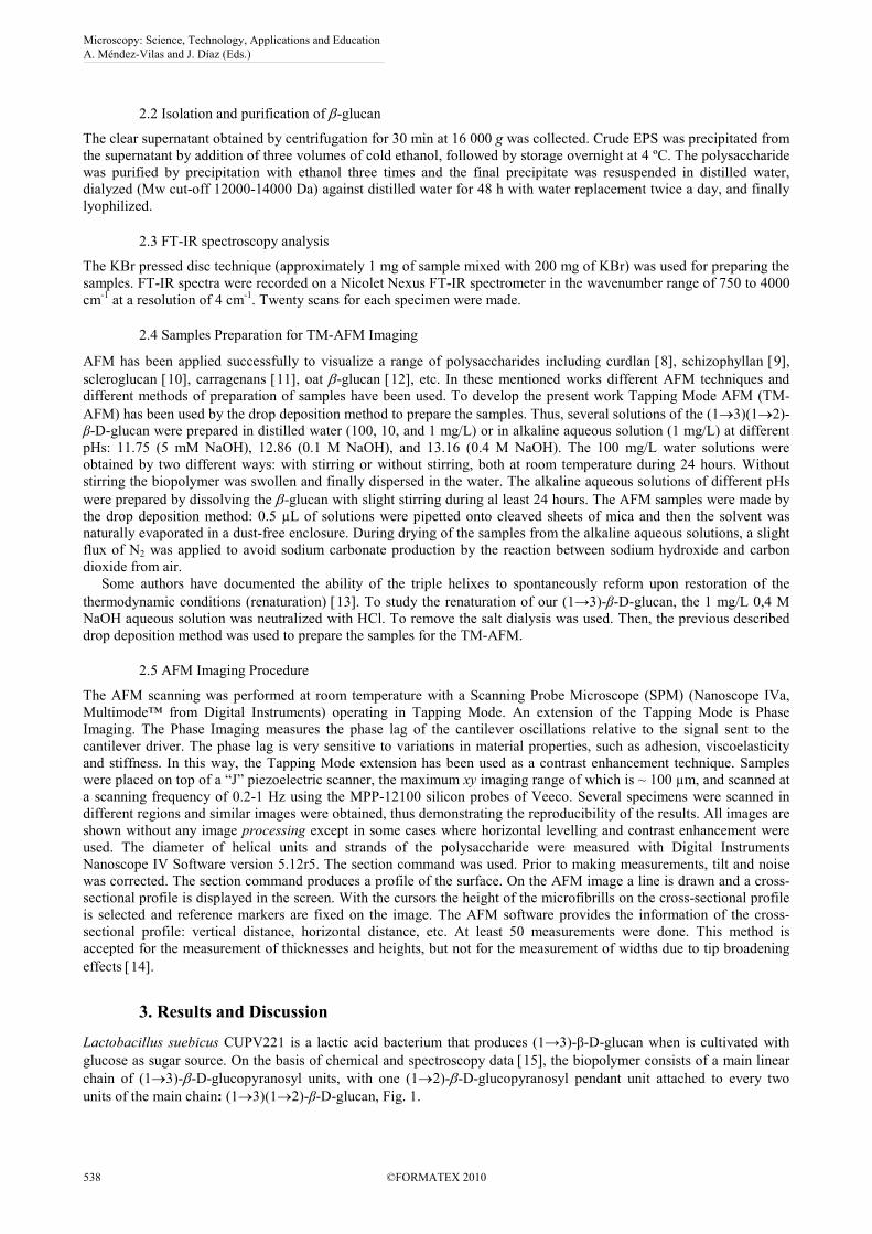
Lactobacillus suebicus CUPV221 is a lactic acid bacterium that produces (1→3)-β-D-glucan when is cultivated with
glucose as sugar source. On the basis of chemical and spectroscopy data [15], the biopolymer consists of a main linear
chain of (1→3)-β-D-glucopyranosyl units, with one (1→2)-β-D-glucopyranosyl pendant unit attached to every two
units of the main chain: (1→3)(1→2)-β-D-glucan, Fig. 1.
Microscopy: Science, Technology, Applications and Education A. Méndez-Vilas and J. Díaz (Eds.)
538 ©FORMATEX 2010
______________________________________________
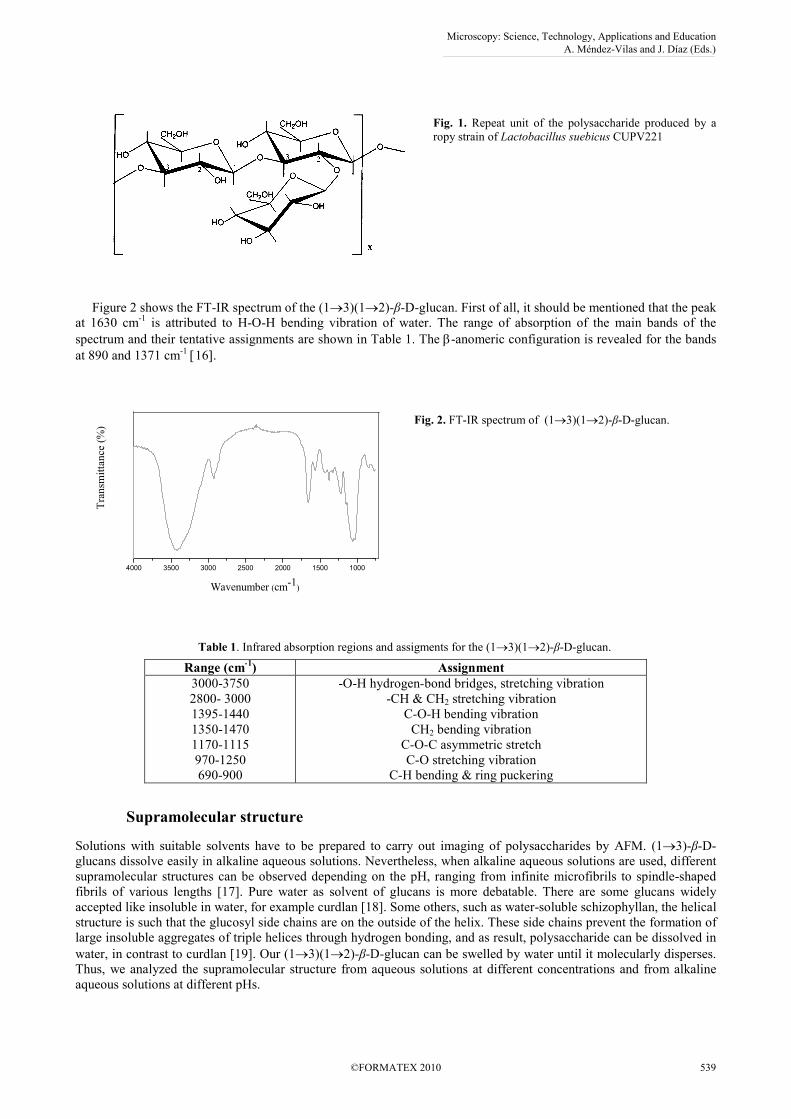
Fig. 1. Repeat unit of the polysaccharide produced by a
ropy strain of Lactobacillus suebicus CUPV221
Figure 2 shows the FT-IR spectrum of the (1→3)(1→2)-β-D-glucan. First of all, it should be mentioned that the peak
at 1630 cm-1
is attributed to H-O-H bending vibration of water. The range of absorption of the main bands of the
spectrum and their tentative assignments are shown in Table 1. The β-anomeric configuration is revealed for the bands
at 890 and 1371 cm-1
[16].
4000 3500 3000 2500 2000 1500 1000
60
80
100
Tra
nsm
itta
nce
(%
)
Wavenumber (cm-1)
Fig. 2. FT-IR spectrum of (1→3)(1→2)-β-D-glucan.
Table 1. Infrared absorption regions and assigments for the (1→3)(1→2)-β-D-glucan.
Range (cm-1) Assignment
3000-3750
2800- 3000
1395-1440
1350-1470
1170-1115
970-1250
690-900
-O-H hydrogen-bond bridges, stretching vibration
-CH & CH2 stretching vibration
C-O-H bending vibration
CH2 bending vibration
C-O-C asymmetric stretch
C-O stretching vibration
C-H bending & ring puckering
Supramolecular structure
Solutions with suitable solvents have to be prepared to carry out imaging of polysaccharides by AFM. (1→3)-β-D-
glucans dissolve easily in alkaline aqueous solutions. Nevertheless, when alkaline aqueous solutions are used, different
supramolecular structures can be observed depending on the pH, ranging from infinite microfibrils to spindle-shaped
fibrils of various lengths [17]. Pure water as solvent of glucans is more debatable. There are some glucans widely
accepted like insoluble in water, for example curdlan [18]. Some others, such as water-soluble schizophyllan, the helical
structure is such that the glucosyl side chains are on the outside of the helix. These side chains prevent the formation of
large insoluble aggregates of triple helices through hydrogen bonding, and as result, polysaccharide can be dissolved in
water, in contrast to curdlan [19]. Our (1→3)(1→2)-β-D-glucan can be swelled by water until it molecularly disperses.
Thus, we analyzed the supramolecular structure from aqueous solutions at different concentrations and from alkaline
aqueous solutions at different pHs.
Microscopy: Science, Technology, Applications and Education A. Méndez-Vilas and J. Díaz (Eds.)
©FORMATEX 2010 539
______________________________________________
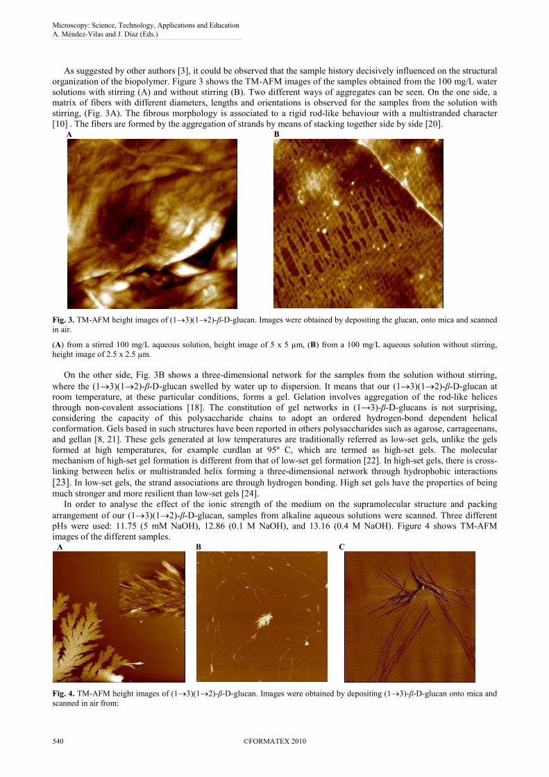
As suggested by other authors [3], it could be observed that the sample history decisively influenced on the structural
organization of the biopolymer. Figure 3 shows the TM-AFM images of the samples obtained from the 100 mg/L water
solutions with stirring (A) and without stirring (B). Two different ways of aggregates can be seen. On the one side, a
matrix of fibers with different diameters, lengths and orientations is observed for the samples from the solution with
stirring, (Fig. 3A). The fibrous morphology is associated to a rigid rod-like behaviour with a multistranded character
[10] . The fibers are formed by the aggregation of strands by means of stacking together side by side [20]. A
B
Fig. 3. TM-AFM height images of (1→3)(1→2)-β-D-glucan. Images were obtained by depositing the glucan, onto mica and scanned
in air.
(A) from a stirred 100 mg/L aqueous solution, height image of 5 x 5 µm, (B) from a 100 mg/L aqueous solution without stirring,
height image of 2.5 x 2.5 µm.
On the other side, Fig. 3B shows a three-dimensional network for the samples from the solution without stirring,
where the (1→3)(1→2)-β-D-glucan swelled by water up to dispersion. It means that our (1→3)(1→2)-β-D-glucan at
room temperature, at these particular conditions, forms a gel. Gelation involves aggregation of the rod-like helices
through non-covalent associations [18]. The constitution of gel networks in (1→3)-β-D-glucans is not surprising,
considering the capacity of this polysaccharide chains to adopt an ordered hydrogen-bond dependent helical
conformation. Gels based in such structures have been reported in others polysaccharides such as agarose, carrageenans,
and gellan [8, 21]. These gels generated at low temperatures are traditionally referred as low-set gels, unlike the gels
formed at high temperatures, for example curdlan at 95º C, which are termed as high-set gels. The molecular
mechanism of high-set gel formation is different from that of low-set gel formation [22]. In high-set gels, there is cross-
linking between helix or multistranded helix forming a three-dimensional network through hydrophobic interactions
[23]. In low-set gels, the strand associations are through hydrogen bonding. High set gels have the properties of being
much stronger and more resilient than low-set gels [24].
In order to analyse the effect of the ionic strength of the medium on the supramolecular structure and packing
arrangement of our (1→3)(1→2)-β-D-glucan, samples from alkaline aqueous solutions were scanned. Three different
pHs were used: 11.75 (5 mM NaOH), 12.86 (0.1 M NaOH), and 13.16 (0.4 M NaOH). Figure 4 shows TM-AFM
images of the different samples. A
B
C
Fig. 4. TM-AFM height images of (1→3)(1→2)-β-D-glucan. Images were obtained by depositing (1→3)-β-D-glucan onto mica and
scanned in air from:
Microscopy: Science, Technology, Applications and Education A. Méndez-Vilas and J. Díaz (Eds.)
540 ©FORMATEX 2010
______________________________________________
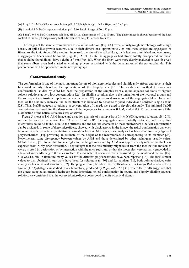
(A) 1 mg/L 5 mM NaOH aqueous solution, pH 11.75, height image of 40 x 40 µm and 5 x 5 µm.
(B) 1 mg/L 0.1 M NaOH aqueous solution, pH 12.86, height image of 50 x 50 µm.
(C) 1 mg/L 0.4 M NaOH aqueous solution, pH 13.16, phase image of 10 x 10 µm. (The phase image is shown because of the high
contrast in the height image owing to the difference of heights between features).
The images of the sample from the weakest alkaline solution, (Fig. 4A) reveal a fairly rough morphology with a high
density of spike-like growth features. Due to their dimensions, approximately 25 nm, these spikes are aggregates of
fibers. As the ionic force of the medium increased, the size of the spike-like growth features diminished and even many
disaggregated fibers could be found, (Fig. 4B). At pH 13.86, the aggregates had almost totally disappeared and those
that could be found did not have a definite form, (Fig. 4C). When the fibers were more deeply analyzed, it was observed
that some fibers even had started unwinding, process associated with the denaturation of the polysaccharide. This
phenomenon will be approached in the next paragraph.
Conformational study
The conformation is one of the most important factors of biomacromolecules and significantly affects and governs their
functional activity, therefore the applications of the biopolymers [25]. The established method to carry out
conformational studies by AFM has been the preparation of the samples from alkaline aqueous solutions or organic
solvent solutions at very low concentrations [26]. In alkaline solutions due to the ionization of the hydroxyl groups and
the subsequent electrostatic repulsion between chains [27], a previous dissociation of the aggregates takes places and
then, as the alkalinity increase, the helix structure is believed to denature to yield individual disordered single chains
[28]. Thus, NaOH aqueous solutions at a concentration of 1 mg/L were used to develop the study. The minimal NaOH
concentration required for the dissociation of the aggregates to occur was 0.1 M, and at 0.4 M the beginning of the
dissociation of the helical structures was observed.
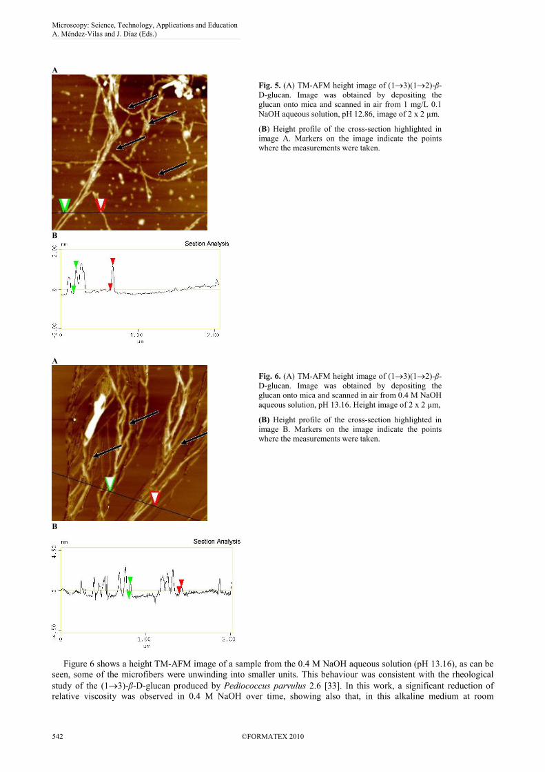
Figure 5 shows a TM-AFM image and a section analysis of a sample from 0.1 M NaOH aqueous solution, pH 12.86.
As can be seen in the image, Fig. 5A at a pH of 12.86, the aggregates were partially detached, and many free
microfibers could be found. Due to the stiffness and the rodlike character of these microfibers a helical conformation
can be assigned. In some of these microfibers, showed with black arrows in the image, the spiral conformation can even
be seen. In order to obtain quantitative information from AFM images, trace analysis has been done for many types of
polysaccharides [14], providing an estimate of the height of the macromolecule corresponding to its diameter [26].
Nevertheless, some discrepancy between values by AFM and those determined by other techniques usually exists.
McIntire et al., [28] found that for scleroglucan, the height measured by AFM was approximately 67% of the thickness
expected from X-ray fiber diffraction. They thought that the dissimilarity might result from the fact that the molecules
were distorted by desiccation or by interaction with the mica substrate, or that the molecules were partially embedded in
a layer of water adhering to the mica surface. The diameter of our microfibers measured by the mentioned method (Fig.
5B) was 1.8 nm. In literature many values for the different polysaccharides have been reported [14]. The most similar
values to that obtained in our work have been for scleroglucan [30] and for xanthan [31], both polysaccharides exist
mainly as linear helical structures [32]. Keeping in mind, besides, the results obtained in Congo Red analysis for a
similar (1→3)-β-D-glucan studied in our laboratory, produced by P. parvulus 2.6 [33], where the results suggested that
the glucan adopted an ordered hydrogen-bond dependent helical conformation in neutral and slightly alkaline aqueous
solution, we considered that the observed microfibers correspond to units of helical strands.
Microscopy: Science, Technology, Applications and Education A. Méndez-Vilas and J. Díaz (Eds.)
©FORMATEX 2010 541
______________________________________________
A
Fig. 5. (A) TM-AFM height image of (1→3)(1→2)-β-
D-glucan. Image was obtained by depositing the
glucan onto mica and scanned in air from 1 mg/L 0.1
NaOH aqueous solution, pH 12.86, image of 2 x 2 µm.
(B) Height profile of the cross-section highlighted in
image A. Markers on the image indicate the points
where the measurements were taken.
B
A
Fig. 6. (A) TM-AFM height image of (1→3)(1→2)-β-
D-glucan. Image was obtained by depositing the
glucan onto mica and scanned in air from 0.4 M NaOH
aqueous solution, pH 13.16. Height image of 2 x 2 µm,
(B) Height profile of the cross-section highlighted in
image B. Markers on the image indicate the points
where the measurements were taken.
B
Figure 6 shows a height TM-AFM image of a sample from the 0.4 M NaOH aqueous solution (pH 13.16), as can be
seen, some of the microfibers were unwinding into smaller units. This behaviour was consistent with the rheological
study of the (1→3)-β-D-glucan produced by Pediococcus parvulus 2.6 [33]. In this work, a significant reduction of
relative viscosity was observed in 0.4 M NaOH over time, showing also that, in this alkaline medium at room
Microscopy: Science, Technology, Applications and Education A. Méndez-Vilas and J. Díaz (Eds.)
542 ©FORMATEX 2010
______________________________________________
temperature, no hydrolysis took place. The viscosity decrease was associated to a denaturation process of the ordered
conformation of the polysaccharide, suggesting that the hydrogen-bonding structure broke down gradually. The image
of Fig. 6A is a direct evidence of this suggestion as the helices consisting of helical strands are unwinding and keeping
even in some strands the helical form (black arrows in the image of Fig. 6A). The diameter of the strands was 0.8 nm,
(Fig. 6B). In view of values by AFM of diameters of strands given by other authors for similar glucans with triple-helix
conformation [14], we propose that our (1→3)-β-D-glucan could have a triple-helix conformation. Nevertheless, more
precise quantification techniques, such as X-ray diffraction, must be used to confirm this statement.
It should be mentioned that the particles which can be seen in the images from the NaOH aqueous solutions, (Fig. 5
and 6) are due to sodium carbonate precipitation, in spite of the slight N2 flux used during the drying of the samples.
For (1→3)-β-D-glucans the structure in units of helical conformation is one important basis for the interesting
properties mentioned in the introduction of the present paper. However, though the helical conformation are stable for a
wide range of solvents and physical conditions, when the helical (1-3)-β-D-glucans are exposed to high pH (as has been
seen above) or also by increasing the temperature above the helix melting temperature, a conformational transition takes
place and the multi-helical units dissociate into single chains or stranded random coils (denaturation). The denaturation
is due to breaking of intermolecular hydrogen bonds resulting in strand separation. However, the multi-helical structure
can be regenerated when denatured samples are restored to thermodynamic conditions (renaturation) [3]. Different
structures have been mentioned in literature for renatured species: linear, circular, hairpin, multichain (aggregated)
structures… [3, 14]. The formation of one or other structure is governed by thermodynamical aspects. Thus, aggregated
structures predominate at higher polymer concentrations and circular and hairpin structures at lower concentrations
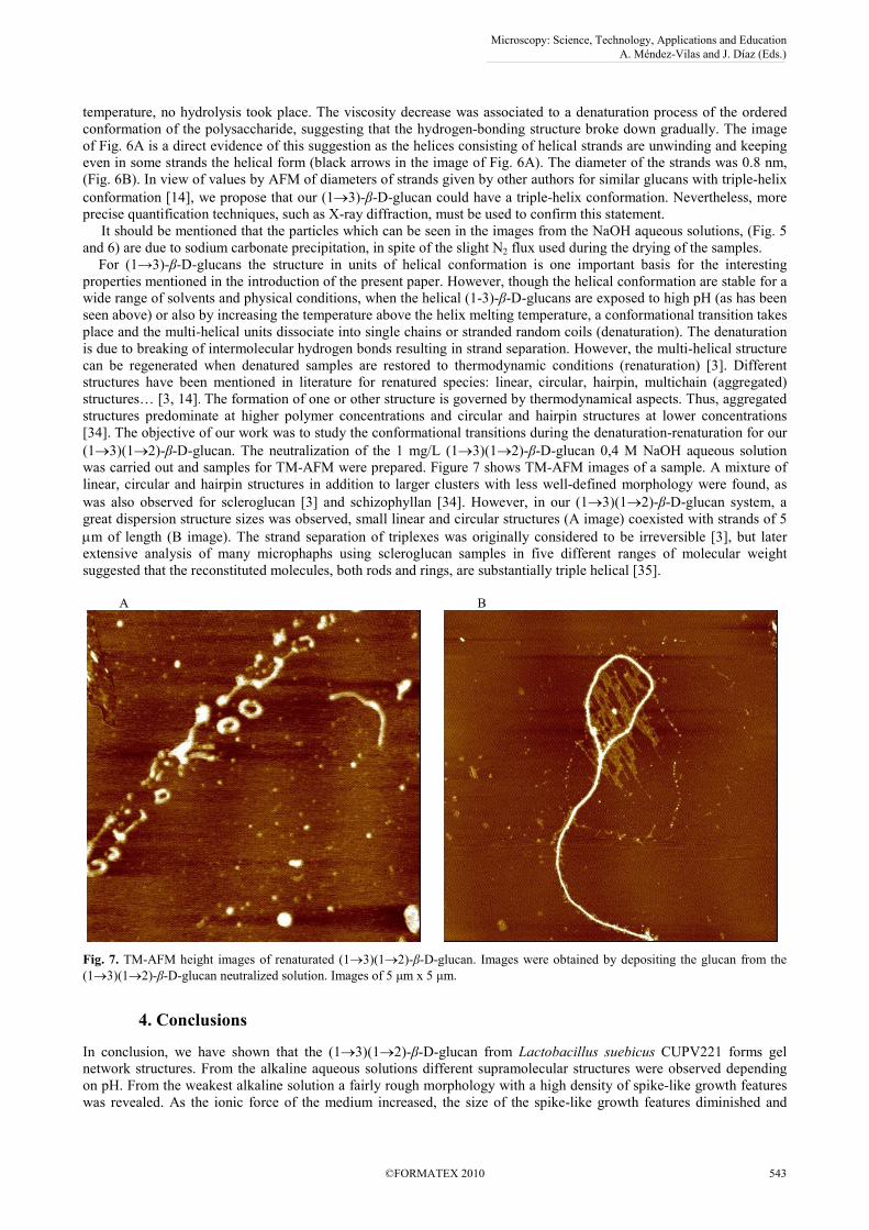
[34]. The objective of our work was to study the conformational transitions during the denaturation-renaturation for our
(1→3)(1→2)-β-D-glucan. The neutralization of the 1 mg/L (1→3)(1→2)-β-D-glucan 0,4 M NaOH aqueous solution
was carried out and samples for TM-AFM were prepared. Figure 7 shows TM-AFM images of a sample. A mixture of
linear, circular and hairpin structures in addition to larger clusters with less well-defined morphology were found, as
was also observed for scleroglucan [3] and schizophyllan [34]. However, in our (1→3)(1→2)-β-D-glucan system, a
great dispersion structure sizes was observed, small linear and circular structures (A image) coexisted with strands of 5
µm of length (B image). The strand separation of triplexes was originally considered to be irreversible [3], but later
extensive analysis of many microphaphs using scleroglucan samples in five different ranges of molecular weight
suggested that the reconstituted molecules, both rods and rings, are substantially triple helical [35].
A
B
Fig. 7. TM-AFM height images of renaturated (1→3)(1→2)-β-D-glucan. Images were obtained by depositing the glucan from the
(1→3)(1→2)-β-D-glucan neutralized solution. Images of 5 µm x 5 µm.
4. Conclusions
In conclusion, we have shown that the (1→3)(1→2)-β-D-glucan from Lactobacillus suebicus CUPV221 forms gel
network structures. From the alkaline aqueous solutions different supramolecular structures were observed depending
on pH. From the weakest alkaline solution a fairly rough morphology with a high density of spike-like growth features
was revealed. As the ionic force of the medium increased, the size of the spike-like growth features diminished and
Microscopy: Science, Technology, Applications and Education A. Méndez-Vilas and J. Díaz (Eds.)
©FORMATEX 2010 543
______________________________________________
even many disaggregated fibers could be found. At pH 13.16 the aggregates had disappeared almost totally. Due to the
stiffness and the rodlike character of fibers, a helical conformation is assigned. At 0.4 M, pH 13.16, the dissociation of
the helical structures takes place.
(1→3)(1→2)-β-D-glucan from Lactobacillus suebicus CUPV221 after exposure to a denaturation-renaturation cycle
forms various molecular topologies: linear, circular and hairpin structures in addition to larger clusters with less well-
defined morphology.
References
[1] Deslandes Y, Marchessault RH, Sarko A. Triple-helical structure of (1,3)-β-D-glucan. Macromolecules. 1980;13:1466-1471.
[2] Zekovic DB, Kwlatkowski S, Vrvic MM, Jakovijevic D, Moran C. A. Natural and modified (1→3)-β-D-glucans in health
promotion and disease alleviation. Crit. Rev. Biotech. 2005;25:205-230.
[3] Sletmoen M, Stokke, BT, Higher order structure of (1,3)-β-D-glucans and its influence on their biological activities and
complexation abilities. Biopolymers. 2008;89:310-320.
[4] Duboc P, Mollet B. Applications of exopolysaccharides in the dairy industry. Int. Dairy J. 2001;11:759-768.
[5] Cerning J. Exocellular polysaccharides produced by lactic acid bacteria. FEMS Microbiol. Rev. 1990;87: 113-130.
[6] Dueñas M, Rodriguez MA, Tejero P, Franco G, Espartero JL, Irastorza A, Gil A. Structural analysis of the exopolysaccharide
produced by Pediococcus damnosus 2.6. Carbohydr. Res. 1997;303: 453-458.
[7] Llaubères RM, Richard B, Lonvaud A, Dubourdieu D. Structure of an exocellular β-D-glucan from Pediococcus sp., a wine lactic
bacteria. Carbohydr. Res. 1990;203:103–107.
[8] Ikeda S, Shishido Y. Atomic force microscopy studies on heat-induced gelation of curdlan. J. Agric. Food Chem. 2005;53:786-
791.
[9] Bae AH, Lee SW, Ikeda M, Sano M, Shinkai S, Sakurai K. Rod-like architecture and helicity of the poly(C)/schizophyllan
complex observed by AFM and SEM. Carbohydr. Res. 2004;339:251-258.
[10] McIntire TM, Brant DA. Observations of the (1→3)-β-D-glucan linear triple helix to macrocycle interconversion using
noncontact atomic force microscopy. J. Am. Chem. Soc. 1998;120:6909-6919.
[11] McIntire TM, Brant DA. Imaging of carrageenan macrocycles and amylose using noncontact atomic force microscopy. Int. J.
Biolog. Macromol. 1999;26:303-310.
[12] Wu J, Zhang Y, Wang L, Xie B, Wang H, Deng S. Visualization of single and aggregated hulless oat (Avena nuda L.)
(1→3)(1→4)-β-D-glucan molecules by atomic force microscopy and confocal scanning laser microscopy. J. Agric. Food Chem.
2006;54:925-934.
[13] Sletmoen M, Stokke, BT. Review Higher order structure of (1, 3)-β-D-Glucans and its influence on their biological activities
and complexation abilities. Biopolymers. 2008;89:310-320.
[14] Abu-Lail NI, Camesano TA. Polysaccharide properties probed with atomic force microscopy. J. Microscopy. 2003;212:217-238.
[15] Ibarburu I. Bacterias lácticas productoras de exopolisacáridos: análisis estructural de polisacáridos y detección molecular de
estirpes que sintetizan β-(1→3)(1→2)-D-glucanos. PhD thesis, 2009. Universidad del País Vasco (UPV/EHU), Donostia-San
Sebastián (Spain).
[16] Corradi da Silva ML, Fukuda EF, Vasconcelos A. F. D, Dekker RF, Matias AC, Monteiro NK., Cardoso, M. S., Barbosa, A. M.,
Silveira, J. L. M., Sassaki, G. L., & Carbonero, E. R. (2008). Carbohydr. Res 343, 793-798.
[17] Koreeda K, Harada T, Ogawa K, Sato S, Kasai N. Study of the ultrastructure of gel-forming (1, 3)-β-D-glucan (curdlan-type
polysaccharide). Carbohydr. Res. 1974; 33:396-399.
[18] McIntosh M, Stone BA, Stanisich VA. Curdlan and other bacterial (1→3)-β-D–glucans. Appl. Microbiol. Biothechnol.
2005;68:163-173.
[19] Bot A, Smorenburg HE, Vreeker R, Paques M, Clarck AH. Melting behaviour of schizophyllan and scleroglucan
exopolysaccharide gels at temperatures between 5 and 20 °C. Carbohydr. Polym. 2001;45:363-372.
[20] Gawronski M, Park JT, Magee AS, Conrad H. Microfibrillar structure of PGG-Glucan in aqueous solution as triple-helix
aggregates by small angle x-ray scattering. Biopolymers 1999;50;569-578.
[21] Guenet JM. Thermorevesible gelation of Polymers and Biopolymers. 1992. Academic Press Inc. San Diego, USA.
[22] Konno A, Harada T. Thermal properties of curdlan in aqueous suspension and curdlan gel. Food Hydrocoll. 1991;5:427-434.
[23] Funami T, Funami M, Yada H, Nakao Y. Rheological and thermal studies on gelling characteristics of curdlan. J. Food Sci.
1999;64:129-132.
[24] Stone BA, Clark AE. Chemistry and Biology of (1-3)-β-D-glucans. 1992. La Trobe University Press. Victoria, Australia.
[25] Bohn JA, BeMiller JN. (1→3)-β-D-Glucans as biological response modifiers: a review of structure-functional activity
relationships. Carbohydr. Polym. 1995;28:3-14.
[26] Jin Y, Zhang H, Yin Y, Nishinari K. Conformation of curdlan as observed by tapping mode atomic force microscopy. Colloid
Polym. Sci. 2006;284:1371-1377.
[27] Wang SC, Bligh SW, Zhu CL, Shi SS, Wang ZY, Hu ZB. Crowder, J. Sulfated β-glucan derived from oat bran with potent anti-
HIV activity. J. Agric. Food Chem. 2008;56:2624-2629.
[28] Futatsuyama H, Yui T, Ogawa K. Viscometry of Curdlan, a Linear (1→3)-β-D-glucan, in DMSO or alkaline Solutions. Biosci.
Biotechnol. Biochem. 1999;63:1481-1483.
[29] McIntire TM, Brant DA. Imaging of individual biopolymers and supramolecular assemblies using noncontact atomic force
microscopy. Biopolymers 1997;42:133-146.
[30] McIntire TM, Penner RM, Brant DA. Observations of a circular, triple-helical polysaccharide using noncontact atomic force
microscopy. Macromolecules 1995;28:6375-6377.
[31] Kirby AR, Gunning AP, Morris VJ. Imaging polysaccharides by atomic force microscopy. Biopolymers 1996;38:355-366.
Microscopy: Science, Technology, Applications and Education A. Méndez-Vilas and J. Díaz (Eds.)
544 ©FORMATEX 2010
______________________________________________

[32] Camesano TA, Wilkinson KJ. Single molecule study of xanthan conformation using atomic force microscopy.
Biomacromolecules 2001;2:1184-1191.
[33] Velasco S, Irastorza A, Dueñas M, Santamaría A, Muñoz ME. Chemical and Rheological properties of the beta-glucan produced
by Pediococcus parvulus 2.6. J. Agric. Food Chem. 2009;57:1827–1834.
[34] Stokke BT, Elgsaeter A, Brant DA, Kuge T, Kitamura A. Macromolecular cyclization of (1→6)-branched-(1→3)-β-D-glucans
observed after denaturation-renaturation of the triple-helical structure. Biopolymers 1993; 33:193-198.
[35] Brant DA. Versatile Polysaccharides: Ropes, Rings, and Rods, AIM Magazine, 2005;60;4049.
Microscopy: Science, Technology, Applications and Education A. Méndez-Vilas and J. Díaz (Eds.)
©FORMATEX 2010 545
______________________________________________