Mridusmita Kakati, Dorothy Das, Pundarikaksha Das, Airy ... · The functions of many proteins have...
5
Mridusmita Kakati, Dorothy Das, Pundarikaksha Das, Airy Sanjeev, Venkata Satish Kumar Mattaparthi Page | 779 Effect of ethanol as molecular crowding agent on the conformational dynamics of α- synuclein Mridusmita Kakati 1 , Dorothy Das 1 , Pundarikaksha Das 1 , Airy Sanjeev 1 , Venkata Satish Kumar Mattaparthi 1* 1 Molecular Modelling and Simulation Laboratory, Department of Molecular Biology and Biotechnology, Tezpur University, Tezpur-784 028, Assam, India *corresponding author e-mail address: [email protected], [email protected] | Scopus ID 54962670000 ABSTRACT The functions of many proteins have been directly connected to their conformational changes. The macromolecular crowding environment inside the cell is known to have a significant impact on the equilibria and transition rates between different conformations of the protein. Here we demonstrate the effect of ethanol as crowders on the conformational dynamics of α- synuclein protein, a primary component of the fibrillar neuronal inclusions, and known as Lewy bodies that are diagnostic of Parkinson’s disease. We observed the α- synuclein protein to experience stronger crowding effects with an increase in concentration of ethanol, the crowding agent. The findings that we obtained from this simulation study would serve as valuable guides for expected crowding effects on conformational dynamics of α- synuclein. Keywords: Parkinson’s disease; Macromolecular crowding; presynaptic, aggregation. 1. INTRODUCTION In the recent past, many research studies have highlighted the importance of the protein dynamics as a valuable platform to understand the association between the structure and function [1- 6]. The protein dynamics leads to the sampling of alternative conformations. Because of ligand binding [7] and post- translational modifications like phosphorylation [8], the conformational changes in the protein molecule gets initiated. As a result, the protein molecule adopts different conformations at varying functional states. From these structures, the conformational changes at atomistic level can be studied. We generally see that biophysical characterizations of conformational changes in protein have been studied mostly under dilute and lesser densed medium. But the proteins perform their biological functions inside the cell which is highly crowded with macromolecules. For example, the cytoplasm of Escherichia coli contains high concentration of macromolecules (about 300–400 g/l and 30% of the total volume occupancy)[9]. Because of crowding in cell membranes, membrane proteins occupy a similar level of the total surface area [10]. However, the impact of crowding environment in cell on the equilibria and transition rates of diverse conformations of proteins are not understood well. The macromolecular crowding in the cell also likely to alter the energy landscapes of conformational changes in a protein resulting in more compact structures over more open structures [11]. Such effects of crowding have been verified experimentally [8]. Molecular Dynamics (MD) simulations have also been used as a tool to investigate the energy landscapes of a number of proteins in a crowding environment, in the context of either conformational change [12] or folding-unfolding transition [13- 15]. In our study, we have investigated the consequences of ethanol as a crowding medium on the conformational dynamics of α-synuclein. We have found the crowding environment to affect the secondary structure content of α- synuclein to a greater extent. 2. MATERIALS AND METHODS The initial 3-D structure of α-synuclein was taken from Protein Data Bank (PDB). In order to study the effect of different concentrations of ethanol, the crowding agent on the conformational dynamics of α-synuclein, we have employed MD simulation using the explicit solvent model. MD simulations were performed using periodic boundary conditions. In carrying out this experiment, cubic simulation boxes were filled with different proportions of water-ethanol mixtures using Packmol. In all these cases, the protein molecule was placed at the center of the simulation box using Leap module of AmberTools 14 program. The protein molecules are then overlaid by equilibrated triple point charge (TIP3P) boxes in order to solvate the molecule of interest in the respective cubic simulation boxes. In addition, positively charged Na+ counter-ions were added into the system to neutralize the negative charge on the protein molecules. The volume occupied by ethanol was set up to about 0%, 5%, 10%, 20%, 50% and 100% of total volume. To study the structural dynamics of intrinsically disordered proteins (IDPs), MD simulations have been extensively in use. The AMBER14 package was used to perform MD simulation while protein and water molecules are described by parameters from ff99SB force field and TIP3P water molecules in the system. In each system, the charge of the protein was neutralized by adding Na+/Cl- counter ions. An isobaric–isothermal ensemble was applied using Langevin dynamics [16] along with Berendsen- thermostat [17] for temperature control. The system was subjected to one stage minimization to ensure the stability of the structure. The integration time step was set to 1 fs. To further take the system to Volume 9, Issue 1, 2020, 779 - 783 ISSN 2284-6808 Open Access Journal Received: 12.12.2019 / Revised: 20.01.2020 / Accepted: 28.01.2020 / Published on-line: 04.02.2020 Original Research Article Letters in Applied NanoBioScience https://nanobioletters.com/ https://doi.org/10.33263/LIANBS91.779783
Transcript of Mridusmita Kakati, Dorothy Das, Pundarikaksha Das, Airy ... · The functions of many proteins have...
Mridusmita Kakati, Dorothy Das, Pundarikaksha Das, Airy Sanjeev, Venkata Satish Kumar Mattaparthi
Page | 779
Effect of ethanol as molecular crowding agent on the conformational dynamics of α-
synuclein
Mridusmita Kakati 1, Dorothy Das
1, Pundarikaksha Das 1, Airy Sanjeev
1, Venkata Satish Kumar
Mattaparthi 1*
1Molecular Modelling and Simulation Laboratory, Department of Molecular Biology and Biotechnology, Tezpur University, Tezpur-784 028, Assam, India
*corresponding author e-mail address: [email protected], [email protected] | Scopus ID 54962670000
ABSTRACT
The functions of many proteins have been directly connected to their conformational changes. The macromolecular crowding
environment inside the cell is known to have a significant impact on the equilibria and transition rates between different conformations
of the protein. Here we demonstrate the effect of ethanol as crowders on the conformational dynamics of α- synuclein protein, a primary
component of the fibrillar neuronal inclusions, and known as Lewy bodies that are diagnostic of Parkinson’s disease. We observed the α-
synuclein protein to experience stronger crowding effects with an increase in concentration of ethanol, the crowding agent. The findings
that we obtained from this simulation study would serve as valuable guides for expected crowding effects on conformational dynamics of
α- synuclein.
Keywords: Parkinson’s disease; Macromolecular crowding; presynaptic, aggregation.
1. INTRODUCTION
In the recent past, many research studies have highlighted
the importance of the protein dynamics as a valuable platform to
understand the association between the structure and function [1-
6]. The protein dynamics leads to the sampling of alternative
conformations. Because of ligand binding [7] and post-
translational modifications like phosphorylation [8], the
conformational changes in the protein molecule gets initiated. As a
result, the protein molecule adopts different conformations at
varying functional states.
From these structures, the conformational changes at
atomistic level can be studied. We generally see that biophysical
characterizations of conformational changes in protein have been
studied mostly under dilute and lesser densed medium. But the
proteins perform their biological functions inside the cell which is
highly crowded with macromolecules. For example, the cytoplasm
of Escherichia coli contains high concentration of macromolecules
(about 300–400 g/l and 30% of the total volume occupancy)[9].
Because of crowding in cell membranes, membrane proteins
occupy a similar level of the total surface area [10]. However, the
impact of crowding environment in cell on the equilibria and
transition rates of diverse conformations of proteins are not
understood well. The macromolecular crowding in the cell also
likely to alter the energy landscapes of conformational changes in
a protein resulting in more compact structures over more open
structures [11]. Such effects of crowding have been verified
experimentally [8].
Molecular Dynamics (MD) simulations have also been
used as a tool to investigate the energy landscapes of a number of
proteins in a crowding environment, in the context of either
conformational change [12] or folding-unfolding transition [13-
15]. In our study, we have investigated the consequences of
ethanol as a crowding medium on the conformational dynamics of
α-synuclein. We have found the crowding environment to affect
the secondary structure content of α- synuclein to a greater extent.
2. MATERIALS AND METHODS
The initial 3-D structure of α-synuclein was taken from
Protein Data Bank (PDB). In order to study the effect of different
concentrations of ethanol, the crowding agent on the
conformational dynamics of α-synuclein, we have employed MD
simulation using the explicit solvent model. MD simulations were
performed using periodic boundary conditions. In carrying out this
experiment, cubic simulation boxes were filled with different
proportions of water-ethanol mixtures using Packmol. In all these
cases, the protein molecule was placed at the center of the
simulation box using Leap module of AmberTools 14 program.
The protein molecules are then overlaid by equilibrated
triple point charge (TIP3P) boxes in order to solvate the molecule
of interest in the respective cubic simulation boxes. In addition,
positively charged Na+ counter-ions were added into the system to
neutralize the negative charge on the protein molecules. The
volume occupied by ethanol was set up to about 0%, 5%, 10%,
20%, 50% and 100% of total volume. To study the structural
dynamics of intrinsically disordered proteins (IDPs), MD
simulations have been extensively in use. The AMBER14 package
was used to perform MD simulation while protein and water
molecules are described by parameters from ff99SB force field
and TIP3P water molecules in the system. In each system, the
charge of the protein was neutralized by adding Na+/Cl- counter
ions. An isobaric–isothermal ensemble was applied using
Langevin dynamics [16] along with Berendsen- thermostat [17] for
temperature control. The system was subjected to one stage
minimization to ensure the stability of the structure. The
integration time step was set to 1 fs. To further take the system to
Volume 9, Issue 1, 2020, 779 - 783 ISSN 2284-6808
Open Access Journal Received: 12.12.2019 / Revised: 20.01.2020 / Accepted: 28.01.2020 / Published on-line: 04.02.2020
Original Research Article
Letters in Applied NanoBioScience https://nanobioletters.com/
https://doi.org/10.33263/LIANBS91.779783
Mridusmita Kakati, Dorothy Das, Pundarikaksha Das, Airy Sanjeev, Venkata Satish Kumar Mattaparthi
Page | 780
room temperature, heating was gradually performed to bring the
temperature of the system to 298 K over a time of 10 ps. To ensure
the equilibration of the system, pressure, density, temperature, root
mean square deviation (RMSD), potential energy, kinetic energy
and total energy of the initial structure of α-synuclein were plotted
as a function of simulation time. The trajectories were collected
and visualized by VMD [18] package after intervals of 10ns for a
total MD run of 50ns and analysed using cpptraj [19] program
from AMBER tools.
3. RESULTS
In order to compare the conformational dynamics of α-
synuclein in different concentrations of ethanol, we carried out all
atom molecular dynamics simulation using the PMEMD module
of AMBER14 software package with ff99SB force field.
Our results demonstrate that α-synuclein folds in a
multiphasic manner in the presence of ethanol as a crowding
agent. We noticed the folding pathway of α-synuclein to vary with
the concentration of ethanol. Although the mechanism of folding
changes with the concentration of ethanol, yet it was characterized
by a common first stage that actually leads to the partially folded
intermediate [20-24]. The nature of the solvent is responsible to
decide for the subsequent fate of this intermediate. It has been
seen that the higher concentrations of ethanol gave rise to an α-
helical conformation. These observations infer that, depending on
the environment, the partially folded intermediate may undergo
self-association to form dimers, soluble oligomers or amorphous
aggregates and fibrils. We also observed that α-synuclein in higher
concentrations of ethanol revealed significant ordered secondary
structure The capacity of concentrated organic solvent inducing
the structural changes in the native globular proteins have been
reported. Typically, alcohol-induced denaturation of globular
proteins is accompanied by a characteristic increase in α-helix
content [25-38]. Much less is currently known about the behaviour
of natively unfolded proteins in water/organic mixtures [39-41];
however, one would expect similar effects. The structural
transformations and oligomerization of α-synuclein in simple
alcohols were driven by the increase in solvent hydrophobicity.
These results, therefore, exclude contributions from specific
protein alcohol interactions, indicating that water/alcohol mixtures
might be useful models for the effect of hydrophobic membrane
surfaces (the membrane field effect) on the conformation of α-
synuclein and other natively unfolded proteins.
3.1. Conformational dynamics of α-synuclein in different
concentrations of crowding agent.
To study the conformational dynamics of α-synuclein in
different concentrations of ethanol, the crowding agent, we have
analyzed the RMSDs of the Cα atoms, radius of gyration (Rg),
Root Mean Square Fluctuation (RMSF) and Solvent Accessible
Surface Area (SASA) over the course of simulation time as shown
in Figure 1A, 1B, 1C and 1D. Assessment of the structural drift
was carried out by analyzing the Cα atom RMSDs. We observed
the RMSD profile in each case of the protein to be varying
(Figure 1A). The backbone RMSD of the protein attained almost
stable conformation from the initial stage in case of 100% ethanol,
whereas in case of 0%, 5%, 10%, 20%, 50% ethanol the structure
had undergone conformational changes through the initial time
period of around 4.5ns, 4.5ns, 4ns, 2.5ns and 2ns respectively, and
then reached equilibration. RMSD value of α-synuclein in 100%
ethanol corresponds to 7.5 Å, 50% ethanol is 11 Å, 20% ethanol is
13 Å, 10% ethanol is 15 Å, 5% ethanol is 19 Å and 0% ethanol is
21 Å. From these RMSD analyses we can infer that α-synuclein
reaches equilibration very quickly with increase in concentration
of crowding. The stability of the protein is due to its ability to
retain its native α-helical conformation even at higher
concentration of ethanol.
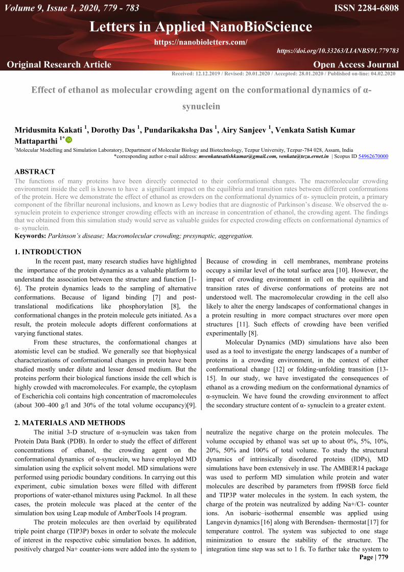
Figure 1. Comparative MD analyses of (A) RMSD, (B) Rg, (C) RMSF
and (D) SASA, for α-synuclein in different concentration of ethanol.
Figure 1B shows the radius of gyration analysis of α-synuclein
protein as a function of time. In the simulation carried out, the
radius of gyration oscillated to a greater degree before 2 ns, further
confirming that the peptide structure remained stable after 2 ns
from the simulation being initiated. From the Rg plots, we can see
that with an increase in ethanol concentration, the structure of α-
synuclein was found to be less compact. The size of the molecule
was bigger in a higher concentration of ethanol. α-synuclein in
100% ethanol depicts the highest Rg value as observed from the
plot. So, it can be inferred that α-synuclein retains its structure
having more helical content in the higher crowding environment
that makes it lesser prone to aggregation. Thus, it is evident that
the aggregation propensities of α-synuclein decreases with
increasing proportions of ethanol. To obtain information on local
structural flexibility, thermal stability and heterogeneity of
macromolecules, root mean-square fluctuations (RMSF) of α-
synuclein were studied (Figure1C). RMSF values obtained for the
backbone C-α atom in different concentrations of ethanol were
calculated from the corresponding MD simulation trajectories and
were plotted against their residue numbers. It can be inferred from
the plot that fluctuation in conformational dynamics of α-
synuclein was found to decrease with an increase in ethanol
concentration. In order to get information regarding the buried and
exposed area present in the protein structure, SASA analysis was
carried out (Figure 1D). The overall solvent accessible area of the
protein molecule was analyzed in different concentrations of
ethanol.
From Figure 1D, it can be seen that α-synuclein in 0%
ethanol has higher SASA values while in 100% ethanol the SASA
value was found to decrease. The SASA of α-synuclein decreases
with an increase in the ethanol concentration.
3.2. Secondary Structure Analysis of α-synuclein in different
concentrations of crowding agent.
The secondary structure analysis was carried out for α-
synuclein in the crowding medium using the Kabsch and Sander
algorithm incorporated in their Dictionary of Secondary Structure
Effect of Molecular Crowding on the Conformational Dynamics of α-Synuclein
Page | 781
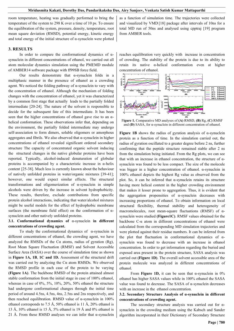
for Protein (DSSP) program [42]. The probability score graph
results were in good agreement with the assessment that crowding
supports in retaining the native structure of α-synuclein. (Figure
2A). From the graph we can observe that most of the residues
retained their α-helical conformation in presence of 100% ethanol.
Figure 2. (A) Probability score of secondary structure for each residue in
α-synuclein in 0%, 5%, 10%, 20%, 50% and 100% ethanol concentration.
Figure 2. (B) Time evolution of Secondary structure of α-synuclein in
presence of 0%, 5%, 10%, 20%, 50% and 100% ethanol.
The plot shows the secondary structural variation of each
residue during the course of simulation time. In each case, the
alpha helical portion of the structure was increased with an
increase in concentration of ethanol, the crowding medium. The
protein in 100% ethanol tends to have higher helical content in
comparison to other proportions. So, the existence and rapid
changes in structural dynamics of α-synuclein in crowding media
were clearly visible from secondary structure analysis.
We also calculated the percentage of individual secondary
structure content in α-synuclein across all conformations using
YASARA software [43] that were sampled during the production
job of trajectories and the results were summarized in Table 1.
From Table 1, we observed that α-synuclein in 100%
ethanol contains a higher amount of α-helix than the other
systems. So, these observations support that higher helical
conformation in α-synuclein which is predominant in case of
100% ethanol, to be actually responsible for preventing fibrillation
process as this structural characteristic feature would induce a
similar conformation that restricts fibrillation as proposed earlier.
Figure 2B shows the classification of the trajectories in terms of
secondary-structure elements obtained by the software tool DSSP
which assigns secondary structures to the amino acids of a protein,
by identifying the intra-backbone hydrogen bonds of the protein.
From the plot we can see the stability (or de-stability) of
secondary structure elements as a function of time. Examination of
Figure 2B shows that the main features of the α -helix structure
were largely retained in the case of 100% ethanol as compared to
other systems.
Table 1. Secondary structure content of α-synuclein in 0%, 5%, 10%,
20%, 50% and 100% ethanol. Concentration
of Ethanol
α-Helix Sheet Turn Coil 310
helix
Π-
helix
0% 4.3% 0.0% 34.3% 61.4% 0.0% 0.0%
5% 8.6% 0.0% 37.1% 54.3% 0.0% 0.0%
10% 5.0% 1.4% 30.0% 60.0% 3.6% 0.0%
20% 22.1% 0.0% 8.6% 63.6% 5.7% 0.0%
50% 20.0% 2.1% 30.0% 47.9% 0.0% 0.0%
100% 55.0% 0.0% 5.7% 39.3% 0.0% 0.0
%
Table 2. Diffusion Coefficient values of -synuclein in different
concentrations of ethanol.
Concentrations
of Ethanol
Diffusion Coefficient
(x10-10
cm2/s)
0% Ethanol 0.5829
5% Ethanol 0.3703
10% Ethanol 0.2516
20% Ethanol 0.2148
50% Ethanol 0.2699
100% Ethanol 0.4446
3.3. Conformers of α-synuclein at different concentrations of
ethanol.
In Figure 3, we can see the snapshots of α-synuclein at 0%,
5%, 10%, 20%, 50% and 100% concentration of ethanol. We
observed most of the residues to be in α-helical conformation with
increasing concentration of ethanol.
3.4. Analysis of Diffusion Coefficient.
The values of diffusion coefficient for α-synuclein in different
concentrations of ethanol were summarized in the Table 2.
Figure 3.Snapshots of α-synuclein conformers in presence of 0%, 5%,
10%, 20%, 50% and 100% ethanol during the time course of simulation.
Diffusion coefficient values tends to decrease with
increasing concentration of ethanol but again at higher
concentrations the corresponding values increase. This is so
because of the shifts in the dielectric medium and structural
changes of the molecule. In the beginning, the structure of α-
synuclein was more compact and its diffusion was affected by the
presence of water molecules. But, gradually with an increase in
ethanol concentration, the intermolecular interaction between
water and α-synuclein decreases. In 50% and 100% ethanol, the
number of water molecules eventually decreases and becomes
negligible, for which the attraction between the water molecules
and the protein diminishes and thus the diffusion coefficient value
escalates. This facilitates the sudden change in the pattern of the
diffusion coefficient values with respect to increasing
concentrations of ethanol.
4. CONCLUSIONS
In this work, the effect of molecular crowding on the
conformational dynamics of α-synuclein was studied. The
conformational changes and fluctuations in α-synuclein was found
to decrease gradually with an increase in the concentration of

Mridusmita Kakati, Dorothy Das, Pundarikaksha Das, Airy Sanjeev, Venkata Satish Kumar Mattaparthi
Page | 782
ethanol, the crowding agent. We also noticed the solvent
accessible surface area of α-synuclein protein to decrease and the
3-D structure to become less compact at higher concentration of
ethanol, which explains its decreasing tendency towards
aggregation. Diffusion coefficient of α-synuclein was found to be
dependent on concentration of the ethanol, the crowding agent.
With an increase in concentration of ethanol, the value of diffusion
coefficient decreases initially but again increases at higher
concentrations due to the change in dielectric medium,
intermolecular interactions and structural changes in α-synuclein.
The intermolecular interactions between the solvent water
molecules and α-synuclein protein were found to decrease with an
increase in concentration of ethanol. Our results show that along
with excluded volume effect, the co-solute properties of crowded
intracellular environment need to be considered to understand α-
synuclein dynamics in cells.
5. REFERENCES
1. Kohen, A. Cannio, R. Bartolucci, S. Klinman, J.P. Enzyme
dynamics and hydrogen tunnelling in a thermophilic alcohol
dehydrogenase. Nature 1999, 399, 496,
https://doi.org/10.1038/20981.
2. Agarwal, P.K. Role of protein dynamics in reaction rate
enhancement by enzymes. J. Am. Chem. Soc. 2005, 127, 15248-
15256, https://doi.org/10.1021/ja055251s.
3. Eisenmesser, E.Z.; Millet, O.; Labeikovsky, W.; Korzhnev,
D.M.; Wolf-Watz, M.; Bosco, D.A.; Skalicky, J.J.; Kay, L.L.;
Kern, D. Intrinsic dynamics of an enzyme underlies catalysis.
Nature 2005, 438, 117, https://doi.org/10.1038/nature04105.
4. Masgrau, L.; Roujeinikova, A.; Johannissen, L.O.; Hothi, P.;
Basran, J.; Ranaghan, K.E.; Mulholland, A.J.; Sutcliffe, M.J.;
Scrutton, N.S.; Leys, D. Atomic description of an enzyme
reaction dominated by proton tunneling. Science, 2006, 312,
237-241, https://doi.org/10.1126/science.1126002
5. Wang, L.; Goodey, N.M.; Benkovic, S.J.; Kohen, A.
Coordinated effects of distal mutations on environmentally
coupled tunneling in dihydrofolate reductase. Proceedings of the
National Academy of Sciences 2006, 103, 15753-15758, https://doi.org/10.1073/pnas.0606976103.
6. Sytina, O.A.; Heyes, D.J.; Hunter, C.N.; Alexandre, M.T.;
van Stokkum, I.H.; van Grondelle, R.; Groot, M.L.
Conformational changes in an ultrafast light-driven enzyme
determine catalytic activity. Nature 2008, 456, 1001, https://doi.org/10.1038/nature07354.
7. Qasba, P.K.; Ramakrishnan, B.; Boeggeman, E. Substrate-
induced conformational changes in glycosyltransferases.
Trends Biochem. Sci. 2005, 30, 53-62,
https://doi.org/10.1016/j.tibs.2004.11.005.
8. Groban, E.S.; Narayanan, A.; Jacobson, M.P.
Conformational changes in protein loops and helices induced by
post-translational phosphorylation. PLoS Comput. Biol. 2006, 2,
e32, https://doi.org/10.1371/journal.pcbi.0020032. 9. Zimmerman, S.B.; Trach, S.O. Estimation of macromolecule
concentrations and excluded volume effects for the cytoplasm of
Escherichia coli. J. Mol. Biol. 1991, 222, 599-620,
https://doi.org/10.1016/0022-2836(91)90499-v. 10. Zhou, H.X. Crowding effects of membrane proteins.
J. Phys. Chem. B. 2009, 113, 7995-8005,
https://doi.org/10.1021/jp8107446.
11. Zhou, H.X.; Rivas, G.; Minton, A.P. Macromolecular
crowding and confinement: biochemical, biophysical, and
potential physiological consequences.
Annu. Rev. Biophys. 2008, 37, 375-397,
https://doi.org/10.1146/annurev.biophys.37.032807.125817.
12. Minh, D.D.; Chang, C.E.; Trylska, J.; Tozzini, V.;
McCammon, J.A. The influence of macromolecular crowding on
HIV-1 protease internal dynamics. J. Am. Chem. Soc. 2006, 128,
6006-6007, https://doi.org/10.1021/ja060483s.
13. Cheung, M.S.; Klimov, D.; Thirumalai, D. Molecular
crowding enhances native state stability and refolding rates of
globular proteins. Proceedings of the National
Academy of Sciences 2005, 102, 4753-4758,
https://doi.org/10.1073/pnas.0409630102.
14. Pincus, D.L.; Thirumalai, D. Crowding effects on the
mechanical stability and unfolding pathways of ubiquitin.
J. Phys. Chem. B 2008, 113, 359-368,
https://doi.org/10.1021/jp807755b
15. Mittal, J.; Best, R.B. Dependence of protein folding stability
and dynamics on the density and composition of
macromolecular crowders. Biophys. J. 2010, 98, 315-320,
https://doi.org/10.1016/j.bpj.2009.10.009.
16. Wu, X.; Brooks, B.R. Self-guided Langevin dynamics
simulation method. Chem. Phys. Lett. 2003, 381, 512-518,
https://doi.org/10.1016/j.cplett.2003.10.013.
17. Mudi, A.; Chakravarty, C. Effect of the Berendsen
thermostat on the dynamical properties of water.
Mol. Phys. 2004, 102, 681-685,
https://doi.org/10.1080/00268970410001698937.
18. Humphrey, W.; Dalke, A.; Schulten, K. VMD: visual
molecular dynamics. J. Mol. Graphics 1996, 14, 33-38,
https://doi.org/10.1016/0263-7855(96)00018-5
19. Roe, D.R.; Cheatham, III.T.E. PTRAJ and CPPTRAJ:
software for processing and analysis of molecular dynamics
trajectory data. J. Chem. Theory. Comput. 2013, 9, 3084-3095,
https://doi.org/10.1021/ct400341p. 20. Uversky, V.N.; Li, J.; Fink, A.L. Evidence for a partially
folded intermediate in α-synuclein fibril formation.
J. Biol. Chem. 2001, 276, 10737-10744,
https://doi.org/10.1074/jbc.m010907200.
21. Uversky, V.N.; Li, J.; Fink, A.L. Metal-triggered structural
transformations, aggregation, and fibrillation of human α-
synuclein a possible molecular link between parkinson′ s disease
and heavy metal exposure. J. Biol. Chem. 2001, 276, 44284-
44296, https://doi.org/10.1074/jbc.m105343200.
22. Uversky, V.N.; Li, J.; Fink, A.L. Pesticides directly
accelerate the rate of α‐synuclein fibril formation: a possible
factor in Parkinson's disease. FEBS Lett. 2001, 500, 105-108,
https://doi.org/10.1016/s0014-5793(01)02597-2.
23. Manning-Bog, A.B.; McCormack, A.L.; Li, J.; Uversky,
V.N.; Fink, A.L.; Di Monte, D.A. The herbicide paraquat causes
up-regulation and aggregation of α-synuclein in mice paraquat
and α-synuclein. J. Biol. Chem. 2002, 277, 1641-1644,
https://doi.org/10.1074/jbc.c100560200.
24. Uversky, V.N.; Li, J.; Fink, A.L. Trimethylamine‐N‐oxide‐
induced folding of α‐synuclein. FEBS Lett. 2001, 509, 31-35,
https://doi.org/10.1016/s0014-5793(01)03121-0.
25. Bychkova, V.E.; Dujsekina, A.E.; Klenin, S.I.; Tiktopulo,
E.I.; Uversky, V.N.; Ptitsyn, O.B. Molten globule-like state of
cytochrome c under conditions simulating those near the
membrane surface. Biochemistry 1996, 35, 6058-6063,
https://doi.org/10.1021/bi9522460.
26. Uversky, V.N.; Narizhneva, N.V.; Kirschstein, S.O.; Winter,
S.; Löber, G. Conformational transitions provoked by organic
solvents in β-lactoglobulin: can a molten globule like
Effect of Molecular Crowding on the Conformational Dynamics of α-Synuclein
Page | 783
intermediate be induced by the decrease in dielectric constant?
Folding Des. 1997, 2, 163-172, https://doi.org/10.1016/s1359-
0278(97)00023-0.
27. Kamatari, Y.O.; Konno, T.; Kataoka, M.; Akasaka, K. The
methanol-induced globular and expanded denatured states of
cytochromec: a study by CD fluorescence, NMR and small-
angle X-ray scattering. J. Mol. Biol. 1996, 259, 512-523,
https://doi.org/10.1006/jmbi.1996.0336.
28. Narizhneva, N.V.; Uversky, V.N. Human a-Fetoprotein is in
the Molten Globule State under Conditions Modelling Protein
Environment near the Membrane Surface. Protein Pept. Lett.
1997, 4, 243-250.
29. Dufour, E.; Bertrand‐Harb, C.; Haertlé, T. Reversible effects
of medium dielectric constant on structural transformation of β‐
lactoglobulin and its retinol binding. Biopolymers 1993, 33, 589-
598, https://doi.org/10.1002/bip.360330408 30. Tanford, C.; De, P.K.; Taggart, V.G. The Role of the α-Helix
in the Structure of Proteins. Optical Rotatory Dispersion of β-
Lactoglobulin1a. J. Am. Chem. Soc. 1960, 82, 6028-6034,
https://doi.org/10.1021/ja01508a015.
31. Arakawa, T.; Goddette, D. The mechanism of helical
transition of proteins by organic solvents. Arch. Biochem.
Biophys. 1985, 240, 21-32, https://doi.org/10.1016/0003-
9861(85)90004-9.
32. Wilkinson, K.D.; Mayer, A.N. Alcohol-induced
conformational changes of ubiquitin. Arch. Biochem. Biophys.
1986, 250, 390-399, https://doi.org/10.1016/0003-
9861(86)90741-1.
33. Jackson, M.; Mantsch, H.H. Halogenated alcohols as
solvents for proteins: FTIR spectroscopic studies. Biochim.
Biophys. Acta, Protein Struct. Mol. Enzymol. 1992, 1118, 139-
143, https://doi.org/10.1016/0167-4838(92)90141-y.
34. Buck, M.; Radford, S.E.; Dobson, C.M. A partially folded
state of hen egg white lysozyme in trifluoroethanol: structural
characterization and implications for protein folding.
Biochemistry 1993, 32, 669-678, https://doi.org/10.1021/bi00053a036.
35. Fan, P., Bracken, C., and Baum, J. Structural characterization
of monellin in the alcohol-denatured state by NMR: Evidence
for. beta.-sheet to. alpha.-helix conversion. Biochemistry
1993, 32, 1573-1582, https://doi.org/10.1021/bi00057a023.
36. Thomas, P.D.; Dill, K.A. Local and nonlocal interactions in
globular proteins and mechanisms of alcohol
denaturation. Protein Sci., 1993, 2, 2050-2065,
https://doi.org/10.1002/pro.5560021206.
37. .Alexandrescu, A.T.; Ng, Y.L.; Dobson, C.M.
Characterization of a trifluoroethanol-induced partially folded
state of α-lactalbumin. J. Mol. Biol. 1994, 235, 587-599,
https://doi.org/10.1006/jmbi.1994.1015.
38. Hamada, D.; Kuroda, Y.; Tanaka, T.; Goto, Y. High helical
propensity of the peptide fragments derived from β-
lactoglobulin, a predominantly β-sheet protein. J. Mol. Biol.
1995, 254, 737-746, https://doi.org/10.1006/jmbi.1995.0651.
39. Dahlman-Wright, K.; Baumann, H.; McEwan, I.J.; Almlöf,
T.; Wright, A.P.; Gustafsson, J.A.; Härd, T. Structural
characterization of a minimal functional transactivation domain
from the human glucocorticoid receptor. Proceedings of the
National Academy of Sciences 1995, 92, 1699-1703,
https://doi.org/10.1073/pnas.92.5.1699.
40. Schmitz, M.L.; dos Santos Silva, M.A.; Altmann, H.; Czisch,
M.; Holak, T.A.; Baeuerle, P.A. Structural and functional
analysis of the NF-kappa B p65 C terminus. An acidic and
modular transactivation domain with the potential to adopt an
alpha-helical conformation. J. Biol. Chem. 1994, 269, 25613-
25620.
41. Donaldson, L.; Capone, J.P. Purification and characterization
of the carboxyl-terminal transactivation domain of Vmw65 from
herpes simplex virus type 1. J Biol. Chem. 1992, 267, 1411-
1414.
42. Kabsch, W.; Sander, C. Dictionary of protein secondary
structure: pattern recognition of hydrogen‐bonded and
geometrical features. Biopolymers 1983, 22, 2577-2637,
https://doi.org/10.1002/bip.360221211.
43. Krieger, E.; Vriend, G.; Spronk, C. YASARA–Yet Another
Scientific Artificial Reality Application. YASARA. Org 2013,
993.
6. ACKNOWLEDGEMENTS
This work was supported by the Science and Engineering Research Board (SERB), Government of India (EMR/2017/005383).
MK and DD thanks DST-SERB for the fellowship.
© 2020 by the authors. This article is an open access article distributed under the terms and conditions of the
Creative Commons Attribution (CC BY) license (http://creativecommons.org/licenses/by/4.0/).


![Glossar Deutsch- Griechisch - praxis.gr · 7a Jägerschnitzel, das, - σνίτσελ του κυνηγού [με μανιτάρια] 7a Kalbfleisch, das μοσχαρίσιο κρέας](https://static.fdocument.org/doc/165x107/5e06fe20a88f4c06457803b9/glossar-deutsch-griechisch-7a-jgerschnitzel-das-ff-.jpg)