BIOCHEMISTRY LECTURES BY RASAQ, Noer.unimed.edu.ng/LECTURE NOTES/3/2/RASAQ-NURUDEEN-OLAJID… ·...
21
BIOCHEMISTRY LECTURES BY RASAQ, N.O
Transcript of BIOCHEMISTRY LECTURES BY RASAQ, Noer.unimed.edu.ng/LECTURE NOTES/3/2/RASAQ-NURUDEEN-OLAJID… ·...
BIOCHEMISTRY LECTURES BY
RASAQ, N.O
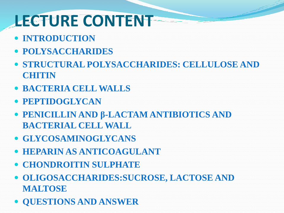
LECTURE CONTENT INTRODUCTION
POLYSACCHARIDES
STRUCTURAL POLYSACCHARIDES: CELLULOSE AND
CHITIN
BACTERIA CELL WALLS
PEPTIDOGLYCAN
PENICILLIN AND β-LACTAM ANTIBIOTICS AND
BACTERIAL CELL WALL
GLYCOSAMINOGLYCANS
HEPARIN AS ANTICOAGULANT
CHONDROITIN SULPHATE
OLIGOSACCHARIDES:SUCROSE, LACTOSE AND
MALTOSE
QUESTIONS AND ANSWER
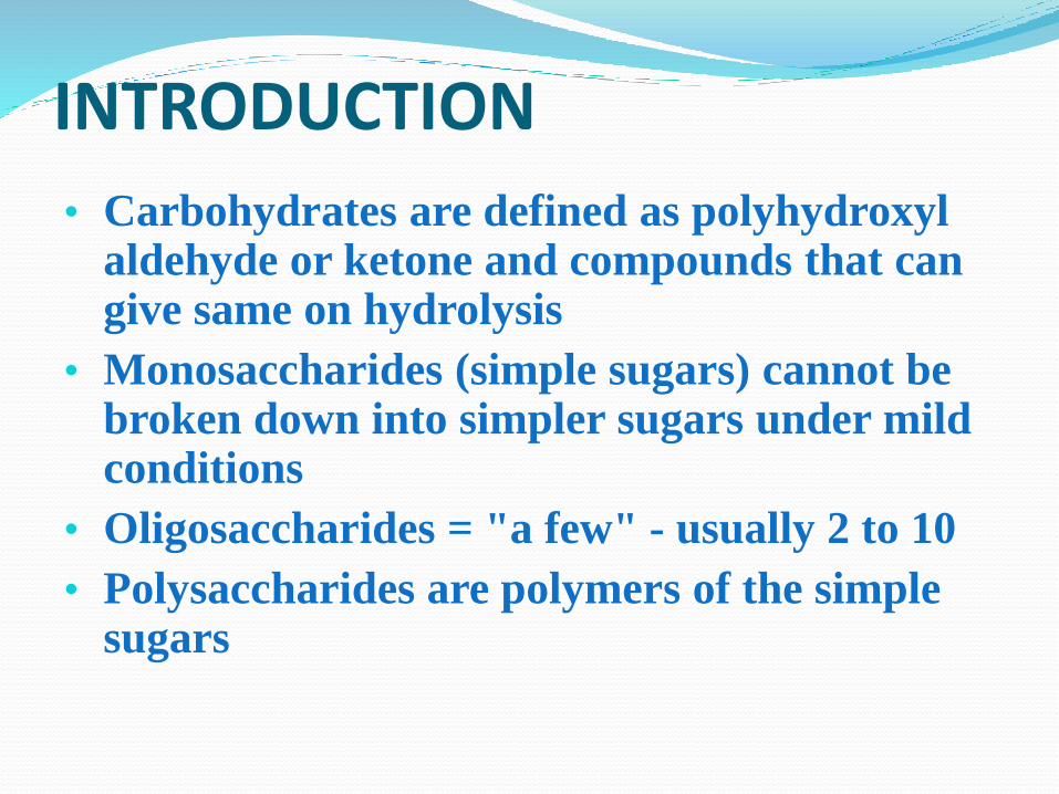
INTRODUCTION
• Carbohydrates are defined as polyhydroxyl aldehyde or ketone and compounds that can give same on hydrolysis
• Monosaccharides (simple sugars) cannot be broken down into simpler sugars under mild conditions
• Oligosaccharides = "a few" - usually 2 to 10
• Polysaccharides are polymers of the simple sugars
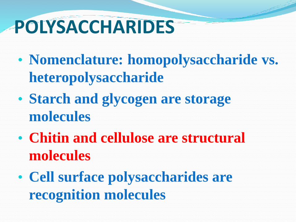
POLYSACCHARIDES
• Nomenclature: homopolysaccharide vs.
heteropolysaccharide
• Starch and glycogen are storage
molecules
• Chitin and cellulose are structural
molecules
• Cell surface polysaccharides are
recognition molecules
STRUCTURAL
POLYSACHHARIDES:CELLULOSE Cellulose is the most abundant natural polymer on
earth
Cellulose is the principal strength and support of
trees and plants
Cellulose is a linear polymer of up to 15000 D-
glucose residues (a glucan) linked by β(1-4)
glycosidic bonds in contrast to the α(1-4) bonds of
amylose.
This difference gives cellulose and amylose very
different structural and physical properties
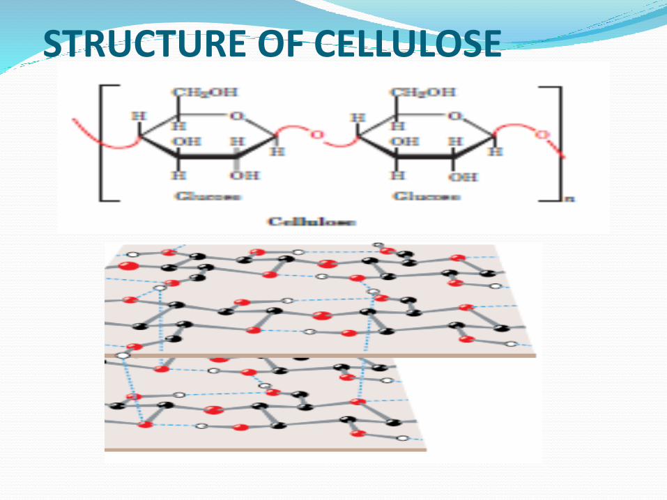
STRUCTURE OF CELLULOSE
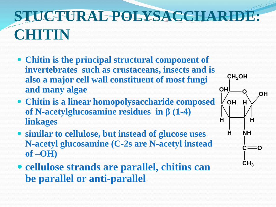
STUCTURAL POLYSACCHARIDE:
CHITIN
Chitin is the principal structural component of invertebrates such as crustaceans, insects and is also a major cell wall constituent of most fungi and many algae
Chitin is a linear homopolysaccharide composed of N-acetylglucosamine residues in β (1-4) linkages
similar to cellulose, but instead of glucose uses N-acetyl glucosamine (C-2s are N-acetyl instead of –OH)
cellulose strands are parallel, chitins can be parallel or anti-parallel
O
CH2OH
NH
OH
H
OH
H
OH
H
H
C O
CH3
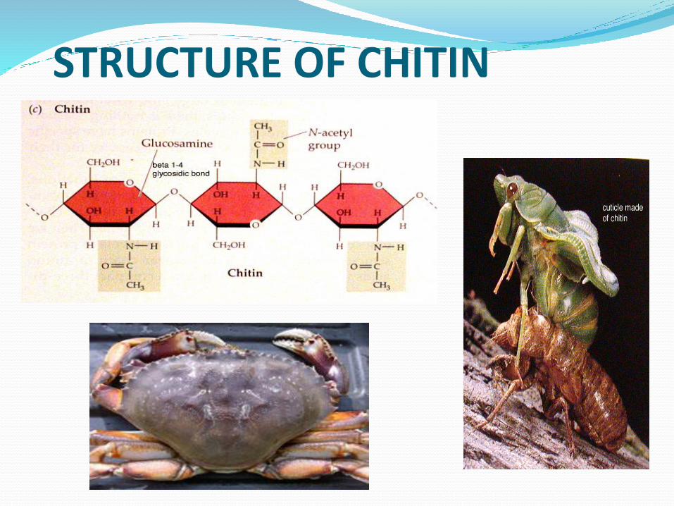
STRUCTURE OF CHITIN
BACTERIAL CELL WALLS
Bacteria are surrounded by rigid cell walls that
give them their characteristics shapes and
permit them to live in hypotonic environments
that would otherwise cause them to swell
osmotically until their plasma (cell)membranes
lysed (burst)
Bacterial cell walls are of considerable medical
significance because they are responsible for
bacterial virulence (disease-evoking power)
Bacteria are classified as gram-positive or
gram-negative depending on whether or not
they take up gram stain
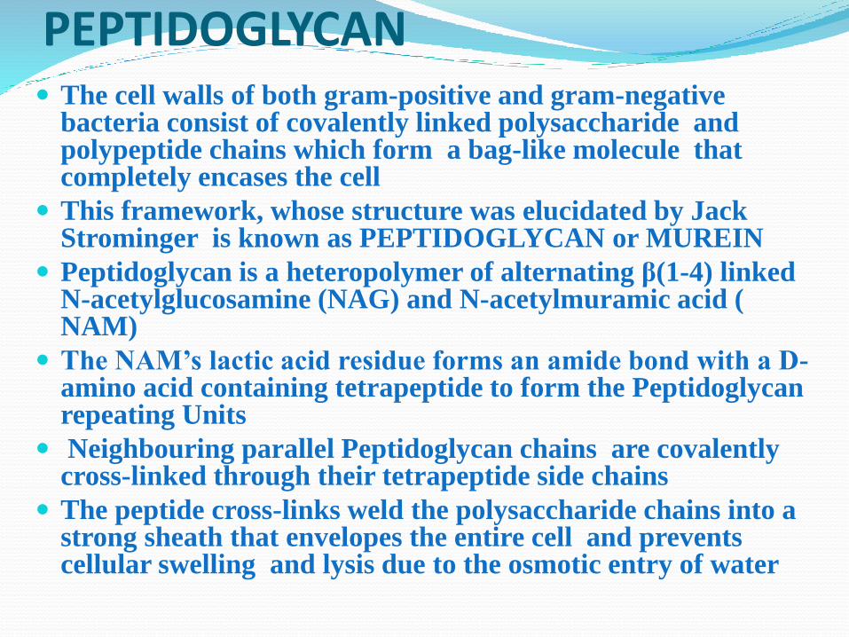
PEPTIDOGLYCAN The cell walls of both gram-positive and gram-negative
bacteria consist of covalently linked polysaccharide and polypeptide chains which form a bag-like molecule that completely encases the cell
This framework, whose structure was elucidated by Jack Strominger is known as PEPTIDOGLYCAN or MUREIN
Peptidoglycan is a heteropolymer of alternating β(1-4) linked N-acetylglucosamine (NAG) and N-acetylmuramic acid ( NAM)
The NAM’s lactic acid residue forms an amide bond with a D-amino acid containing tetrapeptide to form the Peptidoglycan repeating Units
Neighbouring parallel Peptidoglycan chains are covalently cross-linked through their tetrapeptide side chains
The peptide cross-links weld the polysaccharide chains into a strong sheath that envelopes the entire cell and prevents cellular swelling and lysis due to the osmotic entry of water
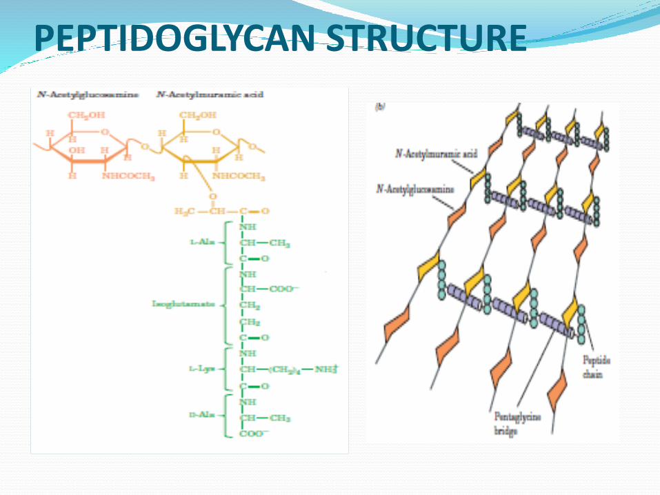
PEPTIDOGLYCAN STRUCTURE
PENICILLIN AND β-LACTAM ANTIBIOTICS
AND BACTERIAL CELLWALL Penicillin and related antibiotics kill bacteria by preventing synthesis of the
cross-links leaving the cell wall too weak to resist osmolytic lysis
Penicillin specifically binds to and inactivates enzymes (transpeptidase)that function to cross-link the peptidoglycan strands of bacterial cell walls
It is this reaction that is inhibited by penicillin and related compounds all of which mimic one conformation of the D-ala-D-ala segment of the peptidoglycan precursor
The peptide bond in the precursor is replaced by a highly reactive β-lactamring of the penicillin .
The β-lactam ring of the penicillin forms an adduct(complex) with the transpeptidase enzyme thereby inactivating it. This in turn blocks synthesis of the bacterial cell wall and most bacteria die as the fragile innermembraneburst under osmotic presuure
Human use of penicillin and its derivates has lead to the evolution of strains of pathogenic bacteria that express β-lactamases (penicillinase), enzymes that cleave β-lactam antiboitics, rendering them inactive. The bacteria thereby become resistant to the antibiotics
This has led to improvement and modification in the initial structure of penicillin to produce antibiotics such as penicillin V, Amoxicillin and Ampicillin but certain strains of bacteria have developed resistance to them
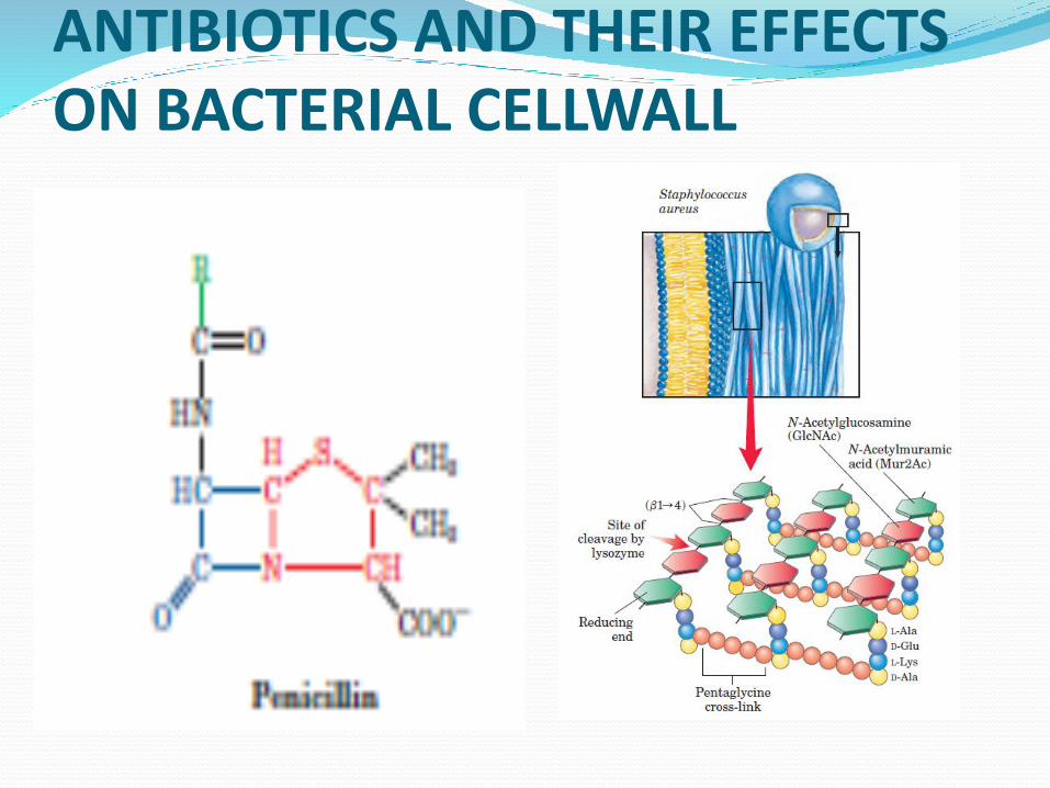
ANTIBIOTICS AND THEIR EFFECTS ON BACTERIAL CELLWALL
GLYCOSAMINOGLYCANS The extracellular spaces, particularly those of connective
tissues such as cartilage, tendon, skin and blood vessel walls,
consist of collagen and elastin fibres embedded in a gel-like
matrix known as GRUOND SUBSTANCE.
Ground substance is composed largely of glycosaminoglycans (
alternatively, MUCOPOLYSACCHARIDES
They are polysaccharides of alternating Uronic and
hexasamine residues.
Solutions of glycosaminoglycans have a slimy, Mucus like
consistency that results from their high viscosity and elasticity
Heparin and chondroitin sulphates are popular examples of
glycosaminoglycans
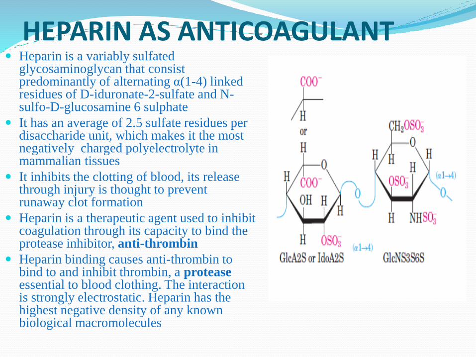
HEPARIN AS ANTICOAGULANT Heparin is a variably sulfated
glycosaminoglycan that consist predominantly of alternating α(1-4) linked residues of D-iduronate-2-sulfate and N-sulfo-D-glucosamine 6 sulphate
It has an average of 2.5 sulfate residues per disaccharide unit, which makes it the most negatively charged polyelectrolyte in mammalian tissues
It inhibits the clotting of blood, its release through injury is thought to prevent runaway clot formation
Heparin is a therapeutic agent used to inhibit coagulation through its capacity to bind the protease inhibitor, anti-thrombin
Heparin binding causes anti-thrombin to bind to and inhibit thrombin, a proteaseessential to blood clothing. The interaction is strongly electrostatic. Heparin has the highest negative density of any known biological macromolecules
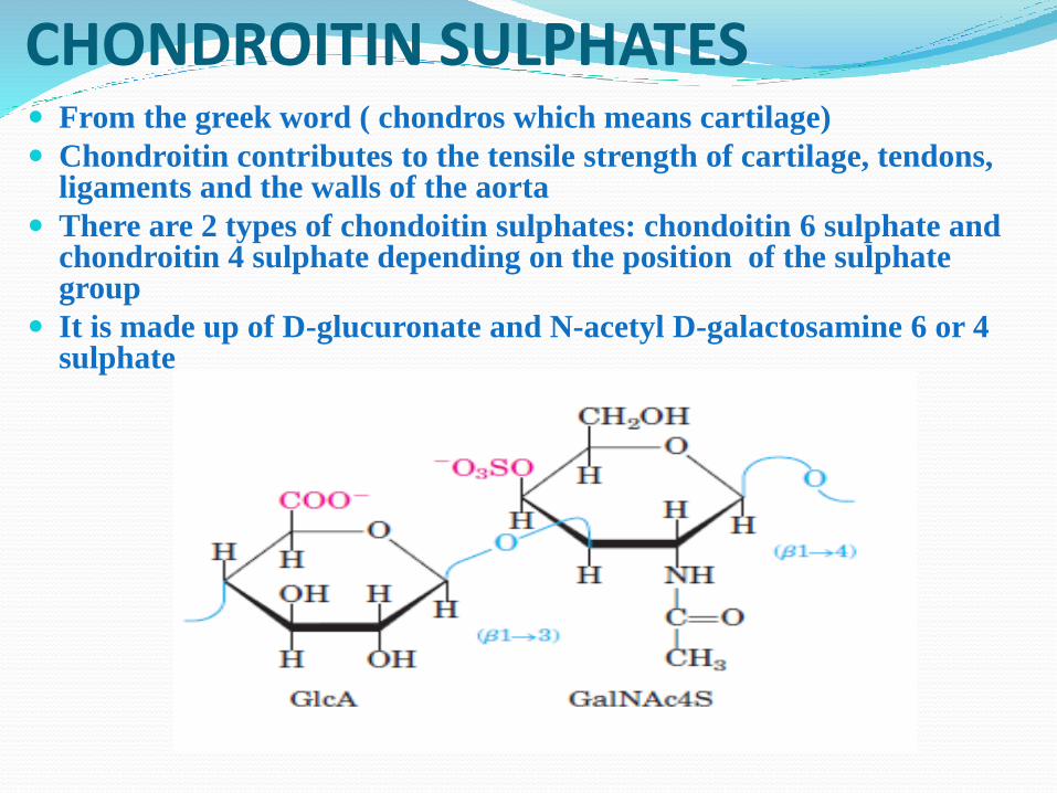
CHONDROITIN SULPHATES From the greek word ( chondros which means cartilage)
Chondroitin contributes to the tensile strength of cartilage, tendons, ligaments and the walls of the aorta
There are 2 types of chondoitin sulphates: chondoitin 6 sulphate and chondroitin 4 sulphate depending on the position of the sulphate group
It is made up of D-glucuronate and N-acetyl D-galactosamine 6 or 4 sulphate
OLIGOSACCHARIDESOligosaccharides consist of short chain
monosaccharide units or residues joined
by characteristic linkages called glycosidic
bonds
The most abundant are the disaccharides
with two monosaccharide units
Sucrose, maltose and lactose are examples
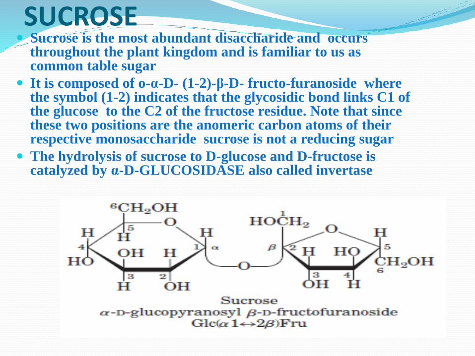
SUCROSE Sucrose is the most abundant disaccharide and occurs
throughout the plant kingdom and is familiar to us as common table sugar
It is composed of o-α-D- (1-2)-β-D- fructo-furanoside where the symbol (1-2) indicates that the glycosidic bond links C1 of the glucose to the C2 of the fructose residue. Note that since these two positions are the anomeric carbon atoms of their respective monosaccharide sucrose is not a reducing sugar
The hydrolysis of sucrose to D-glucose and D-fructose is catalyzed by α-D-GLUCOSIDASE also called invertase
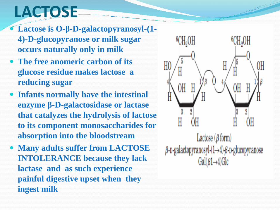
LACTOSE Lactose is O-β-D-galactopyranosyl-(1-
4)-D-glucopyranose or milk sugar
occurs naturally only in milk
The free anomeric carbon of its
glucose residue makes lactose a
reducing sugar
Infants normally have the intestinal
enzyme β-D-galactosidase or lactase
that catalyzes the hydrolysis of lactose
to its component monosaccharides for
absorption into the bloodstream
Many adults suffer from LACTOSE
INTOLERANCE because they lack
lactase and as such experience
painful digestive upset when they
ingest milk
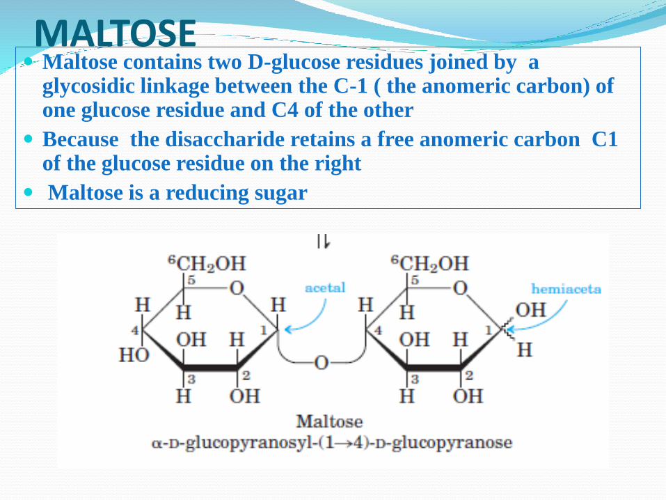
MALTOSE Maltose contains two D-glucose residues joined by a
glycosidic linkage between the C-1 ( the anomeric carbon) of one glucose residue and C4 of the other
Because the disaccharide retains a free anomeric carbon C1 of the glucose residue on the right
Maltose is a reducing sugar
QUESTIONS AND COMMENTS


![Primul cuvânt D · Primul cuvânt 342 D d, D, s.m. "litera d/D "; "sunetul [d]" "litera §/» "; "sunetul [§]" "grupul de litere dh/DH " "sunetul [dh/ δ]" d, D , s.f. invar.: cu](https://static.fdocument.org/doc/165x107/5e4b02b8ccbf8f281c58ecc6/primul-cuvnt-d-primul-cuvnt-342-d-d-d-sm-litera-dd-sunetul.jpg)