Alteration of cartilage mechanical properties in absence of β1 integrins revealed by rheometry and...
8
Alteration of cartilage mechanical properties in absence of β1 integrins revealed by rheometry and FRAP analyses Carole Bougault a , Livia Cueru b , Jonathan Bariller b , Marilyne Malbouyres a , Anne Paumier a , Attila Aszodi c , Yves Berthier b , Frédéric Mallein-Gerin a,1 , Ana-Maria Trunfio-Sfarghiu b,n,1 a Institut de Biologie et Chimie des Protéines, FRE3310 CNRS, Université de Lyon, 7 Passage du Vercors, F-69367 Lyon Cedex 07, France b Université de Lyon, CNRS INSA-Lyon, LaMCoS UMR5259, F-69621, France c Experimental Surgery and Regenerative Medicine, Department of Surgery, Ludwig-Maximilians-University (LMU), Nussbaumstr. 20, 80336 Munich, Germany article info Article history: Accepted 14 April 2013 Keywords: Cartilage Rheometry FRAP Chondrocyte Integrin abstract Context: Mechanical properties are essential for biological functions of the hyaline cartilage such as energy dissipation and diffusion of solutes. Mechanical properties are primarily dependent on the hierarchical organization of the two major extracellular matrix (ECM) macromolecular components of the cartilage: the fibrillar collagen network and the glycosaminoglycan (GAG)-substituted proteoglycan, mainly aggrecan, aggregates. Interaction of chondrocytes, the only cell type in the tissue, with the ECM through adhesion receptors is involved in establishing mechanical stability via bidirectional transduction of both mechanical forces and chemical signals. In this study, we aimed to determine the role of the transmembrane β1 integrin adhesion receptors in cartilage biomechanical properties by the use of genetic modification in mice. Methods: Costal cartilages of wild type and mutant mice lacking β1 integrins in chondrocytes were investigated. Cartilage compressive properties and solute diffusion were characterized by rheometric analysis and Fluorescence Recovery After Photobleaching (FRAP), respectively. Cartilage tissue sections were analyzed by histology, immunohistochemistry and transmission electron microscopy (TEM). Results: At the histological level, the mutant costal cartilage was characterized by chondrocyte rounding and loss of tissue polarity. Immunohistochemistry and safranin orange staining demonstrated apparently normal aggrecan and GAG levels, respectively. Antibody staining for collagen II and TEM showed comparable expression and organization of the collagen fibrils between mutant and control cartilages. Despite the lack of gross histological and ultrastructural abnormalities, rheological measurements revealed that the peak elastic modulus in compression of mutant cartilage was 1.6-fold higher than the peak elastic modulus of wild-type sample. Interestingly, the diffusion coefficient within the mutant cartilage tissue was found to be 1.2-fold lower in the extracellular space and 14-fold lower in the pericellular (PCM) space compared to control. Conclusion: The results demonstrate that the absence of β1 integrins on the surface of chondrocytes increases the stiffness and modifies the diffusion properties of costal cartilage. Our data imply that β1 integrins-mediated chondrocyte–matrix interactions directly affect cartilage biomechanics probably by modifying physical properties of individual cells. This study thus highlights the crucial role of β1 integrins in the cartilage function. & 2013 Published by Elsevier Ltd. 1. Introduction Biomechanical properties of the load-bearing cartilaginous tissues have pivotal importance for the muscoskeletal system enabling organ protection and body movements. The hyaline cartilage is an avascular and aneural poroelastic material com- posed of fluid and solid (matrix) components. The extracellular matrix (ECM) of cartilage contains a number of macromolecular components including collagen fibers, proteoglycan aggregates and non-collagenous glycoproteins. The polyanionic, sulfated glycosaminoglycan (GAG) side chains of proteoglycans (mainly aggrecan) attract a large amount of water generating high osmotic (swelling) pressure within the tissue that resists compressive forces in weight-bearing cartilages. The aggrecan–water gel aggre- gates are embedded into a heteropolymeric collagen fibrillar network composed of major (type II) and minor (types IX and Contents lists available at SciVerse ScienceDirect journal homepage: www.elsevier.com/locate/jbiomech www.JBiomech.com Journal of Biomechanics 0021-9290/$ - see front matter & 2013 Published by Elsevier Ltd. http://dx.doi.org/10.1016/j.jbiomech.2013.04.013 n Correspondence to: Université de Lyon, CNRS INSA-Lyon, LaMCoS UMR5259, 18-20 rue des Sciences, F-69621 Villeurbanne Cedex, France. Tel.: +33 4 72 43 72 45; fax: +33 4 78 89 09 80. E-mail address: [email protected] (A.-M. Trunfio-Sfarghiu). 1 FMG and AMTS: equal contribution. Journal of Biomechanics 46 (2013) 1633–1640
Transcript of Alteration of cartilage mechanical properties in absence of β1 integrins revealed by rheometry and...

Journal of Biomechanics 46 (2013) 1633–1640
Contents lists available at SciVerse ScienceDirect
journal homepage: www.elsevier.com/locate/jbiomech
Journal of Biomechanics
0021-92http://d
n Corr18-20 ruTel.: +33
E-m1 FM
www.JBiomech.com
Alteration of cartilage mechanical properties in absence of β1 integrinsrevealed by rheometry and FRAP analyses
Carole Bougault a, Livia Cueru b, Jonathan Bariller b, Marilyne Malbouyres a, Anne Paumier a,Attila Aszodi c, Yves Berthier b, Frédéric Mallein-Gerin a,1, Ana-Maria Trunfio-Sfarghiu b,n,1
a Institut de Biologie et Chimie des Protéines, FRE3310 CNRS, Université de Lyon, 7 Passage du Vercors, F-69367 Lyon Cedex 07, Franceb Université de Lyon, CNRS INSA-Lyon, LaMCoS UMR5259, F-69621, Francec Experimental Surgery and Regenerative Medicine, Department of Surgery, Ludwig-Maximilians-University (LMU), Nussbaumstr. 20, 80336 Munich,Germany
a r t i c l e i n f o
Article history:
Accepted 14 April 2013Context: Mechanical properties are essential for biological functions of the hyaline cartilage such asenergy dissipation and diffusion of solutes. Mechanical properties are primarily dependent on the
Keywords:CartilageRheometryFRAPChondrocyteIntegrin
90/$ - see front matter & 2013 Published by Ex.doi.org/10.1016/j.jbiomech.2013.04.013
espondence to: Université de Lyon, CNRS INe des Sciences, F-69621 Villeurbanne Cedex,4 72 43 72 45; fax: +33 4 78 89 09 80.
ail address: [email protected] (AG and AMTS: equal contribution.
a b s t r a c t
hierarchical organization of the two major extracellular matrix (ECM) macromolecular components ofthe cartilage: the fibrillar collagen network and the glycosaminoglycan (GAG)-substituted proteoglycan,mainly aggrecan, aggregates. Interaction of chondrocytes, the only cell type in the tissue, with the ECMthrough adhesion receptors is involved in establishing mechanical stability via bidirectional transductionof both mechanical forces and chemical signals. In this study, we aimed to determine the role of thetransmembrane β1 integrin adhesion receptors in cartilage biomechanical properties by the use ofgenetic modification in mice.Methods: Costal cartilages of wild type and mutant mice lacking β1 integrins in chondrocytes wereinvestigated. Cartilage compressive properties and solute diffusion were characterized by rheometricanalysis and Fluorescence Recovery After Photobleaching (FRAP), respectively. Cartilage tissue sectionswere analyzed by histology, immunohistochemistry and transmission electron microscopy (TEM).Results: At the histological level, the mutant costal cartilage was characterized by chondrocyte roundingand loss of tissue polarity. Immunohistochemistry and safranin orange staining demonstrated apparentlynormal aggrecan and GAG levels, respectively. Antibody staining for collagen II and TEM showedcomparable expression and organization of the collagen fibrils between mutant and control cartilages.Despite the lack of gross histological and ultrastructural abnormalities, rheological measurementsrevealed that the peak elastic modulus in compression of mutant cartilage was 1.6-fold higher thanthe peak elastic modulus of wild-type sample. Interestingly, the diffusion coefficient within the mutantcartilage tissue was found to be 1.2-fold lower in the extracellular space and 14-fold lower in thepericellular (PCM) space compared to control.Conclusion: The results demonstrate that the absence of β1 integrins on the surface of chondrocytesincreases the stiffness and modifies the diffusion properties of costal cartilage. Our data imply that β1integrins-mediated chondrocyte–matrix interactions directly affect cartilage biomechanics probably bymodifying physical properties of individual cells. This study thus highlights the crucial role of β1 integrinsin the cartilage function.
& 2013 Published by Elsevier Ltd.
1. Introduction
Biomechanical properties of the load-bearing cartilaginoustissues have pivotal importance for the muscoskeletal systemenabling organ protection and body movements. The hyaline
lsevier Ltd.
SA-Lyon, LaMCoS UMR5259,France.
.-M. Trunfio-Sfarghiu).
cartilage is an avascular and aneural poroelastic material com-posed of fluid and solid (matrix) components. The extracellularmatrix (ECM) of cartilage contains a number of macromolecularcomponents including collagen fibers, proteoglycan aggregatesand non-collagenous glycoproteins. The polyanionic, sulfatedglycosaminoglycan (GAG) side chains of proteoglycans (mainlyaggrecan) attract a large amount of water generating high osmotic(swelling) pressure within the tissue that resists compressiveforces in weight-bearing cartilages. The aggrecan–water gel aggre-gates are embedded into a heteropolymeric collagen fibrillarnetwork composed of major (type II) and minor (types IX and

C. Bougault et al. / Journal of Biomechanics 46 (2013) 1633–16401634
XI) collagens. The intrinsic stiffness of the collagen molecules andthe extensive cross-linking between the fibrils provide the mesh-work with tensile strength and restraining stress that counteractstissue swelling. The delicate balance between swelling pressureand restraining stress controls the mechanical properties ofcartilage (Cohen et al., 1998), which in turn are essential forarticulation, loading, energy dissipation and diffusion of solutes.Dissipation of mechanical energy in articular cartilage subjected todynamic loads during gait is essential to protect bone structuresfrom mechanical stress in joints. Likewise, diffusion is fundamen-tal for transporting nutrients, oxygen, waste products, and signal-ing molecules in cartilage owing to the avascular nature of thetissue.
Chondrocytes, the sole cell type that resides in the cartilage, areresponsible for the synthesis, maintenance and degradation of theECM components. Chondrocytes, with increasing distance fromthe cell surface, are surrounded by pericellular, territorial andinterterritorial matrix compartments characterized by distinctcomposition and organization of the ECM macromolecules(Buckwalter and Mankin, 1998). On chondrocytes, various recep-tors interact with either soluble (growth factors) or solid (matrix)molecules in the pericellular zone. Adhesion molecules likeintegrins, annexin V, CD44 and discoidin domain receptors binda range of ECM components, which trigger intracellular signalingcascades influencing the chondrocyte behavior such as prolifera-tion, survival, polarity, gene expression, matrix synthesis anddeposition. Integrins are a key class of cell adhesion moleculesthat connect the ECM to the cytoskeleton and mediate chemical aswell as mechanical signals in and out of the cells. They arecomposed of α and β subunits and chondrocytes express specifi-cally high levels of β1 integrin-containing heterodimers on theirsurface (Loeser, 2000). To assess the functions of β1 integrins incartilage, transgenic mice, where β1 integrin gene (Itgb1) has beenconditionally ablated in chondrocytes, were generated (Aszodiet al., 2003). Most of these mice develop perinatal lethal chondro-dysplasia characterized by chondrocyte proliferation, shape andpolarity defects, as well as moderate ultrastructural abnormalitiesof the collagen fibrillar network in the developing cartilaginousskeleton. Similar phenotype was found in the adult articularcartilage upon limb mesenchyme-specific deletion of Itgb1(Aszodi et al., 2003).
In the present study, we aimed to determine if β1 integrins playa role in cartilage biomechanical properties comparing Itgb1fl/fl–Col2a1cre (mutant) cartilage with normal cartilage obtained fromwild-type (WT) mice. Since Itgb1 deletion in chondrocytes causesearly lethality, we isolated cartilage explants from embryonic ribcage, the most abundant source of cartilage in the mouse embryo.Cartilage compressive properties were characterized by the peakelastic modulus in compression at the tissue-scale by rheometricanalysis. Fluorescence Recovery After Photobleaching (FRAP) wasperformed to determine diffusion properties upon fluorescentdextran loading. The biochemical composition of cartilage matrixwas analyzed by immunohistochemistry and its ultrastructuralorganization by transmission electron microscopy (TEM). Wefound that the lack of β1 integrin impairs the stiffness anddiffusion properties of costal cartilage implying an important roleof β1 integrin heterodimers in cartilage biomechanics.
Fig. 1. Preparation of the cartilage explants. Experimental procedure diagram ofthe cartilage preparation. Cartilage was isolated from the ventral part of rib cages ofmouse embryos at 17.5-dpc. Top right: optical image of a cartilage sample under athin glass plate weighing 1 g (viewed from the top). This image was used tomonitor the contact area and the initial thickness of pre-load samples (0.01 N)before the rheological stress–relaxation tests (scale bar 1 mm).
2. Materials and methods
2.1. Conditional knockout mice and preparation of cartilage samples
To obtain mice lacking β1 integrins in cartilage, we crossed mice carrying thefloxed β1 integrin (Itgb1fl) gene (Potocnik et al., 2000) with mice expressing the crerecombinase under the control of regulatory regions the mouse collagen type II(Col2a1) gene (Sakai et al., 2001). Costal cartilage was isolated from the ventral
parts of the rib cage of wild type (WT) and mutant (Itgb1fl/fl–Col2a1cre) mice at17.5-days post-coitum (17.5-dpc) (Fig. 1). For rheometry, FRAP and TEM analyses,cartilage explants were isolated from the two lowest non-floating ribs. These tworibs offer the advantage of being the longest and having similar geometry. Ablationof the Itgb1 allele was associated with activation of a promoterless lacZ reportergene (Potocnik et al., 2000) allowing to monitor deletion efficiency with X-galstaining as described by Lefebvre et al. (1994). The costal cartilage samples wereanalyzed within 4 days following their isolation. They were stored at 4 1C inphosphate buffered saline solution (PBS) at pH 7.4, with antibiotics (penicillin(1000 U/mL) and streptomycin (0.1 mg/mL), Gibco) until mechanical testing.
2.2. Real-time PCR analysis
The absence of β1 integrin mRNA expression was confirmed by using a real-time PCR. Chondrocytes were isolated from costal cartilage as described byBougault et al. (2009). Total RNA was extracted from isolated cells by theNucleospin RNA II kit according to the manufacturer instructions (Macherey-Nagel). For cDNA synthesis, 1 mg of total mRNA and SuperScriptTM II ReverseTranscriptase (Invitrogen) were used. Real-time PCR reactions were performed inan I-Cycler apparatus (Biorad) using an iQ SYBR Green Supermix (Biorad) andprimer pairs specific for β1 integrin (Itgb1), α10 integrin (Itga10) and the internalstandard ribosomal protein L13a (Rpl13a). The primer sequences were:
Itgb1 s-ATGCAGGTTGCGGTTTGT, as-CATCCGTGGAAAACACCAGItga10 s-ATGGCTGCTCTTCCTGATCT, as-CTGAACTGCTGACCTGGGTARpl13a s-ATCCCTCCACCCTATGACAA, as-GCCCCAGGTAAGCAAACTT.
2.3. Mechanical tests using a rheometric system
The mechanical resistance of the cartilage samples was measured by compres-sion testing in an unconfined configuration. Step-wise compression tests wereperformed with an Advanced Rheometric Expansion System (ARES, TA Instru-ments). In order to control the boundary conditions, the platens were sonicatedtwice for 10 min at 25 1C in ethanol, before use. The costal cartilage was placed inthe center of a 10 mL drop of PBS to maintain sample hydration. In order to have auniform contact area and thickness for all samples, a pre-load of 0.01 N was appliedunder a rheometer. The samples were then allowed to relax for 5 min. After thisrelaxation, the normal stress level measured by the rheometer was close to 0, sothat initial conditions were similar for all samples. A total of 18 cartilage sampleswere used for the step-wise compression tests (n¼8 for WT and n¼10 formutants).
The thickness of each pre-loaded sample was noted before rheological tests. Forthe contact area measurement, the pre-load of 0.01 N was applied with a thin glass

C. Bougault et al. / Journal of Biomechanics 46 (2013) 1633–1640 1635
plate (1 g) under a binocular microscope (Fig. 1). In addition, we tested theuniformity of the thickness under a confocal microscope, by measuring the widthof the pre-loaded samples at 10 different points.
2.4. Mechanical data acquisition and analysis
Starting from the initial condition (0.01 N pre-load), a cycle of five strains wereperformed with an imposed speed deformation of 0.02 mm/s and a compressionlevel representing 10% of the sample thickness. Every compression strain ramp wasfollowed by a period of stress relaxation of 250 s to reach the balance of stressequilibrium characteristic of biphasic materials. The normal force and the gapthickness were directly measured by the rheometer sensors. Stress–relaxationcurves were generated for each sample: compressive and relaxation stresses,calculated as normal force divided by the costal cartilage area, were drawn asfunctions of time (Fig. 2).
Stress–strain curves were generated by taking only into consideration thecompression peak stress, where the compressive strain (ε) was calculated as
ε¼ h0−hi
h0ð1Þ
where h0 is the initial gap (pre-load) and hi is the controlled gap.According to Hooke's law, the linear regression slope for the significant
compressive peaks of stress–strain curve was used to estimate the peak compres-sion modulus (En) for each cartilage sample.
2.5. Diffusion tests using Fluorescence Recovery After Photobleaching (FRAP)
Fluorescence Recovery After Photobleaching (FRAP) was applied for measuringthe molecular diffusion speed with a confocal microscope (Axelrod et al., 1976). Weused 70 kDa dextran labeled with neutral fluorescein (Sigma) at 0.1 mg/mL in PBS.Each cartilage sample was immersed in the fluorescein-labeled 70 kDa dextransolution at 4 1C for at least 48 h prior to FRAP measurements, to allow the system toequilibrate and to allow dextran to fully permeate the cartilage matrix.
Samples were sandwiched between glass coverslips and were kept moist in thelabeling solution during the FRAP experiments. FRAP measurements were per-formed on a TCS SP5 Leica confocal microscope using a 63�1.20 water objective.Photobleaching was performed with an argon laser at 70% laser power and at 100%transmission and imaging with the same laser at 15% transmission (to preventbleaching). Circular areas were uniformly bleached (regions of interest are termedROI). Each sample was analyzed in multiple zones (2–20 ROI). ROI's size of 50 or100 mm diameter was used to estimate diffusion in the matrix or solution. 10 mmdiameter ROI was used to estimate diffusion at the PCM level (Fig. 3A). Eightcartilage explants were tested (n¼3 for WT and n¼5 for mutants). For eachexperiment, approximately 250 images were acquired at 1.32 s intervals, includingthree pre-bleach images, 50 bleach images and between 150 and 200 post-bleachimages (the post-bleach time was adjusted to achieve the highest degree offluorescence recovery), to track the fluorescence recovery. The pre-bleach images
Fig. 2. Stress–relaxation test. Stress–relaxation responses of WT and mutantcartilage explants under unconfined compression. A cycle of five strains wereapplied; each compression (reaching 10% of the sample thickness) was followed bya period of stress–relaxation of 250 s (dotted line). Stress–relaxation curves weregenerated; compressive stress (normal force divided by the costal cartilage area)was plotted as a function of time for WT (black line) or mutant (gray line) samples.
are used to establish the initial intensity for normalization of the data series(Fig. 3B). For each experiment, a fluorescent dextran solution of 0.1 mg/mL in PBSwas also subjected to FRAP analysis, as control. All measurements were made atroom temperature (2571 1C).
2.6. Diffusion data acquisition and analysis
Fluorescent intensity analysis of FRAP experiments was performed on the basisof the manufacturer's handbook of the confocal microscope (Leica, Eq. (2)). For eachphotobleached zone, a “reference ROI” not subjected to photobleaching wasrecorded. The fluorescence intensity of the experimental curve (IROI(t)) was dividedby a correction factor in order to bring the IROI at the same pre-bleached intensitylevel as the reference ROI (Ireference) at initial conditions. Thus, the correction factorcorresponds to the ratio of the initial fluorescence intensity (pre-bleached intensity,I(0)) of the experimental curve to the reference curve. The corrected IROI wassubtracted from the reference curve's fluorescent intensity (Ireference(t)) in order toeliminate the photobleaching due to image acquisition. The result is represented asa curve fd(t) that we call “normalized difference fluorescence intensity” (Fig. 3C).
f dðtÞ ¼ IreferenceðtÞ−IROIðtÞ
IROIð0Þ=Ireferenceð0Þð2Þ
The normalized difference fluorescence intensity curve was fitted using anexponential mathematical law (Fig. 3C):
yðtÞ ¼ ae−t=b þ c ð3Þ
The equation for the curve was used to determine the half-recovery time (t1/2):
t1=2 ¼ ln2b ð4Þ
The half-recovery time (t1/2), and the radius of the photobleached area (w) wereused to determine the diffusion coefficient (D), using the following simplifiedequation (Axelrod et al., 1976):
D¼ w2
4t1=2ð5Þ
2.7. Cell viability
Cell death within the costal cartilage was measured using the CytotoxicityDetection Kit (Roche Applied Science), as described by the manufacturer. Absor-bance was measured at 490 nm. Samples representing 100% viability were storedonly for 1 day. Samples representing 100% mortality were treated with Triton X100(Sigma Aldrich) 1% in PBS.
2.8. Histological analysis of the cartilage matrix
For histological analysis, costal cartilage explants were fixed in 4% formalde-hyde (pH 7.4) for 24 h at 4 1C. The samples were embedded in paraffin, sectioned at7 μm and stained with Mayer's hematoxylin. For histochemical detection of sulfatedproteoglycans, sections were stained with 0.5% safranin-O in 0.1 M sodium acetateat pH 7.4 for 10 min. Safranin-O is a cationic dye that binds GAGs and stains orangein an aqueous mounting medium (Rosenberg, 1971). Mayer's hematoxylin stainingwas used to counterstain nuclei in chondrocytes. Immunofluorescence detection oftype II collagen and aggrecan in the ECM was performed as previously described byBougault et al. (2009).
2.9. Ultrastructural analysis of the cartilage matrix
Costal cartilage explants were fixed at room temperature in a mixture of 2.5%glutaraldehyde and 2% paraformaldehyde (EM grade, Delta Microscopies) in 0.2 Msodium cacodylate buffer (pH 7.4) for 2 h. After rinsing in 0.15 M sodiumcacodylate, explants were post-fixed with 1% osmium tetroxide in 0.1 M sodiumcacodylate for 45 min. Samples were then dehydrated in graded series of ethanol(from 30 to 100%), and embedded in epoxy resin as previously described byChanut-Delalande et al. (2004). Ultra-thin, 70 nm sections were cut using a LeicaUC7 ultramicrotome. The sections were treated with 7% uranyl acetate in methanoland lead citrate, and then examined with a Philips CM120 electron microscopeequipped with a Gatan Orius 200 2K�2K digital camera (“Centre Technologiquedes Microstructures”, Université Lyon I, Villeurbanne, France).
2.10. Statistical analysis
The data are expressed as the mean7standard deviation (SD). Statisticalsignificance was assessed using the Student T-test and po0.05 was consideredsignificant.

Fig. 3. FRAP diffusion tests. (A) Confocal fluorescence image of a cartilage sample (on the left) or reference solution (on the right) before FRAP analysis. ROI with 10 mmdiameter (gray circle) was used to measure PCM diffusion, while ROI with 50 mm diameter (black circle) was used to measure matrix diffusion. (B) Diagram of the FRAPexperimental procedure (top) and curve of fluorescence recovery after photobleaching of costal cartilage with 100 mm diameter ROI (bottom). Relative fluorescence intensitywas plotted as a function of time for bleached ROI (black line) and non-bleached reference (gray line). (C) Adjusted regression curve (100 μm diameter ROI) for costalcartilage (black line), fitted with the displaced exponential law (gray dotted line). The exponential equation of the fitted curve and the coefficient of determination isindicated.
C. Bougault et al. / Journal of Biomechanics 46 (2013) 1633–16401636
3. Results
3.1. Absence of β1 integrins in mutant cartilage and ECM analysis
Costal cartilages of 17.5-dpc WT and mutant mice were firstinvestigated for the expression of β1 integrin. X-gal stainingshowed LacZ activity in all mutant chondrocytes implicating ahighly efficient deletion of the floxed Itgb1 gene (Fig. 4A). RT-PCRanalysis of total RNA isolated from chondrocytes revealed thecomplete lack of Itgb1 mRNA in mutant samples. The expression ofthe Itga10 mRNA encoding the α10 subunit of the collagen-bindingα10β1 integrin heterodimer was comparable between the geno-types (Fig. 4B). Next, the structure and composition of cartilagesamples were analyzed by histology, immunohistochemistry andelectron microscopy. Hematoxylin staining highlighted a markedchange in cell shape of β1 integrin-deficient costal cartilage.Mutant chondrocytes appeared clearly more rounded and largerthan normal chondrocytes (Fig. 5A). Safranin-O staining for thenegatively charged GAG chains and immunofluorescence stainingusing antibodies against the aggrecan and type II collagen demon-strated normal proteoglycan and collagen deposition in mutantcompared to WT (Fig. 5B–D). Moreover, TEM analysis revealedsimilar ultrastructural organization of the collagen/proteoglycannetwork in WT and mutant (Fig. 5E). Collectively, the data suggest
that synthesis and deposition of the major ECM macromoleculesare apparently unaffected by the lack of β1 integrins in 17.5-dpccostal cartilage.
3.2. The lack of β1 integrins alters the compressive propertiesof cartilage
To assess the role of β1 integrins in the mechanical propertiesof cartilage, stress–relaxation tests were performed on WT and β1mutant costal cartilage samples. First, we verified that cell viabilitywithin the explants was not altered by our storage conditions. Themaximum cytotoxicity was estimated at 671% (n¼5), which wasconsidered negligible. Next, we characterized the geometry of thesamples before compression tests, with a pre-load of 0.01 N.The average contact area was 1.9070.48 mm² for WT, and1.7970.45 mm² for mutant samples (n¼10). The average thick-ness of the pre-loaded samples (the initial gap noted h0 in Eq. (1))was 211744 mm for WT, and 195741 mm for mutant (n¼10). Thereduced size of the mutant samples was expected because of theabnormalities in the developing cartilaginous skeleton alreadydescribed in the mutant mice (Aszodi et al., 2003). In addition,within the same pre-loaded sample, the variation in width wasbetween 6% and 10% (n¼9). Importantly, the difference of thick-ness between samples did not affect the peak compression
C. Bougault et al. / Journal of Biomechanics 46 (2013) 1633–1640 1637
modulus (En) because it was calculated taking into account theratio dh/h0. Similarly, the difference of contact area betweensamples was heeded because the compressive stresses werecalculated as normal force divided by the sample area.
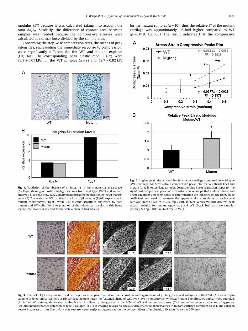
Concerning the step-wise compression tests, the means of peakintensities, representing the immediate response to compression,were significantly different for the WT and mutant explants(Fig. 6A). The corresponding peak elastic moduli (En) were32.770.01 kPa for the WT samples (n¼8) and 53.770.02 kPa
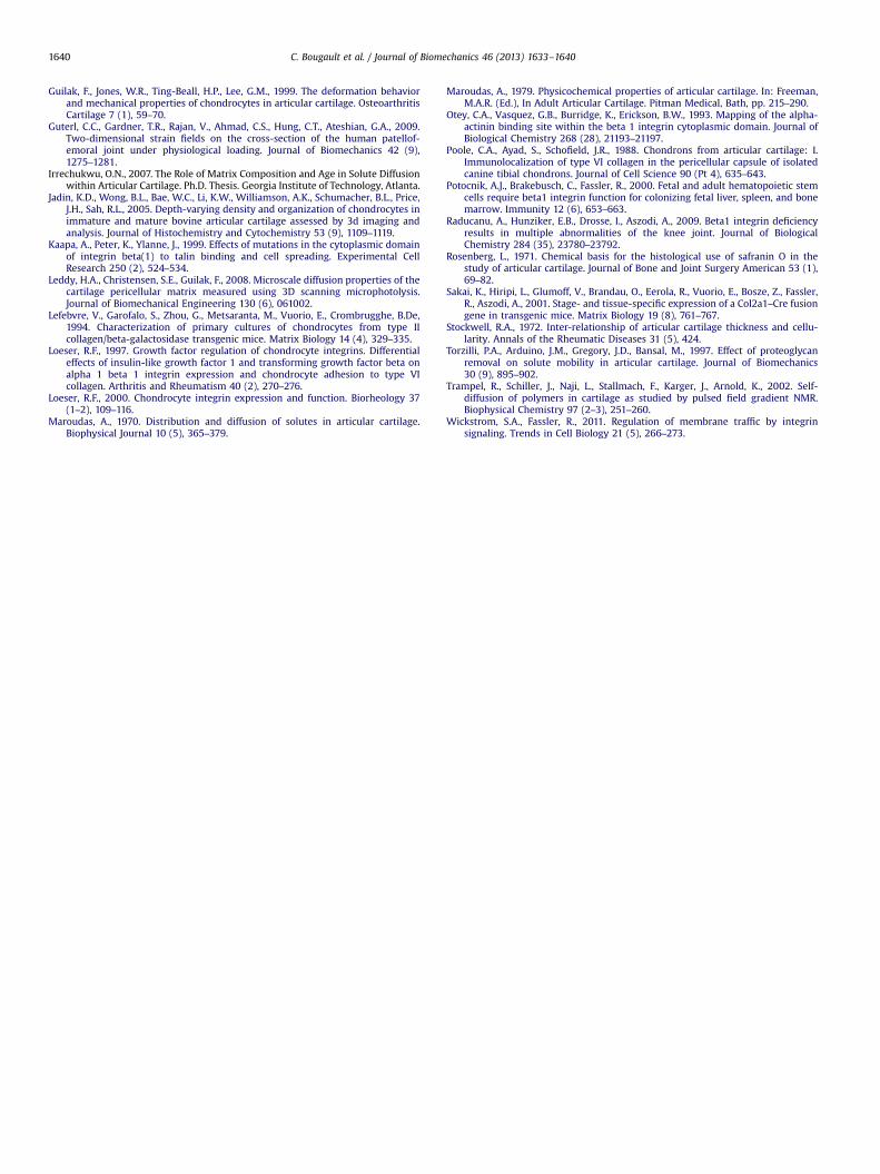
Fig. 4. Validation of the absence of β1 integrins in the mutant costal cartilage.(A) X-gal staining of costal cartilage sections from wild type (WT) and mutantembryos. Blue cells show LacZ activity demonstrating the deletion of the β1 integringene. (B) The real-time PCR confirms the loss of β1 integrin (Itgb1) expression inmutant chondrocytes (right), while α10 integrin (Itga10) is expressed by bothmutant and WT cells. (For interpretation of the references to color in this figurelegend, the reader is referred to the web version of this article).
Fig. 5. The lack of β1 integrins in costal cartilage has no apparent affect on the depositstaining of longitudinal sections of rib cartilage demonstrates the flattened shape of w(B) Safranin-O staining shows comparable levels of sulfated proteoglycans in the EC(D) Immunofluorescence detection of type II collagen. (E) TEM imaging reveals no obvionetwork appears as thin fibers, dark dots represent proteoglycans aggregated on the co
for the mutant samples (n¼10); thus the relative En of the mutantcartilage was approximately 1.6-fold higher compared to WT(p¼0.018, Fig. 6B). The result indicates that the compressive
ion and organization of proteoglycans and collagens in the ECM. (A) Hematoxylinild type (WT) chondrocytes; whereas mutant chondrocytes appear more rounded.M of WT and mutant cartilages. (C) Immunofluorescence detection of aggrecan.us ultrastructural abnormalities in mutant cartilage compared to WT. The collagenllagen fibers after chemical fixation (scale bar 500 nm).
Fig. 6. Higher peak elastic modulus in mutant cartilage compared to wild type(WT) cartilage. (A) Stress–strain compressive peaks plot for WT (black line) andmutant (gray line) cartilage samples. Corresponding linear regression slopes for thesignificant compressive peaks of stress–strain curve are plotted as dotted lines; andlinear equations and coefficients of determination are indicated on the right. Slopecoefficient was used to estimate the apparent elastic modulus of each costalcartilage (mean7SD, npo0.05, nnpo0.01, mutant versus WT).(B) Relative peakelastic modulus for mutant (gray bar) and WT (black bar) cartilage samples(mean7SD, npo0.05, mutant versus WT).

C. Bougault et al. / Journal of Biomechanics 46 (2013) 1633–16401638
properties of cartilage are altered in absence of β1 integrins. Thisdifference could be explained by the biphasic structure of carti-lage, composed of a poroelastic solid phase represented by thematrix and the cells, and a uncompressible fluid phase representedby interstitial water and dissolved electrolytes (Ateshian, 1997). Inorder to investigate whether the mechanical difference was due tothe solid or the fluid phase, we next examined diffusion propertiesof the WT and mutant cartilages.
3.3. Alteration of cartilage diffusion in absence of β1 integrins
FRAP experiments were performed on costal cartilage explantsisolated from WT and mutant mouse embryos. The cytotoxicityafter 4 days of storage was negligible and was not furtherincreased by incubation with the fluorescein-labeled 70 kDa dex-tran solution (471%, n¼5). In WT cartilage, the matrix diffusioncoefficient was estimated to be 7.770.6 mm²/s and the PCMdiffusion coefficient was estimated to be 2.670.8 mm²/s. Bothcoefficients were significantly lower in mutant cartilage (Fig. 7).Noticeably, the decrease of the mutant values was much morepronounced at the PCM level (14-fold decrease, p¼10−13) than atthe ECM level (1.2-fold decrease, p¼0.007). Thus, the diffusionproperties of cartilage seem altered in absence of β1 integrins,particularly at the level of the surrounding PCM.
4. Discussion
The aim of the present study was to evaluate the impact of β1integrin-deficiency on the mechanical behavior of rib cartilageisolated from genetically modified mouse embryos. Our resultsshow that the peak elastic modulus (En) of embryonic costalcartilage assessed by unconfined compression is about 30 foldslower than the values found in literature for the mature humanarticular cartilage (Boschetti and Peretti, 2008). This discrepancycan be explained either by species- and location-specific differ-ences of the cartilage or by the fact that the immature cartilage hasabout twice the cellularity of mature cartilage (Jadin et al., 2005).Importantly, we observed 1.6-fold increase of En in the mutantsamples implicating that the adhesion receptor β1 integrinsinfluence cartilage mechanical properties at cellular and/or extra-cellular matrix levels.
Cellularity of mouse cartilages is high compared to largerspecies such as bovine and man (Stockwell, 1972), thereforechondrocytes might significantly contribute to the bulk mechan-ical properties of the tissue. Histological analysis of costal cartilagehas confirmed a shape change of chondrocytes from flattened andoriented (wild type) to rounded and non-oriented (mutant)
Fig. 7. Lower diffusion coefficient in mutant cartilage compared to wild type (WT)cartilage. Diffusion coefficient calculated from FRAP tests on mutant (gray bars) andWT (black bars) cartilage samples using fluorescein-labeled 70 kDa dextran 0.1 mg/mL in PBS. Matrix and solution diffusion coefficients were calculated for ROI to be50 and 100 mm diameters respectively, and PCM diffusion coefficients werecalculated for ROI to be 10 mm diameter (mean7SD, nnpo0.01, nnnpo0.001,mutant versus WT).
phenotype in agreement with previous studies published on β1integrin-deficient cartilages (Aszodi et al., 2003; Raducanu et al.,2009). The cytoplasmic tail of β1 integrins binds to the cytoske-leton via adapter proteins (Kaapa et al., 1999; Otey et al., 1993)forming an essential link between the ECM and inside of the cell.The bidirectional, matrix–integrin–cytoskeleton connection is fun-damental for the chondrocyte geometry through the control ofmatrix attachment and organization of the cortical actin ringbeneath the plasma membrane (Aszodi et al., 2003). Uncouplingthe cytoskeleton and cell membrane in β1 integrin-deficientchondrocytes may result in a decrease of cellular deformabilityand an increase of cellular rigidity due to alterations of thecytoskeletal mechanics (Fig. 8). To validate this hypothesis, cell-scaled experiments should be considered to study more preciselythe mechanical properties of individual chondrocytes isolatedfrom WT and mutant cartilages.
The macroscale biomechanical properties of the cartilage matrixare attributed primarily to the collagen fibrillar meshwork and thenegatively charged proteoglycan aggrecan. In accordance with aprevious study on embryonic limb cartilage (Aszodi et al., 2003), wefound no obvious alterations in type II collagen or aggrecandeposition and organization in β1 integrin mutant costal cartilageby immunohistochemistry and TEM. Important to note, however,that adult cartilages lacking β1 integrins on chondrocytes displayultrastructural abnormalities characterized by changes in thedensity and diameter of collagen fibrils (Aszodi et al., 2003;Raducanu et al., 2009). Thus, we cannot exclude the possibility thatmild structural changes in the fibrillar network of 17.5-dpc costalcartilage, which could be masked by the harsh chemical fixationand embedding procedure of the specimens for TEM analysis,contribute to the increased stiffness of mutant samples. Alterna-tively, higher GAG content can lead to higher stiffness in cartilage(Guilak et al., 1999). In our study, histochemical staining for GAGswith safranin-O indicated no significant difference in the amount ofsulfated proteoglycans between wild type and mutant cartilages,however, this staining modality does not precisely reflect the GAG/proteoglycan content of the cartilage ECM (Camplejohn and Allard,1988). Similarly, the chemical fixation used for TEM precipitatesproteoglycans, thus the original proteoglycan network cannot beestimated by the conventional ultrastructural analysis.
Diffusion of solutes in cartilage is dependent on specificproperties of the solute and the ECM; thus the mobility of aparticular solute can correlate with the composition, structure andbiomechanics of the tissue. For example, proteoglycan depletion inarticular cartilage by enzymatic digest increases diffusivity of largeuncharged solutes such as 70 kDa dextran (Torzilli et al., 1997) andreduces compressive stiffness (Guterl et al., 2009). To explain thedifferent mechanical behaviors between wild-type and mutantcostal cartilage samples, the diffusion coefficient of fluorescein-labeled 70 kDa dextran within the ECM and PCM was measuredusing the FRAP technique (Fetter et al., 2006). The large bleachspots (50 or 100 mm diameter) were used to estimate the diffusionin the ECM, where the contribution of the cells can be neglected(Maroudas, 1970, 1979). The diffusion coefficient of 7.770.6 μm²/sthat we measured in mouse cartilage matches well with theliterature data. For example, using the FRAP technique, the diffu-sion coefficient of 70 kDa dextran in pig cartilage was estimated at3.8 μm²/s (Greene et al., 2008) and using the pulsed field gradientNMR technique, the diffusion coefficient of 70 kDa dextran inbovine cartilage was measured as 3 μm²/s (Trampel et al., 2002).Our results revealed that the diffusion coefficient was only slightlylower in the matrix of mutant cartilage compared to WT. Thisobservation hence argues against significant changes in thecomposition and/or structure of the mutant cartilage ECM.
Next, we used smaller bleach spots (10 mm diameter) to assessdiffusivity at the pericellular matrix (PCM) level and found 3-fold

Fig. 8. Alterations of biomechanical properties of individual chondrocytes lacking β1 integrins may cause an increase in rigidity at the tissue level. The deformation of mutantcartilage explants in response to normal force is lower compared to wild-type samples. We suggest that the increased compressive stiffness of cartilage lacking β1 integrins iscaused by decrease in matrix permeability and possibly by increase in rigidity of each chondrocyte. The alterations of cellular biomechanical properties may be due to defectsin cytoskeleton–cell–matrix interactions. The normal forces applied to the cartilage explants could partially convert into rolling forces around chondrocytes lacking β1integrin receptors.
C. Bougault et al. / Journal of Biomechanics 46 (2013) 1633–1640 1639
lower diffusion coefficient compared to ECM in the wild-typespecimen. In agreement, the PCM surrounding the chondrocyteswas shown significantly less permeable than the distant inter-territorial matrix in healthy bovine cartilage using the FRAPmethod (Irrechukwu, 2007). However, FRAP may not be the moreappropriate method to estimate the diffusion at the PCM levelbecause of the inadequate initial and boundary conditions(Axelrod et al., 1976). Yet, similar results were obtained using theaccurate technique of 3D scanning microphotolysis: the diffusivityof the PCM in healthy porcine articular cartilage was shown 1.2-fold lower than that of the adjacent ECM, with 70 kDa dextran(Leddy et al., 2008). The difference in diffusivity between PCM andECM is likely attributable to the differences in the structure andcomposition of the PCM, which contains finer collagen fibers and ahigher concentration of proteoglycans relative to the ECM (Pooleet al., 1988).
The great difference of diffusion coefficient at PCM levelbetween mutant and wild-type (14-fold reduction) suggests lesspermeability of the PCM to macromolecular florescent solutes(�8 nm radius of gyration) in mutant specimen. β1 integrinsinteract with various matrix proteins of the cartilage (Loeser,2000; Wickstrom and Fassler, 2011), therefore β1 integrin-deficiency may affect the composition and/or organization of thePCM (Buckwalter and Mankin, 1998; Loeser, 1997; Poole et al.,1988). Indeed, we have previously reported that the PCM com-partment is enlarged and has reduced collagen VI content inβ1-null articular cartilage (Raducanu et al., 2009). Such alterationsmay decrease the friction coefficient between cell and PCM, whichin turn changes the boundary conditions for the mechanicalbehavior. This change might correlate with chondrocyte morphol-ogy in the tissue. Wild-type chondrocytes are more flattened thanmutants owing to the importance of integrin-mediated interac-tions on cell deformation induced by internal mechanical stress(Fig. 8).
5. Conclusion
Taken together, our results demonstrate that disturbing thecytoskeleton–cell–matrix interactions by deleting integrin β1receptors in chondrocytes alters the biomechanical properties ofcartilage. The observed increase in compressive stiffness at thetissue level is most likely caused by alterations in the mechanicalbehavior of the cells and/or the PCM. This study unravels the
crucial role of β1 integrins in the mechanical properties ofcartilage.
Author contributions
Study design: CB, LC, YB, FMG, and AMTS.Acquisition of data: CB, LC, JB, MM, and AP.Analysis and interpretation of data: CB, LC, AA, JB, AP, and
AMTS.Manuscript preparation: CB, LC, AA, YB, FMG, and AMTS.
Conflict of interest
The authors declare no conflict of interest.
References
Aszodi, A., Hunziker, E.B., Brakebusch, C., Fassler, R., 2003. Beta1 integrins regulatechondrocyte rotation, G1 progression, and cytokinesis. Genes and Development17 (19), 2465–2479.
Ateshian, G.A., 1997. Finite deformation biphasic material properties of bovinearticular cartilage from confined compression experiments. Journal of Biome-chanics 30, 1157–1164.
Axelrod, D., Koppel, D.E., Schlessinger, J., Elson, E., Webb, W.W., 1976. Mobilitymeasurement by analysis of fluorescence photobleaching recovery kinetics.Biophysical Journal 16 (9), 1055–1069.
Boschetti, F., Peretti, G.M., 2008. Tensile and compressive properties of healthy andosteoarthritic human articular cartilage. Biorheology 45 (3–4), 337–344.
Bougault, C., Paumier, A., Aubert-Foucher, E., Mallein-Gerin, F., 2009. Investigatingconversion of mechanical force into biochemical signaling in three-dimensionalchondrocyte cultures. Nature Protocols 4 (6), 928–938.
Buckwalter, J.A., Mankin, H.J., 1998. Articular cartilage: tissue design and chon-drocyte–matrix interactions. Instructional course lectures 47, 477–486.
Camplejohn, K.L., Allard, S.A., 1988. Limitations of safranin ‘O’ staining inproteoglycan-depleted cartilage demonstrated with monoclonal antibodies.Histochemistry 89 (2), 185–188.
Chanut-Delalande, H., Bonod-Bidaud, C., Cogne, S., Malbouyres, M., Ramirez, F.,Fichard, A., Ruggiero, F., 2004. Development of a functional skin matrix requiresdeposition of collagen V heterotrimers. Molecular and Cellular Biology 24 (13),6049–6057.
Cohen, N.P., Foster, R.J., Mow, V.C., 1998. Composition and dynamics of articularcartilage: structure, function, and maintaining healthy state. Journal of Ortho-paedic and Sports Physical Therapy 28 (4), 203–215.
Fetter, N.L., Leddy, H.A., Guilak, F., Nunley, J.A., 2006. Composition and transportproperties of human ankle and knee cartilage. Journal of Orthopaedic Research24 (2), 211–219.
Greene, G.W., Zappone, B., Zhao, B., Soderman, O., Topgaard, D., Rata, G., Israelach-vili, J.N., 2008. Changes in pore morphology and fluid transport in compressedarticular cartilage and the implications for joint lubrication. Biomaterials29 (33), 4455–4462.
C. Bougault et al. / Journal of Biomechanics 46 (2013) 1633–16401640
Guilak, F., Jones, W.R., Ting-Beall, H.P., Lee, G.M., 1999. The deformation behaviorand mechanical properties of chondrocytes in articular cartilage. OsteoarthritisCartilage 7 (1), 59–70.
Guterl, C.C., Gardner, T.R., Rajan, V., Ahmad, C.S., Hung, C.T., Ateshian, G.A., 2009.Two-dimensional strain fields on the cross-section of the human patellof-emoral joint under physiological loading. Journal of Biomechanics 42 (9),1275–1281.
Irrechukwu, O.N., 2007. The Role of Matrix Composition and Age in Solute Diffusionwithin Articular Cartilage. Ph.D. Thesis. Georgia Institute of Technology, Atlanta.
Jadin, K.D., Wong, B.L., Bae, W.C., Li, K.W., Williamson, A.K., Schumacher, B.L., Price,J.H., Sah, R.L., 2005. Depth-varying density and organization of chondrocytes inimmature and mature bovine articular cartilage assessed by 3d imaging andanalysis. Journal of Histochemistry and Cytochemistry 53 (9), 1109–1119.
Kaapa, A., Peter, K., Ylanne, J., 1999. Effects of mutations in the cytoplasmic domainof integrin beta(1) to talin binding and cell spreading. Experimental CellResearch 250 (2), 524–534.
Leddy, H.A., Christensen, S.E., Guilak, F., 2008. Microscale diffusion properties of thecartilage pericellular matrix measured using 3D scanning microphotolysis.Journal of Biomechanical Engineering 130 (6), 061002.
Lefebvre, V., Garofalo, S., Zhou, G., Metsaranta, M., Vuorio, E., Crombrugghe, B.De,1994. Characterization of primary cultures of chondrocytes from type IIcollagen/beta-galactosidase transgenic mice. Matrix Biology 14 (4), 329–335.
Loeser, R.F., 1997. Growth factor regulation of chondrocyte integrins. Differentialeffects of insulin-like growth factor 1 and transforming growth factor beta onalpha 1 beta 1 integrin expression and chondrocyte adhesion to type VIcollagen. Arthritis and Rheumatism 40 (2), 270–276.
Loeser, R.F., 2000. Chondrocyte integrin expression and function. Biorheology 37(1–2), 109–116.
Maroudas, A., 1970. Distribution and diffusion of solutes in articular cartilage.Biophysical Journal 10 (5), 365–379.
Maroudas, A., 1979. Physicochemical properties of articular cartilage. In: Freeman,M.A.R. (Ed.), In Adult Articular Cartilage. Pitman Medical, Bath, pp. 215–290.
Otey, C.A., Vasquez, G.B., Burridge, K., Erickson, B.W., 1993. Mapping of the alpha-actinin binding site within the beta 1 integrin cytoplasmic domain. Journal ofBiological Chemistry 268 (28), 21193–21197.
Poole, C.A., Ayad, S., Schofield, J.R., 1988. Chondrons from articular cartilage: I.Immunolocalization of type VI collagen in the pericellular capsule of isolatedcanine tibial chondrons. Journal of Cell Science 90 (Pt 4), 635–643.
Potocnik, A.J., Brakebusch, C., Fassler, R., 2000. Fetal and adult hematopoietic stemcells require beta1 integrin function for colonizing fetal liver, spleen, and bonemarrow. Immunity 12 (6), 653–663.
Raducanu, A., Hunziker, E.B., Drosse, I., Aszodi, A., 2009. Beta1 integrin deficiencyresults in multiple abnormalities of the knee joint. Journal of BiologicalChemistry 284 (35), 23780–23792.
Rosenberg, L., 1971. Chemical basis for the histological use of safranin O in thestudy of articular cartilage. Journal of Bone and Joint Surgery American 53 (1),69–82.
Sakai, K., Hiripi, L., Glumoff, V., Brandau, O., Eerola, R., Vuorio, E., Bosze, Z., Fassler,R., Aszodi, A., 2001. Stage- and tissue-specific expression of a Col2a1–Cre fusiongene in transgenic mice. Matrix Biology 19 (8), 761–767.
Stockwell, R.A., 1972. Inter-relationship of articular cartilage thickness and cellu-larity. Annals of the Rheumatic Diseases 31 (5), 424.
Torzilli, P.A., Arduino, J.M., Gregory, J.D., Bansal, M., 1997. Effect of proteoglycanremoval on solute mobility in articular cartilage. Journal of Biomechanics30 (9), 895–902.
Trampel, R., Schiller, J., Naji, L., Stallmach, F., Karger, J., Arnold, K., 2002. Self-diffusion of polymers in cartilage as studied by pulsed field gradient NMR.Biophysical Chemistry 97 (2–3), 251–260.
Wickstrom, S.A., Fassler, R., 2011. Regulation of membrane traffic by integrinsignaling. Trends in Cell Biology 21 (5), 266–273.