A Viscoelastic Chitosan-Modified Three-Dimensional Porous ... · viscoelastic porous scaffolds...
20
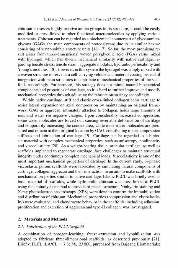
Journal of Biomaterials Science 23 (2012) 405–424 brill.nl/jbs A Viscoelastic Chitosan-Modified Three-Dimensional Porous Poly( L-Lactide-co-ε-Caprolactone) Scaffold for Cartilage Tissue Engineering Chao Li a , Lili Wang a , Zheng Yang b , Gonhyung Kim c , Haifeng Chen a and Zigang Ge a,d,∗ a Department of Biomedical Engineering, College of Engineering, Peking University, Beijing 100871, P. R. China b Tissue Engineering Program, Life Sciences Institute, National University of Singapore, 117510 Singapore c Department of Veterinary Surgery, College of Veterinary Medicine, Chungbuk National University, Cheongju 361763, South Korea d Center for Biomaterials and Tissue Engineering, Academy for Advanced Interdisciplinary Studies, Peking University, Beijing 100871, P. R. China Received 12 July 2010; accepted 14 December 2010 Abstract Biomaterials have been playing important roles in cartilage regeneration. Although many scaffolds have been reported to enhance cartilage regeneration, none of the scaffolds available are optimal regarding me- chanical properties, integration with host cartilage and providing proper micro-environment for chondrocyte attachment, proliferation and differentiation. In the current study, chitosan-modified poly(L-lactide-co-ε- caprolactone) (PLCL) scaffolds were fabricated to simulate the main biochemical components of cartilage, as well as their interaction with the aim to endow them with viscoelasticity similar to native cartilage. Porous PLCL scaffolds were fabricated with porogen-leaching, freeze-extraction and freeze-gelation before chitosan was cross-linked. The acquired porous scaffolds had pore sizes ranging from 200 to 500 μm and about 85% porosity with good interconnection between individual pores. Chitosan was successfully cross- linked to PLCL scaffolds, as validated by ninhydrin staining and X-ray photoelectron spectroscopy (XPS). The viscoelasticity of the scaffolds was similar to that of bovine cartilage and they had a relatively good recovery ratio from compression deformation, while the Young’s modulus was one order of magnitude less than cartilage. Not only could the chitosan-modified PLCL scaffolds promote cell adhesion and prolifera- tion, but also they could significantly enhance excretion of aggrecan and type-II collagen, as testified by both histology and quantitative PCR, compared with PLCL scaffolds. With the fabrication of biomimetic scaffolds, it is possible to make scaffolds for cartilage tissue engineering, which are not only biocompatible, but also have mechanical properties similar to native cartilage. © Koninklijke Brill NV, Leiden, 2012 * To whom correspondence should be addressed. Tel.: (86-10) 6275-6736; Fax: (86-10) 6275-7545; e-mail: [email protected] © Koninklijke Brill NV, Leiden, 2012 DOI:10.1163/092050610X551970
Transcript of A Viscoelastic Chitosan-Modified Three-Dimensional Porous ... · viscoelastic porous scaffolds...
Journal of Biomaterials Science 23 (2012) 405–424brill.nl/jbs
A Viscoelastic Chitosan-Modified Three-Dimensional PorousPoly(L-Lactide-co-ε-Caprolactone) Scaffold for Cartilage
Tissue Engineering
Chao Li a, Lili Wang a, Zheng Yang b, Gonhyung Kim c, Haifeng Chen a
and Zigang Ge a,d,∗
a Department of Biomedical Engineering, College of Engineering, Peking University,Beijing 100871, P. R. China
b Tissue Engineering Program, Life Sciences Institute, National University of Singapore,117510 Singapore
c Department of Veterinary Surgery, College of Veterinary Medicine, Chungbuk National University,Cheongju 361763, South Korea
d Center for Biomaterials and Tissue Engineering, Academy for Advanced Interdisciplinary Studies,Peking University, Beijing 100871, P. R. China
Received 12 July 2010; accepted 14 December 2010
AbstractBiomaterials have been playing important roles in cartilage regeneration. Although many scaffolds havebeen reported to enhance cartilage regeneration, none of the scaffolds available are optimal regarding me-chanical properties, integration with host cartilage and providing proper micro-environment for chondrocyteattachment, proliferation and differentiation. In the current study, chitosan-modified poly(L-lactide-co-ε-caprolactone) (PLCL) scaffolds were fabricated to simulate the main biochemical components of cartilage,as well as their interaction with the aim to endow them with viscoelasticity similar to native cartilage.Porous PLCL scaffolds were fabricated with porogen-leaching, freeze-extraction and freeze-gelation beforechitosan was cross-linked. The acquired porous scaffolds had pore sizes ranging from 200 to 500 µm andabout 85% porosity with good interconnection between individual pores. Chitosan was successfully cross-linked to PLCL scaffolds, as validated by ninhydrin staining and X-ray photoelectron spectroscopy (XPS).The viscoelasticity of the scaffolds was similar to that of bovine cartilage and they had a relatively goodrecovery ratio from compression deformation, while the Young’s modulus was one order of magnitude lessthan cartilage. Not only could the chitosan-modified PLCL scaffolds promote cell adhesion and prolifera-tion, but also they could significantly enhance excretion of aggrecan and type-II collagen, as testified byboth histology and quantitative PCR, compared with PLCL scaffolds. With the fabrication of biomimeticscaffolds, it is possible to make scaffolds for cartilage tissue engineering, which are not only biocompatible,but also have mechanical properties similar to native cartilage.© Koninklijke Brill NV, Leiden, 2012
* To whom correspondence should be addressed. Tel.: (86-10) 6275-6736; Fax: (86-10) 6275-7545; e-mail:[email protected]
© Koninklijke Brill NV, Leiden, 2012 DOI:10.1163/092050610X551970
406 C. Li et al. / Journal of Biomaterials Science 23 (2012) 405–424
KeywordsCartilage tissue engineering, PLCL, chitosan, biomimetic, viscoelasticity
1. Introduction
Progress in cartilage tissue engineering is relatively slow, compared with bone tis-sue engineering. Many reasons contribute to this disparity, such as poor blood andnerve supply, lack of adult stem cells or progenitor cells, existing chondrocytes’inferior ability to proliferate, complex physiological mechanical environment, andpoor integration between de novo cartilage and host cartilage [1]. Autologous chon-drocyte implantation (ACI) has been a powerful tool to deal with focal cartilagedefects of young patients [2, 3], while arthroscopic lavage, abrasion arthroplasty,subchondral drilling and microfracture all lead to inferior regeneration [3]. Bioma-terials and scaffolds have been playing constructive roles in cartilage regeneration inthe forms of micrometer-scale beads as platform of cartilage formation [4], as cell-carrying vehicles of autologous chondrocyte implantation [5], as scaffolds to trapliving cells in vivo [6], or as supporting structures, as well as integration with hostcartilage [7]. Individual scaffolds have been fabricated with pre-designed propertiesto account for one or several challenges faced by cartilage regeneration. The me-chanical properties of scaffolds are one of the most challenging issues faced by denovo cartilage. During both original and degradation stages, the difference betweenthe mechanical properties between neo-cartilage (or scaffolds) and host cartilageoften leads to the deterioration of neo-cartilage during daily joint movement.
Although the importance of mechanical compliance of scaffolds with native car-tilage has long been recognized, few of the existing scaffolds meet the requirement.Proper elastic properties similar to surrounding natural cartilage are critical for scaf-folds used for cartilage regeneration and cyclic mechanical stimuli could regulatethe phenotype of chondrocytes through three-dimensional scaffolds [8]. Severalbiodegradable elastomers have been synthesized, such as poly(1,8-octanediol cit-rate) (POC), poly(glycerol sebacate) (PGS) and poly(L-lactide-co-ε-caprolactone)(PLCL) (molar ratio 7:3), which were proven to be potentially better suited for useas a biodegradable scaffold to improve the quality of engineered cartilage whenapplying long-term cyclic compression on the cell–scaffold construct in vitro [9].PLCL is a highly elastic biodegradable polymer with a relatively mild degradation,which avoids abrupt pH value drops like those reported in PLA and PGA scaffolds[10–12]. It can be used to simulate the elastic collagen in the extracellular matrixof native cartilage.
Biochemical factors are also very important for cartilage regeneration. Althoughthe fabricated PLCL scaffolds could transfer external mechanical signals to cellsattached to them through their high elasticity, they usually lack the necessary mi-croenvironment for cell differentiation [13–15]. Chitosan is a derivative of a naturalmacromolecular compound easily obtained and has been broadly applied to tis-sue engineering due to its relatively good biocompatibility and ease of use. Since
C. Li et al. / Journal of Biomaterials Science 23 (2012) 405–424 407
chitosan possesses highly reactive amine groups in its structure, it could be easilymodified or cross-linked to other functional macromolecules by applying varioustreatments. Chitosan can be regarded as a biochemical counterpart of glycosamino-glycans (GAGs, the main components of proteoglycan) due to its similar hexoseconsisting of water-soluble structure units [16, 17]. So far, the most promising re-sult arises from three-dimensional woven polyglycolic acid (PGA) yarns mixedwith hydrogel, which has shown mechanical similarity with native cartilage, re-garding tensile stress, tensile strain, aggregate modulus, hydraulic permeability andYoung’s modulus [18]. However, in this system the hydrogel was simply mixed witha woven structure to serve as a cell-carrying vehicle and material coating instead ofintegration with main structures to contribute to mechanical properties of the scaf-folds accordingly. Furthermore, this strategy does not mimic natural biochemicalcomponents and properties of cartilage, so it is hard to further improve and modifymechanical properties through adjusting the fabrication strategy accordingly.
Within native cartilage, stiff and elastic cross-linked collagen helps cartilage toresist lateral expansion on axial compression by maintaining an original frame-work. GAG or aggrecan, intimately attached to collagen, holds large amounts ofions and water via negative charges. Upon considerably increased compression,some water molecules are forced out, causing reversible deformation of cartilageand temporarily increasing the contact area, while most water molecules are pres-sured and remain at their original location by GAG, contributing to the compressionstiffness and lubrication of cartilage [19]. Cartilage can be regarded as a bipha-sic material with complex mechanical properties, such as anisotropy, nonlinearityand viscoelasticity [20]. As a weight-bearing tissue, articular cartilage, as well asscaffolds implanted to regenerate cartilage, face challenges to maintain structuralintegrity under continuous complex mechanical loads. Viscoelasticity is one of themost important mechanical properties of cartilage. In the current study, bi-phasicviscoelastic porous scaffolds were fabricated by simulating natural components ofcartilage, collagen, aggrecan and their interaction, in an aim to make scaffolds withmechanical properties similar to native cartilage. Elastic PLCL was briefly used asbasal material of scaffolds, while hydrophilic chitosan was cross-linked to PLCLusing the aminolysis method to provide bi-phasic structure. Ninhydrin staining andX-ray photoelectron spectroscopy (XPS) were done to confirm the immobilizationand distribution of chitosan. Mechanical properties (compression and viscoelastic-ity) were evaluated, and chondrocyte behavior in the scaffolds, including adhesion,proliferation and excretion of aggrecan and type-II collagen, was investigated.
2. Materials and Methods
2.1. Fabrication of the PLCL Scaffold
A combination of porogen-leaching, freeze-extraction and lyophilization wasadopted to fabricate three-dimensional scaffolds, as described previously [21].Briefly, PLCL (LA/CL = 7:3; Mw 23 000; purchased from Daigang Biomaterials)
408 C. Li et al. / Journal of Biomaterials Science 23 (2012) 405–424
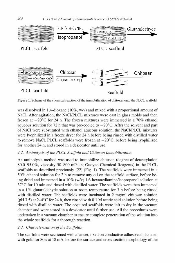
Figure 1. Scheme of the chemical reaction of the immobilization of chitosan onto the PLCL scaffold.
was dissolved in 1,4-dioxane (10%, w/v) and mixed with a proportional amount ofNaCl. After agitation, the NaCl/PLCL mixtures were cast in glass molds and thenfrozen at −20◦C for 24 h. The frozen mixtures were immersed in a 70% ethanolaqueous solution for 72 h that was pre-cooled to −20◦C. After the solvent and partof NaCl were substituted with ethanol aqueous solution, the NaCl/PLCL mixtureswere lyophilized in a freeze dryer for 24 h before being rinsed with distilled waterto remove NaCl. PLCL scaffolds were frozen at −20◦C, before being lyophilizedfor another 24 h, and stored in a desiccator until use.
2.2. Aminolysis of the PLCL Scaffold and Chitosan Immobilization
An aminolysis method was used to immobilize chitosan (degree of deacetylation80.0–95.0%, viscosity 50–800 mPa · s; Guoyao Chemical Reagents) in the PLCLscaffolds as described previously [22] (Fig. 1). The scaffolds were immersed in a50% ethanol solution for 2 h to remove any oil on the scaffold surface, before be-ing dried and immersed in a 10% (w/v) 1,6-hexanediamine/isopropanol solution at37◦C for 10 min and rinsed with distilled water. The scaffolds were then immersedin a 1% glutaraldehyde solution at room temperature for 3 h before being rinsedwith distilled water. The scaffolds were incubated in 2 mg/ml chitosan solution(pH 3.5) at 2–4◦C for 24 h, then rinsed with 0.1 M acetic acid solution before beingrinsed with distilled water. The acquired scaffolds were left to dry in the vacuumchamber and were stored in a dessicator until further use. All the procedures wereundertaken in a vacuum chamber to ensure complete penetration of the solution intothe whole scaffolds for a thorough reaction.
2.3. Characterization of the Scaffolds
The scaffolds were sectioned with a lancet, fixed on conductive adhesive and coatedwith gold for 80 s at 18 mA, before the surface and cross-section morphology of the
C. Li et al. / Journal of Biomaterials Science 23 (2012) 405–424 409
scaffolds were observed by scanning electron microscopy (SEM; Quanta 200FEG,FEI) at an accelerating voltage of 15 kV.
Distribution of amine groups in modified PLCL scaffolds was evaluated by nin-hydrin staining. Briefly, the scaffolds were immersed in 1.0 M ninhydrin/ethanolsolution for 1 min, before being transferred into a glass tube and incubated at 80◦Cfor 10 min to accelerate the reaction. The photographs of the scaffolds were takenwith a Sony digital camera (DSC-H200).
Immobilization of chitosan onto the PLCL scaffolds was confirmed by X-rayphotoelectron spectroscopy (XPS) (AXIS-Ultra, Kratos Analytical). XPS was per-formed using monochromatic Al Kα radiation (225 W, 15 mA, 15 kV). The operat-ing pressure during analysis was maintained at about 10−9 Torr. To compensate forsurface charge effects, binding energies were calibrated using the C1s hydrocarbonpeak at 284.80 eV.
2.4. Swelling Ratio and Porosity
Freeze-dried scaffolds were immersed in phosphate-buffered saline (PBS) at 37◦Cfor 2 h until equilibrium of swelling was reached. The swollen scaffolds were takenout and immediately weighed with a microbalance after the excess water on thesurfaces of the scaffold was carefully removed with tissue paper. The swelling ratiowas calculated and expressed as ER (ER = (Ws − Wd)/Wd, where Ws and Wd arethe weight of the swollen and dried scaffolds, respectively).
Porosity was estimated with a similar methodology [23]. The exact sizes of thescaffolds were measured with a vernier caliper, while the mass of the scaffolds wastaken carefully. The scaffolds were immersed in absolute alcohol for 2 h beforeweighted again. Porosity was calculated as (Ws − Wd)/(ρ/V ), where ρ is the den-sity of alcohol and V the volume of the scaffolds, respectively.
2.5. Mechanical Tests
A compression test creep test, and stress relaxation test of the scaffolds were under-taken with bovine articular cartilage as the control. Fresh quadrate bovine articularcartilage explants (10 mm × 10 mm) were harvested from the weight-bearing areaof the femoral head of bovines and kept wet with PBS before testing. All PLCLscaffolds, chitosan-modified PLCL scaffolds and cartilage explants were immersedin PBS for 24 h at room temperature prior to testing. The length, width and thick-ness of the samples were measured with a vernier caliper and 6 tests were performedfor each group.
2.5.1. Compression TestAn unconfined compression test was carried out with an Instron 5843 mechanicaltest instrument. Compressive loads were applied to individual specimen in a PBSbath using a stainless steel indenter. All the samples were pre-loaded three timesto a 10% strain before a constant loading at a displacement of 0.5 mm/min until45% strain was reached. The measured thickness was converted to the strain of the
410 C. Li et al. / Journal of Biomaterials Science 23 (2012) 405–424
sample (ε = 1 − (L/L0), where L0 and L are the thickness before and after com-pression, respectively). Young’s modulus (E = σ/ε, where σ and ε denote the stressand strain of the sample respectively) was determined directly on a Instron 5843.The thickness of each scaffold was measured to calculate the compressive recoveryratio within 1 min after removal of the compressive load. The recovery ratio wasused to assess the capability of the scaffolds to recover from the deformation andexpressed as recovery ratio (recovery ratio = L1/L0, where L1 and L0 are the finaland initial thickness of the scaffolds after and before compression, respectively).
2.5.2. Creep TestCreep experiments were carried out using an Electronic Universal Testing Machine(WDW3020) in a static unconfined compression. The samples with precompres-sion treatment were loaded at 10 kPa, and the strain was recorded continuously atspecified time intervals for 300 s.
2.5.3. Stress Relaxation TestA stress relaxation test was performed using an Electronic Universal Testing Ma-chine (WDW3020) under a constant strain. After the samples were compressed to20 and 30% of the original thickness, the strain was maintained, and the resultingstress values were recorded over time for 600 s.
2.6. Chondrocyte Culture
Chondrocytes (C28-I2; 15–20 passages), kindly provided by Mary Goldring atChildren’s Hospital Boston, were cultured in tissue culture polystyrene (TCPS,Costar) with culture medium (Dulbecco’s modified Eagle medium (DMEM,Gibco)) containing 10% fetal bovine serum (FBS), 100 IU/ml penicillin and100 µg/ml streptomycin (Gibco) at 37◦C in 5% CO2/95% air.
2.6.1. Biocompatibility3-(4,5-Dimethyl-2-thiazolyl)-2,5-diphenyl-2H-tetrazolium bromide (MTT, M2128, Sigma) was applied to evaluate the biocompatibility of the scaffolds at 24 and48 h, respectively, with TCPS wells as positive the control and latex rubber (HaimenYangzi Medical) as the negative control. Test materials were cut into 5 × 5 × 2 mmsamples and sterilized in 70% ethanol aqueous solution for 2 h before being air-dried and sterilized under UV light for 30 min. The scaffolds were then rinsed andimmersed in PBS for another 30 min. Chondrocytes (5000 cells/well) were cul-tured in a 96-well plate with and without the test materials. MTT solution (100 µl,5 mg/ml) was added in each well at 24 and 48 h, respectively, and incubated at 37◦Cfor 3 h. All wells were emptied before 150 µl DMSO was added. Optical density at540 nm wavelength was measured to determine the percentage of viable cells. Thevalue was compared and expressed as a percentage of the data on the TCPS wells.Six samples from each group were measured.
2.6.2. Cell Attachment and ProliferationProliferation of chondrocytes within scaffolds was evaluated quantitatively withHoechst 33258 (H6024, Sigma). DNA content was analyzed after chondrocytes
C. Li et al. / Journal of Biomaterials Science 23 (2012) 405–424 411
were seeded onto the scaffolds (5 × 5 × 2 mm) for 6 h (for cell attachment) and3 days (for cell proliferation). Cell suspension was added drop-wise onto the top ofthe scaffold at a density of 2× 10⁵ cells/scaffold in 500 µl culture medium (DMEMmedium with 10% FBS). Culture medium was carefully added to the Petri dish(12 wells, Costar) to cover the scaffolds after 30 min incubation. For analysis, cellson the scaffolds were digested in 3 mg/ml proteinase K (H10091, Merck) overnightat 57◦C. H33258 in TRIS-EDTA · 2Na (TNE) buffer (0.1 µg/ml) was used to dye thedigested solution which was loaded in black 96-well plates. Blanks and a series ofDNA standards were also loaded in order to permit calibration of the fluorescencereading. Fluorescence intensity was measured on a microplate reader at excitationand emission wavelengths of 360 and 465 nm, respectively. A calibration curvewas obtained from DNA standard solutions with known concentrations. The DNAconcentration is proportional to the cell number because each cell has a fixed DNAcontent. Therefore, the DNA concentrations provide a reliable means for comparingthe cell numbers grown on the chitosan-modified PLCL scaffolds and plain PLCLscaffolds. Four repeats for each sample were averaged.
2.6.3. Cell Distribution and MorphologyDistribution and morphology of cells on the scaffolds were observed by confocallaser scanning microscopy (CLSM, LSM510, Zeiss). After the chondrocytes seed-ing for 6 h and 3 days, cell-seeded scaffolds were rinsed three times in PBS andincubated with 100 µl of 2 µg/ml Fluorescein Diacetate Solution (FDA, F7378,Sigma) at 37◦C for 15 min. The samples were rinsed in phosphate-buffered saline(PBS) thoroughly, before they were incubated with 100 µl of 5 µg/ml propidium io-dide solution (PI, P4170, Sigma) at 37◦C for 5 min. After washing with PBS threetimes, the samples were observed by CLSM at an excitation wavelength of 488 nmand emission wavelength of 550–670 nm.
2.7. Primary Chondrocyte Isolation and Culture
Chondrocytes were isolated from pig articular cartilage. Briefly, cartilage slicescollected from femoral condyle were digested first with 0.25% TrypLE (Invitro-gen) for 30 min, then with 0.25% (w/v) type-II collagenase solution (Invitrogen) inDMEM for 12–16 h at 37◦C. Isolated chondrocytes were expanded in DMEM +10% FBS. Expanded chondrocytes were seeded onto the scaffold at a density of1 × 107 cells/ml, and cultured in chondrogenic media containing high glucoseDMEM supplemented with 10−7 M dexamethasone, 1% ITS + premix, 50 mg/mlascorbic acid, 1 mM sodium pyruvate, 4 mM proline and 10 ng/ml TGFβ3 (R&DSystems). The cell/scaffold composites were cultured at 37◦C in 5% CO2 over aperiod of up to 3 weeks. Medium was changed every 3 days.
2.8. RNA Analysis and Real-Time PCR
Total RNA was extracted with RNeasy Mini Kit (Qiagen) following the manu-facturer’s instructions. cDNA was reverse transcribed using the iScript™ cDNAsynthesis kit (Bio-Rad), following the manufacturer’s instructions. Real-time
412 C. Li et al. / Journal of Biomaterials Science 23 (2012) 405–424
PCR was performed using the Power SYBR Green PCR Master Mix (AppliedBiosystems) on a Applied Biosystems 7500 Real-Time PCR System at 95◦C for15 min, followed by 40 cycles of 15 s denaturation at 94◦C, 30 s annealing at55◦C and 30 s elongation at 72◦C. Each sample was amplified three times foreach gene of interest. Genes of interest were normalized to the reference geneglutaraldehyde-3-phosphate dehydrogenase (GAPDH). The level of target gene ex-pression was calculated as 2−��Ct. The following forward and reverse primers wereused for amplification: for GAPDH, forward 5′-ATGGTGAAGGTCGGAGTGAA-3′, reverse 5′-AATGAAGGGGTCATTGATGG-3′; for aggrecan, forward, 5′-CATCACCGAGGGTGAAGC-3′, reverse 5′-CCAGGGGCAAATGTAAAGG-3′;for type-II collagen, forward 5′-TGAGAGGTCTTCCTGGCAAA-3′, reverse 5′-GAAGTCCCTGGAAGCCAGAT-3′.2.9. Histological and Immunohistological Assessment
The samples were washed with PBS and embedded with Tissue Freezing Medium®
(Leica, Cat. No. 020108926). Cryosections of 10 µm were prepared using a LeicaCryostat Microtome (CM3050 S). The cut sections were then fixed in an ice-coldmixture of acetone and methanol (1:1, v/v) before processing for histological andimmunological staining.
For Alcian blue staining, the tissue sections were incubated with 0.5% Alcianblue (Sigma-Aldrich) in 0.1 M HCl for 30 min and counterstained with nuclearfast red (Sigma-Aldrich). For immunohistochemistry staining, endogenous peroxi-dase in the tissue sections was first blocked with hydrogen peroxide before pepsintreatment for 20 min. Monoclonal antibodies for type-II collagen (Clone 6B3,Chemicon) diluted factor 1:500 were applied for 1 h, followed by incubation withbiotinylated goat anti-mouse (Lab Vision) for 30 min. Streptavidin peroxidase wasadded for 45 min, 3,3′-diaminobenzidine was used as a chromogenic agent andcounterstaining was done with Gill’s hematoxylin. The slides were dehydrated be-fore coverslipping. The control mouse IgG isotype was from Zymed Laboratories.
2.10. Statistical Analysis
One-way analysis of variance (ANOVA) was used to compare the difference be-tween each group, and all data were expressed as mean ± SD. Statistical signifi-cance was reported at the 95% confidence level (P < 0.05) for all tests.
3. Results
3.1. Characterization of the Scaffolds
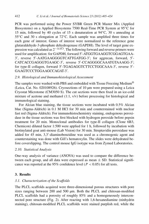
The PLCL scaffolds acquired were three-dimensional porous structures with poresizes ranging between 200 and 500 µm. Both the PLCL and chitosan-modifiedPLCL scaffolds had a porosity of roughly 85% and a homogeneously intercon-nected pore structure (Fig. 2). After reacting with 1,6-hexanediamine (ninhydrinstaining), chitosan-modified PLCL scaffolds were stained purplish red, while the
C. Li et al. / Journal of Biomaterials Science 23 (2012) 405–424 413
Figure 2. SEM images of a PLCL scaffold and a chitosan-modified PLCL scaffold. (A) Surface of thePLCL scaffold; (B) surface of the chitosan-modified scaffold; (C) cross-section of the PLCL scaffold;(D) cross-section of the chitosan-modified scaffold. Scale bar = 300 µm.
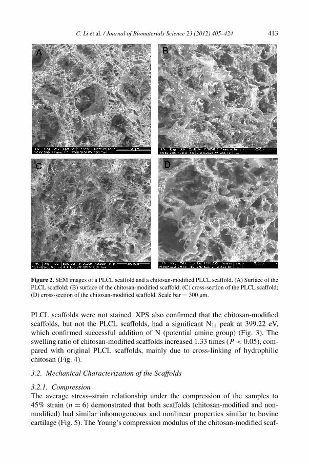
PLCL scaffolds were not stained. XPS also confirmed that the chitosan-modifiedscaffolds, but not the PLCL scaffolds, had a significant N1s peak at 399.22 eV,which confirmed successful addition of N (potential amine group) (Fig. 3). Theswelling ratio of chitosan-modified scaffolds increased 1.33 times (P < 0.05), com-pared with original PLCL scaffolds, mainly due to cross-linking of hydrophilicchitosan (Fig. 4).
3.2. Mechanical Characterization of the Scaffolds
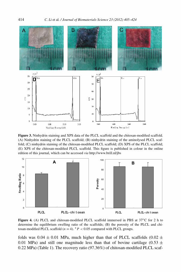
3.2.1. CompressionThe average stress–strain relationship under the compression of the samples to45% strain (n = 6) demonstrated that both scaffolds (chitosan-modified and non-modified) had similar inhomogeneous and nonlinear properties similar to bovinecartilage (Fig. 5). The Young’s compression modulus of the chitosan-modified scaf-
414 C. Li et al. / Journal of Biomaterials Science 23 (2012) 405–424
Figure 3. Ninhydrin staining and XPS data of the PLCL scaffold and the chitosan-modified scaffold.(A) Ninhydrin staining of the PLCL scaffold; (B) ninhydrin staining of the aminolysed PLCL scaf-fold; (C) ninhydrin staining of the chitosan-modified PLCL scaffold; (D) XPS of the PLCL scaffold;(E) XPS of the chitosan-modified PLCL scaffold. This figure is published in colour in the onlineedition of this journal, which can be accessed via http://www.brill.nl/jbs
Figure 4. (A) PLCL and chitosan-modified PLCL scaffold immersed in PBS at 37◦C for 2 h todetermine the equilibrium swelling ratio of the scaffolds; (B) the porosity of the PLCL and chi-tosan-modified PLCL scaffold (n = 4). ∗P < 0.05 compared with PLCL groups.
folds was 0.04 ± 0.01 MPa, much higher than that of PLCL scaffolds (0.02 ±0.01 MPa) and still one magnitude less than that of bovine cartilage (0.53 ±0.22 MPa) (Table 1). The recovery ratio (97.36%) of chitosan-modified PLCL scaf-
C. Li et al. / Journal of Biomaterials Science 23 (2012) 405–424 415
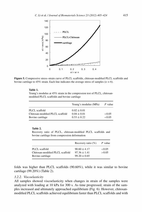
Figure 5. Compressive stress–strain curve of PLCL scaffolds, chitosan-modified PLCL scaffolds andbovine cartilage to 45% strain. Each line indicates the average stress of samples (n = 6).
Table 1.Young’s modulus at 45% strain in the compression test of PLCL, chitosan-modified PLCL scaffolds and bovine cartilage
Young’s modulus (MPa) P value
PLCL scaffold 0.02 ± 0.01Chitosan-modified PLCL scaffold 0.04 ± 0.01 <0.05Bovine cartilage 0.53 ± 0.22 <0.05
Table 2.Recovery ratio of PLCL, chitosan-modified PLCL scaffolds andbovine cartilage from compression deformation
Recovery ratio (%) P value
PLCL scaffold 90.60 ± 4.17 <0.05Chitosan-modified PLCL scaffold 97.36 ± 1.41 >0.05Bovine cartilage 99.20 ± 0.65
folds was higher than PLCL scaffolds (90.60%), while it was similar to bovinecartilage (99.20%) (Table 2).
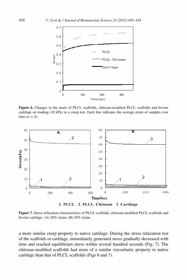
3.2.2. ViscoelasticityAll samples showed viscoelasticity when changes in strain of the samples wereanalyzed with loading at 10 kPa for 300 s. As time progressed, strain of the sam-ples increased and ultimately approached equilibrium (Fig. 6). However, chitosan-modified PLCL scaffolds achieved equilibrium faster than PLCL scaffolds and with
416 C. Li et al. / Journal of Biomaterials Science 23 (2012) 405–424
Figure 6. Changes in the strain of PLCL scaffolds, chitosan-modified PLCL scaffolds and bovinecartilage on loading (10 kPa) in a creep test. Each line indicates the average strain of samples overtime (n = 4).
Figure 7. Stress relaxation characteristics of PLCL scaffolds, chitosan-modified PLCL scaffolds andbovine cartilage. (A) 20% strain; (B) 30% strain.
a more similar creep property to native cartilage. During the stress relaxation testof the scaffolds or cartilage, immediately generated stress gradually decreased withtime and reached equilibrium stress within several hundred seconds (Fig. 7). Thechitosan-modified scaffolds had more of a similar viscoelastic property to nativecartilage than that of PLCL scaffolds (Figs 6 and 7).
C. Li et al. / Journal of Biomaterials Science 23 (2012) 405–424 417
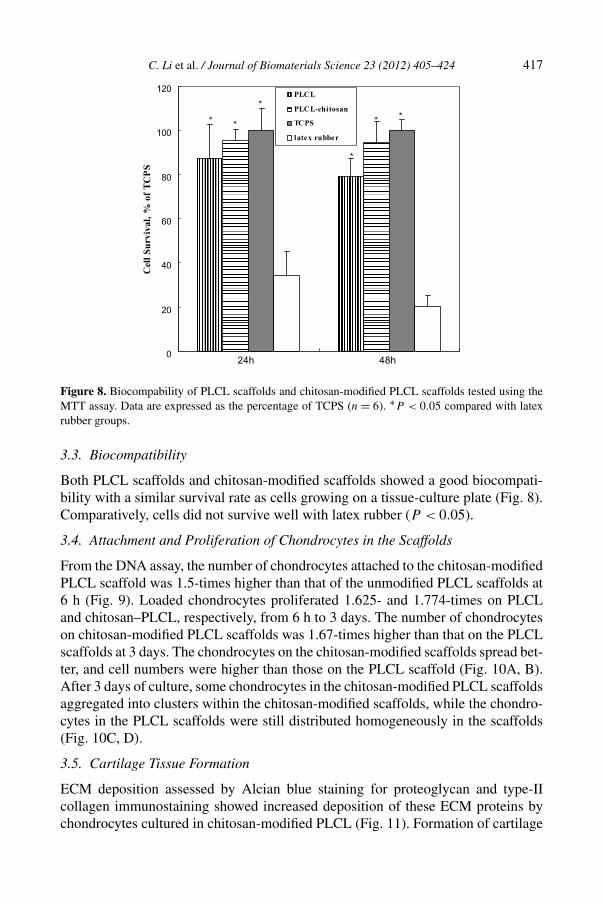
Figure 8. Biocompability of PLCL scaffolds and chitosan-modified PLCL scaffolds tested using theMTT assay. Data are expressed as the percentage of TCPS (n = 6). ∗P < 0.05 compared with latexrubber groups.
3.3. Biocompatibility
Both PLCL scaffolds and chitosan-modified scaffolds showed a good biocompati-bility with a similar survival rate as cells growing on a tissue-culture plate (Fig. 8).Comparatively, cells did not survive well with latex rubber (P < 0.05).
3.4. Attachment and Proliferation of Chondrocytes in the Scaffolds
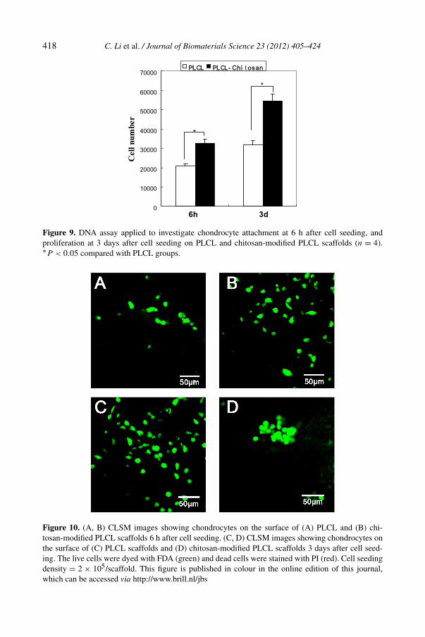
From the DNA assay, the number of chondrocytes attached to the chitosan-modifiedPLCL scaffold was 1.5-times higher than that of the unmodified PLCL scaffolds at6 h (Fig. 9). Loaded chondrocytes proliferated 1.625- and 1.774-times on PLCLand chitosan–PLCL, respectively, from 6 h to 3 days. The number of chondrocyteson chitosan-modified PLCL scaffolds was 1.67-times higher than that on the PLCLscaffolds at 3 days. The chondrocytes on the chitosan-modified scaffolds spread bet-ter, and cell numbers were higher than those on the PLCL scaffold (Fig. 10A, B).After 3 days of culture, some chondrocytes in the chitosan-modified PLCL scaffoldsaggregated into clusters within the chitosan-modified scaffolds, while the chondro-cytes in the PLCL scaffolds were still distributed homogeneously in the scaffolds(Fig. 10C, D).
3.5. Cartilage Tissue Formation
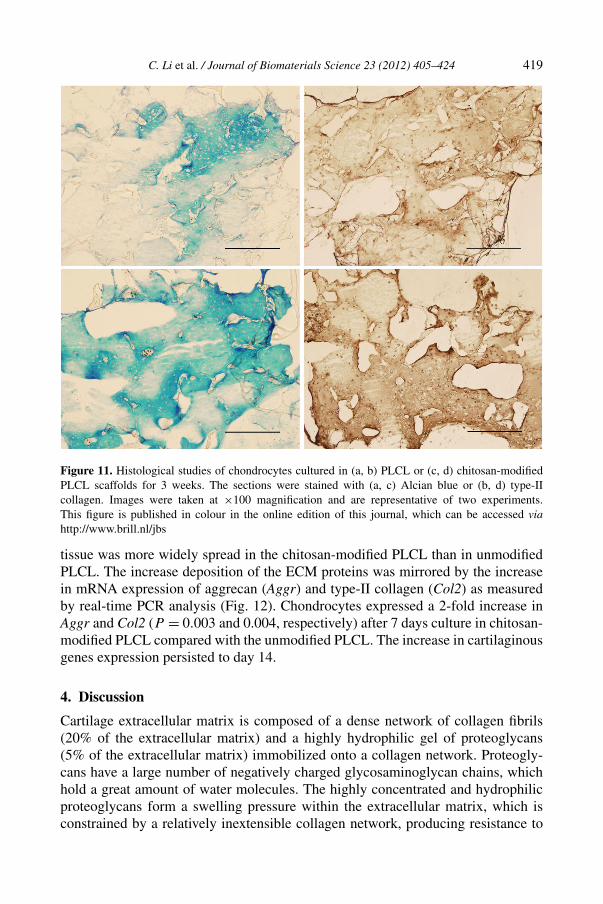
ECM deposition assessed by Alcian blue staining for proteoglycan and type-IIcollagen immunostaining showed increased deposition of these ECM proteins bychondrocytes cultured in chitosan-modified PLCL (Fig. 11). Formation of cartilage
418 C. Li et al. / Journal of Biomaterials Science 23 (2012) 405–424
Figure 9. DNA assay applied to investigate chondrocyte attachment at 6 h after cell seeding, andproliferation at 3 days after cell seeding on PLCL and chitosan-modified PLCL scaffolds (n = 4).∗P < 0.05 compared with PLCL groups.
Figure 10. (A, B) CLSM images showing chondrocytes on the surface of (A) PLCL and (B) chi-tosan-modified PLCL scaffolds 6 h after cell seeding. (C, D) CLSM images showing chondrocytes onthe surface of (C) PLCL scaffolds and (D) chitosan-modified PLCL scaffolds 3 days after cell seed-ing. The live cells were dyed with FDA (green) and dead cells were stained with PI (red). Cell seedingdensity = 2 × 10⁵/scaffold. This figure is published in colour in the online edition of this journal,which can be accessed via http://www.brill.nl/jbs
C. Li et al. / Journal of Biomaterials Science 23 (2012) 405–424 419
Figure 11. Histological studies of chondrocytes cultured in (a, b) PLCL or (c, d) chitosan-modifiedPLCL scaffolds for 3 weeks. The sections were stained with (a, c) Alcian blue or (b, d) type-IIcollagen. Images were taken at ×100 magnification and are representative of two experiments.This figure is published in colour in the online edition of this journal, which can be accessed viahttp://www.brill.nl/jbs
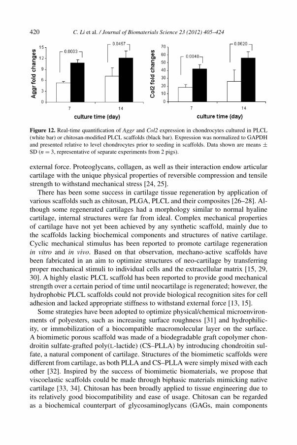
tissue was more widely spread in the chitosan-modified PLCL than in unmodifiedPLCL. The increase deposition of the ECM proteins was mirrored by the increasein mRNA expression of aggrecan (Aggr) and type-II collagen (Col2) as measuredby real-time PCR analysis (Fig. 12). Chondrocytes expressed a 2-fold increase inAggr and Col2 (P = 0.003 and 0.004, respectively) after 7 days culture in chitosan-modified PLCL compared with the unmodified PLCL. The increase in cartilaginousgenes expression persisted to day 14.
4. Discussion
Cartilage extracellular matrix is composed of a dense network of collagen fibrils(20% of the extracellular matrix) and a highly hydrophilic gel of proteoglycans(5% of the extracellular matrix) immobilized onto a collagen network. Proteogly-cans have a large number of negatively charged glycosaminoglycan chains, whichhold a great amount of water molecules. The highly concentrated and hydrophilicproteoglycans form a swelling pressure within the extracellular matrix, which isconstrained by a relatively inextensible collagen network, producing resistance to
420 C. Li et al. / Journal of Biomaterials Science 23 (2012) 405–424
Figure 12. Real-time quantification of Aggr and Col2 expression in chondrocytes cultured in PLCL(white bar) or chitosan-modified PLCL scaffolds (black bar). Expression was normalized to GAPDHand presented relative to level chondrocytes prior to seeding in scaffolds. Data shown are means ±SD (n = 3, representative of separate experiments from 2 pigs).
external force. Proteoglycans, collagen, as well as their interaction endow articularcartilage with the unique physical properties of reversible compression and tensilestrength to withstand mechanical stress [24, 25].
There has been some success in cartilage tissue regeneration by application ofvarious scaffolds such as chitosan, PLGA, PLCL and their composites [26–28]. Al-though some regenerated cartilages had a morphology similar to normal hyalinecartilage, internal structures were far from ideal. Complex mechanical propertiesof cartilage have not yet been achieved by any synthetic scaffold, mainly due tothe scaffolds lacking biochemical components and structures of native cartilage.Cyclic mechanical stimulus has been reported to promote cartilage regenerationin vitro and in vivo. Based on that observation, mechano-active scaffolds havebeen fabricated in an aim to optimize structures of neo-cartilage by transferringproper mechanical stimuli to individual cells and the extracellular matrix [15, 29,30]. A highly elastic PLCL scaffold has been reported to provide good mechanicalstrength over a certain period of time until neocartilage is regenerated; however, thehydrophobic PLCL scaffolds could not provide biological recognition sites for celladhesion and lacked appropriate stiffness to withstand external force [13, 15].
Some strategies have been adopted to optimize physical/chemical microenviron-ments of polyesters, such as increasing surface roughness [31] and hydrophilic-ity, or immobilization of a biocompatible macromolecular layer on the surface.A biomimetic porous scaffold was made of a biodegradable graft copolymer chon-droitin sulfate-grafted poly(L-lactide) (CS–PLLA) by introducing chondroitin sul-fate, a natural component of cartilage. Structures of the biomimetic scaffolds weredifferent from cartilage, as both PLLA and CS–PLLA were simply mixed with eachother [32]. Inspired by the success of biomimetic biomaterials, we propose thatviscoelastic scaffolds could be made through biphasic materials mimicking nativecartilage [33, 34]. Chitosan has been broadly applied to tissue engineering due toits relatively good biocompatibility and ease of usage. Chitosan can be regardedas a biochemical counterpart of glycosaminoglycans (GAGs, main components
C. Li et al. / Journal of Biomaterials Science 23 (2012) 405–424 421
of proteoglycan) due to its similar hexose consisting of water-soluble structuralunits. PLCL is a highly elastic synthetic macromolecular compound which simu-lated an elastic collagen fiber network in native cartilage. It was chosen in placeof the broadly adopted PLA and PLGA, mainly due to its intrinsic elasticity. Fur-thermore, when PLCL degrades into relatively mild acidic products compared toPLGA, chitosan could neutralize the acidic products from the degradation of PLCL.In the current study, chitosan was immobilized onto the PLCL scaffold by aminol-ysis methods to simulate the biochemical components of the cartilage extracellularmatrix [22, 35]. In the aminolysis process, ester groups of PLCL backbone reactedwith one amino group of 1,6-hexanediamine to form a covalent bond while the otheramino group remained free to cross-link with chitosan through glutaraldehyde. Suc-cessful cross-linking of chitosan to PLCL was confirmed by ninhydrin staining andXPS, which qualitatively showed presence of the NH2 group in the scaffolds [27,36, 37].
Practically, the chitosan-modified PLCL scaffolds could possess mechanicalproperties similar to native cartilage. Under compression stress, water moleculesheld by ionic groups of chitosan inside the scaffolds would endow the scaffoldswith viscoelasticity following a similar principle found within native cartilage. Anincrease in viscoelasticity of the chitosan–PLCL scaffolds was confirmed by thecreep and stress relaxation test. Usually aminolysised PLCL scaffolds should havea lower stiffness than PLCL scaffolds due to fragmentation of the PLCL macro-molecular chain, but in the current study, both the Young’s modulus and recoveryratio of chitosan-modified scaffolds increased compared to unmodified PLCL scaf-folds. It is likely that the cross-link of chitosan has increased the internal stiffnessof the structures. Although the Young’s modulus of the chitosan-modified PLCLscaffolds was higher than that of unmodified PLCL scaffolds, it was still one or-der of magnitude less than bovine cartilage. To date, the most successful scaffolds,regarding mechanical properties, are poly(glycolic acid) (PGA) woven structuresmixed with hydrogels [18]. The current chitosan-modified PLCL scaffolds havea Young’s modulus similar to the composite scaffold as mentioned above (0.005and 0.1 MPa). While both the scaffolds did not have sufficient stiffness/Young’smodulus to fully withstand applied high stress, the deformation recovery ratio ofchitosan-modified PLCL scaffolds (97.36%) was in a range very similar to nativecartilage and could potentially deliver proper physiological mechanical signals tothe attached chondrocytes.
Differences in biochemical components and physical/chemical properties lead todisparities in mechanical properties between scaffolds and native cartilage. Whenstress is loaded on the native cartilage, the forces generated from the native car-tilage, including the fluid flow and osmotic pressure derived from the interactionof ions with the proteoglycans, can balance external forces. In the current study,when chitosan-modified PLCL scaffolds were loaded in a PBS-immersed envi-ronment, hydrophilic NH2 groups within the scaffolds absorbed large amounts ofwater resulting in a relatively high osmotic pressure to resist external pressure.
422 C. Li et al. / Journal of Biomaterials Science 23 (2012) 405–424
Multiple causes contribute to the mechanical differences between the biomimeticscaffolds and native cartilage. Firstly, structures of biomacromolecules in carti-lage and scaffolds are different. In cartilage, three collagen molecules self-assembleinto a collagen fiber with natural triple-helical domains and eventually form the se-quential arrangement of collagen fibrils, which brings about the high elasticity ofcollagen network while the PLCL molecules of current scaffolds are in a disorderedstructure. Secondly, electronegative aggrecans in cartilage are cross-linked into thecollagen network; however, in the current biomimetic scaffolds inexpensive andelectropositive chitosan was covalently bonded only to the surface of PLCL scaf-fold. Thirdly, the ratio of chitosan and PLCL in the current scaffolds is not the sameas that of aggrecan and collagen in native cartilage. Therefore, there is still roomto improve the mechanical properties of current biomimetic scaffolds through fur-ther chemical engineering. In the current study, chitosan-modified PLCL scaffoldshad a good recovery ratio similar to native cartilage, which could possibly supplya mechanically compatible microenvironment for cell adhesion, proliferation anddifferentiation.
It has been known that biochemical components of biomaterials are criticalfor potential chondrogenesis [38]. More chondrocytes attached to the chitosan-modified PLCL scaffolds than to the unmodified scaffolds, which can be attributedto a fairly good biocompatibility of chitosan. It is also evidenced by enhanced geneexpression and extracellular matrix excretion of aggrecan and type-II collagen ofchondrocytes within chitosan-modified PLCL scaffolds, compared with PLCL scaf-folds. Chondrocytes spread better when compared with unmodified PLCL scaffoldsat 6 h. Interestingly, a majority of chondrocytes on the chitosan-modified PLCLscaffolds aggregated as clusters after 3 days, in contrast to chondrocytes on thePLCL scaffolds which were distributed homogeneously. As proper aggregation iscritical for chondrocyte function and chondrogenesis, chitosan may help to promotechondrogenesis through regulating proper chondrocyte aggregation [39].
The main purpose of this study was to design viscoelastic three-dimensionalporous scaffolds that simulate the structure and components of native cartilage.Current scaffolds are biocompatible as well as biodegradable and could provide agood microenvironment for cell adhesion, proliferation and extracellular matrix ex-cretion. They are potentially good for cartilage regeneration by providing propermechanical and biochemical signals for chondrocyte adhesion and differentiation.In a future study, efforts should be made to improve stiffness of the chitosan-modified PLCL scaffolds through different chemical modifications while studyingthe mechanical properties of the resulting cartilage tissue. The mechanism of cellaggregation induced by chitosan should also be further investigated.
5. Conclusions
Current chitosan-modified PLCL scaffolds have similar viscoelastic properties withnative cartilage, a good recovery ratio and relatively good biocompatibility for tis-
C. Li et al. / Journal of Biomaterials Science 23 (2012) 405–424 423
sue engineering and regeneration. With the design of similar internal structures andcomponents of native cartilage, the mechanical properties of the scaffolds can befurther improved to meet the requirements for both research and clinical applica-tions. The scaffolds could not only serve as a model for cell mechanical study butcould also be directly implanted in vivo as a cellular inductive, as well as a support-ing structure.
Acknowledgements
This study was supported by the National Natural Science Foundation of China(grants 30970881 and 31011140348) and a Seed Grant from the Wallace H. CoulterFoundation. The authors thank Song Yun for assistance with environmental scan-ning electronic microscope and Prof. Chun Yang for advice on the mechanical test.The authors also thank Dr. Mary Goldring for kindly providing the chondrocyte lineC28-I2. The authors declare they have no competing interests.
References
1. L. A. Setton, D. M. Elliott and V. C. Mow, Osteoarthr. Cartil. 7, 2 (1999).2. M. Brittberg, A. Lindahl, A. Nilsson, C. Ohlsson, O. Isaksson and L. Peterson, N. Engl. J. Med.
331, 889 (1994).3. L. Hangody, G. Vasarhelyi, L. R. Hangody, Z. Sukosd, G. Tibay, L. Bartha and G. Bodo, Injury
39 (Suppl. 1), S32 (2008).4. Y. N. Wu, Z. Yang, J. H. Hui, H. W. Ouyang and E. H. Lee, Biomaterials 28, 4056 (2007).5. M. H. Zheng, C. Willers, L. Kirilak, P. Yates, J. Xu, D. Wood and A. Shimmin, Tissue Eng. 13,
737 (2007).6. F. Ito, K. Usui, D. Kawahara, A. Suenaga, T. Maki, S. Kidoaki, H. Suzuki, M. Taiji, M. Itoh,
Y. Hayashizaki and T. Matsuda, Biomaterials 31, 58 (2010).7. D. A. Wang, S. Varghese, B. Sharma, I. Strehin, S. Fermanian, J. Gorham, D. H. Fairbrother,
B. Cascio and J. H. Elisseeff, Nature Mater. 6, 385 (2007).8. Y. Jung, S. H. Kim, S. H. Kim, Y. H. Kim, J. Xie, T. Matsuda and B. G. Min, J. Biomater. Sci.
Polymer Edn 19, 61 (2008).9. C. G. Jeong and S. J. Hollister, Biomaterials 31, 4304 (2010).
10. V. Kesireddy, J. Ringe, M. Endres, S. Stich, D. Klose, R. Bodmeier and M. Sittinger, Tissue Eng.12, 1073 (2006).
11. C. W. Lou, C. H. Yao, Y. S. Chen, T. C. Hsieh, J. H. Lin and W. H. Hsing, Textil. Res. J. 78, 958(2008).
12. Y. Jung, S. H. Kim, H. J. You, S. H. Kim, Y. H. Kim and B. G. Min, J. Biomater. Sci. Polymer Edn19, 1073 (2008).
13. S. I. Jeong, B. S. Kim, Y. M. Lee, K. J. Ihn, S. H. Kim and Y. H. Kim, Biomacromolecules 5, 1303(2004).
14. Y. Kang, J. Yang, S. Khan, L. Anissian and G. A. Ameer, J. Biomed. Mater. Res. A 77, 331 (2006).15. J. Xie, M. Ihara, Y. M. Jung, I. K. Kwon, S. H. Kim, Y. H. Kim and T. Matsuda, Tissue Eng. 12,
449 (2006).16. G. R. Ragetly, G. J. Slavik, B. T. Cunningham, D. J. Schaeffer and D. J. Griffon, J. Biomed. Mater.
Res. A 93, 46 (2010).
424 C. Li et al. / Journal of Biomaterials Science 23 (2012) 405–424
17. J. K. Suh and H. W. Matthew, Biomaterials 21, 2589 (2000).18. F. T. Moutos, L. E. Freed and F. Guilak, Nature Mater. 6, 162 (2007).19. S. Tepic, T. Macirowski and R. W. Mann, Proc. Natl. Acad. Sci. USA 80, 3331 (1983).20. L. A. Setton, W. B. Zhu and V. C. Mow, J. Biomech. 26, 581 (1993).21. M. H. Ho, P. Y. Kuo, H. J. Hsieh, T. Y. Hsien, L. T. Hou, J. Y. Lai and D. M. Wang, Biomaterials
25, 129 (2004).22. Y. B. Zhu, C. Y. Gao, X. Y. Liu, T. He and J. C. Shen, Tissue Eng. 10, 53 (2004).23. X. Liu and P. X. Ma, Biomaterials 30, 4094 (2009).24. P. J. Basser, R. Schneiderman, R. A. Bank, E. Wachtel and A. Maroudas, Arch. Biochem. Biophys.
351, 207 (1998).25. J. K. Suh and M. R. DiSilvestro, J. Appl. Mech. Trans. ASME 66, 528 (1999).26. Y. Jung, M. S. Park, J. W. Lee, Y. H. Kim, S. H. Kim and S. H. Kim, Biomaterials 29, 4630 (2008).27. D. L. Nettles, S. H. Elder and J. A. Gilbert, Tissue Eng. 8, 1009 (2002).28. K. Uematsu, K. Hattori, Y. Ishimoto, J. Yamauchi, T. Habata, Y. Takakura, H. Ohgushi, T. Fukuchi
and M. Sato, Biomaterials 26, 4273 (2005).29. S. Even-Ram, V. Artym and K. M. Yamada, Cell 126, 645 (2006).30. V. Vogel and M. Sheetz, Nature Rev. Mol. Cell Biol. 7, 265 (2006).31. T. W. Chung, D. Z. Liu, S. Y. Wang and S. S. Wang, Biomaterials 24, 4655 (2003).32. C. T. Lee, C. P. Huang and Y. D. Lee, Biomacromolecules 7, 2200 (2006).33. K. A. Athanasiou, M. P. Rosenwasser, J. A. Buckwalter, T. I. Malinin and V. C. Mow, J. Orthop.
Res. 9, 330 (1991).34. W. C. Hayes and L. F. Mockros, J. Appl. Physiol. 31, 562 (1971).35. Y. B. Zhu, K. S. Chian, M. B. Chan-Park, P. S. Mhaisalkar and B. D. Ratner, Biomaterials 27, 68
(2006).36. Y. C. Kuo and I. N. Ku, Biomacromolecules 9, 2662 (2008).37. J. K. F. Suh and H. W. T. Matthew, Biomaterials 21, 2589 (2000).38. J. K. Mouw, N. D. Case, R. E. Guldberg, A. H. Plaas and M. E. Levenston, Osteoarthr. Cartil. 13,
828 (2005).39. D. R. Albrecht, G. H. Underhill, T. B. Wassermann, R. L. Sah and S. N. Bhatia, Nature Methods
3, 369 (2006).