The protective effect of MT-α-glucan against streptozotocin (STZ)-induced NIT-1 pancreatic β-cell...
7
Carbohydrate Polymers 92 (2013) 1211–1217 Contents lists available at SciVerse ScienceDirect Carbohydrate Polymers jo u rn al hom epa ge: www.elsevier.com/locate/carbpol The protective effect of MT--glucan against streptozotocin (STZ)-induced NIT-1 pancreatic -cell damage Lei Hong a,∗ , Wang Qin a , Guo Shuzhen a , Han Juncheng a , Sun Hanju a , Zhang Xiaoxiang a , Wu Wutong b a School of Biology and Food Engineering, Hefei University of Technology, Hefei 230009, China b School of Life Science and Technology, China Pharmaceutical University, Nanjing 210009, China a r t i c l e i n f o Article history: Received 2 July 2012 Received in revised form 14 October 2012 Accepted 15 October 2012 Available online 23 October 2012 Keywords: G. frondosa Polysaccharide Alpha-glucan NIT-1 pancreatic -cell Streptozotocin (STZ) Protective effect a b s t r a c t The protective effect of an alpha-glucan (designated here as MT--glucan) from fruit body of maitake (Grifola frondosa) on NIT-1 pancreatic -cells damaged by streptozotocin (STZ) in vitro was investigated. The cell viability, insulin secretion, the activity of superoxide dismutase (SOD), glutathione peroxidase (GSHpx) and the content of reduced glutathione (GSH) increased significantly when the cells were incu- bated with MT--glucan (400, 200 g ml −1 ). The content of malondialdehyde (MDA), nitric oxide (NO) production, and the activity of NO synthase (NOS), inducible NOS (iNOS) decreased significantly when the cells were incubated with MT--glucan. The destructive changes of NIT-1 cells ameliorated when incubated with MT--glucan under microscopic observation. These data suggested that MT--glucan had obvious protective effect on NIT-1 pancreatic -cells damaged by STZ, which might be related to its effects of decreasing levels of -cell-destroying factors such as oxidative stress and NO synthesis. © 2012 Elsevier Ltd. All rights reserved. 1. Introduction Maitake mushrooms (Grifola frondosa) belong to Basidiomycetes in fungi and have been praised and consumed by Chinese people for hundreds of years as of their enticing tastes. Moreover, the medic- inal properties of maitake have been claimed for years and some of them have been demonstrated scientifically and experimentally. For instance, maitake has been shown to have antitumor effect (Liu, Chen, & Wu, 2005), immune regulatory activity (Inoue, Kodoma, & Nanba, 2002), anti-hyperliposis (Kubo, 1997), anti-common and specific infections effects such as hepatitis (Kubo & Nanba, 1998; Ooi, 1996) and AIDS/HIV (Nanba, Kodama, Schar, & Turner, 2000). Previous studies have shown that ingesting maitake mush- rooms, or some of its extracts, influenced glucose/lipid metabolism and had anti-diabetic effect (Kubo, 1994, 1997). But studies on its active part and mechanism of action have not been carried out. Based on previous studies, a new kind of alpha-glucan extracted and purified from the fruit body of maitake, designated here as MT- -glucan, was prepared in our laboratory. Previous studies in our laboratory have shown that MT--glucan has hypoglycemic activ- ity in KK-Ay mice (Lei, Ma, & Wu, 2007) and experimental type 2 ∗ Corresponding author at: School of Biology and food engineering, Hefei Univer- sity of Technology, Tunxi Road 193#, Hefei, Anhui 230009, China. Tel.: +86 551 2901395; fax: +86 551 2901395. E-mail address: [email protected] (L. Hong). diabetes mice (Lei et al., 2012), by ameliorating peripheral insulin resistance and protecting pancreatic islets in vivo. The present study was therefore designed to explore the protective effect of MT--glucan on NIT-1 pancreatic -cells damaged by streptozo- tocin (STZ) in vitro. Moreover, its mechanisms of action concerning protection of pancreatic cells from destruction by oxidative stress and nitric oxide (NO) in vitro were also investigated. 2. Materials and methods 2.1. Preparation, purity and structure of MT-˛-glucan The procedure of preparation of MT--glucan and its purity and primary structure were studied in our laboratory previously and had been reported (Lei et al., 2007; Ma et al., 2007), which were described briefly below. MT--glucan was extracted and purified from the fruit body of maitake (G. frondosa), manufactured in Zhejiang province and iden- tified by China Pharmaceutical University. Dried powdered fruit bodies of maitake were refluxed with diethylether and ethylalco- hol mixture at 70 ◦ C for 6 h, and then centrifuged and the residue collected and extracted by distilled water at 121 ◦ C for 30 min. The extract was centrifuged and the supernatant was collected and pre- cipitated in 95% ethyl alcohol at 4 ◦ C for 12 h. This was followed by centrifugation and the precipitate was collected and re-dissolved in distilled water, then fractionated by DEAE Sepharose Fast Flow chromatography. The fraction was collected, concentrated and 0144-8617/$ – see front matter © 2012 Elsevier Ltd. All rights reserved. http://dx.doi.org/10.1016/j.carbpol.2012.10.037
Transcript of The protective effect of MT-α-glucan against streptozotocin (STZ)-induced NIT-1 pancreatic β-cell...
Carbohydrate Polymers 92 (2013) 1211– 1217
Contents lists available at SciVerse ScienceDirect
Carbohydrate Polymers
jo u rn al hom epa ge: www.elsev ier .com/ locate /carbpol
The protective effect of MT-�-glucan against streptozotocin (STZ)-induced
NIT-1 pancreatic �-cell damage
Lei Honga,∗, Wang Qina, Guo Shuzhena, Han Junchenga, Sun Hanjua, Zhang Xiaoxianga, Wu Wutongb
a School of Biology and Food Engineering, Hefei University of Technology, Hefei 230009, Chinab School of Life Science and Technology, China Pharmaceutical University, Nanjing 210009, China
a r t i c l e i n f o
Article history:
Received 2 July 2012
Received in revised form 14 October 2012
Accepted 15 October 2012
Available online 23 October 2012
Keywords:
G. frondosa
Polysaccharide
Alpha-glucan
NIT-1 pancreatic �-cell
Streptozotocin (STZ)
Protective effect
a b s t r a c t
The protective effect of an alpha-glucan (designated here as MT-�-glucan) from fruit body of maitake
(Grifola frondosa) on NIT-1 pancreatic �-cells damaged by streptozotocin (STZ) in vitro was investigated.
The cell viability, insulin secretion, the activity of superoxide dismutase (SOD), glutathione peroxidase
(GSHpx) and the content of reduced glutathione (GSH) increased significantly when the cells were incu-
bated with MT-�-glucan (400, 200 �g ml−1). The content of malondialdehyde (MDA), nitric oxide (NO)
production, and the activity of NO synthase (NOS), inducible NOS (iNOS) decreased significantly when
the cells were incubated with MT-�-glucan. The destructive changes of NIT-1 cells ameliorated when
incubated with MT-�-glucan under microscopic observation. These data suggested that MT-�-glucan
had obvious protective effect on NIT-1 pancreatic �-cells damaged by STZ, which might be related to its
effects of decreasing levels of �-cell-destroying factors such as oxidative stress and NO synthesis.
© 2012 Elsevier Ltd. All rights reserved.
1. Introduction
Maitake mushrooms (Grifola frondosa) belong to Basidiomycetes
in fungi and have been praised and consumed by Chinese people for
hundreds of years as of their enticing tastes. Moreover, the medic-
inal properties of maitake have been claimed for years and some
of them have been demonstrated scientifically and experimentally.
For instance, maitake has been shown to have antitumor effect (Liu,
Chen, & Wu, 2005), immune regulatory activity (Inoue, Kodoma,
& Nanba, 2002), anti-hyperliposis (Kubo, 1997), anti-common and
specific infections effects such as hepatitis (Kubo & Nanba, 1998;
Ooi, 1996) and AIDS/HIV (Nanba, Kodama, Schar, & Turner, 2000).
Previous studies have shown that ingesting maitake mush-
rooms, or some of its extracts, influenced glucose/lipid metabolism
and had anti-diabetic effect (Kubo, 1994, 1997). But studies on its
active part and mechanism of action have not been carried out.
Based on previous studies, a new kind of alpha-glucan extracted
and purified from the fruit body of maitake, designated here as MT-
�-glucan, was prepared in our laboratory. Previous studies in our
laboratory have shown that MT-�-glucan has hypoglycemic activ-
ity in KK-Ay mice (Lei, Ma, & Wu, 2007) and experimental type 2
∗ Corresponding author at: School of Biology and food engineering, Hefei Univer-
sity of Technology, Tunxi Road 193#, Hefei, Anhui 230009, China.
Tel.: +86 551 2901395; fax: +86 551 2901395.
E-mail address: [email protected] (L. Hong).
diabetes mice (Lei et al., 2012), by ameliorating peripheral insulin
resistance and protecting pancreatic islets in vivo. The present
study was therefore designed to explore the protective effect of
MT-�-glucan on NIT-1 pancreatic �-cells damaged by streptozo-
tocin (STZ) in vitro. Moreover, its mechanisms of action concerning
protection of pancreatic � cells from destruction by oxidative stress
and nitric oxide (NO) in vitro were also investigated.
2. Materials and methods
2.1. Preparation, purity and structure of MT-˛-glucan
The procedure of preparation of MT-�-glucan and its purity and
primary structure were studied in our laboratory previously and
had been reported (Lei et al., 2007; Ma et al., 2007), which were
described briefly below.
MT-�-glucan was extracted and purified from the fruit body of
maitake (G. frondosa), manufactured in Zhejiang province and iden-
tified by China Pharmaceutical University. Dried powdered fruit
bodies of maitake were refluxed with diethylether and ethylalco-
hol mixture at 70 ◦C for 6 h, and then centrifuged and the residue
collected and extracted by distilled water at 121 ◦C for 30 min. The
extract was centrifuged and the supernatant was collected and pre-
cipitated in 95% ethyl alcohol at 4 ◦C for 12 h. This was followed by
centrifugation and the precipitate was collected and re-dissolved
in distilled water, then fractionated by DEAE Sepharose Fast Flow
chromatography. The fraction was collected, concentrated and
0144-8617/$ – see front matter © 2012 Elsevier Ltd. All rights reserved.http://dx.doi.org/10.1016/j.carbpol.2012.10.037
1212 L. Hong et al. / Carbohydrate Polymers 92 (2013) 1211– 1217
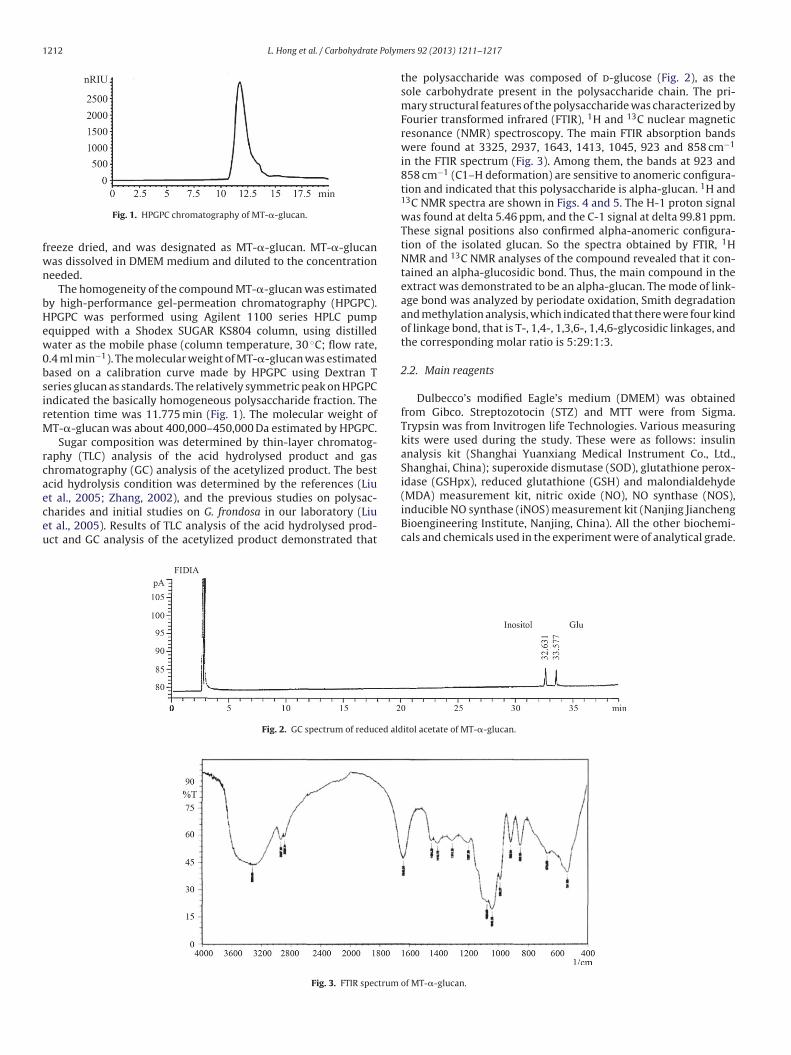
Fig. 1. HPGPC chromatography of MT-�-glucan.
freeze dried, and was designated as MT-�-glucan. MT-�-glucan
was dissolved in DMEM medium and diluted to the concentration
needed.
The homogeneity of the compound MT-�-glucan was estimated
by high-performance gel-permeation chromatography (HPGPC).
HPGPC was performed using Agilent 1100 series HPLC pump
equipped with a Shodex SUGAR KS804 column, using distilled
water as the mobile phase (column temperature, 30 ◦C; flow rate,
0.4 ml min−1). The molecular weight of MT-�-glucan was estimated
based on a calibration curve made by HPGPC using Dextran T
series glucan as standards. The relatively symmetric peak on HPGPC
indicated the basically homogeneous polysaccharide fraction. The
retention time was 11.775 min (Fig. 1). The molecular weight of
MT-�-glucan was about 400,000–450,000 Da estimated by HPGPC.
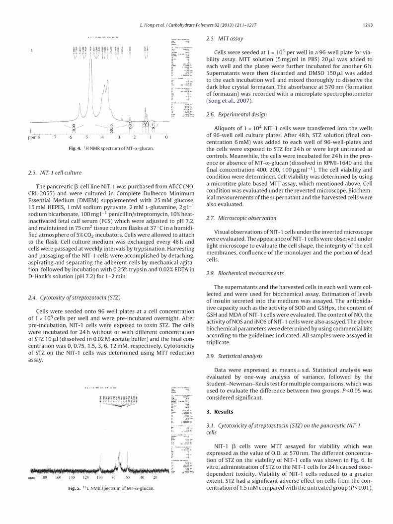
Sugar composition was determined by thin-layer chromatog-
raphy (TLC) analysis of the acid hydrolysed product and gas
chromatography (GC) analysis of the acetylized product. The best
acid hydrolysis condition was determined by the references (Liu
et al., 2005; Zhang, 2002), and the previous studies on polysac-
charides and initial studies on G. frondosa in our laboratory (Liu
et al., 2005). Results of TLC analysis of the acid hydrolysed prod-
uct and GC analysis of the acetylized product demonstrated that
the polysaccharide was composed of d-glucose (Fig. 2), as the
sole carbohydrate present in the polysaccharide chain. The pri-
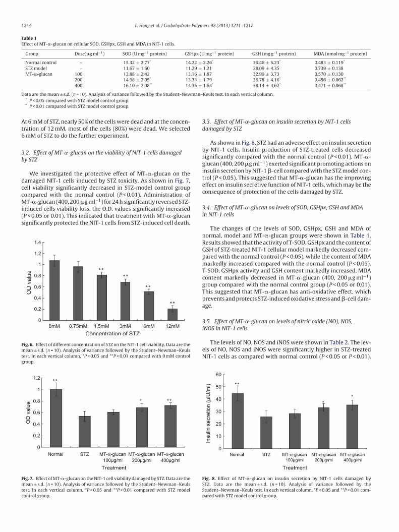
mary structural features of the polysaccharide was characterized by
Fourier transformed infrared (FTIR), 1H and 13C nuclear magnetic
resonance (NMR) spectroscopy. The main FTIR absorption bands
were found at 3325, 2937, 1643, 1413, 1045, 923 and 858 cm−1
in the FTIR spectrum (Fig. 3). Among them, the bands at 923 and
858 cm−1 (C1–H deformation) are sensitive to anomeric configura-
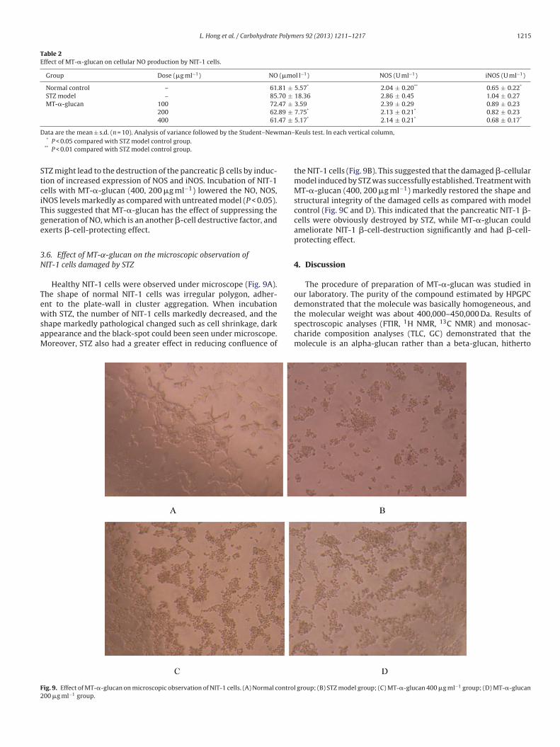
tion and indicated that this polysaccharide is alpha-glucan. 1H and13C NMR spectra are shown in Figs. 4 and 5. The H-1 proton signal
was found at delta 5.46 ppm, and the C-1 signal at delta 99.81 ppm.
These signal positions also confirmed alpha-anomeric configura-
tion of the isolated glucan. So the spectra obtained by FTIR, 1H
NMR and 13C NMR analyses of the compound revealed that it con-
tained an alpha-glucosidic bond. Thus, the main compound in the
extract was demonstrated to be an alpha-glucan. The mode of link-
age bond was analyzed by periodate oxidation, Smith degradation
and methylation analysis, which indicated that there were four kind
of linkage bond, that is T-, 1,4-, 1,3,6-, 1,4,6-glycosidic linkages, and
the corresponding molar ratio is 5:29:1:3.
2.2. Main reagents
Dulbecco’s modified Eagle’s medium (DMEM) was obtained
from Gibco. Streptozotocin (STZ) and MTT were from Sigma.
Trypsin was from Invitrogen life Technologies. Various measuring
kits were used during the study. These were as follows: insulin
analysis kit (Shanghai Yuanxiang Medical Instrument Co., Ltd.,
Shanghai, China); superoxide dismutase (SOD), glutathione perox-
idase (GSHpx), reduced glutathione (GSH) and malondialdehyde
(MDA) measurement kit, nitric oxide (NO), NO synthase (NOS),
inducible NO synthase (iNOS) measurement kit (Nanjing Jiancheng
Bioengineering Institute, Nanjing, China). All the other biochemi-
cals and chemicals used in the experiment were of analytical grade.
Fig. 2. GC spectrum of reduced alditol acetate of MT-�-glucan.
Fig. 3. FTIR spectrum of MT-�-glucan.
L. Hong et al. / Carbohydrate Polymers 92 (2013) 1211– 1217 1213
Fig. 4. 1H NMR spectrum of MT-�-glucan.
2.3. NIT-1 cell culture
The pancreatic �-cell line NIT-1 was purchased from ATCC (NO.
CRL-2055) and were cultured in Complete Dulbecco Minimum
Essential Medium (DMEM) supplemented with 25 mM glucose,
15 mM HEPES, 1 mM sodium pyruvate, 2 mM l-glutamine, 2 g l−1
sodium bicarbonate, 100 mg l−1 penicillin/streptomycin, 10% heat-
inactivated fetal calf serum (FCS) which were adjusted to pH 7.2,
and maintained in 75 cm2 tissue culture flasks at 37 ◦C in a humidi-
fied atmosphere of 5% CO2 incubators. Cells were allowed to attach
to the flask. Cell culture medium was exchanged every 48 h and
cells were passaged at weekly intervals by trypsination. Harvesting
and passaging of the NIT-1 cells were accomplished by detaching,
aspirating and separating the adherent cells by mechanical agita-
tion, followed by incubation with 0.25% trypsin and 0.02% EDTA in
D-Hank’s solution (pH 7.2) for 1–2 min.
2.4. Cytotoxity of streptozotocin (STZ)
Cells were seeded onto 96 well plates at a cell concentration
of 1 × 10⁵ cells per well and were pre-incubated overnight. After
pre-incubation, NIT-1 cells were exposed to toxin STZ. The cells
were incubated for 24 h without or with different concentration
of STZ 10 �l (dissolved in 0.02 M acetate buffer) and the final con-
centration was 0, 0.75, 1.5, 3, 6, 12 mM, respectively. Cytotoxicity
of STZ on the NIT-1 cells was determined using MTT reduction
assay.
Fig. 5. 13C NMR spectrum of MT-�-glucan.
2.5. MTT assay
Cells were seeded at 1 × 10⁵ per well in a 96-well plate for via-
bility assay. MTT solution (5 mg/ml in PBS) 20 �l was added to
each well and the plates were further incubated for another 6 h.
Supernatants were then discarded and DMSO 150 �l was added
to the each incubation well and mixed thoroughly to dissolve the
dark blue crystal formazan. The absorbance at 570 nm (formation
of formazan) was recorded with a microplate spectrophotometer
(Song et al., 2007).
2.6. Experimental design
Aliquots of 1 × 10⁴ NIT-1 cells were transferred into the wells
of 96-well cell culture plates. After 48 h, STZ solution (final con-
centration 6 mM) was added to each well of 96-well-plates and
the cells were exposed to STZ for 24 h or were kept untreated as
controls. Meanwhile, the cells were incubated for 24 h in the pres-
ence or absence of MT-�-glucan (dissolved in RPMI-1640 and the
final concentration 400, 200, 100 �g ml−1). The cell viability and
condition were determined. Cell viability was determined by using
a microtitre plate-based MTT assay, which mentioned above. Cell
condition was evaluated under the reverted microscope. Biochem-
ical measurements of the supernatant and the harvested cells were
also evaluated.
2.7. Microscopic observation
Visual observations of NIT-1 cells under the inverted microscope
were evaluated. The appearance of NIT-1 cells were observed under
light microscope to evaluate the cell shape, the integrity of the cell
membranes, confluence of the monolayer and the portion of dead
cells.
2.8. Biochemical measurements
The supernatants and the harvested cells in each well were col-
lected and were used for biochemical assay. Estimation of levels
of insulin secreted into the medium was assayed. The antioxida-
tive capacity such as the activity of SOD and GSHpx, the content of
GSH and MDA of NIT-1 cells were evaluated. The content of NO, the
activity of NOS and iNOS of NIT-1 cells were also assayed. The above
biochemical parameters were determined by using commercial kits
according to the guidelines indicated. All samples were assayed in
triplicate.
2.9. Statistical analysis
Data were expressed as means ± s.d. Statistical analysis was
evaluated by one-way analysis of variance, followed by the
Student–Newman–Keuls test for multiple comparisons, which was
used to evaluate the difference between two groups. P < 0.05 was
considered significant.
3. Results
3.1. Cytotoxicity of streptozotocin (STZ) on the pancreatic NIT-1
cells
NIT-1 � cells were MTT assayed for viability which was
expressed as the value of O.D. at 570 nm. The different concentra-
tion of STZ on the viability of NIT-1 cells was shown in Fig. 6. In
vitro, administration of STZ to the NIT-1 cells for 24 h caused dose-
dependent toxicity. Viability of NIT-1 cells reduced to a greater
extent. STZ had a significant adverse effect on cells from the con-
centration of 1.5 mM compared with the untreated group (P < 0.01).
1214 L. Hong et al. / Carbohydrate Polymers 92 (2013) 1211– 1217
Table 1Effect of MT-�-glucan on cellular SOD, GSHpx, GSH and MDA in NIT-1 cells.
Group Dose(�g ml−1) SOD (U mg−1 protein) GSHpx (U mg−1 protein) GSH (mg g−1 protein) MDA (nmol mg−1 protein)
Normal control – 15.32 ± 2.77* 14.22 ± 2.26* 36.46 ± 5.23* 0.483 ± 0.119*
STZ model – 11.67 ± 1.60 11.29 ± 1.21 28.09 ± 4.35 0.739 ± 0.138
MT-�-glucan 100 13.88 ± 2.42 13.16 ± 1.87 32.99 ± 3.73 0.570 ± 0.130
200 14.98 ± 2.05* 13.33 ± 1.79 36.78 ± 4.16* 0.456 ± 0.062**
400 16.10 ± 2.08** 14.35 ± 1.64* 38.14 ± 4.62* 0.471 ± 0.068**
Data are the mean ± s.d. (n = 10). Analysis of variance followed by the Student–Newman–Keuls test. In each vertical column,* P < 0.05 compared with STZ model control group.
** P < 0.01 compared with STZ model control group.
At 6 mM of STZ, nearly 50% of the cells were dead and at the concen-
tration of 12 mM, most of the cells (80%) were dead. We selected
6 mM of STZ to do the further experiment.
3.2. Effect of MT-˛-glucan on the viability of NIT-1 cells damaged
by STZ
We investigated the protective effect of MT-�-glucan on the
damaged NIT-1 cells induced by STZ toxicity. As shown in Fig. 7,
cell viability significantly decreased in STZ-model control group
compared with the normal control (P < 0.01). Administration of
MT-�-glucan (400, 200 �g ml−1) for 24 h significantly reversed STZ-
induced cells viability loss, the O.D. values significantly increased
(P < 0.05 or 0.01). This indicated that treatment with MT-�-glucan
significantly protected the NIT-1 cells from STZ-induced cell death.
Fig. 6. Effect of different concentration of STZ on the NIT-1 cell viability. Data are the
mean ± s.d. (n = 10). Analysis of variance followed by the Student–Newman–Keuls
test. In each vertical column, *P < 0.05 and **P < 0.01 compared with 0 mM control
group.
Fig. 7. Effect of MT-�-glucan on the NIT-1 cell viability damaged by STZ. Data are the
mean ± s.d. (n = 10). Analysis of variance followed by the Student–Newman–Keuls
test. In each vertical column, *P < 0.05 and **P < 0.01 compared with STZ model
control group.
3.3. Effect of MT-˛-glucan on insulin secretion by NIT-1 cells
damaged by STZ
As shown in Fig. 8, STZ had an adverse effect on insulin secretion
by NIT-1 cells. Insulin production of STZ-treated cells decreased
significantly compared with the normal control (P < 0.01). MT-�-
glucan (400, 200 �g ml−1) exerted significant promoting actions on
insulin secretion by NIT-1 �-cell compared with the STZ model con-
trol (P < 0.05). This suggested that MT-�-glucan has the improving
effect on insulin secretive function of NIT-1 cells, which may be the
consequence of protection of the cells damaged by STZ.
3.4. Effect of MT-˛-glucan on levels of SOD, GSHpx, GSH and MDA
in NIT-1 cells
The changes of the levels of SOD, GSHpx, GSH and MDA of
normal, model and MT-�-glucan groups were shown in Table 1.
Results showed that the activity of T-SOD, GSHpx and the content of
GSH of STZ-treated NIT-1 cellular model markedly decreased com-
pared with the normal control (P < 0.05), while the content of MDA
markedly increased compared with the normal control (P < 0.05).
T-SOD, GSHpx activity and GSH content markedly increased, MDA
content markedly decreased in MT-�-glucan (400, 200 �g ml−1)
group compared with the normal control group (P < 0.05 or 0.01).
This suggested that MT-�-glucan has anti-oxidative effect, which
prevents and protects STZ-induced oxidative stress and �-cell dam-
age.
3.5. Effect of MT-˛-glucan on levels of nitric oxide (NO), NOS,
iNOS in NIT-1 cells
The levels of NO, NOS and iNOS were shown in Table 2. The lev-
els of NO, NOS and iNOS were significantly higher in STZ-treated
NIT-1 cells as compared with normal control (P < 0.05 or P < 0.01).
Fig. 8. Effect of MT-�-glucan on insulin secretion by NIT-1 cells damaged by
STZ. Data are the mean ± s.d. (n = 10). Analysis of variance followed by the
Student–Newman–Keuls test. In each vertical column, *P < 0.05 and **P < 0.01 com-
pared with STZ model control group.
L. Hong et al. / Carbohydrate Polymers 92 (2013) 1211– 1217 1215
Table 2Effect of MT-�-glucan on cellular NO production by NIT-1 cells.
Group Dose (�g ml−1) NO (�mol l−1) NOS (U ml−1) iNOS (U ml−1)
Normal control – 61.81 ± 5.57* 2.04 ± 0.20** 0.65 ± 0.22*
STZ model – 85.70 ± 18.36 2.86 ± 0.45 1.04 ± 0.27
MT-�-glucan 100 72.47 ± 3.59 2.39 ± 0.29 0.89 ± 0.23
200 62.89 ± 7.75* 2.13 ± 0.21* 0.82 ± 0.23
400 61.47 ± 5.17* 2.14 ± 0.21* 0.68 ± 0.17*
Data are the mean ± s.d. (n = 10). Analysis of variance followed by the Student–Newman–Keuls test. In each vertical column,* P < 0.05 compared with STZ model control group.
** P < 0.01 compared with STZ model control group.
STZ might lead to the destruction of the pancreatic � cells by induc-
tion of increased expression of NOS and iNOS. Incubation of NIT-1
cells with MT-�-glucan (400, 200 �g ml−1) lowered the NO, NOS,
iNOS levels markedly as compared with untreated model (P < 0.05).
This suggested that MT-�-glucan has the effect of suppressing the
generation of NO, which is an another �-cell destructive factor, and
exerts �-cell-protecting effect.
3.6. Effect of MT-˛-glucan on the microscopic observation of
NIT-1 cells damaged by STZ
Healthy NIT-1 cells were observed under microscope (Fig. 9A).
The shape of normal NIT-1 cells was irregular polygon, adher-
ent to the plate-wall in cluster aggregation. When incubation
with STZ, the number of NIT-1 cells markedly decreased, and the
shape markedly pathological changed such as cell shrinkage, dark
appearance and the black-spot could been seen under microscope.
Moreover, STZ also had a greater effect in reducing confluence of
the NIT-1 cells (Fig. 9B). This suggested that the damaged �-cellular
model induced by STZ was successfully established. Treatment with
MT-�-glucan (400, 200 �g ml−1) markedly restored the shape and
structural integrity of the damaged cells as compared with model
control (Fig. 9C and D). This indicated that the pancreatic NIT-1 �-
cells were obviously destroyed by STZ, while MT-�-glucan could
ameliorate NIT-1 �-cell-destruction significantly and had �-cell-
protecting effect.
4. Discussion
The procedure of preparation of MT-�-glucan was studied in
our laboratory. The purity of the compound estimated by HPGPC
demonstrated that the molecule was basically homogeneous, and
the molecular weight was about 400,000–450,000 Da. Results of
spectroscopic analyses (FTIR, 1H NMR, 13C NMR) and monosac-
charide composition analyses (TLC, GC) demonstrated that the
molecule is an alpha-glucan rather than a beta-glucan, hitherto
Fig. 9. Effect of MT-�-glucan on microscopic observation of NIT-1 cells. (A) Normal control group; (B) STZ model group; (C) MT-�-glucan 400 �g ml−1 group; (D) MT-�-glucan
200 �g ml−1 group.
1216 L. Hong et al. / Carbohydrate Polymers 92 (2013) 1211– 1217
reported to be most commonly produced by this mushroom strain
(Kubo, 1994; Kubo & Nanba, 1998). So this molecule is unique
to maitake among mushrooms according to references we have
searched. Initial studies on primary structure of MT-�-glucan
showed that the glycosidic linkage bond is mainly alpha-1,4-
glycosidic linkages, containing a few 1,3,6-,1,4,6-linkage bonds.
Further studies on primary structure of MT-�-glucan such as partial
hydrolysis with acid are under investigation in our laboratory. The
biological activity of MT-�-glucan may be related to the different
kind of alpha glycosidic linkages. The mechanism of action of the
relationship between polysaccharide structure and properties will
be studied in the following research.
The NIT-1 cell line was established from the insulinomas that
developed in the transgenic NOD mice, and the cells were trans-
formed with a hybrid rat insulin promoter/SV40 large T-antigen.
Preliminary work on the NIT-1 cell line showed that it possessed
many characteristics and ultrastructural features of normal differ-
entiated mouse pancreatic � cells, such as well developed rough
endoplasmic reticulum, extensive golgi apparatus and beta gran-
ules (Hamaguchi, Gaskins, & Leiter, 1991). NIT-1 cells provide a
substantial supply of immortalized � cells, which are normally
difficult to obtain.
Streptozotocin (STZ), an antibiotic produced by Streptomyces
achromogenes, has the �-cell cytotoxic effect and is a kind of com-
mon used agent in experimental diabetes. In STZ induced diabetes,
hyperglycemia and �-cell destruction have been implicated in the
etiology and pathology of diabetes (Luo et al., 1998). Although the
mechanism of �-cell cytotoxic action of STZ is not fully established,
it is thought to act through oxidative damage, mediated by the inhi-
bition of free radical scavenger-enzymes and thereby enhancing
the production of the superoxide radical and NO. Chemicals with
antioxidant properties and free radical scavengers were shown to
prevent pancreatic islets against cytotoxic effects of STZ (Jia, Xin,
Hu, Ren, & Wang, 2000).
We have recently demonstrated that treatment with MT-�-
glucan (300, 100 mg kg−1) attenuated the degree of injured �-cells
of pancreatic islets of type 2 diabetes experimental mice induced
by high-fat diets and STZ in vivo (Lei et al., 2012). In the present
study, we adopted STZ-induced NIT-1 cell-injury model to investi-
gate the protective effect of MT-�-glucan on � cells in vitro. Results
of the present study showed that NIT-1 cells incubated with STZ
for 24 h suffered concentration-dependent decrease in cell via-
bility measured by MTT assay. Incubation with MT-�-glucan for
24 h protected NIT-1 cells from being damaged by STZ toxicity.
NIT-1 cells showed significantly higher viabilities co-incubation
with MT-�-glucan. Microscopic observation results also showed
that the protective effect of MT-�-glucan on cellular integrity and
apparent shape destroyed by STZ. MT-�-glucan treatment in vitro
also improved insulin secretion by NIT-1 � cells, suggesting that
MT-�-glucan could directly improve the function of pancreatic �cells.
In vitro and in vivo studies have suggested the implication of
oxidative stress in the progression of �-cell dysfunction in type
2 diabetes (Zhang, He, Yuan, & Lin, 2003). It has been generally
accepted that pancreas is especially susceptible to STZ-induced free
radical damage and low levels of key enzymes scavenging oxy-
gen free radicals. Recently, we reported that MT-�-glucan has the
anti-oxidative effect on KKay mice, a kind of type 2 diabetes ani-
mal model (Lei et al., 2007). In the present study, we investigate
the effects of MT-�-glucan on the antioxidative activity of NIT-1
cells damaged by STZ in vitro. In order to determine the changes
of cellular antioxidant defense system, antioxidant enzymes such
as GSHPx, SOD activities and antioxidative molecule GSH content
were measured. Results showed that STZ induced the increased
lipid per-oxidation and the decreased antioxidant enzyme activ-
ity significantly. Treatment with MT-�-glucan could reduce the
content of the lipid per-oxidation product MDA in the cells, and
increase the activity of the anti-oxidative defense system and thus
inhibit free radical generation.
It has been known that NO is involved in the pathogenesis of
diabetes and the functional impairment of islet � cells and insulin
production (Hirotada, Noriko, & Hiroaki, 2000). In addition to the
macrophages which may be the major source of NO production
in the process of islet inflammation, �-cells themselves have been
shown to induce NOS and generate NO (Ji, Ji, & Won, 2006). We have
recently reported that administration of MT-�-glucan to T2DM
mice could inhibit NO production by macrophages in vivo (Lei
et al., 2012). In the present study we investigated whether MT-
�-glucan may protect NIT-1 �-cells from the destructive actions of
NO induced by STZ in vitro. Our results demonstrated that MT-�-
glucan prevented the generation of NO by NIT-1 cells induced by
STZ, and could inhibit the NOS, iNOS activity of pancreatic �-cells in
vitro. Taken together these data suggests that MT-�-glucan medi-
ates protection of �-cells not only from the cellular oxygen radical
defense system but also from attenuation of NO toxicity.
5. Conclusion
Our study confirms that MT-�-glucan could protect pancreatic
� cells damaged by streptozotocin (STZ) in vitro. The mecha-
nisms may be related to its promoting cellular defense system by
decreasing lipid per-oxidation and NO toxicity, increasing antiox-
idant enzyme activity, and consequently, preserving the integrity
and function of pancreatic � cells.
Acknowledgment
We acknowledge the funding of this research by the National
Natural Science Foundation of China (Grant No. 31101265).
References
Hamaguchi, K., Gaskins, H. R., & Leiter, E. H. (1991). NIT-1, a pancreatic beta-cell lineestablished from a transgenic NOD/Lt mouse. Diabetes, 40, 842–849.
Hirotada, K., Noriko, K., & Hiroaki, N. (2000). Activities of polysaccharides obtainedfrom Grifola frondosa on insulin-dependent diabetes mellitus induced by strep-tozotocin in mice. Mycoscience, 41, 473–480.
Inoue, A., Kodoma, N., & Nanba, N. (2002). Effect of maitake (Grifola frondosa) d-fraction on the control of the T lymph node Th-1/Th-2 proportion. Chemical andPharmaceutical Bulletin, 25, 536–540.
Ji, H. P., Ji, H. K., & Won, H. K. (2006). Association of the endothelial nitric oxidesynthase (ecNOS) gene polymorphism with carotidatherosclerosis in type 2diabetes. Diabetes Research and Clinical Practice, 72, 322–327.
Jia, Y. J., Xin, Z. L., Hu, H. T., Ren, H. M., & Wang, W. X. (2000). Melatonin protects�-cell from streptozotocin induced injury. Journal of Xi’an Medical University, 21,13–15.
Kubo, K. (1997). Anti-hyperliposis effect of maitake fruit body (Grifola frondosa).Biological and Pharmaceutical Bulletin, 20, 781–785.
Kubo, K. (1994). Anti-diabetic activity present in the fruit body of Grifola frondosa(maitake). Biological and Pharmaceutical Bulletin, 17, 1106–1110.
Kubo, K., & Nanba, H. (1998). Modification of cellular immune responses in experi-mental autoimmune hepatitis in mice by maitake (Grifola frondosa). Mycoscience,39, 351–360.
Lei, H., Guo, S. Z., Han, J. C., Wang, Q., Zhang, X. X., & Wu, W. T. (2012). Hypoglycemicand hypolipidemic activities of MT-�-glucan and its effect on immune functionof diabetic mice. Carbohydrate Polymers, 89, 245–250.
Lei, H., Ma, X., & Wu, W. T. (2007). Antidiabetic effect of a �-glucan from fruit body ofmaitake (Grifola frondosa) on KK-Ay mice. Journal of Pharmacy and Pharmacology,59, 575–582.
Liu, X. W., Chen, X., & Wu, D. W. T. (2005). Separation, purification and characteri-zation of polysaccharide from Grifola frondosa. Pharmaceutical Biotechnology, 3,175–178.
Luo, J., Quan, J., Joyce, T., Christina, K., Cynthia, S., Richard, H., et al. (1998). Nongeneticmouse models of non-insulin-dependent diabetes mellitus. Metabolism: Clinicaland Experimental, 47, 663–668.
Ma, X., Lei, H., Li, Q., Gao, M. F., Kong, Y., & Wu, W. T. (2007). Anti-diabetic effectsof polysaccharide extracted from Grifola frondosa (maitake). PharmaceuticalBiotechnology, 14, 328–333.
Nanba, H., Kodama, N., Schar, D., & Turner, D. (2000). Effects of maitake (Grifolafrondosa) glucan in HIV-infected patients. Mycoscience, 41, 293–295.

L. Hong et al. / Carbohydrate Polymers 92 (2013) 1211– 1217 1217
Ooi, V. (1996). Hepatoprotective effect of some edible mushroom. PhytotherapyResearch, 6, 536–538.
Song, I. K., Kap, S. K., Seok, J. S., Myung, S. K., Dae, Y. K., Sun, L. K., et al. (2007). Anti-inflammatory effect of Ulmus davidiana Planch (Ulmaceae) on collagen-inducedinflammation in rats. Environmental Toxicology and Pharmacology, 23, 102–110.
Zhang, H. N., He, J. H., Yuan, L., & Lin, Z. B. (2003). In vitro and in vivo protec-tive effect of Ganoderma lucidum polysaccharides on alloxan-induced pancreaticislets damage. Life Sciences, 73, 2307–2319.
Zhang, W. J. (2002). The biochemical research technology of compound polysaccha-ride. Shanghai Science and Technology Publishing House, 155–171.