ELUCIDATION OF THE PROTECTIVE MECHANISM OF of … · ELUCIDATION OF THE PROTECTIVE MECHANISM OF of...
105
ELUCIDATION OF THE PROTECTIVE MECHANISM OF of α CRYSTALLIN B in CARDIOMYOCYTES by Roxana Chis A thesis submitted in conformity with the requirements for the degree of Masters of Science Department of Physiology University of Toronto © Copyright by Roxana Chis 2012
-
Upload
hoangduong -
Category
Documents
-
view
220 -
download
1
Transcript of ELUCIDATION OF THE PROTECTIVE MECHANISM OF of … · ELUCIDATION OF THE PROTECTIVE MECHANISM OF of...
ELUCIDATION OF THE PROTECTIVE MECHANISM OF of α CRYSTALLIN B in CARDIOMYOCYTES
by
Roxana Chis
A thesis submitted in conformity with the requirements for the degree of Masters of Science
Department of Physiology University of Toronto
© Copyright by Roxana Chis 2012
ii
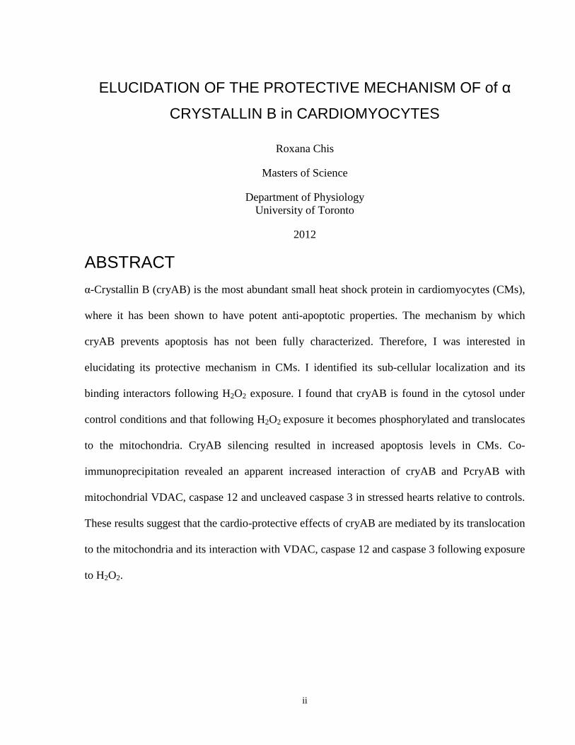
ELUCIDATION OF THE PROTECTIVE MECHANISM OF of α
CRYSTALLIN B in CARDIOMYOCYTES
Roxana Chis
Masters of Science
Department of Physiology
University of Toronto
2012
ABSTRACT
α-Crystallin B (cryAB) is the most abundant small heat shock protein in cardiomyocytes (CMs),
where it has been shown to have potent anti-apoptotic properties. The mechanism by which
cryAB prevents apoptosis has not been fully characterized. Therefore, I was interested in
elucidating its protective mechanism in CMs. I identified its sub-cellular localization and its
binding interactors following H2O2 exposure. I found that cryAB is found in the cytosol under
control conditions and that following H2O2 exposure it becomes phosphorylated and translocates
to the mitochondria. CryAB silencing resulted in increased apoptosis levels in CMs. Co-
immunoprecipitation revealed an apparent increased interaction of cryAB and PcryAB with
mitochondrial VDAC, caspase 12 and uncleaved caspase 3 in stressed hearts relative to controls.
These results suggest that the cardio-protective effects of cryAB are mediated by its translocation
to the mitochondria and its interaction with VDAC, caspase 12 and caspase 3 following exposure
to H2O2.

iii
ACKNOWLEDGEMENTS
I would like to thank my supervisor, Dr. Anthony Gramolini, for all his guidance and support
during the past two years. Dr. Gramolini, your intelligence has yet to cease to amaze me. You
have profoundly augmented my expectations of others and of myself. I am very grateful to you
for providing me with a different measuring stick for my future achievements.
I would also like to thank our research team. Dr. Parveen Sharma and Dr. Nicolas Bousette have
offered their constant support throughout my time in the lab. What has been a challenging
journey would have been unimaginably more difficult without you, Parv and Nic.
I cannot forget my lab-mates -Thiru, Melissa, Tim, Wen-Ping, Tetsuaki, Aaron and Jake - thank
you for your help and great advice.
I am grateful to my advisory committee: Dr. Backx and Dr. von Harsdorf and to Dr. Jurisiocova
for joining my committee at a moment’s notice and providing very important guidance and
discussion. Dr. Backx, I cannot thank you enough for your encouraging words during my
committee meetings and all your invaluable advice and suggestions to help me achieve my career
goals.
Finally, I would like to acknowledge my funding support from the Department of Physiology and
the Ontario Graduate Scholarship Program.
iv
TABLE OF CONTENTS
ABSTRACT .................................................................................................................................... ii
ACKNOWLEDGEMENTS ........................................................................................................... iii
TABLE OF CONTENTS ............................................................................................................... iv
LIST OF COMMON ABREVIATIONS ..................................................................................... viii
LIST OF FIGURES ........................................................................................................................ x
LIST OF TABLES ........................................................................................................................ xii
CHAPTER ONE: INTRODUCTION ............................................................................................. 1
1.1 Cardiovascular Disease and Cardiomyocyte Loss .............................................................. 1
1.1.1 Apoptosis ................................................................................................................ 1
1.1.2 Necrosis ................................................................................................................... 1
1.1.3 Autophagy ............................................................................................................... 2
1.2 Apoptosis in the heart ......................................................................................................... 3
1.2.1 Apoptosis in ischemia/reperfusion injury ............................................................... 4
1.2.2 Apoptosis in heart failure ........................................................................................ 5
1.3 Apoptosis pathways in cardiomyocytes .............................................................................. 5
1.3.1 Death receptor pathway of apoptosis ...................................................................... 6
1.3.2 Mitochondrial pathway of apoptosis ....................................................................... 6
1.3.3 Cross-talk between the death receptor and mitochondrial pathways of
apoptosis ................................................................................................................. 7
1.4 Modulators of apoptosis ...................................................................................................... 9
1.4.1 Bcl-2 family ............................................................................................................ 9
1.4.2 Caspase inhibitors ................................................................................................... 9
1.4.3 Antioxidants .......................................................................................................... 10
1.4.4 Heat shock proteins ............................................................................................... 10
1.5 α Crystallin B .................................................................................................................... 11
v
1.5.1 CryAB in the heart ................................................................................................ 14
1.5.2 CryAB in apoptosis ............................................................................................... 15
1.6 Rationale of current study ................................................................................................. 16
CHAPTER TWO: HYPOTHESIS AND AIMS ........................................................................... 17
CHAPTER THREE: MATERIALS AND METHODS ............................................................... 18
3.1 Lentivector Production and Transduction of Neonatal Cardiomyocytes ........................... 18
3.1.1 Chemical Transformation of DH5-α cells ............................................................ 18
3.1.2 Amplification and Maxi Preparations of cryAB cDNA ....................................... 19
3.2 Detection of Sub-cellular Distribution .............................................................................. 19
3.2.1 Sub-cellular Fractionation of Adult Mouse Hearts ............................................... 19
3.2.2 Sub-cellular Fractionation of Cultured Cardiomyocytes ...................................... 20
3.3.3 Sucrose Density Separation .................................................................................. 20
3.3 Immunoblot and Immunostaining Analysis ...................................................................... 20
3.3.1 Immunoblot Detection of Sub-cellular Distribution of CryAB in Adult Whole
Hearts .................................................................................................................... 20
3.3.2 Immunoblot Detection of Sub-cellular Distribution of CryAB in Stressed
Neonatal Cardiomyocytes ..................................................................................... 21
3.3.3 Immunofluorescence ............................................................................................. 22
3.4 Viability Assays ................................................................................................................ 23
3.4.1 General Viability Assay ........................................................................................ 23
3.4.2 Dissipation of Mitochondrial Membrane Potential Assay .................................... 23
3.4.3 Caspase 3 Activity Assay ...................................................................................... 24
3.4.4 TUNEL Assay ....................................................................................................... 24
3.4.5 ROS Detection and Inhibition ............................................................................... 24
3.5 Co-Immunoprecipitation ................................................................................................... 24
CHAPTER FOUR: α CRYSTALLIN B INTERACTS WITH VDAC, CASPASE 3 AND
CASPASE 12 TO PREVENT APOPTOSIS FOLLOWING H2O2 EXPOSURE IN
CARDIOMYOCYTES ............................................................................................................ 26
vi
4.1 Abstract .............................................................................................................................. 27
4.2 Introduction ........................................................................................................................ 28
4.3 Materials and Methods ....................................................................................................... 30
4.4 Results ................................................................................................................................ 34
4.5 Discussion ......................................................................................................................... 42
4.6 Disclosures ........................................................................................................................ 46
4.7 Acknowledgements ........................................................................................................... 46
4.8 Funding ............................................................................................................................. 46
4.9 Author contributions ......................................................................................................... 46
4.10 References ........................................................................................................................ 47
4.11 Figures .............................................................................................................................. 51
CHAPTER FIVE: DISCUSSION ................................................................................................. 57
5.1 CryAB localization and translocation in cardiomyocytes ................................................ 57
5.1.1 Ischemia/Reperfusion injury and hydrogen peroxide in vitro .............................. 57
5.1.2 CryAB translocates from the cytosol to the mitochondria following oxaidative
stress ...................................................................................................................... 58
5.2 CryAB silencing contributes to higher apoptosis levels in CMs ...................................... 60
5.3 Protective mechanism of cryAB in CMs .......................................................................... 61
CHAPTER SIX: LIMITATIONS ................................................................................................. 65
CHAPTER SEVEN: ONGOING WORK .................................................................................... 67
CHAPTER EIGHT: FUTURE DIRECTIONS ............................................................................. 68
CHAPTER NINE: REFERENCES ............................................................................................... 70
APPENDIX: ADDITIONAL DATA ............................................................................................ 80
1.1 Figures ................................................................................................................................ 80
1.2 Results and Discussion ..................................................................................................... 89
1.2.1 Over-expression of cryAB in mouse neonatal CMs ............................................. 89
vii
1.2.2 Protection against apoptosis .................................................................................. 89
1.2.3 Optimization of H2O2 concentration for the induction of apoptosis ..................... 90
1.2.4 Optimization of time-points for apoptosis measurements .................................... 90
1.2.5 Visual representation of cryAB localization and knock-down ............................. 91
1.2.6 Identification of cryAB protective mechanism and binding interactors in the
intrinsic apoptotic cascade .................................................................................... 92
viii
LIST OF COMMON ABREVIATIONS
Apaf-1 Apoptotic Protease-Activating Factor-1
ARC Apoptosis Repressor with Caspase Recruitment Domain
ASK-1 Apoptosis Signal-Regulating Kinase 1
Bad Bcl-2 Associated Death Promoter
Bak Bcl-2 Homologous Antagonist/Killer
Bcl-2 B-cell Lymphoma 2
Bid BH3 Interacting Domain
Ca2+ Calcium
CM Cardiomyocyte
CNA Calcineurin
Co-Ip Co-immunoprecipitation
cryAB Alpha Crystallin B
Daxx Death-Associated Protein 6
DNA Deoxyribonucleic Acid
ER Endoplasmic Reticulum
FADD Fas-Associated via Death Domain
Fas Apoptosis Stimulating Fragment
FBS Fetal Bovine Serum
GAPDH Glyceraldehyde-3-Phosphate Dehydrogenase
GFP-LC3 Green Fusion Protein Light Chain 3
H2O2 Hydrogen Peroxide
hsp Heat Shock Protein
I/R Ischemia/Reperfusion
IAP Inhibitor of Apoptosis
JNK c-Jun Terminal Kinase
KD Knockdown
ix
MAP kinase Mitogen-Activated Protein
MPTP Mitochondrial Permeability Transition Pore
Na/K ATPase Sodium Potassium Adenosine Triphosphatase
NF-kB Nuclear Factor Kappa B
PARP Poly ADP-Polymerase Ribose
PcryAB Phosphorylated (serine 59) Alpha Crystallin B
PDI Protein Disulfide Isomerase
PLN Phospholamban
RIP1 Receptor Interacting Protein
ROS Reactive Oxygen Species
SDS-page Sodium Dodecyl Sulfate Polyacrylamide Gel Electrophoresis
ShRNA Short Hairpin RNA
TNF Tumor Necrosis Factor
TNFR1 Tumor Necrosis Factor Receptor 1
TOM 20 Translocase of Outer Membrane 20 kDa
TRADD Tumor Necrosis Factor Receptor-1-Associated Death Domain
VDAC Voltage Dependent Anion Channel
WT Wild-Type
x
LIST OF FIGURES
Figure 1. Apoptotic Pathways in Cardiomyocytes. ........................................................................ 8
Figure 2. Organization of human cryAB genomic and protein sequence. .................................... 14
Figure 3. CryAB distribution in cardiac cells. .............................................................................. 51
Figure 4. Viability in cryAB-silenced CMs. ................................................................................. 52
Figure 5. Upregulation and translocation of cryAB and PcryAB to the mitochondria under
oxidative stress conditions. ........................................................................................................... 53
Figure 6. Apoptosis induction in CMs. ......................................................................................... 54
Figure 7. Effect of cryAB silencing on mitochondrial ultrastructure in mouse neonatal CMs. ... 55
Figure 8. CryAB protein interactions in the apoptotic cascade. ................................................... 56
Figure 9. Schematic of cryAB protective mechanism. ................................................................. 64
Figure 10. CryAB expression following over-expression and in CNA CMs. .............................. 80
Figure 11. Mitotracker retention in CNA cardiomyocytes. .......................................................... 81
Figure 12. Optimization of H2O2 concentration for induction of apoptosis in CMs. ................... 82
Figure 13. Time dependent dissipation of mitochondrial membrane potential and caspase 3
activity. .......................................................................................................................................... 83
Figure 14. Translocation of cryAB and PcryAB to the mitochondria under oxidative stress
conditions. ..................................................................................................................................... 84
Figure 15. CryAB expression following stress and knock-down. ................................................ 85
Figure 16. Co-immunoprecipitation of cryAB and cryAB. .......................................................... 86
Figure 17. Interaction of cryAB with cytochrome C. ................................................................... 87

xi
Figure 18. CryAB interactions in the apoptotic cascade that do not change following exposure to
stress. ............................................................................................................................................. 88

xii
LIST OF TABLES
Table 1: Different types of cell death and their associated characteristics ..................................... 3
1
CHAPTER ONE: INTRODUCTION
1.1 Cardiovascular Disease and Cardiomyocyte Loss
Cardiovascular disease is the leading cause of death in the world1 and although the number of
deaths from cardiovascular disease decreased by 21.9% during last two decades, it remains the
number one cause of death in North America1. Cardiovascular disease can be initiated by
multiple factors and recently, evidence has accumulated that a major contributor to its initiation
and progression is the loss of cardiomyocytes (CMs)2. The adult myocardium is comprised of
terminally differentiated CMs, which are characterized by a significant loss of replicative
potential or regeneration following injury3. Therefore, most cardiac insults lead to irreversible
damage, which can contribute to the functional decline of the myocardium due to increased
workload for the remaining myocardial cells, thus contributing to heart failure.
Cell death (including CM death) is attributed to at least three distinct processes – apoptosis,
necrosis and autophagy, which have distinct histological and biochemical characteristics4.
1.1.1 Apoptosis
Apoptosis is a programmed cell death, a suicide, that is achieved by the activation of a cascade
of events that culminate in cellular demise5. Apoptosis is a highly organized, energy-dependent
and neat process, which does not cause damage to the surrounding tissue6. Apoptotic cell death is
defined by particular characteristics – cell shrinkage, nuclear condensation and fragmentation,
cleavage of chromosomal DNA and membrane blebbing7. Apoptosis is also marked by the
absence of inflammation around the dying cell, since these are recognized and removed by
phagocytic cells5. Furthermore, it has been shown that the underlying biochemical events that
lead to the morphologic features of apoptosis result from the activation of a family of proteases,
caspases (cysteine proteases), by either death receptor ligation or the release of apoptotic
mediators from the mitochondria8.
1.1.2 Necrosis
In contrast to apoptosis, necrosis is a rapid, unregulated and irreversible process that occurs when
cells are severely damaged. Necrosis involves swelling of the cell and its organelles, disruption
of mitochondria, membrane breakdown cell lysis and an induction of inflammation around the
2
dying cell attributable to the release of cellular contents and pro-inflammatory molecules6.
Although the causative elements of necrosis are unclear, several mediators, organelles and
cellular processes have been implicated in its occurrence. These include mitochondrial
alterations, lysosomal changes, nuclear changes, lipid degradation9 and increases in the cytosolic
concentration of calcium (Ca2+
) that result in mitochondrial overload and activation of calpains
and cathepsins10
. In several (but not all) cases of necrotic cell death, the serine/threonine kinase,
Receptor Interacting Protein (RIP1) plays an important role11
.
1.1.3 Autophagy
Autophagy is a distinct form of non-apoptotic death that is separate from necrosis. It is
morphologically defined as a type of cell death that occurs in the absence of chromatin
condensation but with significant autophagic vacuolization of the cytoplasm and with no
association with phagocytes12,13
. Autophagy is characterized by massive sequestration of
portions of the cytoplasm within autophagosomes for bulk degradation by lysosomes, giving the
cell a characteristic vacuolated aspect14
. Autophagosomes are bound by two membranes and
contain degenerating cytoplasmic organelles or cytosol which allows them to be distinguished by
transmission electron microscopy from other types of vesicles such as endosomes, lysosomes or
apoptotic blebs4. The fusion between autophagosomes and lysosomes generates autolysosomes,
in which both the autophagosome inner membrane and its luminal content are degraded by acidic
lysosomal hydrolases. This catabolic process marks the completion of the autophagic pathway9.
One technique commonly employed to detect autophagy relies on the redistribution of GFP-LC3
fusion proteins into vesicular structures15
. However, doubts have been shed on the existence of
cell death by autophagy in the mammals9, as in most cases described to date in which autophagy
is suppressed by genetic knockout/knockdown of essential autophagy genes, cell death is not
inhibited but rather occurs at an accelerated pace system, pointing towards autophagy as a cell
survival mechanism5. Furthermore, there are only a few examples of cell death in model
organisms that can be largely prevented by genetic inhibition of autophagy16,17
.
3
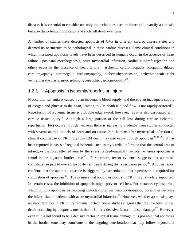
Table 1: Different types of cell death and their associated characteristics
The table below outlines the different types of cellular death and their characteristics.
1.2 Apoptosis in the heart
It is now widely accepted that apoptosis occurs in the heart, however, its contribution to cardiac
pathology remains controversial due to the difficulty of distinguishing between different types of
cell death in cardiac tissue. This poses problems in quantifying the relative contribution of each
to disease. Another obstacle to studying apoptosis of cardiomyocytes rests with determining the
‘true’ rate of apoptosis in disease. Some studies measured the value of apoptosis in the heart and
found it to be very variable and in some cases very high18
. However, different studies have
provided seemingly low values (<1%)19
, attesting to the difficulty of measuring apoptosis rates
accurately. The caveat is that such measurements represent only the number of cells undergoing
apoptosis at a single point in time. It is now widely postulated that given the relatively short time
course of apoptosis, the gradual loss of such numbers of myocardial cells could have serious
consequences over an extended period. Thus, when investigating the role of apoptosis in heart
Type of cell death
Apoptosis Necrosis Autophagy
Programmed cell death
Reduction of cellular and nuclear
volume (pyknosis)
Nuclear fragmentation
(karyorrhexis)
DNA cleavage into 200 bp
fragments
Plasma membrane blebbing
Engulfment by resident phagocytes
Uncontrolled cell death
Cytoplasm swelling
(oncosis)
Rupture of plasma
membrane
Swelling of organelles
Chromatin condensation
Cell survival or cell death
mechanism
Lack of chromatin
condensation
Significant vacuolization
of the cytoplasm
Presence of autophagic
vacuoles
No engulfment by
phagocytes
4
disease, it is essential to consider not only the techniques used to detect and quantify apoptosis,
but also the potential implications of such cell death over time.
A number of studies have detected apoptosis of CMs in different cardiac disease states and
deemed its occurrence to be pathological in these cardiac diseases. Some clinical conditions in
which increased apoptosis levels have been described in humans occur in the absence of heart
failure - postnatal morphogenesis, acute myocardial infarction, cardiac allograft rejection and
others occur in the presence of heart failure – ischemic cardiomyopathy, idiopathic dilated
cardiomyopathy, acromegalic cardiomyopathy, diabetes/hypertension, arrhythmogenic right
ventricular dysplasia, myocarditis, hypertrophic cardiomyopathy20
.
1.2.1 Apoptosis in ischemia/reperfusion injury
Myocardial ischemia is caused by an inadequate blood supply, and thereby an inadequate supply
of oxygen and glucose to the heart, leading to CM death if blood flow is not rapidly restored21
.
Reperfusion of ischemic tissue is a double edge sword, however, as it is also associated with
cardiac tissue injury22
. Although a large portion of the cell loss during cardiac ischemic-
reperfusion (I/R) occurs through necrosis, there is increasing evidence from studies conducted
with several animal models of heart and on tissue from humans after myocardial infarction (a
clinical counterpart of I/R injury) that CM death may also occur through apoptosis23,24 ,25
. It has
been reported in cases of regional ischemia such as myocardial infarction that the central area of
infarct, or the most affected area by the stress, is predominantly necrotic, whereas apoptosis is
found in the adjacent border areas26
. Furthermore, recent evidence suggests that apoptosis
contributes in part to overall myocyte cell death during the reperfusion period25
. Another report
confirms that the apoptotic cascade is triggered by ischemia and that reperfusion is required for
completion of apoptosis27
. The premise that apoptosis occurs in I/R injury is widely supported.
In certain cases, the inhibition of apoptosis might prevent cell loss. For instance, cyclosporine,
which inhibits apoptosis by blocking mitochondrial permeability-transition pores, can decrease
the infarct size in patients with acute myocardial infarction28
. However, whether apoptosis plays
an important role in I/R injury remains unclear. Some studies suggests that the low level of cell
death occurring by apoptosis means that it is not a decisive factor in tissue damage27
. However,
even if it is not found to be a decisive factor in initial tissue damage, it is possible that apoptosis
in the border zone may contribute to the ongoing deterioration that may follow myocardial
5
infarction. This is supported by the fact that apoptosis is found in the heart days after the initial
reperfusion therapy26
.
1.2.2 Apoptosis in heart failure
Several cardiac disorders, such as chronic overload such as that caused by hypertension or a
defective heart valve, viral myocarditis, alcoholism, and ischemic heart disease culminate in
heart failure6. In response to increased workload, the myocardium undergoes remodelling, which
generally involves changes in gene expression and hypertrophy of CMs whereby the cells
increase in size29
. Although this structural change is an adaptive response to stress, it can
eventually lead to heart failure. The progression to heart failure seems to be accompanied by
apoptosis30
. Numerous studies support the possible role for apoptosis in heart failure.
Examination of human heart tissue has shown apoptosis in association with conditions such as
idiopathic dilated cardiomyopathy, ischemic cardiomyopathy, arrhythmogenic right ventricular
dysplasia, and hypertrophic cardiomyopathy20
. And although the degree of apoptosis found in
these conditions is low, the gradual loss of CMs over time is thought to contribute to the eventual
progression to heart failure. It has been proposed that apoptosis may have a role in the transition
from mild to end-stage heart failure. In a recent study that measured the levels of proapoptotic
Bax in relation to the severity of heart failure it was found that levels of Bax were significantly
higher in association with mild heart failure compared with moderate or severe heart failure that
CMs were more prone to apoptosis in the earlier stages of the disease thus facilitating the
transition to heart failure31
.
There is little doubt that apoptosis is found in association with myocardial disease given the
increasing evidence that apoptosis is involved in heart failure. And although it may occur
alongside other mechanisms of death modulation of apoptosis is of significant value.
1.3 Apoptosis pathways in cardiomyocytes
A number of different experimental models using different stresses, including work overload,
hypoxia, neuro-humoral overstimulation, free radical stress, viral infection and toxic insults have
have shown that CM can be induced to undergo apoptosis32
. Apoptotic death occurs by two
pathways - the activation of cell surface death receptors by extracellular ligands33
,34
and the

6
activation of mitochondrial-related pro-apoptotic mechanisms in response to unfavorable
changes in the intracellular environment35,36
(Fig.1).
1.3.1 Death receptor pathway of apoptosis
In the extrinsic or death receptor pathway of apoptosis, following ligand binding, death receptors
interact with death domain adaptor proteins, such as Fas-Associated Death Domain (FADD),
Tumor Necrosis Factor Receptor-1-Associated Death Domain (TRADD), Receptor-Interacting
Protein (RIP), and Death-Associated Protein 6 (Daxx), which activate a cascade of cell signaling
pathways (e.g., MAP kinase, NF-κB and Akt) that regulate gene expression and the
phosphorylation status of proteins (e.g., Bcl-2 proteins and IAP proteins)37
. These, in turn,
modulate the activity of different families of proteases (e.g., caspases, cathepsins and calpains)
involved in the execution of the final steps of the apoptotic process33,38,39
.
The death receptor pathway has been found to be activated in the heart. For instance, Fas and Fas
ligand expression has been found in the heart40
. Enhanced expression of Fas is found in
association with increased apoptosis in experimental models of myocardial infarction41
hypoxia42
, and overstretched myocardium43
. Signaling through the TNF receptor has also been
reported in the heart. Like Fas, binding of the TNFR1 by its ligand (TNF-α) results in the
recruitment of adaptor proteins that transduce signals downstream, leading to various
physiological events including inflammation, cell growth, differentiation, and apoptosis,
depending on the different adaptor proteins and downstream signaling events associated with
receptor activation33
. Furthermore, it has been demonstrated that TNF plays a role in the
progression of myocardial disease, with increased TNF-α and TNFR1 expression are found in
association with heart failure44
. Agents that suppress TNF-α have been shown to have positive
therapeutic effects, thus attesting to the negative role of elevated TNF-α45
. As TNF-α can induce
apoptosis of CMs46
, it is thought that at least part of its pathogenic effect in the heart is due to the
induction of cell death.
1.3.2 Mitochondrial pathway of apoptosis
A second pathway of apoptosis that occurs in CMs is the intrinsic, or mitochondrial pathway. In
addition to their established role in energy production, mitochondria are actively involved in the
regulation of apoptosis49
. In essence, stresses like ischemia have been shown to lead to
7
mitochondrial death signaling, where the opening of the mitochondrial permeability transition
pore (MPTP) leads to a change in the permeability of the inner mitochondrial membrane. MPTP
is a mitochondrial inner membrane channel whose opening allows molecules smaller than 1500
Daltons to enter the mitochondrial matrix47
. The entry of small molecules leads to increased
mitochondrial permeability and ultimately to swelling of the mitochondria and rupture of the
outer mitochondrial membrane, thus leading to the release into the cytosol of a number of highly
lethal mitochondrial substances that can initiate apoptosis. One of these is the small electron
transporter cytochrome C that forms an apoptosis-promoting complex, the apoptosome, with
procaspase-9 and its cofactor apoptotic protease-activating factor-1 (Apaf-1), thus activating
caspase-9 and, subsequently, caspase-3 and other caspases48
. Mitochondria are a primary site of
action of the apoptosis regulatory proteins of the Bcl-2 family and a major source of reactive
oxygen species (ROS), which have been implicated in cellular damage and death49
.
Recent studies suggest that cytochrome C-mediated apoptosis is important in CMs. Serum and
glucose deprivation induce cytochrome C release in vitro, resulting in activation of caspases-9
and -3 and apoptosis as determined by nuclear fragmentation, DNA cleavage, and processing of
caspase substrates50
. As serum and glucose deprivation are components of ischemia in vivo,
these results suggest that this pathway may be involved in cell death in relation to heart disease.
ROS have also been implicated in I/R induced damage, and it has been reported that
mitochondrial cytochrome C release, activation of caspase-3, and PARP cleavage are involved in
H2O2-induced cardiomyocyte apoptosis51
. These in vitro studies demonstrate that CM apoptosis
can occur via a cytochrome C-mediated pathway.
1.3.3 Cross-talk between the death receptor and mitochondrial pathways of apoptosis
In certain cases, the two pathways do not occur in isolation, but rather interactions between the
two pathways may take place52
. For example, the pro-apoptotic protein BH3 interacting-domain
(Bid) is cleaved by caspase-8 in response to Fas and TNF receptor activation53
. Following
cleavage, the C-terminal fragment of Bid translocates and binds to the mitochondria, leading to
the release of cytochrome C and activation of downstream caspases. Another instance of cross-
talk is that of the apoptosis signal-regulating kinase 1 (ASK1), which is a member of the MAP
kinase family. This activates JNK and p38 kinases leading to apoptosis mainly by mitochondria-
dependent caspase activation, due to the phosphorylation and inactivation of the anti-apoptotic
8
protein Bcl-2 that prevents the release of cytochrome C54
. Thus, Bcl-2 family proteins are
mediators linking the death receptor signals to the mitochondria-dependent death signals55
.
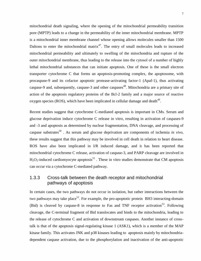
Figure 1. Apoptotic Pathways in Cardiomyocytes.
The extrinsic pathway occurs by ligand binding to the death receptor. Death receptors then interact with death
domain adaptor proteins, which activate a cascade of cell signaling pathways (e.g., MAP kinase, NF-κB and Akt)
that regulate gene expression and the phosphorylation status of proteins (e.g., Bcl-2 proteins and IAP proteins).
These, in turn, modulate the activity of caspases, cathepsins and calpains. The intrinsic pathway is stimulated by
mitochondrial dysfunction. Here, apoptosis-inducing signals, such as I/R, hypoxia or oxidative stress cause the
opening of the mitochondrial permeability transition pore (MPTP). Opening of the pore leads to dissipation of
mitochondrial membrane potential and matrix swelling, which ruptures the outer mitochondrial membrane, leading
to the release of proteins from the intermembrane space, including cytochrome C. This causes the activation of
caspase-9, which in turn activates caspase-3. ER stress activates caspase 12, which also activates caspase 3, leading
to apoptosis. Various intermediary signaling and proteins inhibiting the apoptotic cascade are also shown. Apaf-1,
apoptosis-activating factor 1; Bak, bacille Calmette–Guérin; Bax, BCL-2-associated X protein; Bid, proapoptotic
Bcl-2 family member; tBid, truncated beta interaction domain.
Death Receptor Pathway
Fas L
Fas
FADD
TNF
TNFR
FADD
TRADD
procaspase 8
caspase 8 Bid
Mitochondrial Pathway
cytochrome C
Apaf-1 procaspase 9
caspase 9
caspase 3
caspase12
Apoptosis
Apoptotic Stimuli
Bak
tBid
Bax
Bcl-2
Bcl-xL
9
1.4 Modulators of apoptosis
Apoptosis is a highly orchestrated multistep process with many different points at which it can
be regulated. Regulation of apoptosis is particularly important in fully differentiated and
replication-impaired CMs in order to avoid unnecessary death of salvageable cells. Regulation of
apoptosis is also important in promoting its occurrence in response to irreversible cellular
damage, as opposed to necrosis, which could further harm the myocardium. Hence, mechanisms
allowing the modulation of death signals in CMs are important as they may help to keep the cell
alive.
1.4.1 Bcl-2 family
CMs are known to express the Bcl-2 family of proteins and it has been suggested that these play
a role in modulating apoptosis. A dramatic increase in Bax expression and decreased expression
of Bcl-2 have been reported in the left ventricle in response to chronic pressure overload in the
rat56
, and it is thought that such changes are likely to influence cells to undergo apoptosis. In a
model of ROS-induced apoptosis, H2O2 led to increased expression of pro-apoptotic Bad and
elicited the translocation of Bax and Bad to the mitochondria, resulting in cytochrome C release,
activation of caspase-3, and cleavage of PARP51
. An up-regulation of the Bcl-2 protein family
has also been reported in human end-stage heart failure where increases in pro-apoptotic Bak and
Bax and anti-apoptotic Bcl-2 were observed in association with apoptosis57
. Expression of Bax
was significantly higher than that of the anti-apoptotic proteins, which suggests that in the failing
human heart apoptosis is favoured.
Manipulation of the Bcl-2 system may also provide new treatments to prevent CM apoptosis.
The potential strategies could involve up-regulating the anti-apoptotic Bcl-2 pathway or
inhibiting pro-apoptotic pathways. Indeed, it has been shown that ischemic preconditioning with
reduced apoptosis is associated with up-regulation of Bcl-2 in rats58
and that over-expression of
Bcl-2 in ventricular myocytes prevents apoptosis59,60
.
1.4.2 Caspase inhibitors
The final steps of apoptotic death are mediated by active caspases33,38,39
. ARC (apoptosis
repressor with caspase recruitment domain), an inhibitor of apoptosis that is expressed almost
exclusively in skeletal and cardiac muscle has been shown to interact with caspases-8 and -2 and
10
to attenuate apoptosis induced by stimulation of death receptors61
. The use of synthetic inhibitors
of apoptosis presents another potential therapeutic avenue. broad range caspase inhibitors were
effective in reducing myocardial reperfusion injury in rats, which was attributed in part to the
attenuation of cardiomyocyte apoptosis62
. The same inhibitor is reported to attenuate apoptosis
in rabbit cardiomyocytes63
. However, caspase inhibition has been stipulated to increase ROS
production resulting in secondary toxicity 64
. Specific inhibitors of caspases also seem to have
regulatory effects on apoptosis. One study found that CM DNA fragmentation and caspase
activation were prevented by inhibitors of caspase-1 and -3 without reduction of the infarct size
in I/R rat hearts65
. This contrasts with another study that found that inhibitors of caspase-8, -9,
and -3 all limited infarct size as a result of reperfusion injury66
. The different outcomes of these
studies may reflect the different administration times of the inhibitors, before ischemia and
during early reperfusion, respectively.
1.4.3 Antioxidants
It has been proposed that apoptosis is modulated by oxidative stress and antioxidants play a part
in its prevention67
. Cellular antioxidants act by removing free radicals from the cell and thereby
minimize oxidative stress resulting from a variety of insults, including I/R injury68
. Several
endogenous antioxidants are found in CMs including glutathione (GSH), glutathione peroxidase
(GSHPx), superoxide dismutase (SOD) catalase, and vitamins E and C69
. In relation to apoptosis,
a drop in intracellular levels of GSH is associated with increased oxidative stress. This leads to
damage of cellular macromolecules within the cell and subsequent cell death67
. In the heart,
GSHPx has a role in regulating I/R-induced apoptosis. Experiments with GSHPx knockout mice
demonstrated increased levels of apoptosis in response to ischemia/reperfusion whereas mice
overexpressing GSHPx were more resistant to such damage70
. Over-expression of manganese-
SOD (MnSOD) protects against I/R injury in transgenic mice, in part by modulating apoptosis71
.
1.4.4 Heat shock proteins
It is well established that heat shock proteins (hsps) are synthesized in response to a variety of
stressful stimuli and that their expression coincides with increased resistance to subsequent
cellular damage is due at least in part to interference with apoptosis72
. The hsps are recognized as
important regulators of CM apoptosis. Different families of hsps are expressed in the heart,
including the hsp90, 70, 60, 27, and 10 families; induction of these proteins by a variety of
11
agents including cardiotrophin-173
, heat shock, and ethanol 73
protects CMs against stress such as
ischemia. For instance, over-expression of hsps70 and 27 can protect cells against ceramide,
serum withdrawal, and lethal hypoxia74
, all of which are known to be inducers of apoptosis.
Thus, hsps are likely to be key regulators of apoptosis in the heart during stressful conditions
such as ischemia.
Although hsps play a key role in the regulation of apoptosis, the mechanisms by which they do
so are not fully understood. It is known that hsp70 inhibits apoptosis downstream of cytochrome
C release and upstream of caspase-3 activation75
. Hsp27 can prevent cytochrome C release at the
level of the mitochondria or can interact with cytochrome C or procaspase-3, preventing
apoptosome formation and caspase-3 activation, respectively76,77
. Given the cardioprotective
effect of heat shock protein induction in in vitro and animal studies, these proteins represent a
potentially valuable treatment for cardiac disease.
1.5 α Crystallin B
Alpha-crystallin is a major protein component of the lens in the vertebrate eye, and a key
member of the small heat-shock proteins with the ‘alpha-crystallin domain’ being a common C-
terminal consensus sequence to all the members of the small heat-shock protein super family 78
.
Alpha crystallin was initially discovered in the mammalian lens where ~ 50% of the total dry
mass of the lens is alpha-crystallin protein79
. In the lens, crystallin is involved in maintaining the
necessary refractive index of the lens80
, and maintaining lens transparency 79
, through ensuring
proper protein folding and minimizing protein aggregation. For instance, during aging, and
especially in the center part of the lens, there is increased protein unfolding and denaturing. This
can interfere with vision, through the formation of a cataract, for instance. Alpha-crystallin binds
such unfolded or denatured proteins and suppresses non-specific aggregation80
, thus maintaining
lens transparency.
There are two alpha-crystallin genes, alpha A, and alpha B. In humans, the alps A gene is found
on chromosome 21 and encodes for a 173 amino acid residue protein, while the alpha B gene is
found on chromosome 11, is made up of 3124 base pairs organized within 3 exons and 2 introns
(Fig. 2A) and encodes for a 175 amino-acid residue protein. The amino-acid sequence homology
between alpha A and alpha B is about 57%80
, and the expression pattern of alpha A and alpha B
12
is also quite different. Alpha A crystallin is found mainly in the lens with trace amounts in other
tissues80
, whereas alpha B is constitutively expressed in the lens of the eye , brain, kidney,
skeletal and cardiac muscle81,82
. In mammals, alpha crystallin B associates with alpha crystallin
A to form large hetero-oligomeric structures82
.
Alpha crystallin B (cryAB) or heat shock protein beta 5 (hspB5) is a 20 kDa small heat shock
protein 83
and a molecular chaperone 79
. As a small hsp, cryAB is involved in modulating the
ubiquitin–proteasome pathway and is essential for proper disassembly–assembly of protein
complexes to prevent undesirable interactions and aggregation84
. Like other small hsps cryAB
can form globular oligomeric structures that are characterized, in mammalian cells, by molecular
masses ranging from 50 to approximately 700–800 kDa80
and it is this dynamic organization of
small hsps oligomers that appears to be important in the control of their activity.
Another property of some small hsps, including cryAB, concerns their ability to modulate their
activities in response to a wide variety of stimuli 85,86
, a process known to be dependent on
phosphorylation. CryAB can be phosphorylated at three serine site corresponding to residues 19,
45 and 59 in response to cellular stresses82
(Fig.2B). These phosphorylated serine sites are found
in the N-terminal part of the polypeptides, in the WDPF domain and close to the α-crystallin
domain86
. Although the signaling pathways leading to serine 19 phosphorylation are unknown,
the extracellular signal-regulated protein kinase (ERK) MAPK pathway appears to be
responsible for phosphorylation of serine 45, and the p38 MAPK pathway is responsible for
phosphorylation of serine87
. Other post-translational modifications of cryAB include truncation
of both the N terminus and the C terminus, deamidation, racemization, methionine oxidation,
glycation, disulfide formation, addition of O-GlcNAc, and the addition of 72 mass units to the C-
terminal lysine of B-crystallin88
.
CryAB mutations, as well as modulation of cryAB levels have been associated with human
diseases. For instance, a missense mutation in cryAB changing arginine 120 to glycine (R120G)
leads to misfolded protein aggregation and is associated with cardiomyopathy and cataract
formation89
. Recently, two novel mutations leading to myofibrillar myopathies (Q151X and
464delCT) have been identified in the terminal part of the cryAB coding sequence90
(Fig.2B).
Three mutations in the cryAB gene (P20S, 464delCT and D140N) are also responsive for
dominant cataract 91
and two mutations (R157H and G154S) for cardiomyopathy91
. Another
13
mutation in the alpha B-crystallin gene (450delA) which results in the expression of an aberrant
cryAB protein containing 184 residues in which the 35 residues at the C-terminus are completely
different from the native of protein has been shown to cause a dominant congenital posterior
polar cataract in humans 88
(Fig.2B).
CryAB expression is often increased in many protein conformation diseases. This upregulation
can be observed in Alexander’s disease, Rosenthal fibers, cortical Lewy bodies, Alzheimer
disease plaques, neurofibrillary tangles as well as in synuclein deposit associated to Parkinson
disease or myopathy-associated inclusion body92
. The reason for the association of cryAB with
these structures is potentially linked to its chaperone activity, potentially providing a first line of
defense against misfolded, aggregation-prone proteins. Constitutive expression of cryAB has
been detected in gliomas, prostate cancer, oral squamous cell carcinomas, renal cell carcinomas,
head and neck cancer82
, and increased expression of αB-crystallin was related to more advanced
tumors, potentially attesting to the involvement of cryAB in regulation of cell survival and death.
A high level of αB-crystallin has also been detected in basal-like breast carcinomas and
preinvasive ductal carcinoma that correlated with poor clinical outcome of the patients82
.
Recently, a pathological role of cryAB has again been reported in breast cancer diseases, hence
suggesting that it acts as an oncoprotein93
.

14
A
B
Figure 2. Organization of human cryAB genomic and protein sequence.
(A) Genomic organization of cryAB. Exons are depicted as light blue boxes, introns as grey connecting lines and the
5’ and 3’ untranslated regions (UTRs) are depicted as light grey boxes. The number of base pairs (Bps) are depicted
on the axis. (B) Protein sequence arrangement of cryAB. Red box: α crystallin domain; blue box: WDPF domain; P:
phosphorylated serine residues. Amino acids are indicated. Positions of point mutations that are responsible of
pathologies are indicated by arrows. 464delCT: frame-shift mutant. The resulting mutant is modified from amino
acid 155 and is truncated of 13 residues compared to wild type protein. 450delA: frame-shift mutant. The resulting
mutant is modified from amino acid 160 to amino acid 184. This protein is larger than wild type polypeptide (175
amino acids).
1.5.1 CryAB in the heart
Despite initially being discovered in the lens of the eye, cryAB has been shown to be the most
abundantly expressed small hsp in cardiac muscle, making up 3% of the protein of heart
homogenates81
. Mutations in cryAB have been reported to lead to cardiomyopathies. A missense
mutation in αB-crystallin gene, changing arginine 120 to glycine (R120G), leads to a
myofibrillar myopathy associated with cardiomyopathy and cataract formation89
. The cryAB
R120G mutant decreases the chaperone activities of cryAB in vitro and increases protein
aggregation with the intermediate filament, desmin, causing ‘desmin-related cardiomyopathy’.
Recently, two novel mutations leading to myofibrillar myopathies have been identified in the
C
175
14967NP20S
p p p
Ser19 Ser59Ser45
α crystallin domain
R120G D140N R157H
Q151X 464delCT
450delA
G154S
1 2 3
5’ UTR 3’ UTR
-111,782,473 -111,779,350
Bps from the promoter
15
terminal part of the αB-crystallin coding sequence90
. When it comes to the deletion of cryAB in
cryAB KO, this mice does not lead to decreased viability94
, indicating that cryAB is dispensable
for survival and suggesting that other small hsps might serve redundant roles in vivo. On the
other hand, these KO mice have reported increased sensitivity to I/R injury95
, suggesting that
presence of cryAB is protective against stress. Furthermore, it has been shown that over-
expression of cryAB in cultured rat cardiac myocytes87
or in hearts of transgenic mice 96
protects
from ischemia/reperfusion (I/R) damage.
1.5.2 CryAB in apoptosis
As described above, cryAB has been shown have protective properties. It has been demonstrated
on numerous occasions that cryAB has potent anti-apoptotic properties, protecting cells from
from thermal, osmotic and oxidative insult97
. It has also been shown that cryAB can prevent
induced apoptosis by various factors including staurosporine, TNF, okadaic acid and hydrogen
peroxide97
. Moreover, cryAB negatively regulates apoptosis during myogenic differentiation98
.
The protective mechanism of cryAB in response to stress has been described in part and it
involves the interaction of cryAB with myofilament proteins helping to preserve contractile
protein integrity and myocardial function99
.Furthermore, cryAB has been shown to translocate to
the mitochondria, thus reducing I/R damage in mouse hearts during ex vivo I/R100
. Another
component of the protective mechanism of cryAB seems to involve its phosphorylation on serine
59 101
in a p38-dependent manner 102
in response to I/R and other stresses. Moreover, a
molecular mimic of serine 59-phosphorylated cryAB was shown to enhance the protective
ability of cryAB against several different stresses that imitate I/R; in contrast, a cryAB mutant
which blocks its phosphorylation, increased I/R-mediated myocardial cell death87
.
Several steps in the apoptotic pathway are modulated by cryAB. CryAB has been shown to
directly interact with the precursors of caspase 3 to suppress its activation in an immortalized
rabbit lens epithelial cell line 103
and in breast carcinoma cells104
. In human lens epithelial cells
cryAB has also been shown to interact with the pro-apoptotic proteins Bax and Bcl-Xs,
preventing their translocation from cytosol into mitochondria, thus leading to decreased
apoptosis97
. CryAB also binds pro-apoptotic protein p53, preventing its translocation to the
mitochondria105
. And although the mechanism of protection against apoptosis of cryAB has been
described in part, it remains to be fully elucidated in CMs.
16
1.6 Rationale of current study
Recently, our group confirmed the anti-apoptotic properties of cryAB in a mouse model of
hypertrophy, calcineurin (CNA) hypertrophy106
. In this study, proteomic analysis was carried out
by subjecting fractionated cardiac samples from CNA mice and their WT littermates to gel-free
liquid chromatography linked to shotgun tandem mass spectrometry. We found that 290 were
differentially expressed. Of the proteins whose expression was upregulated, several were
associated with programmed cell death, including regulation of apoptosis106
. This prompted us
to look at the levels of apoptosis, and we found that in the CNA model the rates of apoptosis
were minimal.
We compared the CNA proteome with the proteome from the cardiac tissue of a mouse model
previously described by our group, the PLNR9C
model107
. A main difference between the CNA
and R9C models is that in the PLNR9C
the rates of apoptosis in cardiac tissue are significant106
.
The proteome comparison identified four candidates with differential expression between CNA
and PLNR9C
mice, of which cryAB was significantly upregulated in CNA mice but not in the
PLNR9C
mouse model. Furthermore, the viability of cultured neonatal mouse cardiomyocytes
from CNA mice was higher than WTs after serum starvation, an apoptotic trigger. However
following silencing of cryAB via lentivector-mediated transduction of shRNAs there was a
significant reduction in neonatal cardiomyocyte viability and loss of protection against apoptosis.
Given these findings and the literature available on the anti-apoptotic properties of cryAB, it was
selected for further study. Specifically, I determined the mechanism by which cryAB prevents
apoptosis in CMs, with a specific focus on the intrinsic pathway of apoptosis, since very few
studies have focused on the anti-apoptotic mechanism of cryAB in CMs.
17
CHAPTER TWO: HYPOTHESIS AND AIMS
The following aims were addressed throughout the experiments:
1. To identify the sub-cellular localization of cryAB.
2. To validate the protective effect of cryAB in CMs by knockdown by assessing the effect
of cryAB levels on early and late apoptosis.
3. To identify cryAB binding interactors in the intrinsic apoptosis cascade under oxidative
stress.
I hypothesized that:
1. CryAB regulation will protect CMs from oxidative stress.
2. CryAB modulation in CMs will attenuate apoptosis by binding and inhibiting caspases
and/or mitochondrial proteins thus leading to decreased mitochondrial permeability.
18
CHAPTER THREE: MATERIALS AND METHODS
In this section, I discuss or expand upon pertinent methods and materials that were not included
in the manuscript presented in Chapter 4.
3.1 Lentivector Production and Transduction of Neonatal Cardiomyocytes
Lentivector compatible shRNA clones targeting mouse α-crystallin B and α-crystallin B cDNA
were obtained from Open-Biosystems. The scrambled shRNA construct, used as a negative
control was a kind gift from Dr. Stephane Angers (University of Toronto, Toronto, ON, Canada).
Lentivector production and transduction of neonatal CMs were performed as described
previously106
. Briefly, clones were amplified using the ampicilin resistance marker in DH5-α
cells. Plasmids were then isolated using Qiagen maxi preps according to the manufacturer’s
instructions. The packaging plasmid (pCMV-R8.74psPAX2, 2.5 μg), envelope plasmid (VSV-
G/pMD2.G, 0.3 μg), and the target construct plasmid ((pLKO.1, 2.7 μg) expressing either the
shRNA or scrambled shRNA (as a negative control)) or the target construct with the cryAB
cDNA( pLJM1-cryAB) were simultaneously transfected into HEK-293T cells with Optimem
(Invitrogen) diluted FuGene (Roche). Neonatal cardiomyocytes were incubated with supernatant
from transfected HEK-293T cells for 21 h after which the medium was replaced daily. Because
the lentivector constructs have a puromycin resistance gene, we selected for transduced
cardiomyocytes by incubating with 2 μg/mL puromycin for 48 h to kill off all nontransduced
cells to ensure a homogenous population of transduced cells.
3.1.1 Chemical Transformation of DH5-α cells
The cDNA vectors were then introduced into Escherichia coli strain DH5-α cells by chemical
transformation. Briefly, 3 μL of plasmid DNA was added to 50 μL of DH5-α cells and incubated on
ice for 30 minutes and then heat shocked for 45 seconds at 42°C. The cells were then shaken for 1
hour at 37°C with 950 μL of SOC media (Sigma). Finally, the transformed cells were pelleted by
centrifugation (10000 rpm, 1 minute), re-suspended in 150 μL of SOC media, and spread onto LB
19
agar plates with ampicillin resistance (50 μg/mL) under sterile conditions. The plates were incubated
overnight at 37°C.
3.1.2 Amplification and Maxi Preparations of cryAB cDNA
To amplify the cryAB cDNA, 250 mL of sterile 2x YT liquid culture (BioShop) with ampicillin (50
μg/mL) was inoculated with a sample of DH5-α cells picked from an individual colony from the LB
agar plates. The cultures were shaken overnight at 37°C. To isolate the plasmid DNA, the maxi
preparation method was performed using Qiagen’s Plasmid Maxi Kit. Approximately after 16-18
hours of shaking, the bacterial culture was centrifuged at 4100 rpm for 15 minutes at 4°C and the
supernatant discarded. The pellet was completely resuspended in 10 mL of cold P1 Resuspension
Buffer containing RNAse, with vortexing. 10 mL of P2 Lysis Buffer was then added and
thoroughly mixed by gently inverting the tube several times. Immediately after, the reaction was
terminated with the addition of 10 mL of P3 Neutralization Buffer and mixing by inverting. The
samples were incubated on ice for 20 minutes and then centrifuged at 4100 rpm for 20 minutes at
4°C. The resulting supernatant was applied to a QIAGEN-tip 500 column, which was already
equilibrated by allowing 20 mL of Buffer QBT to drain through by gravity flow. Three
successive washes were performed with 30 mL of Buffer QC to remove all contaminants. 15 mL
of Buffer QF was then added to elute the bound DNA and collected in a centrifuge tube. To
precipitate the DNA, 10.5 mL of isopropanol was added and mixed by inverting and centrifuged
immediately at 12000 rpm for 30 minutes at 4°C. The supernatant was discarded slowly and the
DNA pellet was washed by adding 20 mL of 70% ethanol and centrifuging at 12000 rpm for 15
minutes at 4°C. The supernatant was again removed slowly and the pellet was allowed to air-dry
to remove all traces of ethanol. Finally, the pellet was dissolved in 1 mL of TE Buffer and the
resulting DNA concentration was determined using an Ultrospec™ 2100 pro UV/Visible
Spectrophotometer (GE Healthcare).
3.2 Detection of Sub-cellular Distribution
3.2.1 Sub-cellular Fractionation of Adult Mouse Hearts
Adult mice were euthanized by carbon dioxide asphyxiation. Hearts were harvested and
ventricular tissue was isolated. The tissue was rinsed with ice-cold PBS to remove any remaining
blood. The tissue was placed in an ice-cold lysis buffer (250 mM Sucrose, 50 mM Tris-HCl (pH
7.4), 5 mM MgCl2, 1 mM DTT, 1 mM PMSF). The tissue was dounce-homogenized and
20
differential centrifugation was carried out to isolate cytosolic, microsomal, and mitochondrial
fractions, as described previously108
. Briefly, the lysate was cleared of debris by tabletop
centrifugation at 800 x g for 15 min. Mitochondrial and microsomal fractions were isolated from
the supernatant by further centrifugation at 8000 x g and 100,000 x g, respectively, and the
supernatant served as the soluble cytosolic fraction. Total protein concentration calculated by
using Bradford Reagent (Sigma).
3.2.2 Sub-cellular Fractionation of Cultured Cardiomyocytes
Cardiomyocytes from neonatal mouse were cultured as described above. These were maintained
in culture for five days and on the sixth day they were either maintained in culture or stressed
with 60 µM H2O2 for 24 hours. The CMs were then rinsed with PBS and collected in lysis
buffer, as above. The CMs in lysis buffer were dounce-homogenized and differential
centrifugation was carried out to isolate cytosolic and organellar (including the mitochondria)
fractions, as described above.
3.3.3 Sucrose Density Separation
The cleared homogenized sample described above was also used for sucrose gradient separation,
as described previously109
. Briefly, the supernatant was collected and layered on top of a 20–60%
linear sucrose gradient made up in 10 mM Tris-HCl (pH 7.6), 10 mM EDTA and protease
inhibitors (Roche). Samples were centrifuged at 100,000 x g for 20 hrs in a SW40Ti swinging
bucket rotor. Fractions (750 µl) were collected from the bottom of each gradient and total protein
concentration calculated by using Bradford Reagent (Sigma).
3.3 Immunoblot and Immunostaining Analysis
3.3.1 Immunoblot Detection of Sub-cellular Distribution of CryAB in Adult Whole Hearts
The protein content from the microsomal, cytosolic, nuclear and mitochondrial fractions was
harvested from WT adult mice and total protein concentration calculated by using Bradford
Reagent. Also, the protein content of the fractions collected following sucrose density separation
was measured. These were then subjected to standard Western blotting techniques, via SDS-
Polyacrylamide Gel Electrophoresis. Briefly, 10ml of resolving gel was first added and allowed
to solidify 37.5:1 Acrylamide/Bis Mix (BIO-RAD) (0.2M), 1.5 M Tris (pH 8.8) (VWR
21
International) (1.5M), 12% SDS (EMD) (0.4 M), 10% Ammonium Persulfate (VWR
International), TEMED (EMD)]. Stacking gel was then added and solidified [H2O, 37.5:1
Acyrlamide/Bis Mix (BIO-RAD), 1.5 M Tris (pH 6.8) (VWR International), 12% SDS (EMD),
10% Ammonium Persulfate (VWR International), TEMED (EMD). Next, samples to be resolved
were denatured by the addition of 5x Protein Loading Dye and boiling for 5 minutes before being
loaded onto the gel and resolved by electrophoresis. PageRuler™ Prestained Protein Ladder
(Fermentas) was used as a protein standard to gauge molecular weights. Protein concentrations
were determined using Bradford assay and equal protein loading conditions were verified.
Approximately 30μg of protein from each protein fraction were resolved on the gel. Proteins
were then transferred from the polyacrylamide gel to a nitrocellulose membrane. The membrane
was blocked in 1x PBS with 0.2% Tween 20 (Sigma) (PBS-T) and 5% milk for 30 minutes with
shaking at room temperature. This step was followed by incubating the membrane with primary
antibody diluted in 5% milk-PBS-T solution overnight at 4oC on a shaker. Three 15 minutes
washes with PBS-T were performed the following day, and the membrane was incubated with
HRP-conjugated secondary antibody diluted in 5% milk-PBS-T solution for 1 hour at room
temperature with shaking. Subsequently three 15 minutes washes with PBS-T were performed.
The blots were treated with SuperSignal West Pico Chemiluminescent Substrates (Pierce) for 5
minutes and then either imaged using Fluoro-STM Multi Imager (Bio Rad) or exposed to film in
a dark room setting, which was subsequently developed. All blots were probed using
commercially available antibodies: rabbit polyclonal to α crystallin B (cryAB; 1:1000) and serine
59-phosphorylated α crystallin B (Pser59, 1:1000) (Stressgen), rabbit polyclonal to caspase 3
(1:1000), and caspase 12 (Abcam; 1:1000), mouse monoclonal to caspase 9 (1:1000) and rabbit
polyclonal to cytochrome C (Cell Signalling Technologies; 1:1000), goat polyclonal to Voltage
Dependent Anion Channel (VDAC; 1:100), mouse monoclonal to translocase of outer
mitochondrial membranes 20 kDa (TOM 20; 1:100), mouse monoclonal to cryAB (1:100) and
mouse monoclonal to GAPDH (Santa Cruz Biotechnology; 1:100).
3.3.2 Immunoblot Detection of Sub-cellular Distribution of CryAB in Stressed Neonatal Cardiomyocytes
For detecting the translocation to the mitochondria in cultured neonatal CMs, fraction protein
content was measured from control or stressed neonatal CMs with 60 µM H2O2 for 24 hours and
the protein samples were treated and resolved as above. Following transfer to the nitrocellulose
22
membrane, the membranes were probed with α crystallin B (cryAB; 1:1000) and serine 59-
phosphorylated α crystallin B (Pser59, 1:1000) (Stressgen), VDAC; Santa Cruz Biotechnology;
1:100) and GAPDH (Santa Cruz Biotechnology; 1:100).
3.3.3 Immunofluorescence
3.3.3.1 Neonatal Cardiomyocyte Staining
Lentivector transduced or wild type neonatal CMs were grown on 8-well culture slides (BD-
Falcon) for 5 days. After 5 days, the cells were either maintained under the same culture
conditions or stressed with hydrogen peroxide. After 24 hours, the cells were washed in
phosphate buffered saline (PBS) and fixed in 2% paraformaldehyde in PBS (pH 7.0). The slides
were washed 3 times with 1mL of fresh permeabilization buffer (0.2% Tween-20, 0.5% Triton
X-100 in 1x PBS) at 4°C for 15 minutes each. The washed cells were then incubated in 1 mL of
blocking buffer (5% FBS, 0.2% Tween-20, 0.5% Triton X-100 in 1x PBS) for 30 minutes at
room temperature and then labelled with primary antibody diluted in blocking buffer overnight at
4°C. The following day the slides were washed in 1mL of permeabilization buffer 3 times for 15
minutes each and then incubated with fluorescent secondary antibody diluted in blocking buffer
in the dark for 1 hour at room temperature. Subsequently, three 15-minute washes were
performed with 1mL of 1x PBS in the dark at room temperature, before mounting in
Fluoromount™ medium (Sigma). Images were collected by using a Leica DM IRBE inverted
microscope equipped with a Leica TCS SP laser scanning confocal system. Primary antibodies
used for immunofluorescent analysis were obtained from collaborators or commercially: rabbit
polyclonal to α crystallin B (cryAB; 1:1000) and α-actinin (Santa Cruz Biotechnologies; 1:1000).
Secondary antibodies used for immunofluorescent analysis were obtained commercially: Alexa
488 1:500 and Alexa 633 anti-mouse (Invitrogen) secondary antibodies 1:200, and Alexa 488
1:500 and Alexa 633 anti-rabbit (Invitrogen) secondary antibodies 1:200.
3.3.3.2 Adult Cardiomyocyte Staining
Adult CMs were isolated by placing ventricle pieces in Hank’s solution with 1mg/mL
Collagenase II (Worthington Biochemicals) were subjected to gentle rocking overnight at room
temperature. The next morning, the CMs were dissociated using a magnetic flea while incubated
at 37 °C. The suspended cells were pelleted by centrifugation at 1000 RPM for 5 minutes and
washed twice in PBS. The CMs were then fixed in 90% methanol for 30 minutes at -20°C. The

23
CMs were washed 3 times with 1mL of fresh permeabilization buffer (0.2% Tween-20, 0.5%
Triton X-100 in 1x PBS) at 4°C for 15 minutes each. The washed cells were then incubated in 1
mL of blocking buffer (5% FBS, 0.2% Tween-20, 0.5% Triton X-100 in 1x PBS) for 30 minutes
at room temperature and then labelled with primary antibody diluted in blocking buffer overnight
at 4°C. The following day the slides were washed in 1mL of permeabilization buffer 3 times for
15 minutes each and then incubated with fluorescent secondary antibody diluted in blocking
buffer in the dark for 1 hour at room temperature. Subsequently, three 15-minute washes were
performed with 1mL of 1x PBS in the dark at room temperature, before mounting in
Fluoromount™ medium (Sigma). Primary antibodies used for immunofluorescent analysis were
obtained from collaborators or commercially: rabbit polyclonal to α crystallin B (cryAB; 1:1000)
and serine 59-phosphorylated α crystallin B (Pser59, 1:1000) (Stressgen), rabbit polyclonal to
caspase 3 (1:1000), and caspase 12 (Abcam; 1:1000), rabbit polyclonal to cytochrome C (Cell
Signalling Technologies; 1:1000), goat polyclonal to Voltage Dependent Anion Channel
(VDAC; 1:100), mouse monoclonal to translocase of outer mitochondrial membranes 20 kDa
(TOM 20; 1:100), mouse monoclonal to cryAB (1:100) and mouse monoclonal to GAPDH
(Santa Cruz Biotechnology; 1:100). Secondary antibodies used for immunofluorescent analysis
were obtained commercially: Alexa 488 1:500 and Alexa 633 anti-mouse (Invitrogen) secondary
antibodies 1:200, and Alexa 488 1:500 and Alexa 633 anti-rabbit (Invitrogen) secondary
antibodies 1:200. Images were collected by using a Leica DM IRBE inverted microscope
equipped with a Leica TCS SP laser scanning confocal system.
3.4 Viability Assays
3.4.1 General Viability Assay
Neonatal CMs were subjected to 60 μM H2O2 for 24 hours. Viability assays were carried out
using a commercially available cell counting kit (CCK-8; Dodinjo) according to the
manufacturer’s instructions. Briefly, a WST colorimetric substrate was added to cultured CMs
and incubated at 37 °C for 3 hours. The absorbance, which is proportional to the number of
viable cells was then measured at 450 nm on a plate reader.
3.4.2 Dissipation of Mitochondrial Membrane Potential Assay
Apoptosis was determined based on dissipation of mitochondrial membrane potential, measured
by JC-1 fluorescence (abcam), according to the manufacturer’s instructions. Briefly, the cultured
24
CMs were incubated with 20 µM JC-1 for 10 minutes at 37 °C in the dark. Fluorescence was
measured at 535-590 nm with a plate reader. MitoTracker Red CMXRos (Invitrogen) and
DiOC6(3) (FluoProbes) were also used for detection of respiratory cells. Briefly, the cultured
CMs were incubated with 50 nM DiOC6(3) or 100 nM MitoTracker for 30 minutes 37 °C in the
dark, then washed in PBS and the fluorescence was measured with a plate reader at 488 nm and
657 nm, respectively.
3.4.3 Caspase 3 Activity Assay
Apoptosis was also measured by detecting caspase 3 activity using a caspase 3 activity assay
(R&D Systems) as per manufacturer’s instructions. Briefly, a fluorogenic substrate, DEVD-AFC,
was added to lysed CMs and incubated for 2 hours. The plate was read on a plate reader at 405
nm. Caspase 3 inhibitors (R&D Systems) were used at a concentration of 100 µM, according to
the manufacturer’s instructions.
3.4.4 TUNEL Assay
Lastly, apoptosis was measured by labeling of TUNEL-positive nuclei, carried out at PRP-
Histology laboratory, University Health Network, Toronto, ON.
3.4.5 ROS Detection and Inhibition
The presence of reactive oxygen species in cultured KD and WT neonatal CMs was detected
using CellROX™ Deep Red reagent (Invitrogen), a fluorogenic probe according to the
manufacturer’s instructions. Briefly, the CMs were seeded on 96-well plates and transduced with
scrambled shRNA or cryAB shRNA and maintained in culture or stressed with 60 µM H2O2 for
16 hours. The dye was then added to at a concentration of 5 µM and fluorescence was measured
with plate reader at 640 nm. ROS scavengers, Tiron and sodium pyruvate (Sigma) were used at a
concentration of 1.0 mM.
3.5 Co-Immunoprecipitation
Immunoprecipitations were carried out using Protein A/G-Agarose beads (Thermo Scientific).
Briefly, a post-nuclear fraction was obtained from heart tissue homogenates from control hearts
or hearts exposed to 100 µM H2O2-stressed H2O2. The tissue was collected in lysis buffer, as
described above. The lysate was then cleared by centrifugation for 15 min at 2600 rpm at 4°C.
To allow antibody-protein complex formation, the cleared lysate was incubated at 4°C under
25
continuous rotation with either cryAB (1:100) or PcryAB (1:100) antibody in binding buffer
(140 mM NaCl, 8 mM NaPO4, 2 mM KPO4, 14 mM KCl pH 7.4) and 0.1% Triton-X100, 0.01%
BSA for 2 hours. Protein A/G-Sepharose beads were blocked in 0.1% BSA in binding buffer for
2 hours. The beads were then pelleted and added to protein sample and allowed to rotate
overnight at 4°C. Samples were washed 3 times and then eluted in 0.1 M glycine pH 2.4. The
samples were then resolved on an SDS-PAGE gel and were probed with the following
antibodies: rabbit polyclonal to α crystallin B (cryAB; 1:1000) and serine 59-phosphorylated α
crystallin B (Pser59, 1:1000) (Stressgen), rabbit polyclonal to caspase 3 (Abcam;1:1000), and
caspase 12 (Abcam; 1:1000), rabbit polyclonal to cytochrome C (Cell Signalling Technologies;
1:1000), goat polyclonal to Voltage Dependent Anion Channel (VDAC; 1:100), and mouse
monoclonal to translocase of outer mitochondrial membranes 20 kDa (TOM 20; 1:100).
26
CHAPTER FOUR: α CRYSTALLIN B INTERACTS WITH VDAC, CASPASE 3 AND CASPASE 12 TO PREVENT
APOPTOSIS FOLLOWING H2O2 EXPOSURE IN CARDIOMYOCYTES
Roxana Chisa, Parveen Sharma
a, Nicolas Bousette
a,*, Tetsuaki Miyake
a, Aaron
Wilsona , Peter H. Backx
a,b and Anthony O. Gramolini
a,b.
aDepartment of Physiology, University of Toronto;
bHeart and Stroke/Richard Lewar Centre of
Excellence, University of Toronto.
*Current address: Montreal Heart Institute, University of Montreal.
Running head: α Crystallin B interacts with VDAC, caspase 3 and caspase 12.
Status: Am J Physiol (Heart and Circ Physiol)- (To be submitted).
Author contributions
As presented in the manuscript text: R.Chis, P.Sharma, N.Bousette, T. Miyake, A.Wilson, and
A.O. Gramolini conceived and designed research. R.Chis, P. Sharma, N. Bousette, T. Miyake,
and A. Wilson performed experiments. R.Chis, P.Sharma, N.Bousette, T. Miyake, and A.O.
Gramolini analyzed data. R.Chis, P.Sharma, N.Bousette, T. Miyake, A.Wilson, P.H.Backx and
A.O. Gramolini interpreted results of experiments. R.Chis, P.Sharma, and A.O. Gramolini
prepared figures. R.Chis, P.Sharma, and A.O. Gramolini drafted manuscript. R.Chis, P.Sharma,
and A.O. Gramolini edited and revised the manuscript.
Specifically, P. Sharma assisted me with sub-cellular fractionation, sucrose gradient separation,
confocal microscopy and co-immunoprecipitation experiments. N. Bousette and A. Wilson
performed the lentivector-mediated transductions of my cardiomyocytes. N. Bousette assisted
with optimization of hydrogen peroxide doses, time-points and viability assays. N. Bousette, P.
Sharma, and A.O. Gramolini helped me optimize neonatal cardiomyocyte isolation. T. Miyake
provided immunofluorescent images of cryAB knock-down efficiency (appendix). I performed
all remaining experimental procedures and wrote the paper with Dr Gramolini.
27
4.1 Abstract
α-Crystallin B (cryAB) is the most abundant small heat shock protein in cardiomyocytes (CMs),
which has been shown to have potent anti-apoptotic properties. Because the mechanism by
which cryAB prevents apoptosis has not been fully characterized, we examined its protective
effects at the cellular level by silencing cryAB in mouse neonatal CMs using lentivector
mediated transduction of shRNAs. Sub-cellular fractionation of whole hearts showed that cryAB
is cytosolic under control conditions, and following H2O2 exposure it translocates to the
mitochondria. Phosphorylated cryAB (PcryAB) is associated mainly with the mitochondria, and
any residual cytosolic PcryAB translocates to mitochondria following H2O2 exposure. H2O2
exposure caused increases in cryAB and PcryAB levels and cryAB silencing resulted in
increased levels of apoptosis following exposure to H2O2. Co-immunoprecipitation assays
revealed an apparent increased level of interaction of both cryAB and PcryAB with
mitochondrial VDAC, caspase 12 and caspase 3 in H2O2-treated hearts, compared to controls.
Interactions between cryAB and TOM 20, cytochrome C and caspase 9 were observed, but were
unchanged in H2O2-stressed cardiomyocytes. Our results are consistent with the conclusion that
the cardio-protective effects of cryAB are mediated by its translocation from the cytosol to the
mitochondria under conditions of oxidative stress and that cryAB interactions with VDAC,
caspase 12 and caspase 3 may be part of its protective mechanism.
KEYWORDS: Mitochondria, reactive oxygen species, PcryAB, cardiac cell.
28
4.2 Introduction
α Crystallin B (cryAB) is a member of the small heat shock protein family of molecular
chaperones (34). It is the most abundantly expressed small heat shock protein in cardiac muscle,
comprising 3–5% of total protein in cardiac myocytes (34). Studies have demonstrated that over-
expression of cryAB has potent cardio-protective properties in cultured rat cardiac myocytes (25)
or in hearts of transgenic mice (29) protecting from ischemia/reperfusion (I/R) damage.
Additionally, using in vitro models, cryAB has been shown to prevent apoptosis induced by
various insults, including hydrogen peroxide (H2O2) treatment (22). By contrast, silencing of
cryAB increases sensitivity to I/R injury and leads to increased cell death in mouse hearts (26).
Recently, our group has shown that cryAB is protective against apoptosis in a mouse model of
calcineurin (CNA) hypertrophy, which presents with elevated levels of endoplasmic reticulum
(ER) stress (3).
The protective mechanism of cryAB in response to stress is not fully characterized, but
previous studies have indicated that cryAB binds myofilament proteins, thereby preserving
contractile protein integrity and myocardial function (2). CryAB also translocates to the
mitochondria (24) and is phosphorylated on serine 59 (15) in response to ischemia and other
stresses (14), which is required for limiting myocardial cell apoptosis (25), although the precise
interactions mediating this protection have not been elucidated.
Apoptosis in cells occurs by two pathways, the intrinsic pathway involving mitochondria
or the extrinsic pathway downstream of death receptors. The mitochondrial and death receptor
pathways activate distinct apical caspases (caspase 9 or caspase 8, respectively) that activate the
downstream executioner, caspase 3 (39). Ischemia or other stresses that mimic ischemia activate
the intrinsic apoptotic pathway (21), leading to the opening of the mitochondrial permeability
transition pore (MPTP) and further downstream apoptotic events, culminating in cellular demise
(8). Global ischemia as well as reperfusion have been associated with significant increases in
reactive oxygen species, including myocardial H2O2 content (33) which plays a significant role
in oxidative stress injury (35). H2O2 also leads to apoptosis in CMs, by activating the intrinsic
pathway of apoptosis (21), and thus make H2O2 a very good in vitro model of I/R injury.
The same stress stimuli that trigger apoptosis, such as oxidative stress (9, 37), induce the
synthesis of diverse heat shock proteins that confer a protective effect against a wide range of
29
cellular stresses. Recent evidence indicates that many heat shock proteins are anti-apoptotic by
inhibiting one or more components in the apoptotic cascade (1, 5). In this regard, cryAB has
been shown to interact directly with the precursors of caspase-3 to suppress its activation in an
immortalized rabbit lens epithelial cell line (22) and in breast carcinoma cells (17). In human
lens epithelial cells cryAB has also been shown to interact with the pro-apoptotic proteins Bax
and Bcl-Xs, preventing their translocation from cytosol to mitochondria, thus leading to
decreased apoptosis (23). The anti-apoptotic mechanisms of cryAB in CMs, however, have not
been fully characterized. Thus, the purpose of this study was to elucidate the mechanisms by
which cryAB prevents apoptosis in CMs with a specific focus on the mitochondrial pathway in
response to H2O2-induced oxidative stress, an in vitro model that mimics I/R in which H2O2
plays a significant role in oxidative stress injury (33).
30
4.3 Materials and Methods
Neonatal Cardiomyocyte Isolation and Culture
Neonatal mice were euthanized using isoflurane, in accordance with procedures approved
by our institutional Animal Care and Ethics Committee. Hearts were harvested and placed in ice-
cold Hank’s solution (136 mM NaCl, 4.2 mM KCl, 5.6 mM dextrose, 0.44 mM KH2PO4, 0.34
mM NaH2PO4, 4.2 mM NaHCO3, 5 mM HEPES pH 7.4, and 100 units/mL of
penicillin/streptomycin (Invitrogen)). Atria were removed and discarded and ventricles were cut
into small pieces (2-4/heart), and washed several times with Hank’s solution. Following washes,
the tissues were incubated in fresh Hank’s solution with 1mg/mL Collagenase II (Worthington
Biochemicals) and subjected to gentle rocking for 2 hours at room temperature. The suspended
cells were pelleted by centrifugation at 1000 rotations per minute (RPM) for 5 minutes and
resuspended in 0.25% trypsin for 3 minutes. Fibroblasts were removed through pre-plating in
10% fetal bovine serum (FBS) containing media for one hour.
Lentivector Production and Transduction of Neonatal Cardiomyocytes
Lentivector compatible shRNA clones targeting mouse α-crystallin B were obtained from
Open-Biosystems (Thermo Scientific). The scrambled shRNA construct used as a negative
control was a kind gift from Stephane Angers (University of Toronto, Toronto, ON, Canada).
Lentivector production and transduction of neonatal CMs were performed as described
previously(3). Plasmids were isolated using Qiagen Maxi preps according to the manufacturer’s
instructions. The packaging plasmid (pCMV-R8.74psPAX2, 2.5 μg), envelope plasmid (VSV-
G/pMD2.G, 0.3 μg), and the target construct plasmid (pLKO.1, 2.7 μg) expressing either the
shRNA or scrambled shRNA (as a negative control) were simultaneously transduced into HEK-
293T cells using FuGene (Roche) diluted in Optimem (Invitrogen). Neonatal CMs were
incubated with supernatant from transduced HEK-293T cells for 21 h, after which the medium
was replaced daily. Transduced CMs were selected by incubating with 2 μg/mL puromycin for
48 h to remove all non-transduced cells to ensure a homogenous population of transduced cells.
31
Sub-cellular Fractionation and Sucrose Gradient Separation
Adult mice were euthanized by carbon dioxide asphyxiation. Hearts were harvested and
ventricular tissue was isolated. The tissue was rinsed with ice-cold PBS to remove any remaining
blood and placed in an ice-cold lysis buffer (250 mM Sucrose, 50 mM Tris-HCl (pH 7.4), 5 mM
MgCl2, 1 mM DTT, 1 mM PMSF). The tissue was dounce-homogenized and differential
centrifugation was carried out to isolate nuclear, cytosolic, microsomal, and mitochondrial
fractions, as described previously (6). The homogenized sample was also used for sucrose
gradient separation, as described previously (31).
CMs from neonatal mice were cultured as described above. Cells were maintained in
culture for five days and on the sixth day they were either maintained in culture or stressed with
H2O2 (ranging in concentration from 0 to 200 µM) for 24 hours. The CMs were rinsed with PBS
and collected in lysis buffer, as above. The CMs in lysis buffer were dounce-homogenized and
differential centrifugation was carried out to isolate cytosolic and organellar (including the
mitochondria) fractions, as described previously (6).
Sample Preparation for Transmission Electron Microscopy (TEM)
Cardiomyocyte fixation was performed at the Department of Pathology and Laboratory
Medicine, Mount Sinai Hospital, Toronto. Briefly, neonatal CMs transduced with either
scrambled shRNA (Scram shRNA) or cryAB targeting shRNA (cryAB KD) for TEM were fixed
in 2% glutaraldehyde in 0.1M sodium cacodylate buffer, rinsed in buffer, post-fixed in 1%
osmium tetroxide in buffer, dehydrated in a graded ethanol series followed by propylene oxide,
and embedded in Quetol-Spurr resin. Sections of 100nm thickness were cut on an RMC MT6000
ultramicrotome, stained with uranyl acetate and lead citrate and viewed in an FEI Tecnai 20
TEM.
Immunogold Labelling
Immuno-gold labelling of cryAB was performed at the Department of Pathology and
Laboratory Medicine, Mount Sinai Hospital, Toronto. Whole hearts from adult mice were fixed
in 4% paraformaldehyde and 0.1% glutaraldehyde in 0.1M sodium cacodylate buffer, rinsed in
buffer, dehydrated in a graded ethanol series with progressive lowering of temperature and
32
embedded in LR White resin. Sections of 100nm thickness were cut on an RMC MT6000
ultramicrotome, labelled with 10nm gold-conjugated antibodies (anti-cryAB and anti-PcryAB),
stained with uranyl acetate and lead citrate, and viewed in an FEI Tecnai 20 TEM.
Viability Assays
Neonatal CMs were subjected to 60 μM H2O2 for 24 hours. Viability assays were carried
out using a cell counting kit (CCK-8; Dodinjo) according to the manufacturer’s instructions.
Early apoptosis was determined based on dissipation of mitochondrial membrane potential,
measured by JC-1 fluorescent dye (Abcam), a mitochondrial dye, which was added at a
concentration of 2.0 µM (7) according to the manufacturer’s instructions. Apoptosis was
measured by detecting caspase 3 activity using an assay (R&D Systems) as per manufacturer’s
instructions and by labeling of TUNEL-positive nuclei (PRP-Histology laboratory, University
Health Network, Toronto, ON). The caspase 3 inhibitor, Z-D(OMe)-E(OMe)-V-D(OMe)
fluoromethyl ketone (Z-DEVD-FMK; R&D Systems), was used at a concentration of 100 µM
(4) according to the manufacturer’s instructions.
Reactive Oxygen Species (ROS) Detection and Inhibition
The presence of ROS in cultured WT and KD neonatal CMs was detected using CellROX
Deep Red reagent (Invitrogen), a fluorogenic probe, according to the manufacturer’s instructions.
Briefly, the CMs were seeded on 96-well plates and transduced with scrambled shRNA or
shRNA for cryAB and maintained in culture or stressed with 60 µM H2O2. The dye was then
added at a concentration of 5 µM and fluorescence was measured with a Perkin Elmer plate
reader. The ROS scavengers, Tiron (37), and sodium pyruvate (38) (Sigma) were used at a
concentration of 1.0 mM.
Co-Immunoprecipitation
Immunoprecipitations were carried out using Protein A/G-Agarose beads (Thermo
Scientific). Briefly, heart tissue homogenates were obtained from WT control hearts or hearts
exposed to 100 µM H2O2 for 1 hour (22). The tissue was collected in lysis buffer, as described
above. The lysate was cleared by centrifugation for 15 min at 2600 rpm at 4°C. To allow
antibody-protein complex formation, the cleared lysate was incubated at 4°C under continuous
rotation with antibody in binding buffer (140 mM NaCl, 14 mM KCl pH 7.4) and 0.1% Triton-
33
X100, 0.01% BSA for 2 hours. Protein A/G-agarose beads were blocked in 0.1% BSA in binding
buffer for 2 hours. The beads were pelleted and added to protein sample and rotated overnight at
4°C. Samples were washed 3 times and eluted in 0.1 M glycine pH 2.4.
Immunoblot Analysis
Total cellular protein was harvested from control or stressed with 100 µM H2O2 for 1
hour cardiac ventricular homogenates from WT animals and was subjected to standard
immunoblotting. Protein concentrations were determined by Bradford assay and equal protein
levels were loaded. Following antibodies were used to target specific proteins: rabbit polyclonal
to α crystallin B (Stressgen; 1:1000) and serine 59-phosphorylated α crystallin B (Stressgen;
1:1000), rabbit polyclonal to caspase 3 (Abcam; 1:1000), rabbit polyclonal to caspase 12
(Abcam; 1:1000), mouse monoclonal to caspase 9 (Cell Signalling Technologies; 1:1000) and
rabbit polyclonal to cytochrome C (Cell Signalling Technologies; 1:1000), goat polyclonal to
Voltage Dependent Anion Channel (VDAC; Santa Cruz Biotechnology; 1:100), mouse
monoclonal to translocase of outer mitochondrial membranes 20 kDa (TOM 20; Santa Cruz
Biotechnology; 1:1000), and mouse monoclonal to cryAB (Santa Cruz Biotechnology ; 1:100).
Immunofluorescence
Adult CMs were isolated as described previously (30) and fixed in ice-cold 90%
methanol. Staining was performed as described previously (31). A Leica DM IRBE inverted
microscope equipped with a Leica TCS SP laser scanning confocal system collected the images.
Statistical Analysis
Statistical differences were determined by an analysis of variance (ANOVA) and
unpaired Student’s t-test. Post-hoc Tukey tests were performed when analysis of variance was
significant. Results were considered significant at p < 0.05.
34
4.4 Results
The expression pattern of cryAB in mouse cardiac muscle
CryAB protein is present diffusely in the cytosol under normal physiological conditions
in CMs (20). To determine the endogenous distribution of cryAB protein among different
cellular compartments, sub-cellular fractionation of adult mouse hearts was performed by
differential centrifugation and sucrose gradient separation. Differential centrifugation was used
to generate nuclear, organellar (including the mitochondria), microsomal, and cytosolic fractions.
Positive fraction markers used were GAPDH for the cytosol, histone H3 for the nucleus, Na/K
ATPase for the microsomes and VDAC for the organellar/mitochondrial fraction. As observed in
Figure 1A, the fractions were relatively pure as GAPDH was found exclusively in the cytosolic
fraction, VDAC was found restricted to the mitochondrial fraction, Histone H3 was found
preferentially in the nuclear fraction, and Na/K ATPase was found predominately in the
microsomal fraction with some expression in the organellar/mitochondrial fraction. Immunoblot
analysis for the expression of cryAB in each sub-cellular fractions showed the highest levels
(83% of total levels) seen in the cytosol, in agreement with the literature (20). Remaining levels
were: 8% of total levels in the microsomal fraction, 8% in the nuclear fraction, and negligible
levels (<1%) in the organellar fraction (Fig. 1A). The cytosolic localization of cryAB was further
confirmed by sucrose gradient separation of whole heart lysates. Figure 1B shows that cryAB
was restricted to fractions 4, 5 and 6. This was consistent with GAPDH, a known cytosolic
protein, which was also detected in fractions 4, 5, and 6. Protein disulfide isomerase (PDI), an
ER marker, was isolated within the heavy fractions, corresponding to mainly fraction 1 and
showed some expression in fractions 2, 3 and 4. Conversely, the Na/K ATPase, a plasma
membrane protein found normally within the microsome biochemical fraction (19), was isolated
within the light fractions (with some expression in fractions 8, 9, 10, 11 and 12). Finally, electron
microscopy images depict the distribution of cryAB in the cytosol and PcryAB predominantly in
the mitochondria in adult mouse cardiac muscle. Representative images in Figure 1C-F show that
~75% of gold-labelled cryAB (111 of 149 particles in ten separate images) were found in the
cytosol, 11% in the mitochondria, and 14% associated with the sarcomeres. When similar
experiments were performed using gold-labelled PcryAB antibody, 75% of the gold particles
35
were found in the mitochondria, while 10% were found in the cytosol, and 15% were in the
sarcomeres (73 total particles were observed in ten separate images).
CryAB silencing induces loss of viability in CMs
To determine whether cryAB expression in mouse CMs affects cell viability, we silenced
cryAB using lentivector mediated transduction of shRNA targeting cryAB expression. Figure 2A
shows the reduced levels of cryAB expression following transduction of cryAB-specific shRNA
virus (cyAB KD) relative to a ‘scrambled’ shRNA control vector (Scram shRNA). The
transduction resulted in reduced cryAB expression to 28 ± 2% in the KD CMs compared to
Scram shRNA CMs (Fig. 2A; p < 0.05).
Since cryAB has anti-apoptotic properties (22 26, 30) and previous studies have
illustrated that it prevents apoptosis induced by oxidative insult (22), the neonatal CMs
transduced with either the cryAB targeting lentivectors or the scrambled shRNA control
lentivectors, were analyzed following administration of 60 µM, 100 µM or 200 µM H2O2 for 24
hours (37) to explore the role of cryAB on the viability of CMs exposed to H2O2 oxidative
stress. CryAB KD neonatal CMs showed a significant reduction in viability compared with the
neonatal CMs transduced with the scrambled control construct (Scram shRNA) at all H2O2
concentrations used (Fig. 2B). At 0 µM H2O2, the viability in the cryAB KD CMs was 50 ± 6%
compared to the levels seen in the Scram shRNA CMs (p < 0.05). At 60 µM, the scram shRNA
CMs had a viability of 73 ± 4% compared to reference levels, while the cryAB KD CMs
exhibited a viability of only 29 ± 2% (p < 0.05). At 100 µM H2O2, scrambled-infected cells
showed a reduced viability to 64 ± 2% of the pre-treated values, however, in the cryAB KD CMs
there was a significantly lower viability of only 16 ± 5% (p < 0.05). When cells were treated with
200 µM H2O2, there was a slightly lower level of viability in scram shRNA CMs down to 47 ±
7%; and the cryAB KD CMs remained low with only 20 ± 2% survival rates (p < 0.05).
Altogether, cryAB KD appears to promote cell death at baseline and following exposure to
H2O2-induced oxidative stress and exposure to H2O2 seems to cause reductions in viability in
scrambled and cryAB KD CMs, which are more pronounced in the KD CMs. Since cryAB KD
alone induced significant cell death and treatment with 60 µM H2O2 lead to significant reductions
in viability in both scrambled and cryAB KD CMs, subsequent experiments were performed
using 60 µM H2O2 to exacerbate the cell death initially observed at baseline in KD CMs.
36
To confirm activation of apoptosis at baseline and following exposure to H2O2 we
measured caspase 3 activity. Activity was determined in control and cryAB KD CMs under
resting conditions and following exposure to H2O2 , in the absence or presence of caspase 3
inhibitors (Fig.2C). Caspase 3 activity levels in the scrambled CMs under baseline conditions
were set as a reference level of 1 arbitrary unit (A.U.). In the cryAB KD CMs, caspase 3 activity
levels were significantly higher than in Scram controls (130 ± 2%) at rest (p < 0.05). The
addition of caspase inhibitors had negligible effects on caspase 3 activity levels in the Scram
CMs under basal conditions, bringing the levels down to 90 ± 2% compared to baseline
conditions. In the KD CMs, however, caspase 3 activity levels were significantly lowered to 70 ±
2% of their baseline levels following the addition of caspase 3 inhibitors (p < 0.05). As
expected, following exposure to H2O2, caspase 3 activity levels increased to 240 ± 4% in the
control CMs (p < 0.05), while in the KD CMs they increased ~5-fold with addition of H2O2 (460
±5% relative to Scram CMs baseline levels). The addition of H2O2 together with caspase 3
inhibitors had caspase 3 activity levels close to the levels observed at basal conditions (110 ±
3%) in the Scram shRNA CMs and the KD CMs (120 ± 3% of reference levels). We confirmed
the levels of apoptosis in Scram shRNA CMs and cryAB KD CMs at baseline and following
exposure to 60 µM H2O2 by TUNEL-staining. Analysis of TUNEL-positive nuclei (Fig. 2D)
shows a significantly higher percentage of TUNEL-positive nuclei in the cryAB KD CMs
compared to Scram shRNA CMs at baseline (30 ± 3% vs. 9 ± 2%; p <0.05) and following
exposure to H2O2 (67± 6% vs. 30± 5%, p< 0.05). These results suggest that the KD CMs were
undergoing apoptosis by activation of caspase 3 more readily when compared to Scram CMs at
baseline, an effect that was exacerbated following exposure to H2O2, suggestive of a protective
role for cryAB against apoptosis.
To determine whether cryAB knockdown and exposure to H2O2 are interacting variables
affecting CM viability, we performed a two-way analysis of variance and found that, while
cryAB KD has significant effects on lowering viability (# at p < 0.05) and exposure to H2O2 also
lowers viability significantly at all concentrations tested in scrambled and cryAB KD CMs (*
and ** at p < 0.05), the effects of cryAB KD and H2O2 exposure combined on CMs viability
seem to be additive. In other words, the interaction of cryAB KD and H2O2 exposure only
approaches statistical significance (p =0.1). However, in Fig. 2A we show that cryAB silencing
37
was not 100% efficient, which could account for the higher than expected viability that was
observed in cryAB KD CMs in the presence of extrinsic oxidative stress.
Upregulation and translocation of cryAB to the mitochondria under stress conditions
The expression levels of cryAB have been shown to increase in response to H2O2
exposure (32). To determine whether similar upregulation occurs in our CM model,
cardiomyocytes isolated from adult mice were treated with 100 µM H2O2, and cryAB levels were
determined. In comparison to untreated conditions, following exposure to H2O2 total cryAB
levels were significantly increased (1.8-fold over control levels; p < 0.05). PcryAB levels were
also increased significantly under H2O2 treatment with a 2-fold increase (p < 0.05) in protein
levels (Fig. 3A).
Under conditions of I/R, cryAB translocates to the mitochondria and contractile units
(16). Following I/R, phosphorylation of cryAB on the serine-59 residue enhances protection
against apoptosis and it has been stipulated that cryAB and PcryAB may have different
protective effects in CMs (16). To determine the distribution of cryAB and PcryAB in H2O2-
induced oxidative stress, sub-cellular fractionation of cultured control neonatal mouse CMs, and
CMs exposed to 60 µM H2O2 for 24 hours was performed. Figure 3B and C show higher levels
of cryAB expression in the cytosol and lower levels in the mitochondria. Under normal culture
conditions, a 2:1 cytosolic to mitochondrial ratio distribution of cryAB was observed. In contrast,
upon exposure to 60 μM H2O2, cryAB translocates to the mitochondria resulting in 1:5 cytosolic
to mitochondrial ratio distribution (p < 0.05). PcryAB, on the other hand, was found at very low
levels in the cytosol compared to the levels observed in mitochondria. Expression ratio levels of
PcryAB were calculated to be 1:5 cytosolic to mitochondrial ratio distribution. Under stress
conditions PcryAB was almost exclusively associated with the mitochondria, with only less than
5 ± 0.6 % of total PcryAB detected in the cytosol (p < 0.05; Fig. 3B-C). GAPDH and VDAC
were used as fraction markers for the cytosol and mitochondria, respectively. Under control
conditions, GAPDH and VDAC were found exclusively in the cytosolic and mitochondrial
fraction, respectively. Taken together, these findings suggest that part of the protective
mechanism of cryAB following exposure to oxidative stress may involve its translocation to the
mitochondria.
38
CryAB silencing induces mitochondrial dysfunction
Since CM viability was decreased significantly under control conditions in cryAB KD
CMs, it was of interest to determine the cause for the increased cell death in the absence of a
stressor. The association of cryAB and PcryAB with the mitochondria following exposure to
oxidative stress suggested potential mitochondrial involvement in cryAB knockdown-induced
apoptosis. Furthermore, previously, cryAB R120G transgenic mice were demonstrated to exhibit
desmin-related cardiomyopathy where mitochondrial respiration was compromised leading to
alterations in the permeability transition pore, compromised inner membrane potential and
elevated levels of apoptosis (21). To test whether reduced levels of cryAB might affect the
mitochondria by induction of oxidative stress, the levels of reactive oxygen species (ROS) in
cryAB KD and scrambled CMs were determined at baseline, following exposure to 60 µM H2O2
and in the presence of ROS scavengers (Fig. 4B). At baseline, ROS levels in the cryAB KD CMs
were 1.9-fold higher than the levels observed in the scrambled CMs (# at p < 0.05). With the
addition of ROS scavengers, ROS levels were decreased in the scrambled CMs to 75 ± 2% of the
levels observed in scrambled-infected cells without ROS inhibition, cryAB-KD CMs treated with
the ROS scavengers, ROS levels were decreased to near control levels of 110 ± 3% relative to
baseline levels observed in scrambled CMs (significantly different than untreated cryAB-KD
CMs; p < 0.05). These results suggest that some oxidative stress was associated with the culture
process, but that there is higher production of ROS by the mitochondria in the cryAB KD CMs
with or without scavengers.
Following the addition of H2O2 there was a 2.4-fold increase in ROS levels in the
scrambled CMs (p < 0.05) and much greater 5.8-fold increase in the cryAB KD CMs (p < 0.05)
relative to reference levels in scrambled CMs. In the presence of H2O2 and ROS scavengers, cells
with scrambled shRNA showed a 54 ± 7%, decrease in ROS levels compared to H2O2 treatment
alone, while in KD CMs ROS levels were decreased to 55 ± 4% compared of the levels observed
with H2O2 treatment alone, but still remained elevated compared to the Scram shRNA CMs (p <
0.05, Fig. 4B). These results suggest that the oxidative stress observed at baseline in the Scram
shRNA and KD CMs is exacerbated by the addition of exogenous H2O2.
To determine the effect of reduced expression of cryAB and ROS production on
mitochondrial function, we assessed dissipation of mitochondrial membrane potential, an
39
indicator of early apoptosis, by using the mitochondrial dye, JC-1. Increased dissipation of the
mitochondrial membrane potential in cryAB KD CMs was observed compared to
scrambled/control CMs at baseline. This effect was illustrated by a significant loss of JC-1 red
fluorescence in cryAB KD CMs to 63 ± 8% of the levels that were observed in scrambled/control
CMs at baseline (p < 0.05). In the presence of ROS scavengers under control conditions, JC-1
fluorescence was increased by 12 ± 7% in control CMs relative to initial levels at rest (p < 0.05)
and by 2 ± 0.5% in the KD CMs (p <0.05) relative to their initial levels at rest. These results
suggest that both control and KD CMs were exposed to oxidative stress under cell culturing
conditions, but that dissipation of membrane potential only occurred in the KD CMs in the
absence or presence of ROS scavengers. The increased ROS observed in the KD CMs may occur
alongside mitochondrial structure alterations that cannot be salvaged by the removal of ROS
alone, pointing to an involvement of cryAB at the mitochondrial level. Following exposure to 60
µM H2O2, JC-1 fluorescence decreased to 32 ± 2% in Scram shRNA CMs (p < 0.05) and to only
9 ± 1% (p < 0.05) in the cryAB KD CMs (Fig. 4A). In the presence of H2O2, together with ROS
scavengers, JC-1 fluorescence was 63 ± 3% of its original value in control CMs. In KD CMs, the
fluorescence was 26 ± 3% of its original value (p < 0.05). Together, these results suggest that the
increased ROS levels in the cryAB KD CMs contribute to the loss of mitochondrial membrane
potential, which cannot be overcome with free radical scavengers (Fig. 4C).
Since we observed mitochondrial dysfunction and altered respiration in the cryAB KD
CMs, we investigated whether mitochondrial ultrastructure was disturbed by cryAB silencing.
Electron microscopy images showed that in the CMs transduced with the scrambled shRNA, the
mitochondria were intact, with dense, well-ordered cristae (Fig. 5A-B). However, in the cryAB
KD CMs, approximately half of the observed mitochondria exhibited altered morphology, and
loss of density and organization of cristae, suggestive of mitochondrial swelling and rupture of
the inner membrane (Fig. 5C-D). These results are consistent with our earlier results of
mitochondrial dysfunction and increased ROS production (Fig. 4B-C).
40
Identification of cryAB interactions with proteins involved in apoptosis following exposure to
H2O2
Our results suggest an involvement of cryAB in preventing ROS-induced apoptosis at the
level of the mitochondria. Furthermore, in response to oxidative stress, cryAB and PcryAB have
been observed to associate with the mitochondria (16), thus an interaction with mitochondrial
proteins (such as VDAC and TOM 20) may prevent the mitochondrial permeability pore from
forming and, ultimately, prevent apoptosis. We hypothesized that cryAB potentially also
regulates the apoptotic cascade downstream of the mitochondria via interacting with cytochrome
C, caspase 9, caspase 12 and caspase 3. To mimic and exacerbate further the oxidative stress
observed following cryAB knockdown, we exposed adult WT CMs to treatment with 100 µM
H2O2. A co-localization of cryAB with any of these proteins shown by qualitative fluorescence
images of adult CMs exposed to 100 µM H2O2 stained with cryAB, in green, and TOM 20,
VDAC, cytochrome C or caspase 3, in red (Fig. 5A). The far right panel depicts co-localization
of cryAB with TOM 20, VDAC, cytochrome C and caspase 3, in yellow, suggestive of a
potential interaction of cryAB with these proteins following exposure to H2O2 (Fig. 5A). To
determine whether cryAB interacts with any of these proteins, we immunoprecipitated cryAB
and PcryAB, under control conditions and following exposure to 100 µM H2O2. CryAB and
PcryAB both were found to coprecipitate with mitochondrial protein TOM 20 and with caspase
9. CryAB and PcryAB formed complexes with TOM 20, which were increased 1.2-fold and 1.4-
fold, respectively, under stressed conditions compared to control levels; and increased 1.4-fold
and 1.5-fold with caspase 9. CryAB. PcryAB was found to precipitate with cytochrome C under
both control and stress conditions at a 1:1 ratio (Fig. 5A). Interestingly, cryAB precipitated
VDAC, and for this interaction, there was a significantly higher level of VDAC precipitated
under stress conditions when compared with control of cryAB with VDAC (2877 ± 165 A.U. in
control cells compared to 8761 ± 429 A.U. in stressed cells; * at p < 0.05). PcryAB also
interactions with VDAC, and amount of interacting VDAC with PcryAB increased under stress
conditions, 549 ± 26 A.U. vs 2452 ± 1015 A.U. (p < 0.05). Caspase 12 was found in the cryAB
precipitate and greater band intensities were seen under stress conditions (3684 ± 247 A.U. vs
13838 ± 537 A.U.; p < 0.05). Uncleaved Caspase 3 was seen in the cryAB precipitate although it
did not change significantly between conditions (19702 ± 402 in control vs 31523 ± 1598 under
stress; p > 0.05). Similar results were obtained in PcryAB precipitations as caspase 3 formed
41
immune-complexes with PcryAB increased from 24263 ± 692 A.U. under control conditions to
44274 ± 1067 A.U. under stress conditions, but this did not reach statistical significance (p >
0.05). The interaction of PcryAB with cleaved caspase 3 also seemed to increase following
exposure to hydrogen peroxide when compared to control conditions (2432 ± 311 A.U. from
2002 ± 434 A.U., or a 1.2-fold increase, but this was not statistically significant) (Fig. 5B-C).
However, we did observe a 2.2-fold increase in the interaction of cryAB with cleaved caspase 3
(2375 ± 460 under control conditions and 5225 ± 255 A.U. in stressed cells (p < 0.05)). We
made numerous attempts to determine interactions of either cryAB or PcryAB with Bcl-Xs and
Bax (23), but were unable to detect signals under these conditions.
42
4.5 Discussion
This study provides evidence of a cytosolic to mitochondrial translocation of cryAB and
PcryAB in adult mouse CMs in response to H2O2-induced oxidative stress. Upregulation of total
cryAB levels following H2O2 exposure and the significant reduction in viability in mouse
neonatal cryAB KD CMs suggest that cryAB is protective against apoptosis.
Immunoprecipitation assays indicated that cryAB intervenes at multiple points in the intrinsic
apoptotic cascade by interacting with VDAC, caspase 3 and caspase 12 (activated by ER stress
(27)) and that these interactions may be part of the protective mechanism of cryAB in CMs.
One of the conclusions of this study is that the protective mechanism of cryAB in ROS-
induced cell death may involve its stabilization of the mitochondria. Our results extend earlier
findings regarding cryAB localization (25). PcryAB is associated with the mitochondria even
under control conditions (Fig. 1E-F) and this association increases significantly under conditions
of oxidative stress (Fig. 2). It is possible that through this association PcryAB binds and
stabilizes or modulates the activity of MPTP proteins, preventing pore opening and ensuing
apoptosis. Furthermore, our results indicate that the interaction of cryAB or PcryAB with VDAC
may be important for the protective mechanism, as it is significantly increased under stress
conditions (Fig. 5). VDAC is one of the proteins that make up the MPTP, the opening of which
induces apoptosis (8). By interacting with VDAC, cryAB and/or PcryAB may stabilize the
mitochondrial membrane, preventing the opening of the pore and thus blocking the first step in
the mitochondrial pathway of apoptosis. In agreement with this, studies have shown that MPTP
is activated by unfolding of pore proteins upon stress (12), which could act as a stimulus drawing
chaperones such as cryAB to the mitochondria following exposure to stress. We have shown by
electron microscopy that PcryAB is present in the mitochondria; thus, interaction with proteins
found in the inner mitochondrial membrane would be feasible. Furthermore, studies have shown
that cytosolic heat shock proteins can localize to the interior of the mitochondria- for instance,
Hsp25 binds to and protects mitochondria cytochrome Complex I from oxidative stress in PC12
cells (5). Therefore, the interaction of cryAB with proteins in the inner mitochondrial membrane
may be a central mechanism by which cryAB prevents apoptosis. Interestingly, cryAB has been
shown to interact with pro-apoptotic proteins Bax and Bcl-Xs, preventing their translocation
from cytosol into mitochondria, thus maintaining the mitochondrial integrity and leading to
decreased apoptosis (23).

43
A second conclusion to be drawn from the present study is that cryAB is protective
against oxidative stress-induced apoptosis in CMs at baseline and following extrinsically
exacerbated oxidative stress. Much like other heat shock proteins that confer a protective effect
against a wide range of cellular stresses, cryAB is induced by cellular stresses (9) such as
oxidative stress and is involved in protection against stress. We found that at baseline, cryAB
KD CMs are already subject to oxidative stress by increased ROS production that decreases their
viability. Previously, it was shown that in a heat shock factor 1 (hsf1) knock out mouse, which
has reduced expression of hsp70, hsp25 and cryAB, there is increased mitochondrial oxidative
damage (40) and here we show that silencing of cryAB alone induced increased oxidative
damage at the mitochondrial level as well as loss of mitochondrial membrane potential. The
mitochondria are a major source of ROS and oxidative stress, and they trigger ROS-induced
activation of the intrinsic apoptotic pathway by releasing cytochrome C (9). Hence, KD of
cryAB could contribute to increased mitochondria-induced apoptosis by ROS-induced ROS
production (41). Increased ROS production as a result of alterations to the mitochondrial
membrane potential and electron transport chain would trigger subsequent ROS production,
leading to apoptosis. Electron microscopy images in this study support this speculation, as they
show significantly altered mitochondrial ultrastructure in cryAB-silenced cardiomyocytes,
suggestive of mitochondrial permeabilization and rupture (Fig. 5), which would lead to apoptosis
induction. Furthermore, given the ability of antioxidants to protect against apoptosis in I/R injury
by blocking the increased expression of p53, Bax, and caspase-3, and inhibiting caspase-3
activation (28), it may be that the oxidative stress imposed by cryAB KD functions by the
opposite mechanism, increasing the expression of pro-apoptotic proteins and stimulating
apoptosis.
Downstream of the interactions with mitochondrial proteins, cryAB may also protect
from apoptosis induced by oxidative stress by interacting with caspase 3 and caspase 12, further
suggesting an involvement of cryAB in protection against not only the intrinsic pathway but also
the ER stress-induced apoptosis. Caspase 3 is activated in response to exposure to H2O2, which
induces the intrinsic pathway of apoptosis (37). In our study and others (17, 18, 20), cytosolic
cryAB appears to interact with the uncleaved caspase 3 to prevent its cleavage, therefore, its
activation, thus preventing progression along the apoptotic cascade. In the absence of cryAB,
caspase 3 activity increases. It results in higher levels of apoptosis observed in the cryAB KD
44
CMs. The increased apparent interactions of cryAB with cleaved caspase 3 observed in the
present study which has not been reported previously, to our knowledge. This may be indicative
of the potential ability of cryAB to bind and inhibit active caspase 3, even following its cleavage,
thus potentially inhibiting its proteolytic activity and cleavage of substrates in the cell and
providing an additional point for intervention in preventing apoptosis in CMs. CryAB but not
PcryAB interaction with caspase 12 is significantly different under stress when compared to
control conditions. Caspase 12 is activated by ER stress (27), suggesting that cytosolic cryAB is
also potentially involved in preventing ER stress-induced apoptosis. ER stress also contributes to
apoptosis, by leading to the release of Ca2+
from the ER causing depletion of ER calcium and
activating Ca2+
-dependent endonucleases involved in DNA fragmentation (10). Furthermore,
Ca2+
is also believed to modulate cytochrome C release directly by regulating the MPTP, leading
to apoptosis (11). As a molecular chaperone, cryAB would be induced in response to ER stress,
suggesting that in cryAB-silenced cells ER stress would lead to rampant apoptosis. This comes
in agreement with our previous findings that show an upregulation of cryAB in response to ER
stress, as well as decreased viability of cardiomyocytes in the absence of cryAB under conditions
of augmented ER stress (3). This study does not differentiate between apoptosis by the intrinsic
pathway and apoptosis induced by ER stress, but determining the contribution of ER stress to
apoptosis in the absence of cryAB would be of interest.
Regulating the levels of apoptosis is an attractive target for ameliorating any ischemic
injury in the heart. Our results contribute to delineating the involvement of cryAB in the intrinsic
and ER stress pathway of apoptosis activated by ROS-induced oxidative damage. ROS are
known to play a major role in the pathogenesis of myocardial dysfunction in a variety of
conditions including I/R injury (13), with H2O2, playing a significant role in oxidative stress
injury (35). Therefore, determining where in the apoptotic cascade cryAB intervenes to prevent
H2O2 injury can have therapeutic implications for cardiac disease. For instance, it has been
shown that administration of exogenous cryAB significantly improves murine cardiac function
following I/R injury, and that it decreases caspase 3 activity and apoptosis in hypoxic human
endothelial cells, but not in mouse atrial cardiomyocytes (36). Our study determined the anti-
apoptotic response and mechanism of cryAB in ventricular CMs, a different CM type than the
one tested in the Velotta et al. study and shows that cryAB intervenes and modulates apoptosis
by interacting with VDAC, caspase 3 and caspase 12 in ventricular CMs. This suggests that

45
therapeutic administration of cryAB could target and inhibit the intrinsic and ER stress pathways
of apoptosis in ventricular CMs, thus potentially contributing to improved cardiac function.
Further studies focusing on the extrinsic pathway of apoptosis and alternate death pathways, like
autophagy and necrosis, would therefore prove quite insightful.
46
4.6 Disclosures
The authors have no disclosures to make.
4.7 Acknowledgements
The authors would like to thank Drs. Andrea Jurisicova and Rudiger von Harsorf for
valuable discussions and insight.
4.8 Funding
This work was funded by the Ontario Graduate Scholarship (RC), Heart and Stroke
Foundation of Ontario (AOG: T-6281), an unrestricted grant from Boehringer Ingelheim Canada,
and a Heart and Stroke/Lewar Centre Fellowship (NB). AOG was a New Investigator of HSF
Canada and holds a Canada Research Chair.
4.9 Author contributions
R.Chis, P.Sharma, N.Bousette, T. Miyake, A.Wilson, and A.O. Gramolini conceived and
designed research; R.Chis, P.Sharma, N.Bousette, T. Miyake, and A.Wilson performed
experiments; R.Chis, P.Sharma, N.Bousette, T. Miyake, and A.O. Gramolini analyzed data;
R.Chis, P.Sharma, N.Bousette, T. Miyake, A.Wilson, P.H.Backx and A.O. Gramolini interpreted
results of experiments; R.Chis, P.Sharma, and A.O. Gramolini prepared figures; R.Chis,
P.Sharma, and A.O. Gramolini drafted manuscript; R.Chis, P.Sharma, P.H.Backx, and A.O.
Gramolini edited and revised the manuscript; R.Chis, P.Sharma, N.Bousette, T. Miyake,
A.Wilson, P.H.Backx and A.O. Gramolini approved the final version of manuscript.
47
4.10 References
1. Aschkenasy G, Bromberg Z, Raj N, Deutschman CS, and Weiss YG. Enhanced
Hsp70 Expression Protects against Acute Lung Injury by Modulating Apoptotic Pathways.
Public Library of Science One 6: e26956, 2011.
2. Bennardini F, Wrzosek A, and Chiesi M. Alpha B-crystallin in cardiac tissue.
Association with actin and desmin filaments. Circulation Research 71: 288-294, 1992.
3. Bousette N, Chugh S, Fong V, Isserlin R, Kim K-H, Volchuk A, Backx PH, Liu P,
Kislinger T, MacLennan DH, Emili A, and Gramolini AO. Constitutively active calcineurin
induces cardiac endoplasmic reticulum stress and protects against apoptosis that is mediated by
α-crystallin-B. Proceedings of the National Academy of Sciences 107: 18481-18486, 2010.
4. Coutinho ARS, Assumpção MEO, and Bordignon V. Presence of cleaved caspase 3 in
swine embryos of different developmental capacities produced by parthenogenetic activation.
Molecular Reproduction and Development 78: 673-683, 2011.
5. Downs CA, Jones LR, and Heckathorn SA. Evidence for a Novel Set of Small Heat-
Shock Proteins That Associates with the Mitochondria of Murine PC12 Cells and Protects
NADH:Ubiquinone Oxidoreductase from Heat and Oxidative Stress. Archives of Biochemistry
and Biophysics 365: 344-350, 1999.
6. Gramolini AO, Kislinger T, Alikhani-Koopaei R, Fong V, Thompson NJ, Isserlin R,
Sharma P, Oudit GY, Trivieri MG, Fagan A, Kannan A, Higgins DG, Huedig H, Hess G,
Arab S, Seidman JG, Seidman CE, Frey B, Perry M, Backx PH, Liu PP, MacLennan DH,
and Emili A. Comparative Proteomics Profiling of a Phospholamban Mutant Mouse Model of
Dilated Cardiomyopathy Reveals Progressive Intracellular Stress Responses. Molecular &
Cellular Proteomics 7: 519-533, 2008.
7. Gravance CG, Garner DL, Baumber J, and Ball BA. Assessment of equine sperm
mitochondrial function using JC-1. Theriogenology 53: 1691-1703, 2000.
8. Green DR, and Reed JC.. Mitochondria and Apoptosis. Science 281: 1309-1312, 1998.
9. Green DR, and Kroemer G. The Pathophysiology of Mitochondrial Cell Death. Science
305: 626-629, 2004.
10. Hajnóczky G, Csordás G, Madesh M, and Pacher P. Control of apoptosis by IP3and
ryanodine receptor driven calcium signals. Cell Calcium 28: 349-363, 2000.
11. Halestrap AP, Clarke SJ, and Javadov SA. Mitochondrial permeability transition pore
opening during myocardial reperfusion—a target for cardioprotection. Cardiovascular Research
61: 372-385, 2004.
12. He L, and Lemasters JJ. Regulated and unregulated mitochondrial permeability
transition pores: a new paradigm of pore structure and function? Federation of European
Biochemical Societies Letters 512: 1-7, 2002.
48
13. Hess ML, Manson NH, and Okabe E. Involvement of free radicals in the
pathophysiology of ischemic heart disease. Canadian Journal of Physiology and Pharmacology
60: 1382-1389, 1982.
14. Hoover HE, Thuerauf DJ, Martindale JJ, and Glembotski CC. αB-crystallin Gene
Induction and Phosphorylation by MKK6-activated p38. Journal of Biological Chemistry 275:
23825-23833, 2000.
15. Ito H, Okamoto K, Nakayama H, Isobe T, and Kato K. Phosphorylation of αB-
Crystallin in Response to Various Types of Stress. Journal of Biological Chemistry 272: 29934-
29941, 1997.
16. Jin J-K, Whittaker R, Glassy MS, Barlow SB, Gottlieb RA, and Glembotski CC.
Localization of phosphorylated αB-crystallin to heart mitochondria during ischemia-reperfusion.
American Journal of Physiology - Heart and Circulatory Physiology 294: H337-H344, 2008.
17. Kamradt MC, Chen F, and Cryns VL. The Small Heat Shock Protein αB-Crystallin
Negatively Regulates Cytochrome c- and Caspase-8-dependent Activation of Caspase-3 by
Inhibiting Its Autoproteolytic Maturation. Journal of Biological Chemistry 276: 16059-16063,
2001.
18. Kamradt MC, Chen F, Sam S, and Cryns VL. The Small Heat Shock Protein αB-
crystallin Negatively Regulates Apoptosis during Myogenic Differentiation by Inhibiting
Caspase-3 Activation. Journal of Biological Chemistry 277: 38731-38736, 2002.
19. Kraev A, Chumakov I, and Carafoli E. The Organization of the Human Gene NCX1
Encoding the Sodium–Calcium Exchanger. Genomics 37: 105-112, 1996.
20. Longoni SJ, Chiesi M Cardiac alpha-crystallin. I. Isolation and identification. Molecular
and Cellular Biochemistry 99: 113–120, 1990.
21. Maloyan A, Sanbe A, Osinska H, Westfall M, Robinson D, Imahashi K-i, Murphy E,
and Robbins J. Mitochondrial Dysfunction and Apoptosis Underlie the Pathogenic Process in α-
B-Crystallin Desmin-Related Cardiomyopathy. Circulation 112: 3451-3461, 2005.
22. Mao Y-W, Xiang H, Wang J, Korsmeyer S, Reddan J, and Li DW-C. Human bcl-2
Gene Attenuates the Ability of Rabbit Lens Epithelial Cells against H2O2-induced Apoptosis
through Down-regulation of the αB-crystallin Gene. Journal of Biological Chemistry 276:
43435-43445, 2001.
23. Mao YW, Liu JP, Xiang H, and Li DWC. Human alphaA- and alphaB-crystallins bind
to Bax and Bcl-XS to sequester their translocation during staurosporine-induced apoptosis. Cell
Death and Differentiation 11: 512-526, 2004.
24. Martindale JJ, Wall JA, Martinez-Longoria DM, Aryal P, Rockman HA, Guo Y,
Bolli R, and Glembotski CC. Overexpression of Mitogen-activated Protein Kinase Kinase 6 in
the Heart Improves Functional Recovery from Ischemia in Vitro and Protects against Myocardial
Infarction in Vivo. Journal of Biological Chemistry 280: 669-676, 2005.
49
25. Morrison LE, Hoover HE, Thuerauf DJ, and Glembotski CC. Mimicking
Phosphorylation of αB-Crystallin on Serine-59 Is Necessary and Sufficient to Provide Maximal
Protection of Cardiac Myocytes From Apoptosis. Circulation Research 92: 203-211, 2003.
26. Morrison LE, Whittaker RJ, Klepper RE, Wawrousek EF, and Glembotski CC.
Roles for αB-crystallin and HSPB2 in protecting the myocardium from ischemia-reperfusion-
induced damage in a KO mouse model. American Journal of Physiology - Heart and Circulatory
Physiology 286: H847-H855, 2004.
27. Nakagawa T, Zhu H, Morishima N, Li E, Xu J, Yankner BA, and Yuan J. Caspase-
12 mediates endoplasmic-reticulum-specific apoptosis and cytotoxicity by amyloid-[beta].
Nature 403: 98-103, 2000.
28. Oskarsson HJ, Coppey L, Weiss RM, and Li W-G. Antioxidants attenuate myocyte
apoptosis in the remote non-infarcted myocardium following large myocardial infarction.
Cardiovascular Research 45: 679-687, 2000.
29. Ray PS, Martin JL, Swanson EA, Otani H, Dillman WH, and Das DK. Transgene
overexpression of αB crystallin confers simultaneous protection against cardiomyocyte apoptosis
and necrosis during myocardial ischemia and reperfusion. The Federation of American Societies
for Experimental Biology Journal 15: 393-402, 2001.
30. Sharma P, Shathasivam T, Ignatchenko V, Kislinger T, and Gramolini AO.
Identification of an FHL1 protein complex containing ACTN1, ACTN4, and PDLIM1 using
affinity purifications and MS-based protein-protein interaction analysis. Molecular BioSystems 7:
1185-1196, 2011.
31. Sharma P. IV, Grace K. , Ursprung C. , Kislinger T. , and Gramolini A.O. .
Endoplasmic Reticulum Protein Targeting of Phospholamban: A Common Role for an N-
Terminal Di-Arginine Motif in ER Retention? Public Library of Science One 5: 2010.
32. Shin J-H, Jeong J-Y, Jin Y, Kim I-D, and Lee J-K. p38β MAPK affords
cytoprotection against oxidative stress-induced astrocyte apoptosis via induction of αB-crystallin
and its anti-apoptotic function. Neuroscience Letters 501: 132-137, 2011.
33. Slezak Jea. Hydrogen Peroxide Changes in Ischemic and Reperfused Heart. American
Journal of Pathology 147: 772-781, 1995.
34. Taylor RP, and Benjamin IJ. Small heat shock proteins: a new classification scheme in
mammals. Journal of Molecular and Cellular Cardiology 38: 433-444, 2005.
35. Vandeplassche G, Hermans C, Thoné F, and Borgers M. Mitochondrial hydrogen
peroxide generation by NADH-oxidase activity following regional myocardial ischemia in the
dog. Journal of Molecular and Cellular Cardiology 21: 383-392, 1989.
36. Velotta JB, Kimura N, Chang SH, Chung J, Itoh S, Rothbard J, Yang PC, Steinman
L, Robbins RC, and Fischbein MP. αB-Crystallin Improves Murine Cardiac Function and
Attenuates Apoptosis in Human Endothelial Cells Exposed to Ischemia-Reperfusion. The Annals
of Thoracic Surgery 91: 1907-1913, 2011.
50
37. von Harsdorf R, Li P-F, and Dietz R. Signaling Pathways in Reactive Oxygen Species–
Induced Cardiomyocyte Apoptosis. Circulation 99: 2934-2941, 1999.
38. Wang X, Perez E, Liu R, Yan L-J, Mallet RT, and Yang S-H. Pyruvate protects
mitochondria from oxidative stress in human neuroblastoma SK-N-SH cells. Brain Research
1132: 1-9, 2007.
39. Whelan RS, Kaplinskiy V, and Kitsis RN. Cell Death in the Pathogenesis of Heart
Disease: Mechanisms and Significance. Annual Review of Physiology 72: 19-44, 2010.
40. Yan L-J, Christians ES, Liu L, Xiao X, Sohal RS, and Benjamin IJ. Mouse heat
shock transcription factor 1 deficiency alters cardiac redox homeostasis and increases
mitochondrial oxidative damage. European Molecular Biology Organization Journal 21: 5164-
5172, 2002.
41. Zorov DB, Filburn CR, Klotz L-O, Zweier JL, and Sollott SJ. Reactive Oxygen
Species (Ros-Induced) Ros Release. The Journal of Experimental Medicine 192: 1001-1014,
2000.
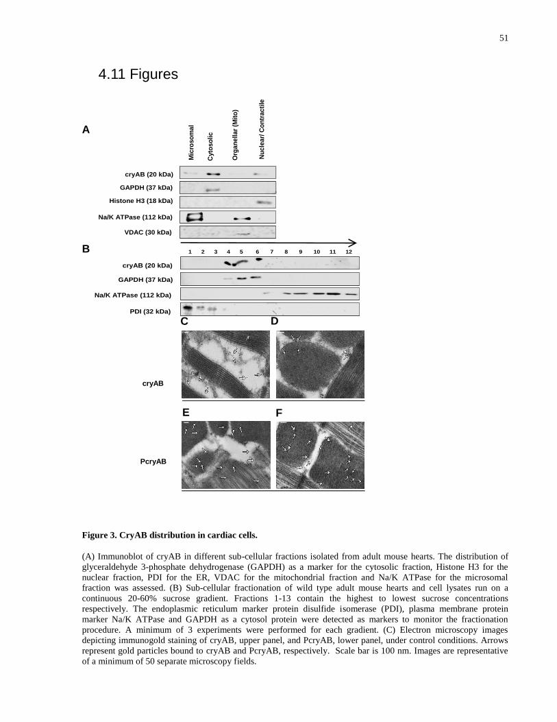
51
4.11 Figures
A
B
Figure 3. CryAB distribution in cardiac cells.
(A) Immunoblot of cryAB in different sub-cellular fractions isolated from adult mouse hearts. The distribution of
glyceraldehyde 3-phosphate dehydrogenase (GAPDH) as a marker for the cytosolic fraction, Histone H3 for the
nuclear fraction, PDI for the ER, VDAC for the mitochondrial fraction and Na/K ATPase for the microsomal
fraction was assessed. (B) Sub-cellular fractionation of wild type adult mouse hearts and cell lysates run on a
continuous 20-60% sucrose gradient. Fractions 1-13 contain the highest to lowest sucrose concentrations
respectively. The endoplasmic reticulum marker protein disulfide isomerase (PDI), plasma membrane protein
marker Na/K ATPase and GAPDH as a cytosol protein were detected as markers to monitor the fractionation
procedure. A minimum of 3 experiments were performed for each gradient. (C) Electron microscopy images
depicting immunogold staining of cryAB, upper panel, and PcryAB, lower panel, under control conditions. Arrows
represent gold particles bound to cryAB and PcryAB, respectively. Scale bar is 100 nm. Images are representative
of a minimum of 50 separate microscopy fields.
E F
cryAB (20 kDa)
Histone H3 (18 kDa)
GAPDH (37 kDa)
Na/K ATPase (112 kDa)
VDAC (30 kDa)
Mic
roso
ma
l
Nu
cle
ar/
Co
ntr
ac
tile
Org
an
ell
ar
(Mit
o)
Cyto
so
lic
1 2 3 4 5 6 7 8 9 10 11 12
cryAB (20 kDa)
PDI (32 kDa)
GAPDH (37 kDa)
Na/K ATPase (112 kDa)
C D
cryAB
PcryAB
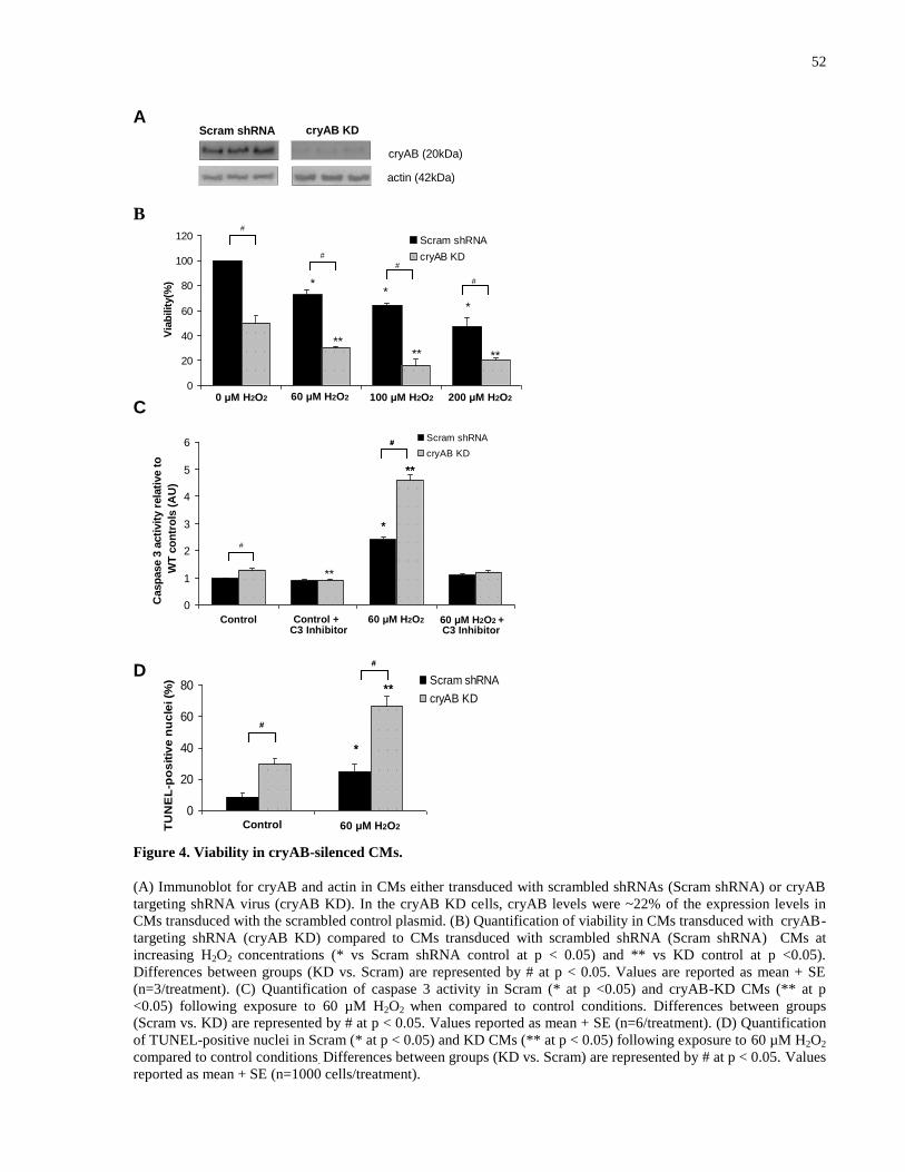
52
A
B
C
D
Figure 4. Viability in cryAB-silenced CMs.
(A) Immunoblot for cryAB and actin in CMs either transduced with scrambled shRNAs (Scram shRNA) or cryAB
targeting shRNA virus (cryAB KD). In the cryAB KD cells, cryAB levels were ~22% of the expression levels in
CMs transduced with the scrambled control plasmid. (B) Quantification of viability in CMs transduced with cryAB-
targeting shRNA (cryAB KD) compared to CMs transduced with scrambled shRNA (Scram shRNA) CMs at
increasing H2O2 concentrations (* vs Scram shRNA control at p < 0.05) and ** vs KD control at p <0.05).
Differences between groups (KD vs. Scram) are represented by # at p < 0.05. Values are reported as mean + SE
(n=3/treatment). (C) Quantification of caspase 3 activity in Scram (* at p <0.05) and cryAB-KD CMs (** at p
<0.05) following exposure to 60 µM H2O2 when compared to control conditions. Differences between groups
(Scram vs. KD) are represented by # at p < 0.05. Values reported as mean + SE (n=6/treatment). (D) Quantification
of TUNEL-positive nuclei in Scram (* at p < 0.05) and KD CMs (** at p < 0.05) following exposure to 60 µM H2O2
compared to control conditions. Differences between groups (KD vs. Scram) are represented by # at p < 0.05. Values
reported as mean + SE (n=1000 cells/treatment).
Scram shRNA cryAB KD
cryAB (20kDa)
actin (42kDa)
0
20
40
60
80
100
120
0 µM H2O2 60 µM H2O2 100 µM H2O2 200 µM H2O2
Via
bilit
y(%
)
Scram shRNA
cryAB KD
**** **
**
*
#
#
#
#
0 μM H2O2 60 μM H2O2 100 μM H2O2 200 μM H2O2
0
20
40
60
80
Control 60 µM H2O2 TU
NE
L-p
os
itiv
e n
uc
lei (%
) Scram shRNA
cryAB KD**
*
#
#
**
*
#
#
Control 60 μM H2O2
Control 60 μM H2O2Control + 60 μM H2O2 + C3 Inhibitor C3 Inhibitor
0
1
2
3
4
5
6
Control Control + C3
Inhibitor
60 µM H2O2 60 µM H2O2 +
C3 Inhibitor
Casp
ase 3
acti
vit
y r
ela
tive t
o
WT
co
ntr
ols
(A
U)
Scram shRNA
cryAB KD
*
**
#
*
**
#
**
#
#
**
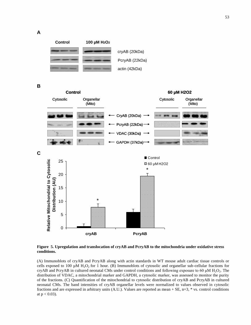
53
A
B
C
Figure 5. Upregulation and translocation of cryAB and PcryAB to the mitochondria under oxidative stress
conditions.
(A) Immunoblots of cryAB and PcryAB along with actin standards in WT mouse adult cardiac tissue controls or
cells exposed to 100 µM H2O2 for 1 hour. (B) Immunoblots of cytosolic and organellar sub-cellular fractions for
cryAB and PcryAB in cultured neonatal CMs under control conditions and following exposure to 60 μM H2O2. The
distribution of VDAC, a mitochondrial marker and GAPDH, a cytosolic marker, was assessed to monitor the purity
of the fractions. (C) Quantification of the mitochondrial to cytosolic distribution of cryAB and PcryAB in cultured
neonatal CMs. The band intensities of cryAB organellar levels were normalized to values observed in cytosolic
fractions and are expressed in arbitrary units (A.U.). Values are reported as mean + SE, n=3, * vs. control conditions
at p < 0.03).
Control 60 µM H2O2
Cytosolic CytosolicOrganellar
(Mito)
PcryAB (22kDa)
CryAB (20kDa)
GAPDH (37kDa)
VDAC (30kDa)
Organellar
(Mito)
Control 60 µM H2O2
Cytosolic CytosolicOrganellar
(Mito)
PcryAB (22kDa)
CryAB (20kDa)
GAPDH (37kDa)
VDAC (30kDa)
Organellar
(Mito)
0
5
10
15
20
25
cryAB PcryAB
Re
lati
ve
Mit
oc
ho
nd
ria
l to
Cy
tos
oli
c
Dis
trib
uti
on
(A
U)
Control
60 µM H2O2
*
*
cryAB (20kDa)
PcryAB (22kDa)
actin (42kDa)
Control 100 µM H2O2
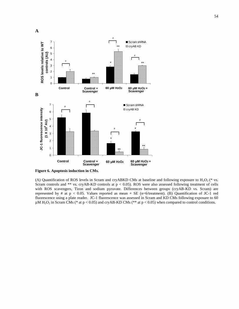
54
A
B
Figure 6. Apoptosis induction in CMs.
(A) Quantification of ROS levels in Scram and cryABKD CMs at baseline and following exposure to H2O2 (* vs.
Scram controls and ** vs. cryAB-KD controls at p < 0.05). ROS were also assessed following treatment of cells
with ROS scavengers, Tiron and sodium pyruvate. Differences between groups (cryAB-KD vs. Scram) are
represented by # at p < 0.05. Values reported as mean + SE (n=6/treatment). (B) Quantification of JC-1 red
fluorescence using a plate reader. JC-1 fluorescence was assessed in Scram and KD CMs following exposure to 60
µM H2O2 in Scram CMs (* at p < 0.05) and cryAB-KD CMs (** at p < 0.05) when compared to control conditions.
Control 60 μM H2O2Control +Scavenger
60 μM H2O2 + Scavenger
0
1
2
3
4
5
6
7
Control Control +
Scavenger
60 µM H2O2 60 µM H2O2 +
Scavenger
RO
S l
evels
rela
tive t
o W
T
co
ntr
ols
(A
U)
Scram shRNA
cryAB KD
#
**
**
*
#
#
**
Control 60 μM H2O2Control +Scavenger
60 μM H2O2 + Scavenger
#
0
1
2
3
4
5
6
7
Control Control +
Scavenger
60 µM H2O2 60 µM H2O2 +
Scavenger
JC
-1 f
luo
res
ce
nc
e in
ten
sit
y
(1 X
10
6 A
U)
Scram shRNA
cryAB KD#
**
*
# *
**
#
#
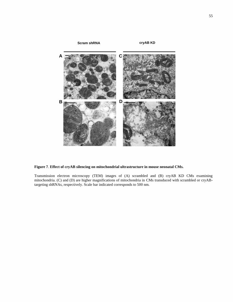
55
Figure 7. Effect of cryAB silencing on mitochondrial ultrastructure in mouse neonatal CMs.
Transmission electron microscopy (TEM) images of (A) scrambled and (B) cryAB KD CMs examining
mitochondria. (C) and (D) are higher magnifications of mitochondria in CMs transduced with scrambled or cryAB-
targeting shRNAs, respectively. Scale bar indicated corresponds to 500 nm.
Scram shRNA cryAB KD
A C
B D
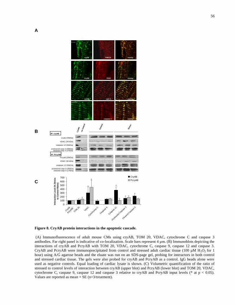
56
A
B
C
Figure 8. CryAB protein interactions in the apoptotic cascade.
(A) Immunofluorescence of adult mouse CMs using cryAB, TOM 20, VDAC, cytochrome C and caspase 3
antibodies. Far right panel is indicative of co-localization. Scale bars represent 4 µm. (B) Immunoblots depicting the
interactions of cryAB and PcryAB with TOM 20, VDAC, cytochrome C, caspase 9, caspase 12 and caspase 3.
CryAB and PcryAB were immunoprecipitated from control and stressed adult cardiac tissue (100 µM H2O2 for 1
hour) using A/G agarose beads and the eluate was run on an SDS-page gel, probing for interactors in both control
and stressed cardiac tissue. The gels were also probed for cryAB and PcryAB as a control. IgG beads alone were
used as negative controls. Equal loading of cardiac lysate is shown. (C) Volumetric quantification of the ratio of
stressed to control levels of interaction between cryAB (upper blot) and PcryAB (lower blot) and TOM 20, VDAC,
cytochrome C, caspase 9, caspase 12 and caspase 3 relative to cryAB and PcryAB input levels (* at p < 0.05).
Values are reported as mean + SE (n=3/treatment).
VDAC
cryAB caspase 3
cryAB cyto C overlay
cryAB TOM 20 overlay
cryAB overlay
overlay
VDAC
cryAB caspase 3
cryAB cyto C overlay
cryAB TOM 20 overlay
cryAB overlay
overlay
0
100
200
300
400
500
600
700
Cry
AB
TOM
20
VDAC
Cyt
ochro
me
C
Casp
ase 9
Casp
ase 1
2
Uncl
eave
d Cas
pase 3
Cle
aved
Cas
pase
3
Inte
rac
tio
n L
ev
el
(% S
tre
ss
to C
on
tro
l R
ati
o)
CryAB
PcryAB
Pcr
yAB
* *
*
*
Con
tro
l
Lysat
e
-ve
contr
ol
Str
ess
caspase 12 (55kDa)
caspase 12 (55kDa)
cryab (20kDa)
VDAC (30 kDa)
IP: cryAB
uncleaved casp 3 (35kDa)cleaved casp 3 (17kDa)
Pcryab (20kDa)
VDAC (30 kDa)
uncleaved casp 3 (35kDa)cleaved casp 3 (17kDa)
IP: PcryAB
57
CHAPTER FIVE: DISCUSSION
In the present study, I demonstrated the following: (1) under control conditions cryAB assumes a
cytosolic localization in adult mouse CMs and that in response to hydrogen peroxide-induced
oxidative stress, an in vitro mimic of I/R, cryB and PcryAB translocate to the mitochondria, (2)
following H2O2 exposure total cryAB levels become upregulated and knock-down of cryAB
leads to a significant reduction in viability in mouse neonatal cryAB KD CMs suggesting that
cryAB is protective against apoptosis, (3) cryAB silencing is accompanied by significant ROS
increases, mitochondrial ultrastructure alterations and dissipation of membrane potential, leading
to a significant reduction in viability in mouse neonatal CMs, by increasing the levels of intrinsic
apoptosis at baseline conditions, (4) cryAB interacts with a number of apoptotic mediators,
TOM 20, VDAC, cytochrome C, and caspases 9, 12 and 3 under both control conditions and
following exposure to H2O2 and (5) a significantly higher level of interaction following exposure
to H2O2 when compared to control conditions was observed between both cryAB and PcryAB
and VDAC and between cryAB and caspase 12, suggesting these interactions may be part of the
cryAB protective mechanism against apoptosis in CMs.
5.1 CryAB localization and translocation in cardiomyocytes
CryAB is a small heat shock protein that has cardio-protective properties87
. It has been shown
that part of the protective mechanism of cryAB involves its translocation to the mitochondria,
thus reducing I/R damage in mouse hearts during ex vivo I/R100
. Moreover, a molecular mimic of
PcryAB enhances the protective ability of cryAB against several different stresses that imitate
I/R87
. Our results extend these earlier findings to an in vitro model of I/R in cultured neonatal
mouse CMs.
5.1.1 Ischemia/Reperfusion injury and hydrogen peroxide in vitro
Myocardial ischemia is caused by an inadequate blood supply, and thereby an inadequate supply
of oxygen and glucose to the heart, leading to CM death if blood flow is not rapidly restored21
.
Reperfusion of ischemic tissue has its dangers as well, as it is also associated with cardiac tissue
injury22
.Reactive oxygen species are known to play a major role in the pathogenesis of
myocardial dysfunction in a variety of conditions including I/R injury110
. Among different

58
activated oxygen species, H2O2, which is not a free radical, has a relatively longer half-life and
may play a significant role in oxidative stress injury111
. Different oxidases of the heart, especially
xanthine oxidase, monoamine oxidase, and NADH oxidase produce H2O2, which directly and/or
indirectly through the production of highly reactive hydroxyl radicals can cause myocardial
injury111
. Global ischemia as well as reperfusion have been associated with significant increases
in myocardial H2O2 content112
. As a result, the addition of H2O2 to cells in culture provides a
model for the study of I/R injury in vitro. Furthermore, I/R has been associated with the
induction of apoptosis in the myocardium23,24 ,25
. H2O2 has also been shown to lead to apoptosis
in CMs, by activating the intrinsic pathway of apoptosis51
. Its abundant presence in I/R injury
and its ability to induce apoptosis in CMs led us to use optimally determined levels of H2O2 in
our in vitro model of I/R injury to determine the anti-apoptotic mechanism of cryAB in CMs.
5.1.2 CryAB translocates from the cytosol to the mitochondria following oxaidative stress
Following sub-cellular fractionation of adult mouse hearts, by differential centrifugation and
sucrose gradient separation, generating nuclear, organellar (including the mitochondria),
microsomal, and cytosolic fractions, we found that under control conditions, a majority of cryAB
(83%, Fig. 4) was found in the cytosolic fraction, in agreement with the literature113
. It has been
shown previously that under conditions of I/R, cryAB translocates to the mitochondria and
contractile units114
. Also, following I/R stress, phosphorylation of cryAB on the serine-59
residue enhances protection against apoptosis114
. Following sub-cellular fractionation and
immunofluorescence analysis, cryAB and PcryAB seemed to translocate to the mitochondria.
These results suggest that part of the protective mechanism of cryAB in the face of H2O2-
induced cell death involves its translocation and the translocation of any phosphorylated pools of
cryAB to the mitochondria.
With regards to the function of the association of cryAB and PcryAB with the mitochondria, it is
possible that, as a molecular chaperone, it exerts its protective effects by binding to other
proteins in or on the mitochondria. For instance, PcryAB is associated with the mitochondria
even under control conditions (Fig. 3 and 5) and this association increases significantly under
conditions of oxidative stress. It is possible that this association is due to PcryAB binding and
stabilizing or modulating the activity of MPTP proteins. This speculation is supported by studies

59
that have shown that over-expression of PcryAB in rat CMs decreases calcium-induced swelling
of the mitochondria, which is used to examine permeability transition pore opening115
.
A question that remains to be answered involves the nature of the stimulus that draws cryAB and
PcryAB to the mitochondria, as cryAB lacks a mitochondrial targeting sequence. Potential
stimuli that attract cryAB and PcryAB to the mitochondria could include the unfolding of
mitochondrial proteins. For example, TOM 20, a member of the outer mitochondrial membrane
protein translocase is prone to unfolding in response to I/R in the myocardium47
. By maintaining
TOM 20 structure, other hsps (hsp 70) contribute to protection of the myocardium in ischemic
preconditioning47
. Furthermore, the unfolding of proteins in the MPTP may act as a stimulus to
attract cryAB and PcryAB to the mitochondria. Consistent with this possibility are studies
showing that the unfolding of these proteins contributes to the activation of MPT116
. Our results
are in agreement with this possibility, namely the ability of PcryAB to interact with the proteins
of the MPTP, as we have shown by EM that PcryAB can also enter the mitochondria, in addition
to binding to its surface (Fig. 2C). Other studies have also shown that other cytosolic hsps can
localize to the interior of the mitochondria. For instance, hsp25 binds to and protects cytochrome
Complex I of the mitochondria from oxidative stress in PC12 cells117
. Our results extend the
findings that hsps can interact with MPTP proteins and stabilize the mitochondria, as they
indicate that the interaction of cryAB or PcryAB with VDAC may be important for the
protective mechanism, as it is significantly increased under stress conditions (Fig. 7). VDAC is
one of the proteins that make up the MPTP, the opening of which induces apoptosis49
. By
interacting with VDAC, cryAB and/or PcryAB may stabilize the mitochondrial membrane,
preventing the opening of the pore and thus blocking the first step in the mitochondrial pathway
of apoptosis. Therefore, the interaction of cryAB with proteins in the inner mitochondrial
membrane may be a central mechanism by cryAB prevents apoptosis. This contention would
work in concert with previously established interactions of cryAB with pro-apoptotic proteins
Bax and Bcl-Xs, preventing their translocation from cytosol into mitochondria, thus maintaining
the mitochondrial integrity and leading to decreased apoptosis97
.
60
5.2 CryAB silencing contributes to higher apoptosis levels in CMs
In in vitro models, it has been shown that cryAB prevents apoptosis induced by various factors
including hydrogen peroxide103
. We found that, much like other heat shock proteins, cryAB is
upregulated in response to stress (Fig. 5A) following exposure to hydrogen peroxide, suggesting
an involvement in protection against cell death. Furthermore, in cryAB KD CMs viability was
significantly decreased at all hydrogen peroxide concentrations, suggestive of the fact that cryAB
expression is protective against cell death under baseline conditions and following exposure to
H2O2-induced oxidative stress. Increased ROS production at baseline in KD CMs contributed to
the higher levels of cell death due to imposed oxidative stress. The results of this project extend
previous findings in a cryAB R120G mouse model of desmin cardiomyopathy. This model
presents with alterations in mitochondrial organization, architecture and function, decreased
complex I activity in the mitochondria by 50%, as well as alterations in the permeability
transition pore, and compromised inner membrane potential, leading to the activation of
apoptosis118
. Since in the presence of a mutated from of cryAB, complex I activity is severely
compromised, it may be possible that a similar effect would take place in the absence of cryAB.
This would explain the increased ROS levels that were observed in the present study.
The mitochondria are a major source of ROS and oxidative stress, but they are also highly
susceptible to ROS damage. Proteins, lipids, and nucleic acids can be altered by ROS, resulting
in covalent changes that affect mitochondrial structure and function119
. It has also been shown
that mitochondrial complex I activity is decreased by oxidative stress119
and reduced complex I
activity has been associated with numerous protein misfolding and neurodegenerative
diseases120
. Complex I dysfunction and the subsequent impairment of mitochondrial respiration
activate the intrinsic apoptotic pathway by directly triggering the release of cytochrome C from
affected mitochondria121
. Hence, KD of cryAB could contribute to increased mitochondria-
induced apoptosis by ROS-induced ROS production 122
leading to apoptosis. Electron
microscopy images in this study support this speculation, as they show significantly altered
mitochondrial ultrastructure in cryAB-silenced cardiomyocytes, suggestive of mitochondrial
permeabilization and rupture (Fig. 7), which would lead to apoptosis induction. The toxic effects
of ROS can be reduced by scavengers, and there is indeed a reduction of ROS levels and
mitochondrial dysfunction with the addition of ROS scavengers (Fig. 6). Given the ability of

61
antioxidants to protect against apoptosis in I/R injury by blocking the increased expression of
p53, Bax, and caspase-3, and inhibiting caspase-3 activation68
, it may be that the oxidative stress
imposed by cryAB KD functions by the opposite mechanism, increasing the expression of pro-
apoptotic proteins and stimulating apoptosis. And in the permanent absence of cryAB the
expectation would be a continuous production of mitochondrial ROS, which would lead to
considerable mitochondrial damage. The exogenous addition of hydrogen peroxide caused
further reductions in viability that were more pronounced in the KD CMs (Fig.4), suggesting that
the KD cells were significantly more prone to undergoing both early apoptosis and mid-stage
apoptosis, as depicted by caspase 3 activity under both baseline and stress conditions, indicating
that presence of cryAB is protective against apoptosis.
5.3 Protective mechanism of cryAB in CMs
The mechanism of protection of cyAB has not been fully elucidated in CMs. CryAB has been
shown to directly interact with the precursors of caspase-3 to suppress its activation in an
immortalized rabbit lens epithelial cell line 103
and in breast carcinoma cells104
. Furthermore,
cryAB interacts with pro-caspase 3 to negatively regulate apoptosis during myogenic
differentiation98
. CryAB also interacts with the pro-apoptotic proteins Bax and Bcl-Xs,
preventing their translocation from cytosol into mitochondria, thus leading to decreased
apoptosis in human lens epithelial cells 97
.
CryAB co-localized with TOM 20, VDAC, cytochrome C and caspase 3 (Figure 8A), suggesting
a potential interaction between cryAB and these proteins following exposure to hydrogen
peroxide. Co-immunoprecipitation results revealed that cryAB interacts with numerous proteins,
TOM 20, VDAC, cytochrome C, and caspase 9, 12 and 3 under both control conditions and
following exposure to hydrogen peroxide (Fig. 7 and 18). As was previously mentioned, cryAB
is the most abundantly expressed small heath shock protein in cardiac muscle 81
and is hence is
involved in protein quality control123
. As a result, its interaction with a high number of proteins is
not surprising. What is significant about these results, however, is the apparent higher level of
interaction under stress conditions when compared to control conditions of cryAB and PcryAB
with VDAC of cryAB with caspase 12 and of cryAB and PcryAB with uncleaved caspase 3,
although this did not reach statistical significance (Fig. 7B-C). These results suggest that the
62
interaction of cryAB and PcryAB with TOM 20 and caspase 9 may not contribute to the
protective mechanism of cryAB, as they are similar under control and stress conditions. The
interaction of cryAB with cytochrome C is also similar under both control and stress conditions,
suggesting that this interaction may also be inconsequential to its protective mechanism.
Furthermore, there was no interaction of PcryAB with cytochrome C. This difference could be
explained by the different distribution of cryAB and PcryAB. Following exposure to stress, some
unphosphorylated cryAB would remain in the cytosol capable of interacting with cytochrome C
to prevent the progression of apoptosis. PcryAB levels, on the other hand, are expected to
increase following stress when PcryAB associates mainly with the mitochondria. Following
mitochondrial outer membrane rupture due to oxidative stress, cytochrome C would be found in
the cytosol 49
and PcryAB in the mitochondria, making an interaction between the two less likely
than an interaction between unphosphorylated cryAB, some of which is still found in cytosol, as
was shown in Figure 5. The interaction between cryAB and PcryAB with uncleaved caspase 3,
although it did not reach statistical significance, may be part of the protective mechanism, by
preventing the cleavage and activation of caspase 3 thus preventing progression to apoptosis.
Furthermore, cryAB has been shown to interact with uncleaved caspase 3 in a breast cancer cell
line124
. The significantly higher interaction of cryAB with cleaved caspase 3 that was observed
in the present study, has not been reported previously, to my knowledge. This may be indicative
of a potential inhibition of the proteolytic activity of caspase 3 by cryAB even following its
cleavage and activation. Therefore, cryAB binding of caspase 3 would prevent its proteolytic
cleavage of substrates in the cell and contribute to diminished levels of apoptosis, thus providing
an additional point for intervention in preventing apoptosis in CMs.
The interaction of cryAB and PcryAB with VDAC seems to be important to the protective
mechanism, as it is significantly increased under stress conditions. VDAC is one of the proteins
that make up the mitochondrial permeability transition pore, the opening of which induces
apoptosis49
. By interacting with VDAC, cryAB and PcryAB may help stabilize the mitochondrial
membrane, preventing the opening of the pore and thus preventing the early stages of apoptosis.
In agreement with this possibility are studies showing that one mechanism by which
mitochondrial permeability transition (MPT) is activated is the unfolding of MPT pore proteins
upon stress116
. Furthermore, Maloyan et al. have previously shown that in the desmin
cardiomyopathy model, VDAC interacts with the mutated R120G cryAB, whereas there is no

63
interaction of VDAC with cryAB in WT hearts118
. This suggests that cryAB does not interact
with VDAC under control conditions, but following the induction of stress (either due to the
R120G mutation or exposure to hydrogen peroxide) cryAB can translocate to the mitochondria
and interact with VDAC to either stabilize or destabilize the MPT pore.
CryAB, but not PcryAB interaction with caspase 12 is significantly different under stress when
compared to control conditions. This suggests that cryAB is also potentially involved in
preventing ER stress-induced apoptosis, since ER stress activates ER resident caspase 12 125
.
This again, is in line with the cytosolic localization of both cryAB and caspase 12 and the mostly
mitochondrial localization of PcryAB. Following exposure to hydrogen peroxide, cryAB may
interact with caspase 12 in the cytosol, thus preventing the cleavage and activation of caspase 3
by caspase 12 and inhibiting the progression of the apoptosis cascade.
In summary, I found that the protective effects of cryAB in the heart are mediated by its
translocation from the cytosol to the mitochondria and its phosphorylation under conditions of
stress. Furthermore, we showed that the interaction of cryAB interactions with VDAC, caspase
12 and caspase 3 may be part of the cryAB protective mechanism against apoptosis following
exposure to H2O2 in the heart, thus showing a protective role of cryAB against intrinsic and ER-
mediated apoptosis. Further studies are needed to delineate the involvement of cryAB in the
extrinsic pathway of apoptosis and alternate death pathways, like autophagy and necrosis.
Regulating the levels of apoptosis is an attractive target for ameliorating any ischemic injury in
the heart. Our results contribute to delineating the involvement of cryAB in the intrinsic and ER
stress pathway of apoptosis activated by ROS-induced oxidative damage. Given the involvement
of ROS and H2O2 in myocardial dysfunction110
determining where in the apoptotic cascade
cryAB intervenes to prevent H2O2 injury can have therapeutic implications for cardiac disease.
Further studies focusing on the extrinsic pathway of apoptosis and alternate death pathways, like
autophagy and necrosis, would therefore prove a worthwhile pursuit.
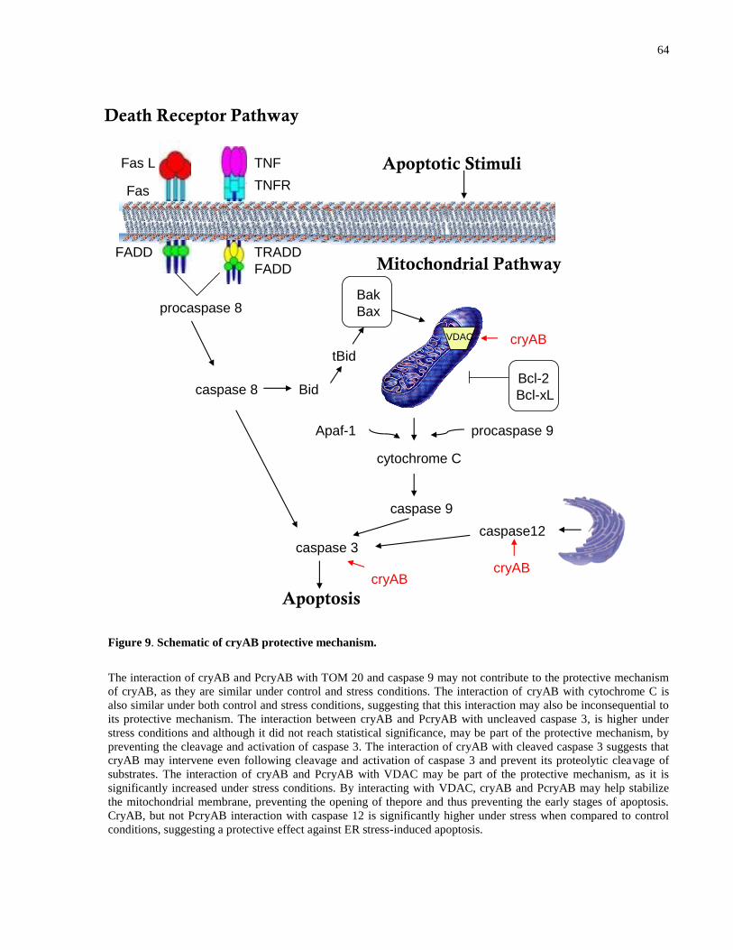
64
Figure 9. Schematic of cryAB protective mechanism.
The interaction of cryAB and PcryAB with TOM 20 and caspase 9 may not contribute to the protective mechanism
of cryAB, as they are similar under control and stress conditions. The interaction of cryAB with cytochrome C is
also similar under both control and stress conditions, suggesting that this interaction may also be inconsequential to
its protective mechanism. The interaction between cryAB and PcryAB with uncleaved caspase 3, is higher under
stress conditions and although it did not reach statistical significance, may be part of the protective mechanism, by
preventing the cleavage and activation of caspase 3. The interaction of cryAB with cleaved caspase 3 suggests that
cryAB may intervene even following cleavage and activation of caspase 3 and prevent its proteolytic cleavage of
substrates. The interaction of cryAB and PcryAB with VDAC may be part of the protective mechanism, as it is
significantly increased under stress conditions. By interacting with VDAC, cryAB and PcryAB may help stabilize
the mitochondrial membrane, preventing the opening of thepore and thus preventing the early stages of apoptosis.
CryAB, but not PcryAB interaction with caspase 12 is significantly higher under stress when compared to control
conditions, suggesting a protective effect against ER stress-induced apoptosis.
Death Receptor Pathway
Fas L
Fas
FADD
TNF
TNFR
FADD
TRADD
procaspase 8
caspase 8 Bid
Mitochondrial Pathway
cytochrome C
Apaf-1 procaspase 9
caspase 9
caspase 3
caspase12
Apoptosis
Apoptotic Stimuli
Bak
tBid
Bax
Bcl-2
Bcl-xL
cryABcryAB
cryABVDAC

65
CHAPTER SIX: LIMITATIONS
The current study made use of lentivector mediated knock-down of cryAB in neonatal CMs to
determine the protective ability and mechanism of protection of cryAB. Initially, I was also
planning on validating the enhanced protective ability of cryAB following its over-expression.
However, following transduction with lentivectors containing the cryAB over-expression
construct, I was unable to achieve up-regulation of cryAB and therefore unable to test the
hypothesis that higher, exogenous cryAB levels would attenuate apoptosis by interacting more
readily with proteins in the apoptotic cascade.
And while I began using cultured neonatal CMs from calcineurin transgenic mice (which present
with an upregulation of cryAB in cardiac tissue) as an alternative to the over-expression model,
it was impossible to attribute any protective effects noticed in the calcineurin CMs to cryAB
alone, due to confounding effects and numerous other changes induced by calcineurin over-
expression.
Another limitation relates to the use of an in vitro model. Cells in culture are not the best
representation of in vivo cells because cells in culture are situated in a different microenvironment.
This became evident throughout our experiments, as we noticed that the cell-culture environment
alone exposed the cells to increased oxidative stress at baseline conditions. Furthermore, in in vivo
models compensatory effects could be noticed due to redundant genes and proteins. Therefore, the
increased cell death observed in our in vitro model could be an over-estimate. For instance, in vivo
knock out of cryAB was not associated with increased mortality at baseline conditions. We, however,
noticed significantly higher cell death of the KD CMs in the absence of a stressor. Extrapolating
from these results, with increased CMs death, we would expect increased likelihood of progression to
heart failure in vivo even at baseline. This suggests that in in vivo models our results would be
attenuated due to compensatory effects of other genes and proteins. To make up for this limitation,
we were initially planning on testing all of our hypotheses on an in vivo mouse model with cardiac
knockdown or over-expression of cryAB, achieved through the direct injection of adeno-associated
virus serotype 9 (AAV9) containing either the knockdown or over-expression cryAB construct into
the hearts of adult mice. AAV9 have a high propensity to the heart (90%) and their expression is
indefinite. Preliminary results of over-expression of GFP-tagged constructs were very promising
(data not shown) but due to time constraints, this objective was not pursued further, although it
remains very worthwhile for future studies.
66
A major drawback for a large portion of this study was the limited CM yield, due to inefficient
harvesting techniques. Neonatal CMs isolation was very challenging throughout this study. Due to
the fact that we were working with primary cells with no replicative capability, large numbers of
pups were euthanized every week to isolate CMs for this study. And despite numerous optimizations
to the protocol, the CMs yield was low with every harvest, making protein isolation difficult. The
initial goal of the study was to perform all experiments in cultured neonatal CMs. However, due to
this limitation, I was unable to do so. The knock-downs and viability assays were performed in
neonatal CMs, since the lentivector transductions were optimized and efficient in neonatal CMs.
However, I was unable to isolate enough protein from cultured CMs to perform immunoprecipitation.
Therefore, I used adult CMs to address the low protein yield. Due to this limitation I was also unable
to perform immunoprecipitation out of KD CMs as a control and all immunoprecipitaitons were
performed in WT CMs.
Lastly, a significant portion of this study relied on the measurement of cell viability, not cell death
(with the exception of TUNEL labeling). Cell viability, caspase 3 and mitochondrial assay
measurements are dependent on the number of live cells. A limitation of this study was the inability
to maintain constant numbers of live cells under control and stress conditions. Due to an inability to
maintain constant cell numbers following transduction and stress (since the KD CMs were already
more sensitive at baseline and potentially more prone to demise), an underestimate of caspase 3, ROS
and JC-1 levels would be expected. This fact could potentially explain why the effects of cryAB KD
and exposure to extrinsic oxidative stress were additive on cell viability. This limitation could be
overcome by either measuring cell death as an alternative (or addition) to cell viability measurements
or by labeling the number of cells present in an assay (for instance, DAPI labeling) and finding the
proportion of live cells in different conditions.
67
CHAPTER SEVEN: ONGOING WORK
Since there was a significant decrease in viability in the KD CMs at baseline due to oxidative stress I
am also trying to over-express cryAB in neonatal cardiomyocytes (an initial objective of this project)
using Lipofectamine transfection to determine whether over-expression of cryAB would rescue cell
viability following treatment with extrinsic hydrogen peroxide, which would mimic the oxidative
stress observed at baseline in the absence of cryAB.
I am also using caspase 9 inihibitors and measuring caspase 3 activity to determine whether
mitochondrial ROS in the cryAB KD CMs are activating the intrinsic pathway of apoptosis. This
would further elucidate the mechanism by which cryAB silencing induces CM death.

68
CHAPTER EIGHT: FUTURE DIRECTIONS
In the present study, I characterized the steps at which cryAB intervenes in the apoptotic cascade
to attenuate apoptosis in the face of hydrogen peroxide-induced oxidative stress in CMs. For a
more complete picture, future directions in which the project could be taken involve the
determination of cryAB involvement in extrinsic apoptosis pathway, as well as other types of
cell death, like autophagy in the H2O2 oxidative stress model. Possible avenues include
determining whether autophagosomes can be detected in cryAB WT and KD CMs and whether
there is an association of cryAB with autophagosomes.
Furthermore, this study identifies a higher level of interaction of cryAB with caspase 12 under
stress conditions when compared to control conditions, suggesting that this interaction may be
part of the protective mechanism of cryAB. Caspase 12 becomes active in response to ER stress.
Therefore, examining ER stress markers in cryAB KD CMs would be of value. Also,
determining the contribution of ER stress to the levels of apoptosis detected by this study, by
specifically inhibiting caspase 12, would be of interest.
ER stress leads to the release of Ca2+
from the ER causing depletion of ER calcium and
activating Ca2+
-dependent endonucleases involved in DNA fragmentation. Furthermore, Ca2+
is
also believed to modulate cytochrome C release directly by regulating the MPTP, a proposed
mechanism for cytochrome C release during apoptosis. Hence, measuring Ca2+
transients in
cryAB KD CMs would confirm the role of Ca2+
and ER stress in triggering apoptosis in the
absence of cryAB.
Lastly, testing all of the hypotheses on an in vivo mouse model with cardiac knockdown or over-
expression of cryAB, achieved through the direct injection of adeno-associated virus serotype 9
(AAV9) containing either the knockdown or over-expression cryAB construct into the hearts of adult
mice remains a worthwhile pursuit. It may be that due to compensatory effects, the consequences of
cryAB KD in vivo would not be as dire as in this study. As I mentioned previously, in vivo knock out
of cryAB was not associated with increased mortality at baseline conditions. Extrapolating from the
significantly higher cell death of the KD CMs at baseline observed in this study, one would expect
69
increased likelihood of progression to heart failure in vivo even at baseline. This suggests that in in
vivo models the results in this study would be attenuated due to compensatory effects of other genes
and proteins, so an in vivo model would be helpful in determining the impact of the findings at the in
vitro level. Furthermore, to date, the only knock out mouse model of cryAB is in fact a double knock
out, of cryAB and hspB2 genes, which are in very close proximity to each other and driven by the
same promoter. This fact makes it impossible to distinguish the contribution of the protein encoded
by cryAB from the protein encoded by the hspB2 gene to cyto-protection. Targeted disruption of
cryAB in the heart through the use of AAV9 would allow for the separation of the roles of cryAB
and hspB2 and it provides an alternative the creation of transgenic mouse lines.

70
CHAPTER NINE: REFERENCES
1. Lloyd-Jones D, Adams RJ, Brown TM, et al. Heart Disease and Stroke Statistics—2010
Update. Circulation. 2010;121(7):e46-e215.
2. Nadal-Ginard B, Kajstura J, Leri A, Anversa P. Myocyte Death, Growth, and
Regeneration in Cardiac Hypertrophy and Failure. Circulation Research. 2003;92(2):139-
150.
3. von Harsdorf R, Poole-Wilson PA, Dietz R. Regenerative capacity of the myocardium:
implications for treatment of heart failure. The Lancet. 2004;363(9417):1306-1313.
4. Shen S, Kepp O, Michaud M, et al. Association and dissociation of autophagy, apoptosis
and necrosis by systematic chemical study. Oncogene. 2011;30(45):4544-4556.
5. Edinger AL, Thompson CB. Death by design: apoptosis, necrosis and autophagy. Current
Opinion in Cell Biology. 2004;16(6):663-669.
6. Gill C, Mestril R, Samali A. Losing heart: the role of apoptosis in heart disease—a novel
therapeutic target? The FASEB Journal. 2002;16(2):135-146.
7. Kerr JF, Wyllie AH, Currie AR. Apoptosis: A Basic Biological Phenomenon with
Wideranging Implications in Tissue Kinetics. British Journal of Cancer. 1972;26(4):239-
257.
8. Thornberry NA, Lazebnik Y. Caspases: Enemies Within. Science. 1998;281(5381):1312-
1316.
9. Kroemer G, Galluzzi L, Vandenabeele P, et al. Classification of cell death:
recommendations of the Nomenclature Committee on Cell Death 2009. Cell Death and
Differentiation. 2008;16(1):3-11.
10. Golstein P, Kroemer G. Cell death by necrosis: towards a molecular definition. Trends in
Biochemical Sciences. 2007;32(1):37-43.
11. Festjens N, Vanden Berghe T, Cornelis S, Vandenabeele P. RIP1, a kinase on the
crossroads of a cell's decision to live or die. Cell Death and Differentiation.
2007;14(3):400-410.
12. Baehrecke EH. Autophagy: dual roles in life and death? Nature Reviews Molecular Cell
Biology. 2005;6(6):505-510.
13. Ziegler U, Groscurth P. Morphological Features of Cell Death. Physiology.
2004;19(3):124-128.
71
14. Shintani T, Klionsky DJ. Autophagy in Health and Disease: A Double-Edged Sword.
Science. 2004;306(5698):990-995.
15. Mizushima N, Yamamoto A, Matsui M, Yoshimori T, Ohsumi Y. In Vivo Analysis of
Autophagy in Response to Nutrient Starvation Using Transgenic Mice Expressing a
Fluorescent Autophagosome Marker. Molecular Biology of the Cell. 2004;15(3):1101-
1111.
16. Berry DL, Baehrecke EH. Growth Arrest and Autophagy Are Required for Salivary
Gland Cell Degradation in Drosophila. Cell. 2007;131(6):1137-1148.
17. Samara C, Syntichaki P, Tavernarakis N. Autophagy is required for necrotic cell death in
Caenorhabditis elegans. Cell Death Differ. 2007;15(1):105-112.
18. Narula J, Haider N, Virmani R, et al. Apoptosis in Myocytes in End-Stage Heart Failure.
New England Journal of Medicine. 1996;335(16):1182-1189.
19. Guerra S, Leri A, Wang X, et al. Myocyte Death in the Failing Human Heart Is Gender
Dependent. Circulation Research. 1999;85(9):856-866.
20. González A, Fortuño MaA, Querejeta R, et al. Cardiomyocyte apoptosis in hypertensive
cardiomyopathy. Cardiovascular Research. 2003;59(3):549-562.
21. Nian M, Lee P, Khaper N, Liu P. Inflammatory Cytokines and Postmyocardial Infarction
Remodeling. Circulation Research. 2004;94(12):1543-1553.
22. Dong JW, Zhu HF, Zhu WZ, Ding HL, Ma TM, Zhou ZN. Intermittent hypoxia
attenuates ischemia/reperfusion induced apoptosis in cardiac myocytes via regulating
Bcl-2/Bax expression. Cell Research. 2003;13(5):385-391.
23. Gottlieb RA, Burleson KO, Kloner RA, Babior BM, Engler RL. Reperfusion injury
induces apoptosis in rabbit cardiomyocytes. The Journal of Clinical Investigation.
1994;94(4):1621-1628.
24. Fliss H, Gattinger D. Apoptosis in Ischemic and Reperfused Rat Myocardium.
Circulation Research. 1996;79(5):949-956.
25. Zhao Z-Q, Nakamura M, Wang N-P, et al. Reperfusion induces myocardial apoptotic cell
death. Cardiovascular Research. 2000;45(3):651-660.
26. Saraste A, Pulkki K, Kallajoki M, Henriksen K, Parvinen M, Voipio-Pulkki L-M.
Apoptosis in Human Acute Myocardial Infarction. Circulation. 1997;95(2):320-323.
27. Freude B, Masters TN, Robicsek F, et al. Apoptosis is Initiated by Myocardial Ischemia
and Executed During Reperfusion. Journal of Molecular and Cellular Cardiology.
2000;32(2):197-208.
28. Piot C, Croisille P, Staat P, et al. Effect of Cyclosporine on Reperfusion Injury in Acute
Myocardial Infarction. New England Journal of Medicine. 2008;359(5):473-481.
72
29. Pfeffer M, Braunwald E. Ventricular remodeling after myocardial infarction.
Experimental observations and clinical implications. Circulation. 1990;81(4):1161-1172.
30. Hilfiker-Kleiner D, Landmesser U, Drexler H. Molecular Mechanisms in Heart Failure:
Focus on Cardiac Hypertrophy, Inflammation, Angiogenesis, and Apoptosis. Journal of
the American College of Cardiology. 2006:j.jacc.2006.2007.2007.
31. Akyürek Ö, Akyürek N, Sayin T, et al. Association between the severity of heart failure
and the susceptibility of myocytes to apoptosis in patients with idiopathic dilated
cardiomyopathy. International Journal of Cardiology. 2001;80(1):29-36.
32. Bishopric NH, Andreka P, Slepak T, Webster KA. Molecular mechanisms of apoptosis in
the cardiac myocyte. Current Opinion in Pharmacology. 2001;1(2):141-150.
33. Ashkenazi A, Dixit VM. Death Receptors: Signaling and Modulation. Science.
1998;281(5381):1305-1308.
34. Hengartner MO. The biochemistry of apoptosis. Nature. 2000;407(6805):770-776.
35. Ferri KF, Kroemer G. Organelle-specific initiation of cell death pathways. Nature Cell
Biology. 2001;3(11):E255-E263.
36. Kroemer G, Reed JC. Mitochondrial control of cell death. Nature Medicine.
2000;6(5):513-519.
37. Lakshmi G. Cell Death Inhibition: Keeping Caspases in Check. Cell. 2001;104(6):805-
808.
38. Turk B, Turk D, Turk V. Lysosomal cysteine proteases: more than scavengers.
Biochimica et Biophysica Acta (BBA) - Protein Structure and Molecular Enzymology.
2000;1477(1-2):98-111.
39. Huang Y, Wang KKW. The calpain family and human disease. Trends in Molecular
Medicine. 2001;7(8):355-362.
40. Yamaguchi S, Yamaoka M, Okuyama M, et al. Elevated circulating levels and cardiac
secretion of soluble fas ligand in patients with congestive heart failure. The American
Journal of Cardiology. 1999;83(10):1500-1503.
41. Kajstura Jea. Apoptotic and necrotic myocyte cell deaths are independent contributing
variables of infarct size in rats. Laboratory Investigations. 1996;74(1):86-107.
42. Tanaka M, Ito H, Adachi S, et al. Hypoxia induces apoptosis with enhanced expression of
Fas antigen messenger RNA in cultured neonatal rat cardiomyocytes. Circulation
Research. 1994;75(3):426-433.
43. Cheng W, Li B, Kajstura J, et al. Stretch-induced programmed myocyte cell death. The
Journal of Clinical Investigation. 1995;96(5):2247-2259.
73
44. Torre-Amione G, Kapadia S, Lee J, et al. Tumor Necrosis Factor-α and Tumor Necrosis
Factor Receptors in the Failing Human Heart. Circulation. 1996;93(4):704-711.
45. Sliwa K, Skudicky D, Candy G, Wisenbaugh T, Sareli P. Randomised investigation of
effects of pentoxifylline on left-ventricular performance in idiopathic dilated
cardiomyopathy. The Lancet. 1998;351(9109):1091-1093.
46. Krown KA, Page MT, Nguyen C, et al. Tumor necrosis factor alpha-induced apoptosis in
cardiac myocytes. Involvement of the sphingolipid signaling cascade in cardiac cell
death. The Journal of Clinical Investigation. 1996;98(12):2854-2865.
47. Bowers M, Ardehali H. TOM20 and the Heartbreakers: Evidence for the role of
mitochondrial transport proteins in cardioprotection. Journal of Molecular and Cellular
Cardiology. 2006;41(3):406-409.
48. Wang X. The expanding role of mitochondria in apoptosis. Genes and Development.
2001;15:2922-2933.
49. Green, John C. Reed DR. Mitochondria and Apoptosis. Science. 1998;281(5381):1309-
1312.
50. Bialik S, Cryns VL, Drincic A, et al. The Mitochondrial Apoptotic Pathway Is Activated
by Serum and Glucose Deprivation in Cardiac Myocytes. Circulation Research.
1999;85(5):403-414.
51. von Harsdorf R, Li P-F, Dietz R. Signaling Pathways in Reactive Oxygen Species–
Induced Cardiomyocyte Apoptosis. Circulation. 1999;99(22):2934-2941.
52. Khosravi-Far R. Death receptor signals to the mitochondria. Cancer Biology & Therapy.
2004;3(11):1051-1057.
53. Li H, Zhu H, Xu C-j, Yuan J. Cleavage of BID by Caspase 8 Mediates the Mitochondrial
Damage in the Fas Pathway of Apoptosis. Cell. 1998;94(4):491-501.
54. Matsuzawa A, Ichijo H. Molecular Mechanisms of the Decision between Life and Death:
Regulation of Apoptosis by Apoptosis Signal-Regulating Kinase 1. Journal of
Biochemistry. 2001;130(1):1-8.
55. Adams JM, Cory S. Life-or-death decisions by the Bcl-2 protein family. Trends in
Biochemical Sciences. 2001;26(1):61-66.
56. Condorelli G, Morisco C, Stassi G, et al. Increased Cardiomyocyte Apoptosis and
Changes in Proapoptotic and Antiapoptotic Genes bax and bcl-2 During Left Ventricular
Adaptations to Chronic Pressure Overload in the Rat. Circulation. 1999;99(23):3071-
3078.
57. Latif N, Khan MA, Birks E, et al. Upregulation of the Bcl-2 family of proteins in end
stage heart failure. Journal of the American College of Cardiology. 2000;35(7):1769-
1777.
74
58. Maulik N, Engelman RM, Rousou JA, Flack JE, Deaton D, Das DK. Ischemic
Preconditioning Reduces Apoptosis By Upregulating Anti-Death Gene Bcl-2.
Circulation. 1999;100(suppl 2):II-369-II-375.
59. Kirshenbaum LA, de Moissac D. The bcl-2 Gene Product Prevents Programmed Cell
Death of Ventricular Myocytes. Circulation. 1997;96(5):1580-1585.
60. Maloyan A, Sayegh J, Osinska H, Chua BHL, Robbins J. Manipulation of Death
Pathways in Desmin-Related Cardiomyopathy. Circulation Research. 2010;106(9):1524-
1532.
61. Koseki T, Inohara N, Chen S, Núñez G. ARC, an inhibitor of apoptosis expressed in
skeletal muscle and heart that interacts selectively with caspases. Proceedings of the
National Academy of Sciences. 1998;95(9):5156-5160.
62. Yaoita H, Ogawa K, Maehara K, Maruyama Y. Attenuation of Ischemia/Reperfusion
Injury in Rats by a Caspase Inhibitor. Circulation. 1998;97(3):276-281.
63. Gottlieb RA, Gruol DL, Zhu JY, Engler RL. Preconditioning rabbit cardiomyocytes: role
of pH, vacuolar proton ATPase, and apoptosis. The Journal of Clinical Investigation.
1996;97(10):2391-2398.
64. Yaoita H, Ogawa K, Maehara K, Maruyama Y. Apoptosis in relevant clinical situations.
Cardiovascular Research. 2000;45(3):630-641.
65. Okamura T, Miura T, Takemura G, et al. Effect of caspase inhibitors on myocardial
infarct size and myocyte DNA fragmentation in the ischemia–reperfused rat heart.
Cardiovascular Research. 2000;45(3):642-650.
66. Mocanu MM, Baxter GF, Yellon DM. Caspase inhibition and limitation of myocardial
infarct size: protection against lethal reperfusion injury. British Journal of Pharmacology.
2000;130(2):197-200.
67. Chandra J, Samali A, Orrenius S. Triggering and modulation of apoptosis by oxidative
stress. Free Radical Biology and Medicine. 2000;29(3-4):323-333.
68. Oskarsson HJ, Coppey L, Weiss RM, Li W-G. Antioxidants attenuate myocyte apoptosis
in the remote non-infarcted myocardium following large myocardial infarction.
Cardiovascular Research. 2000;45(3):679-687.
69. Dhalla NS, Elmoselhi AB, Hata T, Makino N. Status of myocardial antioxidants in
ischemia–reperfusion injury. Cardiovascular Research. 2000;47(3):446-456.
70. Maulik N, Yoshida T, Das DK. Regulation of cardiomyocyte apoptosis in ischemic
reperfused mouse heart by glutathione peroxidase. Molecular and Cellular Biochemistry.
1999;196(1):13-21.
75
71. Chen Z, Siu B, Ho Y-S, et al. Overexpression of MnSOD Protects Against Myocardial
Ischemia/reperfusion Injury in Transgenic Mice. Journal of Molecular and Cellular
Cardiology. 1998;30(11):2281-2289.
72. Samali A, Cotter TG. Heat Shock Proteins Increase Resistance to Apoptosis.
Experimental Cell Research. 1996;223(1):163-170.
73. Stephanou A, Brar B, Heads R, et al. Cardiotrophin-1 Induces Heat Shock Protein
Accumulation in Cultured Cardiac Cells and Protects them from Stressful Stimuli.
Journal of Molecular and Cellular Cardiology. 1998;30(4):849-855.
74. Brar BK, Stephanou A, Wagstaff MJD, et al. Heat Shock Proteins Delivered With a Virus
Vector Can Protect Cardiac Cells Against Apoptosis As Well As Against Thermal or
Hypoxic Stress. Journal of Molecular and Cellular Cardiology. 1999;31(1):135-146.
75. Li C-Y, Lee J-S, Ko Y-G, Kim J-I, Seo J-S. Heat Shock Protein 70 Inhibits Apoptosis
Downstream of Cytochrome c Release and Upstream of Caspase-3 Activation. Journal of
Biological Chemistry. 2000;275(33):25665-25671.
76. Concannon CG, Orrenius S, Samali A. Hsp27 Inhibits Cytochrome c-Mediated Caspase
Activation by Sequestering Both Pro-caspase-3 and Cytochrome c. Gene Expression.
2001;9(4&5):195-201.
77. Bruey J-M, Ducasse C, Bonniaud P, et al. Hsp27 negatively regulates cell death by
interacting with cytochrome c. Nature Cell Biology. 2000;2(9):645-652.
78. de Jong WW, Caspers G-J, Leunissen JAM. Genealogy of the α-crystallin—small heat-
shock protein superfamily. International Journal of Biological Macromolecules.22(3-
4):151-162.
79. Horwitz J. Alpha-crystallin can function as a molecular chaperone. Proceedings of the
National Academy of Sciences. 1992;89(21):10449-10453.
80. Groenen PJTA, Merck KB, De Jong WW, Bloemendal H. Structure and Modifications of
the Junior Chaperone α-Crystallin. European Journal of Biochemistry. 1994;225(1):1-19.
81. Taylor RP, Benjamin IJ. Small heat shock proteins: a new classification scheme in
mammals. Journal of Molecular and Cellular Cardiology. 2005;38(3):433-444.
82. Arrigo A-P, Simon S, Gibert B, et al. Hsp27 (HspB1) and αB-crystallin (HspB5) as
therapeutic targets. FEBS Letters. 2007;581(19):3665-3674.
83. Klemenz R, Fröhli E, Steiger RH, Schäfer R, Aoyama A. Alpha B-crystallin is a small
heat shock protein. Proceedings of the National Academy of Sciences. 1991;88(9):3652-
3656.
84. Lin DI, Barbash O, Kumar KGS, et al. Phosphorylation-Dependent Ubiquitination of
Cyclin D1 by the SCFFBX4-αB Crystallin Complex. Molecular Cell. 2006;24(3):355-
366.
76
85. Launay N, Goudeau B, Kato K, Vicart P, Lilienbaum A. Cell signaling pathways to αB-
crystallin following stresses of the cytoskeleton. Experimental Cell Research.
2006;312(18):3570-3584.
86. Thériault JR, Lambert H, Chávez-Zobel AT, Charest G, Lavigne P, Landry J. Essential
Role of the NH2-terminal WD/EPF Motif in the Phosphorylation-activated Protective
Function of Mammalian Hsp27. Journal of Biological Chemistry. 2004;279(22):23463-
23471.
87. Morrison LE, Hoover HE, Thuerauf DJ, Glembotski CC. Mimicking Phosphorylation of
αB-Crystallin on Serine-59 Is Necessary and Sufficient to Provide Maximal Protection of
Cardiac Myocytes From Apoptosis. Circulation Research. 2003;92(2):203-211.
88. Berry V, Francis P, Reddy MA, et al. Alpha-B Crystallin Gene (CRYAB) Mutation
Causes Dominant Congenital Posterior Polar Cataract in Humans. The American Journal
of Human Genetics. 2001;69(5):1141-1145.
89. Vicart P, Caron A, Guicheney P, et al. A missense mutation in the [agr]B-crystallin
chaperone gene causes a desmin-related myopathy. Nature Genetics. 1998;20(1):92-95.
90. Selcen D, Engel AG. Myofibrillar myopathy caused by novel dominant negative αB-
crystallin mutations. Annals of Neurology. 2003;54(6):804-810.
91. Liu M, Ke T, Wang Z, et al. Identification of a CRYAB Mutation Associated with
Autosomal Dominant Posterior Polar Cataract in a Chinese Family. Investigative
Ophthalmology & Visual Science. 2006;47(8):3461-3466.
92. Muchowski PJ, Wacker JL. Modulation of neurodegeneration by molecular chaperones.
Nature Review Neuroscience. 2005;6(1):11-22.
93. Gruvberger-Saal SK, Parsons R. Is the small heat shock protein αB-crystallin an
oncogene? Journal of Clinical Investigation. 2006;116(1):30-32.
94. Brady JP, Garland DL, Green DE, Tamm ER, Giblin FJ, Wawrousek EF. αB-crystallin in
lens development and muscle integrity: A gene knockout approach. Investigative
Ophthalmology and Visual Science. 2001;42(12):2924-2934.
95. Morrison LE, Whittaker RJ, Klepper RE, Wawrousek EF, Glembotski CC. Roles for αB-
crystallin and HSPB2 in protecting the myocardium from ischemia-reperfusion-induced
damage in a KO mouse model. American Journal of Physiology - Heart and Circulatory
Physiology. 2004;286(3 55-3):H847-H855.
96. Ray PS, Martin JL, Swanson EA, Otani H, Dillman WH, Das DK. Transgene
overexpression of αB crystallin confers simultaneous protection against cardiomyocyte
apoptosis and necrosis during myocardial ischemia and reperfusion. The Federation of
American Societies for Experimental Biology Journal. 2001;15(2):393-402.
77
97. Mao YW, Liu JP, Xiang H, Li DWC. Human [alpha]A- and [alpha]B-crystallins bind to
Bax and Bcl-XS to sequester their translocation during staurosporine-induced apoptosis.
Cell Death and Differentiation. 2004;11(5):512-526.
98. Kamradt MC, Chen F, Sam S, Cryns VL. The Small Heat Shock Protein αB-crystallin
Negatively Regulates Apoptosis during Myogenic Differentiation by Inhibiting Caspase-
3 Activation. Journal of Biological Chemistry. 2002;277(41):38731-38736.
99. Bennardini F, Wrzosek A, Chiesi M. Alpha B-crystallin in cardiac tissue. Association
with actin and desmin filaments. Circulation Research. 1992;71(2):288-294.
100. Martindale JJ, Wall JA, Martinez-Longoria DM, et al. Overexpression of Mitogen-
activated Protein Kinase Kinase 6 in the Heart Improves Functional Recovery from
Ischemia in Vitro and Protects against Myocardial Infarction in Vivo. Journal of
Biological Chemistry. 2005;280(1):669-676.
101. Ito H, Okamoto K, Nakayama H, Isobe T, Kato K. Phosphorylation of αB-Crystallin in
Response to Various Types of Stress. Journal of Biological Chemistry.
1997;272(47):29934-29941.
102. Hoover HE, Thuerauf DJ, Martindale JJ, Glembotski CC. αB-crystallin Gene Induction
and Phosphorylation by MKK6-activated p38. Journal of Biological Chemistry.2000
2000;275(31):23825-23833.
103. Mao Y-W, Xiang H, Wang J, Korsmeyer S, Reddan J, Li DW-C. Human bcl-2 Gene
Attenuates the Ability of Rabbit Lens Epithelial Cells against H2O2-induced Apoptosis
through Down-regulation of the αB-crystallin Gene. Journal of Biological Chemistry.
2001;276(46):43435-43445.
104. Kamradt MC, Chen F, Cryns VL. The Small Heat Shock Protein αB-Crystallin
Negatively Regulates Cytochrome c- and Caspase-8-dependent Activation of Caspase-3
by Inhibiting Its Autoproteolytic Maturation. Journal of Biological Chemistry. May 11,
2001 2001;276(19):16059-16063.
105. Liu S, Li J, Tao Y, Xiao X. Small heat shock protein αB-crystallin binds to p53 to
sequester its translocation to mitochondria during hydrogen peroxide-induced apoptosis.
Biochemical and Biophysical Research Communications. 2007;354(1):109-114.
106. Bousette N, Chugh S, Fong V, et al. Constitutively active calcineurin induces cardiac
endoplasmic reticulum stress and protects against apoptosis that is mediated by α-
crystallin-B. Proceedings of the National Academy of Sciences. 2010;107(43):18481-
18486.
107. MacLennan DH, Kranias EG. Phospholamban: a crucial regulator of cardiac contractility.
Nat Rev Mol Cell Biol. 2003;4(7):566-577.
108. Gramolini AO, Kislinger T, Alikhani-Koopaei R, et al. Comparative Proteomics Profiling
of a Phospholamban Mutant Mouse Model of Dilated Cardiomyopathy Reveals
78
Progressive Intracellular Stress Responses. Molecular & Cellular Proteomics.
2008;7(3):519-533.
109. Sharma P. IV, Grace K. , Ursprung C. , Kislinger T. , and Gramolini A.O. . Endoplasmic
Reticulum Protein Targeting of Phospholamban: A Common Role for an N-Terminal Di-
Arginine Motif in ER Retention? Public Library of Science One 2010;5(7).
110. Hess ML, Manson NH, Okabe E. Involvement of free radicals in the pathophysiology of
ischemic heart disease. Canadian Journal of Physiology and Pharmacology.
1982;60(11):1382-1389.
111. Vandeplassche G, Hermans C, Thoné F, Borgers M. Mitochondrial hydrogen peroxide
generation by NADH-oxidase activity following regional myocardial ischemia in the dog.
Journal of Molecular and Cellular Cardiology. 1989;21(4):383-392.
112. Slezak Jea. Hydrogen Peroxide Changes in Ischemicand Reperfused Heart. American
Journal of Pathology. 1995;147(3):772-781.
113. Longoni SJ, Chiesi M Cardiac alpha-crystallin. I. Isolation and identification. Molecular
and Cellular Biochemistry. 1990;99(1):113–120.
114. Jin JK, Whittaker R, Glassy MS, Barlow SB, Gottlieb RA, Glembotski CC. Localization
of phosphorylated αB-crystallin to heart mitochondria during ischemia-reperfusion. Vol
2942008:H337-H344.
115. Jin J-K, Whittaker R, Glassy MS, Barlow SB, Gottlieb RA, Glembotski CC. Localization
of phosphorylated αB-crystallin to heart mitochondria during ischemia-reperfusion.
American Journal of Physiology - Heart and Circulatory Physiology. 2008;294(1):H337-
H344.
116. He L, Lemasters JJ. Regulated and unregulated mitochondrial permeability transition
pores: a new paradigm of pore structure and function? FEBS Letters. 2002;512(1):1-7.
117. Downs CA, Jones LR, Heckathorn SA. Evidence for a Novel Set of Small Heat-Shock
Proteins That Associates with the Mitochondria of Murine PC12 Cells and Protects
NADH:Ubiquinone Oxidoreductase from Heat and Oxidative Stress. Archives of
Biochemistry and Biophysics. 1999;365(2):344-350.
118. Maloyan A, Sanbe A, Osinska H, et al. Mitochondrial Dysfunction and Apoptosis
Underlie the Pathogenic Process in α-B-Crystallin Desmin-Related Cardiomyopathy.
Circulation. 2005;112(22):3451-3461.
119. Ide T, Tsutsui H, Kinugawa S, et al. Mitochondrial Electron Transport Complex I Is a
Potential Source of Oxygen Free Radicals in the Failing Myocardium. Circulation
Research. 1999;85(4):357-363.
120. Schon EA, Manfredi G. Neuronal degeneration and mitochondrial dysfunction. The
Journal of Clinical Investigation. 2003;111(3):303-312.
79
121. Green DR, Kroemer G. The Pathophysiology of Mitochondrial Cell Death. Science.
2004;305(5684):626-629.
122. Zorov DB, Filburn CR, Klotz L-O, Zweier JL, Sollott SJ. Reactive Oxygen Species (Ros-
Induced) Ros Release. The Journal of Experimental Medicine. 2000;192(7):1001-1014.
123. Wang X, Robbins J. Heart Failure and Protein Quality Control. Circulation Research.
2006;99(12):1315-1328.
124. Kamradt MC CF, and Cryns VL. . The Small Heat Shock Protein αB-Crystallin
Negatively Regulates Cytochrome c- and Caspase-8-dependent Activation of Caspase-3
by Inhibiting Its Autoproteolytic Maturation. . Journal of Biological Chemistry.
2001;276: 16059-16063, .
125. Nakagawa T, Zhu H, Morishima N, et al. Caspase-12 mediates endoplasmic-reticulum-
specific apoptosis and cytotoxicity by amyloid-[beta]. Nature. 2000;403(6765):98-103.
126. Wang F, He Q, Sun Y, Dai X, Yang X-P. Female Adult Mouse Cardiomyocytes Are
Protected Against Oxidative Stress. Hypertension. 2010;55(5):1172-1178.
127. Elmore S. Apoptosis: A Review of Programmed Cell Death. Toxicologic Pathology.
2007;35(4):495-516.
128. Grey JY, Connor MK, Gordon JW, Yano M, Mori M, Hood DA. Tom20-mediated
mitochondrial protein import in muscle cells during differentiation. American Journal of
Physiology - Cell Physiology. 2000;279(5):C1393-C1400.
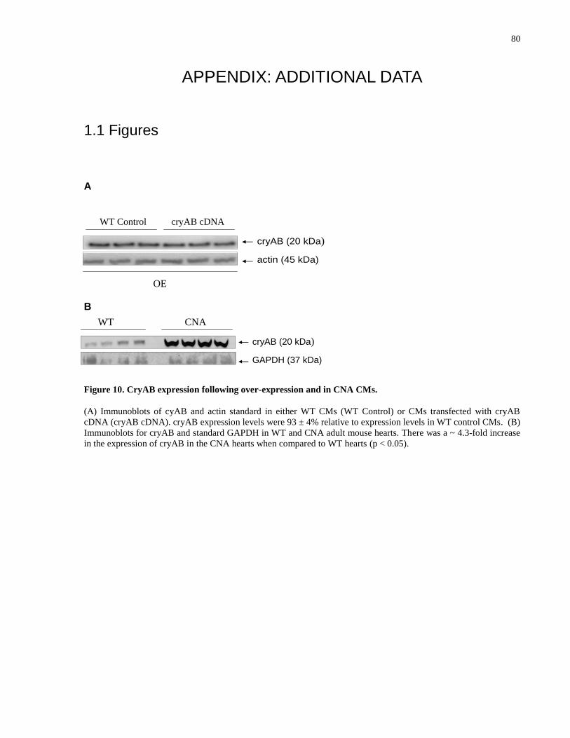
80
APPENDIX: ADDITIONAL DATA
1.1 Figures
A
B
Figure 10. CryAB expression following over-expression and in CNA CMs.
(A) Immunoblots of cyAB and actin standard in either WT CMs (WT Control) or CMs transfected with cryAB
cDNA (cryAB cDNA). cryAB expression levels were 93 ± 4% relative to expression levels in WT control CMs. (B)
Immunoblots for cryAB and standard GAPDH in WT and CNA adult mouse hearts. There was a ~ 4.3-fold increase
in the expression of cryAB in the CNA hearts when compared to WT hearts (p < 0.05).
cryAB (20 kDa)
actin (45 kDa)
Scrambled ScrambledshRNA cDNA
KD OE
WT CNA
cryAB (20 kDa)
GAPDH (37 kDa)
cryAB (20 kDa)
actin (45 kDa)
Scrambled ScrambledshRNA cDNA
KD OE
WT Control cryAB cDNA
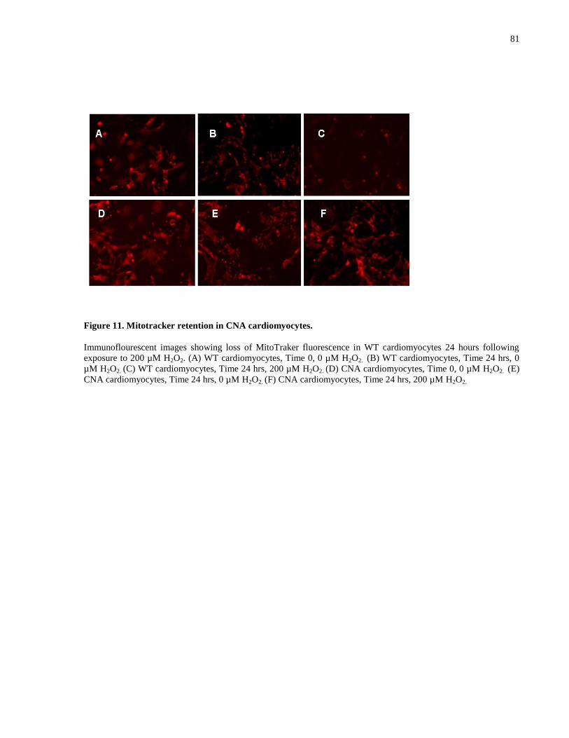
81
Figure 11. Mitotracker retention in CNA cardiomyocytes.
Immunoflourescent images showing loss of MitoTraker fluorescence in WT cardiomyocytes 24 hours following
exposure to 200 µM H2O2. (A) WT cardiomyocytes, Time 0, 0 µM H2O2. (B) WT cardiomyocytes, Time 24 hrs, 0
µM H2O2. (C) WT cardiomyocytes, Time 24 hrs, 200 µM H2O2. (D) CNA cardiomyocytes, Time 0, 0 µM H2O2. (E)
CNA cardiomyocytes, Time 24 hrs, 0 µM H2O2. (F) CNA cardiomyocytes, Time 24 hrs, 200 µM H2O2.
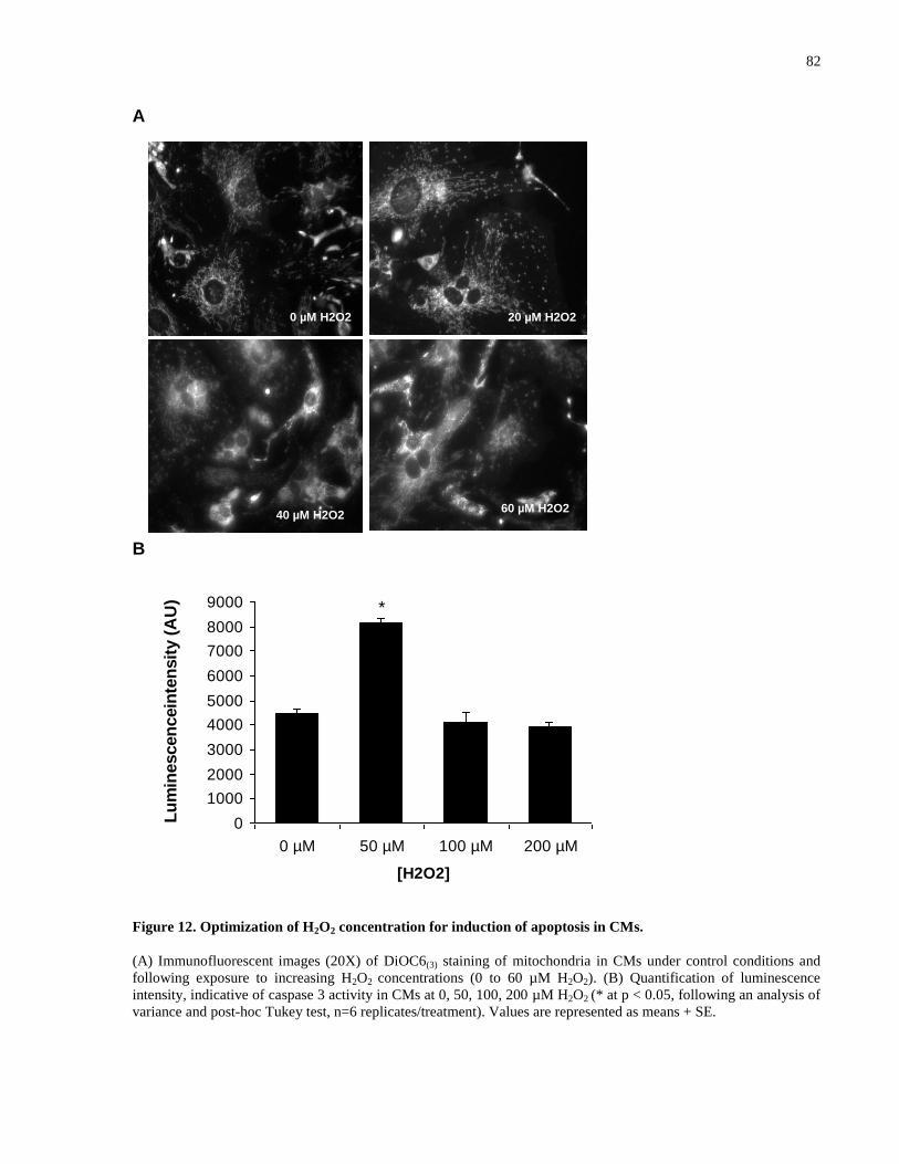
82
A
B
Figure 12. Optimization of H2O2 concentration for induction of apoptosis in CMs.
(A) Immunofluorescent images (20X) of DiOC6(3) staining of mitochondria in CMs under control conditions and
following exposure to increasing H2O2 concentrations (0 to 60 µM H2O2). (B) Quantification of luminescence
intensity, indicative of caspase 3 activity in CMs at 0, 50, 100, 200 µM H2O2 (* at p < 0.05, following an analysis of
variance and post-hoc Tukey test, n=6 replicates/treatment). Values are represented as means + SE.
0 µM H2O2 20 µM H2O2
40 µM H2O260 µM H2O2
0
1000
2000
3000
4000
5000
6000
7000
8000
9000
0 µM 50 µM 100 µM 200 µM
[H2O2]
Lu
min
escen
cein
ten
sit
y (
AU
) *
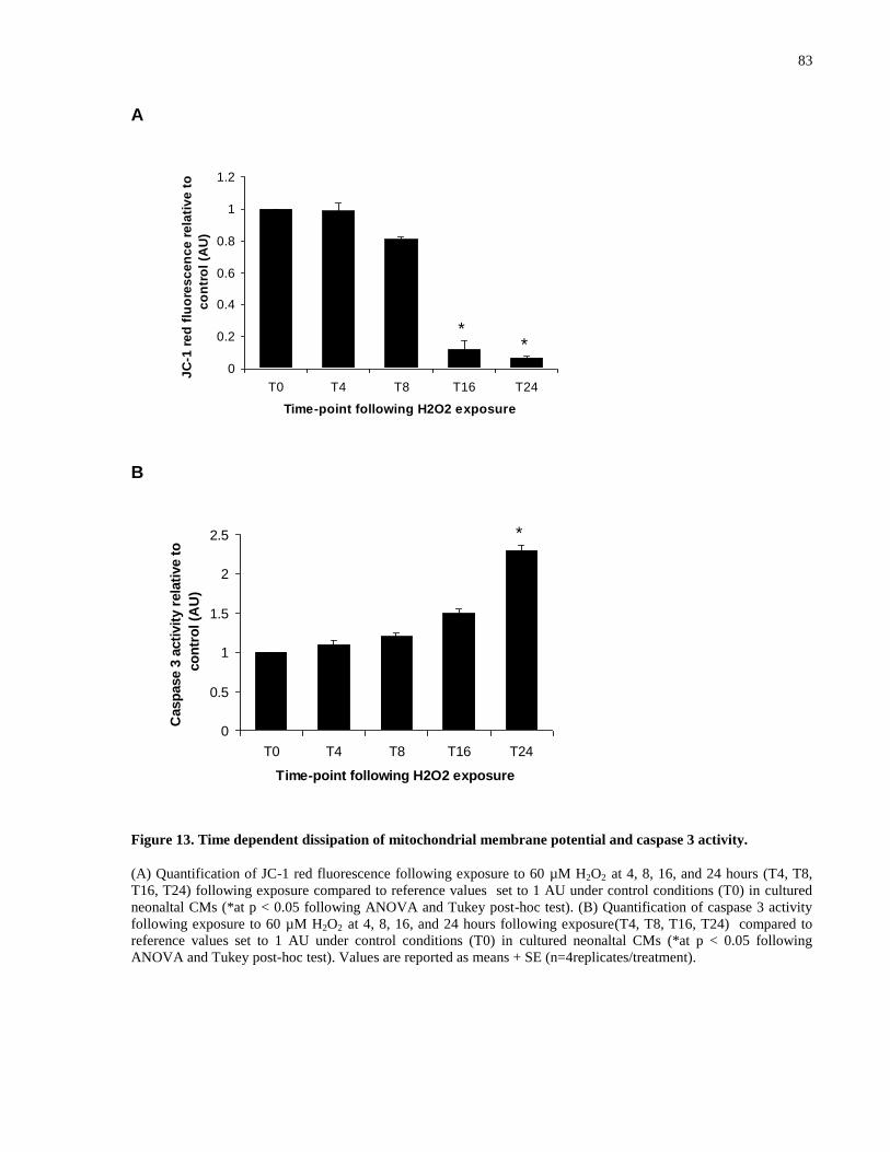
83
A
0
0.2
0.4
0.6
0.8
1
1.2
T0 T4 T8 T16 T24
Time-point following H2O2 exposure
JC
-1 r
ed
flu
ore
sc
en
ce
re
lati
ve
to
co
ntr
ol
(AU
)
B
0
0.5
1
1.5
2
2.5
T0 T4 T8 T16 T24
Time-point following H2O2 exposure
Casp
ase 3
acti
vit
y r
ela
tive t
o
co
ntr
ol (A
U)
Figure 13. Time dependent dissipation of mitochondrial membrane potential and caspase 3 activity.
(A) Quantification of JC-1 red fluorescence following exposure to 60 µM H2O2 at 4, 8, 16, and 24 hours (T4, T8,
T16, T24) following exposure compared to reference values set to 1 AU under control conditions (T0) in cultured
neonaltal CMs (*at p < 0.05 following ANOVA and Tukey post-hoc test). (B) Quantification of caspase 3 activity
following exposure to 60 µM H2O2 at 4, 8, 16, and 24 hours following exposure(T4, T8, T16, T24) compared to
reference values set to 1 AU under control conditions (T0) in cultured neonaltal CMs (*at p < 0.05 following
ANOVA and Tukey post-hoc test). Values are reported as means + SE (n=4replicates/treatment).
**
*
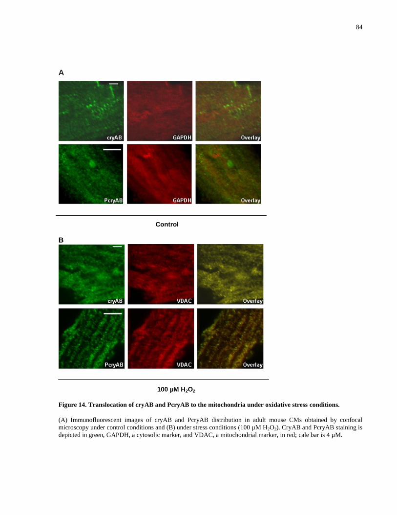
84
A
Control
B
100 µM H2O2
Figure 14. Translocation of cryAB and PcryAB to the mitochondria under oxidative stress conditions.
(A) Immunofluorescent images of cryAB and PcryAB distribution in adult mouse CMs obtained by confocal
microscopy under control conditions and (B) under stress conditions (100 µM H2O2). CryAB and PcryAB staining is
depicted in green, GAPDH, a cytosolic marker, and VDAC, a mitochondrial marker, in red; cale bar is 4 µM.
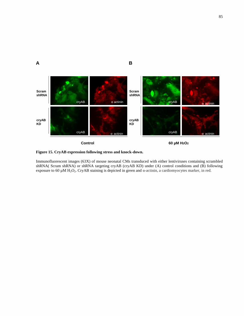
85
A B
Figure 15. CryAB expression following stress and knock-down.
Immunofluorescent images (63X) of mouse neonatal CMs transduced with either lentiviruses containing scrambled
shRNA( Scram shRNA) or shRNA targeting cryAB (cryAB KD) under (A) control conditions and (B) following
exposure to 60 µM H2O2. CryAB staining is depicted in green and α-actinin, a cardiomyocytes marker, in red.
cryAB cryAB
cryAB cryAB
α actininα actinin
α actinin α actinin
Control 60 µM H2O2
Scram
shRNA
Scram
shRNA
cryAB
KD
cryAB
KD
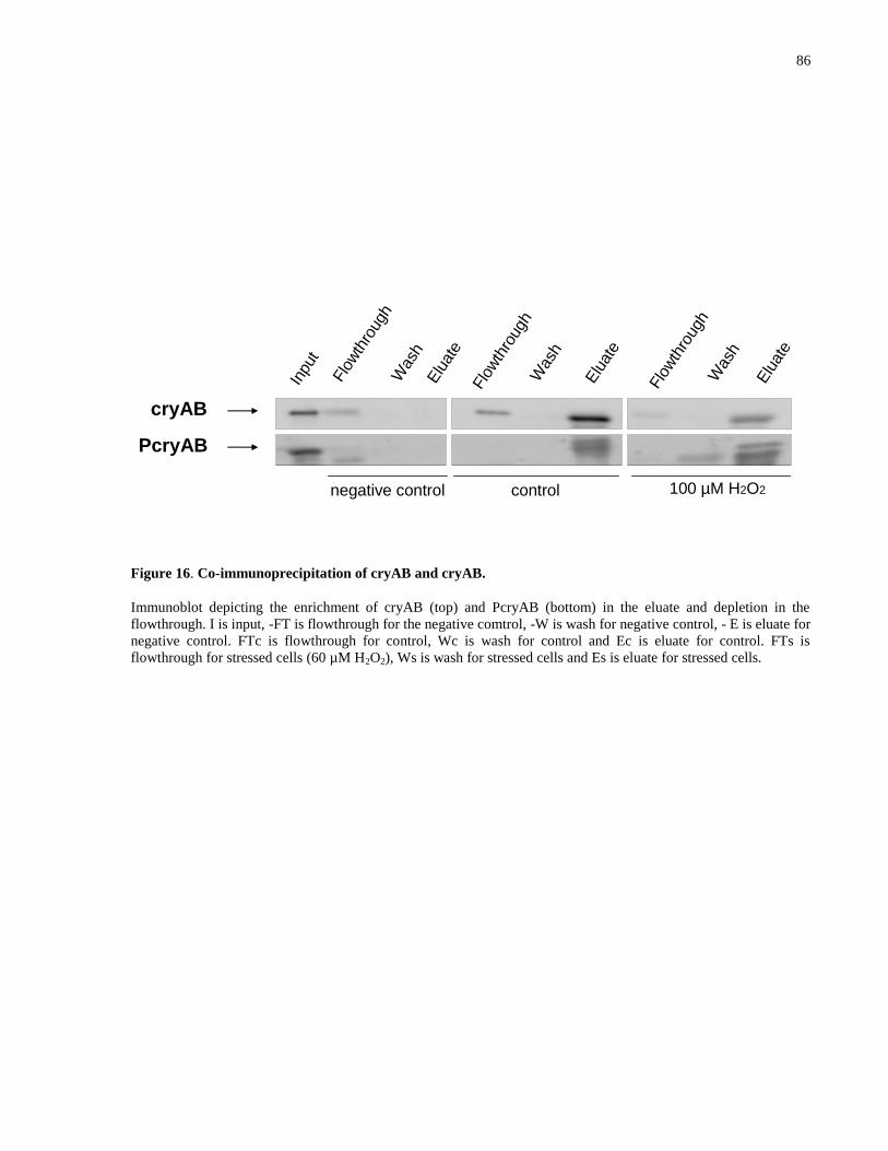
86
Figure 16. Co-immunoprecipitation of cryAB and cryAB.
Immunoblot depicting the enrichment of cryAB (top) and PcryAB (bottom) in the eluate and depletion in the
flowthrough. I is input, -FT is flowthrough for the negative comtrol, -W is wash for negative control, - E is eluate for
negative control. FTc is flowthrough for control, Wc is wash for control and Ec is eluate for control. FTs is
flowthrough for stressed cells (60 µM H2O2), Ws is wash for stressed cells and Es is eluate for stressed cells.
I -FT -W -E FTc Wc Ec FTs Ws Es
cryAB
PcryAB
Inpu
t
Flo
wth
roug
h
Was
hElu
ate
Flo
wth
roug
hW
ash
Elu
ate
Flo
wth
roug
h
Was
h
Elu
ate
negative control control 100 µM H2O2
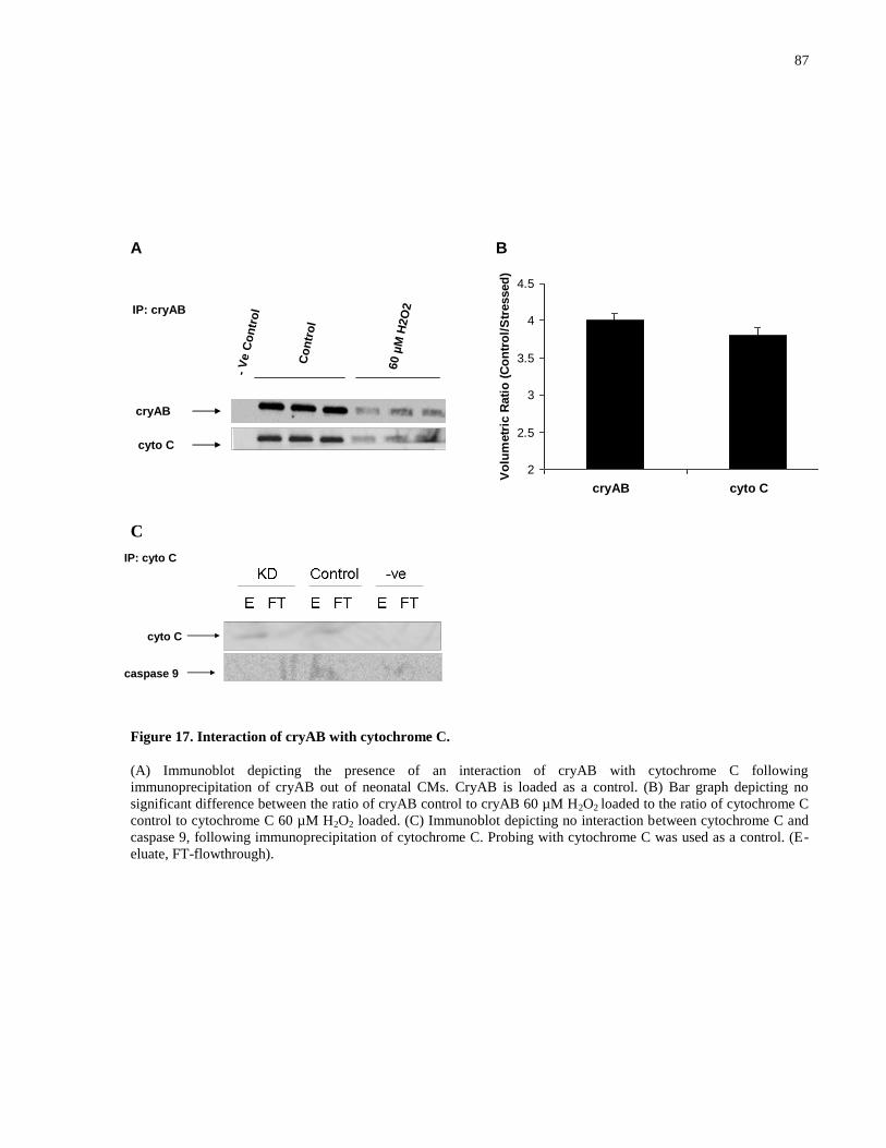
87
A B
C
Figure 17. Interaction of cryAB with cytochrome C.
(A) Immunoblot depicting the presence of an interaction of cryAB with cytochrome C following
immunoprecipitation of cryAB out of neonatal CMs. CryAB is loaded as a control. (B) Bar graph depicting no
significant difference between the ratio of cryAB control to cryAB 60 µM H2O2 loaded to the ratio of cytochrome C
control to cytochrome C 60 µM H2O2 loaded. (C) Immunoblot depicting no interaction between cytochrome C and
caspase 9, following immunoprecipitation of cytochrome C. Probing with cytochrome C was used as a control. (E-
eluate, FT-flowthrough).
Co
ntr
ol
-V
eC
on
tro
l
60 µ
M H
2O
2cryAB
cyto C
IP: cryAB
2
2.5
3
3.5
4
4.5
Cytochrome C CryAB
Vo
lum
etr
ic R
ati
o (
Co
ntr
ol/
Str
essed
)
cryAB cyto C
cyto C
caspase 9
IP: cyto C
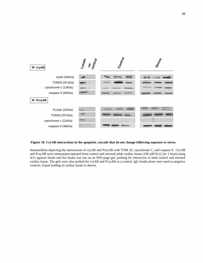
88
Figure 18. CryAB interactions in the apoptotic cascade that do not change following exposure to stress.
Immunoblots depicting the interactions of cryAB and PcryAB with TOM 20, cytochrome C, and caspase 9. CryAB
and PcryAB were immunoprecipitated from control and stressed adult cardiac tissue (100 µM H2O2 for 1 hour) using
A/G agarose beads and the eluate was run on an SDS-page gel, probing for interactors in both control and stressed
cardiac tissue. The gels were also probed for cryAB and PcryAB as a control. IgG beads alone were used as negative
controls. Equal loading of cardiac lysate is shown.
Contr
ol
TOM20 (20 kDa)
cytochrome c (12kDa)
Pcryab (22kDa)
caspase 9 (46kDa)
TOM20 (20 kDa)
cytochrome c (12kDa)
cryab (20kDa)
caspase 9 (46kDa)
Lysate
-ve
co
ntr
ol
Str
ess
IP: cryAB
IP: PcryAB
89
1.2 Results and Discussion
1.2.1 Over-expression of cryAB in mouse neonatal CMs
In order to validate the protective properties of cryAB against apoptosis that we initially
uncovered106
, I over-expressed cryAB in cultured mouse neonatal CMs, using using lentivector
mediated transduction of cryAB cDNA. A scrambled control was also used. Lentivector
production and transduction of neonatal CMs was carried out as described in Chapter 2. The
success of the over-expression transduction was validated via immunoblot analysis. The over-
expression did not lead to upregulation of cryAB in the cardiomyocytes (Fig. 10). Following
transduction, cryAB expression levels were 93 ± 4% relative to expression levels in control CMs
(Fig. 10). Probing for actin was used as a control and it shows a uniform distribution in the WT
CMs and the CMs transduced with the cryAB cDNA construct.
CNA cardiomyocytes in which cryAB is upregulated were as an alternative to the over-
expression model. The upregulation of cryAB was validated by Western Blot analysis of protein
isolated from adult whole heart lysates (Fig. 10). There was a ~ 4.3-fold increase in the
expression of cryAB in the CNA hearts when compared to WT hearts (expression levels of 425 ±
40% in CNA hearts compared to 100% levels observed in WT hearts; p < 0.05).
1.2.2 Protection against apoptosis
To determine whether upregulation of cryAB was protective against apoptosis in CNA CMs, WT
and CNA CMs were exposed to 200 µM H2O2 and Mitotracker fluorescence was examined.
Mitotracker is a cell-permeable dye that accumulates in active mitochondria, where it becomes
oxidized and fluorescent. Mitotracker fluorescence thus gives an indication of actively respiring
cells, or live cells. Upon exposure of CNA and WT CMs to 200 µM H2O2, the CNA CMs
retained Mitotracker fluorescence after 24 hours, whereas WT CMs seemed to have lost almost
all mitochondrial staining (Fig. 11). This suggests that CNA CMs are protected against H2O2-
induced apoptosis. However, it was not possible to single out cryAB as a mediator of this effect,
due to the many other alterations in CNA CMs when compared to WT CMs. Future experiments
focused only on CMs in which cryAB had been silenced (KD cryAB).
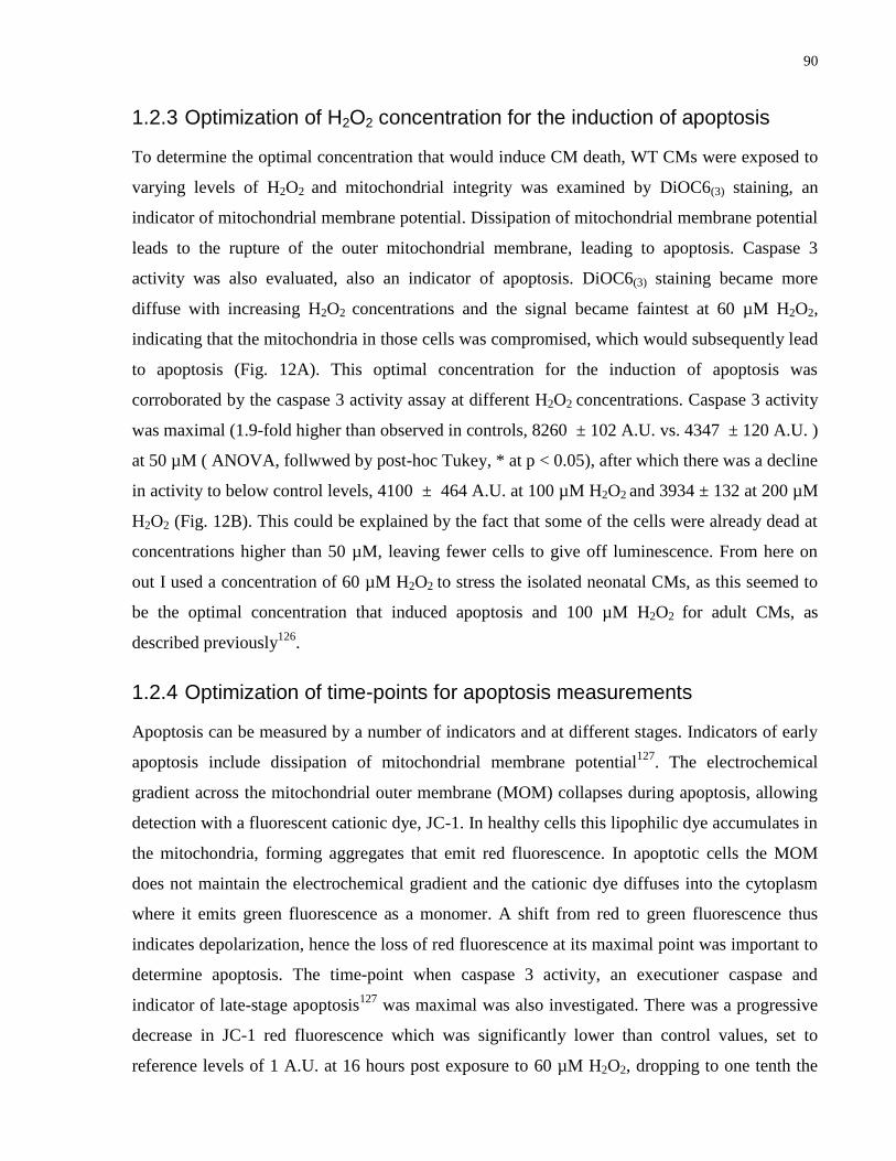
90
1.2.3 Optimization of H2O2 concentration for the induction of apoptosis
To determine the optimal concentration that would induce CM death, WT CMs were exposed to
varying levels of H2O2 and mitochondrial integrity was examined by DiOC6(3) staining, an
indicator of mitochondrial membrane potential. Dissipation of mitochondrial membrane potential
leads to the rupture of the outer mitochondrial membrane, leading to apoptosis. Caspase 3
activity was also evaluated, also an indicator of apoptosis. DiOC6(3) staining became more
diffuse with increasing H2O2 concentrations and the signal became faintest at 60 µM H2O2,
indicating that the mitochondria in those cells was compromised, which would subsequently lead
to apoptosis (Fig. 12A). This optimal concentration for the induction of apoptosis was
corroborated by the caspase 3 activity assay at different H2O2 concentrations. Caspase 3 activity
was maximal (1.9-fold higher than observed in controls, 8260 ± 102 A.U. vs. 4347 ± 120 A.U. )
at 50 µM ( ANOVA, follwwed by post-hoc Tukey, * at p < 0.05), after which there was a decline
in activity to below control levels, 4100 ± 464 A.U. at 100 µM H2O2 and 3934 ± 132 at 200 µM
H2O2 (Fig. 12B). This could be explained by the fact that some of the cells were already dead at
concentrations higher than 50 µM, leaving fewer cells to give off luminescence. From here on
out I used a concentration of 60 µM H2O2 to stress the isolated neonatal CMs, as this seemed to
be the optimal concentration that induced apoptosis and 100 µM H2O2 for adult CMs, as
described previously126
.
1.2.4 Optimization of time-points for apoptosis measurements
Apoptosis can be measured by a number of indicators and at different stages. Indicators of early
apoptosis include dissipation of mitochondrial membrane potential127
. The electrochemical
gradient across the mitochondrial outer membrane (MOM) collapses during apoptosis, allowing
detection with a fluorescent cationic dye, JC-1. In healthy cells this lipophilic dye accumulates in
the mitochondria, forming aggregates that emit red fluorescence. In apoptotic cells the MOM
does not maintain the electrochemical gradient and the cationic dye diffuses into the cytoplasm
where it emits green fluorescence as a monomer. A shift from red to green fluorescence thus
indicates depolarization, hence the loss of red fluorescence at its maximal point was important to
determine apoptosis. The time-point when caspase 3 activity, an executioner caspase and
indicator of late-stage apoptosis127
was maximal was also investigated. There was a progressive
decrease in JC-1 red fluorescence which was significantly lower than control values, set to
reference levels of 1 A.U. at 16 hours post exposure to 60 µM H2O2, dropping to one tenth the
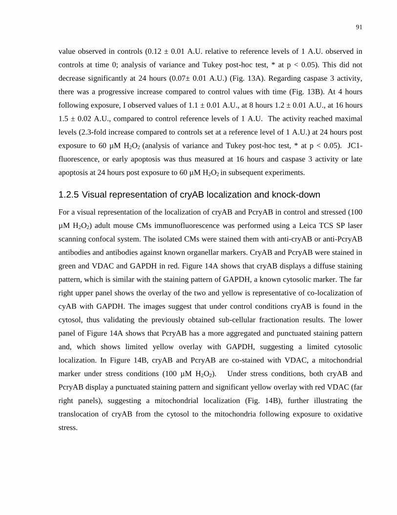
91
value observed in controls (0.12 ± 0.01 A.U. relative to reference levels of 1 A.U. observed in
controls at time 0; analysis of variance and Tukey post-hoc test, * at p < 0.05). This did not
decrease significantly at 24 hours (0.07± 0.01 A.U.) (Fig. 13A). Regarding caspase 3 activity,
there was a progressive increase compared to control values with time (Fig. 13B). At 4 hours
following exposure, I observed values of 1.1 ± 0.01 A.U., at 8 hours 1.2 ± 0.01 A.U., at 16 hours
1.5 ± 0.02 A.U., compared to control reference levels of 1 A.U. The activity reached maximal
levels (2.3-fold increase compared to controls set at a reference level of 1 A.U.) at 24 hours post
exposure to 60 µM H2O2 (analysis of variance and Tukey post-hoc test, * at p < 0.05). JC1-
fluorescence, or early apoptosis was thus measured at 16 hours and caspase 3 activity or late
apoptosis at 24 hours post exposure to 60 µM H2O2 in subsequent experiments.
1.2.5 Visual representation of cryAB localization and knock-down
For a visual representation of the localization of cryAB and PcryAB in control and stressed (100
µM H2O2) adult mouse CMs immunofluorescence was performed using a Leica TCS SP laser
scanning confocal system. The isolated CMs were stained them with anti-cryAB or anti-PcryAB
antibodies and antibodies against known organellar markers. CryAB and PcryAB were stained in
green and VDAC and GAPDH in red. Figure 14A shows that cryAB displays a diffuse staining
pattern, which is similar with the staining pattern of GAPDH, a known cytosolic marker. The far
right upper panel shows the overlay of the two and yellow is representative of co-localization of
cyAB with GAPDH. The images suggest that under control conditions cryAB is found in the
cytosol, thus validating the previously obtained sub-cellular fractionation results. The lower
panel of Figure 14A shows that PcryAB has a more aggregated and punctuated staining pattern
and, which shows limited yellow overlay with GAPDH, suggesting a limited cytosolic
localization. In Figure 14B, cryAB and PcryAB are co-stained with VDAC, a mitochondrial
marker under stress conditions (100 µM H2O2). Under stress conditions, both cryAB and
PcryAB display a punctuated staining pattern and significant yellow overlay with red VDAC (far
right panels), suggesting a mitochondrial localization (Fig. 14B), further illustrating the
translocation of cryAB from the cytosol to the mitochondria following exposure to oxidative
stress.
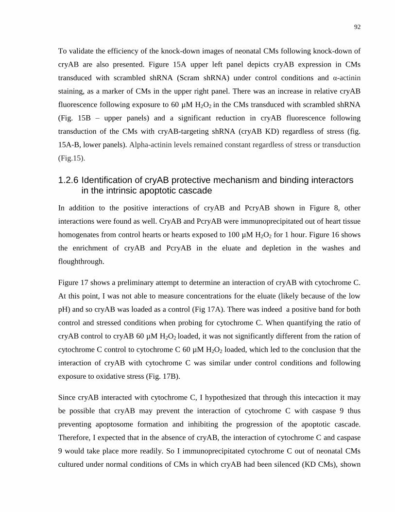
92
To validate the efficiency of the knock-down images of neonatal CMs following knock-down of
cryAB are also presented. Figure 15A upper left panel depicts cryAB expression in CMs
transduced with scrambled shRNA (Scram shRNA) under control conditions and α-actinin
staining, as a marker of CMs in the upper right panel. There was an increase in relative cryAB
fluorescence following exposure to 60 µM H2O2 in the CMs transduced with scrambled shRNA
(Fig. 15B – upper panels) and a significant reduction in cryAB fluorescence following
transduction of the CMs with cryAB-targeting shRNA (cryAB KD) regardless of stress (fig.
15A-B, lower panels). Alpha-actinin levels remained constant regardless of stress or transduction
(Fig.15).
1.2.6 Identification of cryAB protective mechanism and binding interactors in the intrinsic apoptotic cascade
In addition to the positive interactions of cryAB and PcryAB shown in Figure 8, other
interactions were found as well. CryAB and PcryAB were immunoprecipitated out of heart tissue
homogenates from control hearts or hearts exposed to 100 µM H2O2 for 1 hour. Figure 16 shows
the enrichment of cryAB and PcryAB in the eluate and depletion in the washes and
floughthrough.
Figure 17 shows a preliminary attempt to determine an interaction of cryAB with cytochrome C.
At this point, I was not able to measure concentrations for the eluate (likely because of the low
pH) and so cryAB was loaded as a control (Fig 17A). There was indeed a positive band for both
control and stressed conditions when probing for cytochrome C. When quantifying the ratio of
cryAB control to cryAB 60 µM H2O2 loaded, it was not significantly different from the ration of
cytochrome C control to cytochrome C 60 µM H2O2 loaded, which led to the conclusion that the
interaction of cryAB with cytochrome C was similar under control conditions and following
exposure to oxidative stress (Fig. 17B).
Since cryAB interacted with cytochrome C, I hypothesized that through this intecaction it may
be possible that cryAB may prevent the interaction of cytochrome C with caspase 9 thus
preventing apoptosome formation and inhibiting the progression of the apoptotic cascade.
Therefore, I expected that in the absence of cryAB, the interaction of cytochrome C and caspase
9 would take place more readily. So I immunoprecipitated cytochrome C out of neonatal CMs
cultured under normal conditions of CMs in which cryAB had been silenced (KD CMs), shown
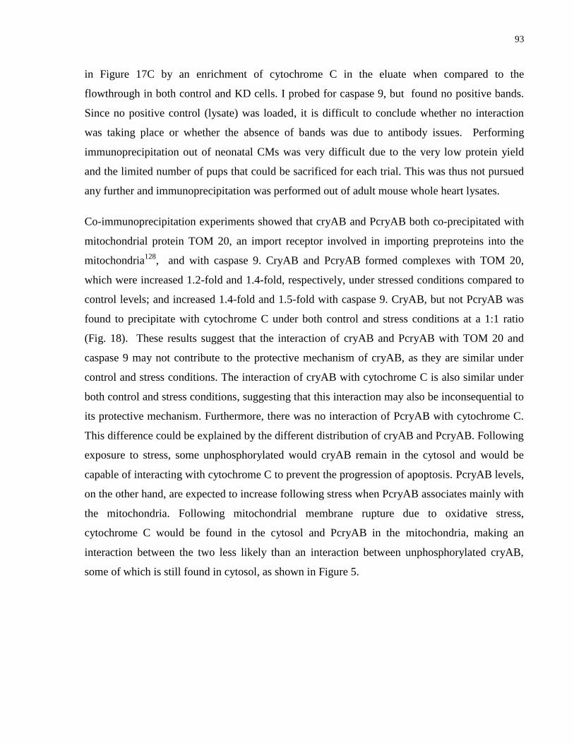
93
in Figure 17C by an enrichment of cytochrome C in the eluate when compared to the
flowthrough in both control and KD cells. I probed for caspase 9, but found no positive bands.
Since no positive control (lysate) was loaded, it is difficult to conclude whether no interaction
was taking place or whether the absence of bands was due to antibody issues. Performing
immunoprecipitation out of neonatal CMs was very difficult due to the very low protein yield
and the limited number of pups that could be sacrificed for each trial. This was thus not pursued
any further and immunoprecipitation was performed out of adult mouse whole heart lysates.
Co-immunoprecipitation experiments showed that cryAB and PcryAB both co-precipitated with
mitochondrial protein TOM 20, an import receptor involved in importing preproteins into the
mitochondria128
, and with caspase 9. CryAB and PcryAB formed complexes with TOM 20,
which were increased 1.2-fold and 1.4-fold, respectively, under stressed conditions compared to
control levels; and increased 1.4-fold and 1.5-fold with caspase 9. CryAB, but not PcryAB was
found to precipitate with cytochrome C under both control and stress conditions at a 1:1 ratio
(Fig. 18). These results suggest that the interaction of cryAB and PcryAB with TOM 20 and
caspase 9 may not contribute to the protective mechanism of cryAB, as they are similar under
control and stress conditions. The interaction of cryAB with cytochrome C is also similar under
both control and stress conditions, suggesting that this interaction may also be inconsequential to
its protective mechanism. Furthermore, there was no interaction of PcryAB with cytochrome C.
This difference could be explained by the different distribution of cryAB and PcryAB. Following
exposure to stress, some unphosphorylated would cryAB remain in the cytosol and would be
capable of interacting with cytochrome C to prevent the progression of apoptosis. PcryAB levels,
on the other hand, are expected to increase following stress when PcryAB associates mainly with
the mitochondria. Following mitochondrial membrane rupture due to oxidative stress,
cytochrome C would be found in the cytosol and PcryAB in the mitochondria, making an
interaction between the two less likely than an interaction between unphosphorylated cryAB,
some of which is still found in cytosol, as shown in Figure 5.