LECTURE: 03 Title: THE IMMUNOLOGICAL PROTECTIVE …
22
1 LECTURE : 03 Title: THE IMMUNOLOGICAL PROTECTIVE MECHANISMS TO BACTERIAL AND FUNGAL INFECTIONS LEARNING OBJECTIVES : The student should be able to: • Identify the types of immunity involve in the mechanisms of protection against bacterial and fungal infections. - Cellular immunity (delayed type "type IV" in case of fungal infections). a. Production of IFN-γ by Th1 which activate phagocytes. b. Involvement of neutrophil, and polymorph in respiratory mycoses. c. Phagocytosis: 1. Oxygen-dependent mechanism. 2. Oxygen-independent mechanism. - Humoral immunity (bacterial infection). a. Neutralizing antibody (Extracellular bacteria) Tcell-independent antigens (cell wall and capsule of most bacteria are polysaccharides). b. By low pH environment on the skin produced as a result of production of lactic acid in sweat glands secretions. c. Enzymes such as lysozyme which is found in the mucous secretions e.g., tears and saliva. d. Acute inflammatory response: 1. C-reactive proteins (CRP). 2. The complement proteins. e. Cytokines: 1. IFN type 1(IFN- α, and β). 2. IL-1 3. TNF- α. 4. IFN- γ 5. IL-2. • List some common serological techniques used in bacterial and fungal diagnosis, providing some examples of bacteria, and fungi. LECTURE REFRENCES : 1. TEXTBOOK: ROITT, BROSTOFF, MALE IMMUNOLOGY. 6 th edition. Chapter 15. pp 245- 256 2. HANDOUT.
Transcript of LECTURE: 03 Title: THE IMMUNOLOGICAL PROTECTIVE …
1
LECTURE: 03
Title: THE IMMUNOLOGICAL PROTECTIVE MECHANISMS
TO BACTERIAL AND FUNGAL INFECTIONS
LEARNING OBJECTIVES: The student should be able to: • Identify the types of immunity involve in the mechanisms of protection
against bacterial and fungal infections. - Cellular immunity (delayed type "type IV" in case of fungal infections).
a. Production of IFN-γ by Th1 which activate phagocytes. b. Involvement of neutrophil, and polymorph in respiratory mycoses. c. Phagocytosis:
1. Oxygen-dependent mechanism. 2. Oxygen-independent mechanism.
- Humoral immunity (bacterial infection).
a. Neutralizing antibody (Extracellular bacteria) Tcell-independent antigens
(cell wall and capsule of most bacteria are polysaccharides). b. By low pH environment on the skin produced as a result of production of
lactic acid in sweat glands secretions. c. Enzymes such as lysozyme which is found in the mucous secretions e.g.,
tears and saliva. d. Acute inflammatory response:
1. C-reactive proteins (CRP). 2. The complement proteins.
e. Cytokines:
1. IFN type 1(IFN- α, and β). 2. IL-1 3. TNF- α. 4. IFN- γ 5. IL-2.
• List some common serological techniques used in bacterial and fungal diagnosis, providing some examples of bacteria, and fungi. LECTURE REFRENCES:
1. TEXTBOOK: ROITT, BROSTOFF, MALE IMMUNOLOGY. 6th edition. Chapter 15. pp 245- 256 2. HANDOUT.
2
Immunity to bacteria and fungi GENERAL INTRODUCTION
Mechanisms of protection from a bacterial species can be deduced from the structure of the organism, particularly its cell wall, and its mode of pathogenicity.
Neutralizing antibody may be all that is needed for protection if the organism is pathogenic only because of a single toxin or adhesion molecule.
Non-specific, phylogeneetically ancient recognition pathways for conserved bacterial structures trigger phagocytosis, the alternative complement pathway and release of cytokines.
Complement can kill some bacteria, particularly those with an exposed outer lipid bilayer such as Gram-negative bacteria.
Phagocytes kill the majority of bacteria following a multistage process of chemotaxis, attachment, uptake and killing.
Successful pathogens have evolved a starting diversity of mechanisms for avoiding the effects of complement, avoiding phagocyte function, or misdirecting the T cell-dependent activation of phagocyte killing mechanisms.
Excessive release of cytokines caused by microorganisms can result in immunopathological syndromes, such as endotoxin shock and the Shwartzman reaction.
Chronic tissue-damaging immunopathology (as in tuberculosis) probably results from an imbalance of cytokine release patterns, leading to inappropriate effector function.
Immunity to fungi is poorly understood but is apparently cell-mediated and similar to immunity to bacteria.
IMMUNITY TO BACTERIA
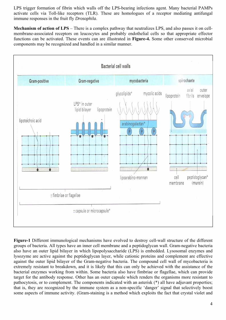
The defense mechanisms appropriate for a particular bacterial infection are related to the structure of the invading bacteria, and hence the immunological mechanisms to which are susceptible, and to the mechanism of their pathogenicity. There are four main types of bacterial cell wall (Figure-1) belonging to the following groups:
• Gram-positive bacteria
• Gram-negative bacteria
• Mycobacteria
• Spirochetes.
The outer lipid bilayer Gram-negative organisms are of particular importance because it is often susceptible to mechanisms that can lyse membranes, such as complement and certain cytotoxic cells. In contrast, killing of the other types of bacteria usually requires uptake by phagocytes.
The outer surface of the bacterium may also contain fimbriac or flagellae or it may be covered by a protective capsule. These can impede the functions of phagocytes or complement, but they also act as targets for the antibody response, the role of which is discussed later.
3
Mechanisms of immunity are related to bacterial mechanisms of pathogenicity
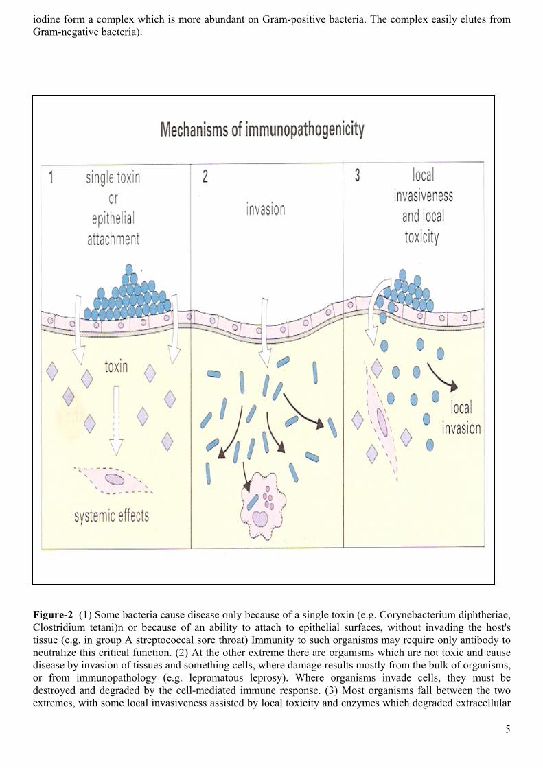
The two extreme patterns of pathogenicity are:
• Toxicity without invasiveness
• Invasiveness without toxicity (Figure-2).
However, most bacteria are intermediate between these extremes, having some invasiveness assisted by some locally acting toxins and spreading factors (tissue-degrading enzymes).
Corynebacterium diphtheriae and Vibria choleae are example of organisms that are toxic but not invasive. Since their pathogenicity depends almost entirely on toxin production, neutralizing antibody to the toxin is probably sufficient for immunity, although antibody, binding to the bacteria and so blocking their adhesion to the epithelium, could also be important.
In contrast, however, the pathogenicity of most invasive organisms does not rely so heavily on a single toxin, so immunity requires killing of the organisms themselves.
The first lines of defense are antibacterial mechanisms that do not depend on antigen recognition
The body’s first line of defense against pathogenic bacteria consists of simple barriers to the entry or establishment of the infection. Thus, the skin and exposed epithelial surface have non-specific or innate protective systems which limit the entry of potentially invasive organisms (Figure-1). Intact skin is impenetrable to most bacteria. Additionally, fatty acids produced by the skin are toxic to many organisms. Indeed, the pathogenicity of some strains correlates with their ability to survive on the skin. Epithelial surfaces are cleansed, for example, by ciliary action in the trachea or by flushing of the urinary tract. Many bacteria are destroyed by pH changes in the stomach and vagina, both of which provide an acidic environment. In the vagina, the epithelium secretes glycogen, which is metabolized by particular species of commensal bacteria, producing lactic acid. More generally, commensals can limit pathogen invasion through production of antibacterial proteins termed colicins. Thus commensals may occupy an ecological niche that would otherwise be occupied by something more unpleasant. When the normal flora are disturbed by antibiotics, infections by Candida or Clostridium difficile can occur, and the latter is a major cause of antibiotic-induced colitis and diarrhea. Several studies suggest that the re-introducing of non-pathogenic ‘probiotic’ organisms such as lactobacilli into the intestinal tract can alleviate the symptoms, presumably by replacing those killed by the antibiotics.
In practice, only a minute proportion of the potentially pathogenic organisms around us ever succeed in gaining access to the tissues.
The second line of defense is mediated via recognition of common bacterial components
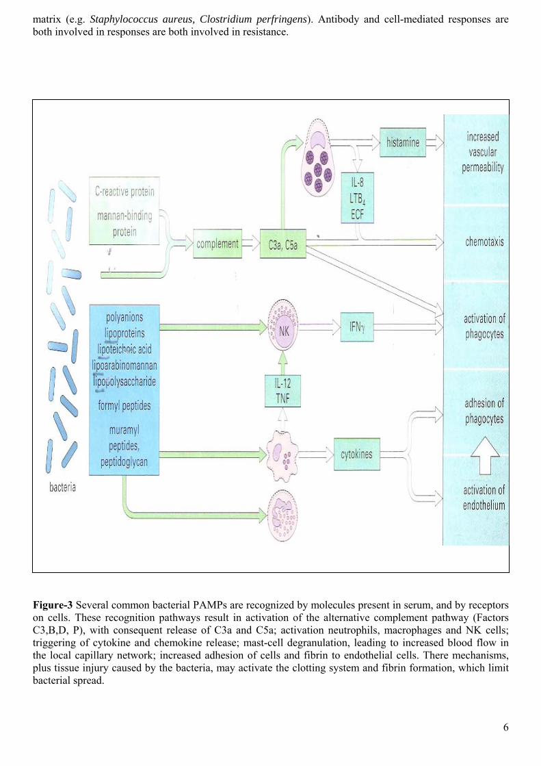
If the organisms do enter the tissues, they can be combated initially by further elements of the innate immune system. Numerous bacterial components are recognized in ways which do not rely on the antigen-specific receptors of either B cells or T cells. These types of recognition are phylogeneetically ancient ‘broad-spectrum’ mechanisms that evolved before antigen-specific T cells and immunoglobulins, allowing protective responses to be triggered by common microbial components bearing so-called ‘pathogen-associated molecular patterns’ (PAMPs) such as lipopolysaccharide and mannan. The host molecules which recognize these microbial components are referred to as the ‘pattern recognition molecules’ of the innate immune system. Many organisms, such as non-pathogenic cocci, are probably removed from the tissues as a consequence of these pathways, without the need for a specific adaptive immune reaction. Figure-3 shows some of the microbial components involved, and the host responses which are triggered. It is interesting to note that the ‘Limulus assay’, which is used to detect contaminating lipopolysaccharide (LPS) in preparations for use in humans is based on one such recognition pathway found in an invertebrate species of
LPS trigger formation of fibrin which walls off the LPS-bearing infections agent. Many bacterial PAMPs activate cells via Toll-like receptors (TLR). These are homologues of a receptor mediating antifungal immune responses in the fruit fly Drosophila.
Mechanism of action of LPS – There is a complex pathway that neutralizes LPS, and also passes it on cell-membrane-associated receptors on leucocytes and probably endothelial cells so that appropriate effector functions can be activated. These events can are illustrated in Figure-4. Some other conserved microbial components may be recognized and handled in a similar manner.
Figure-1 Different immunological mechanisms have evolved to destroy cell-wall structure of the different groups of bacteria. All types have an inner cell membrane and a peptidoglycan wall. Gram-negative bacteria also have an outer lipid bilayer in which lipopolysaccharide (LPS) is embedded. Lysosomal enzymes and lysozyme are active against the peptidoglycan layer, while cationic proteins and complement are effective against the outer lipid bilayer of the Gram-negative bacteria. The compound cell wall of mycobacteria is extremely resistant to breakdown, and it is likely that this can only be achieved with the assistance of the bacterial enzymes working from within. Some bacteria also have fimbriae or flagellae, which can provide target for the antibody response. Other has an outer capsule which renders the organisms more resistant to pathocytosis, or to complement. The components indicated with an asterisk (*) all have adjuvant properties; that is, they are recognized by the immune system as a non-specific ‘danger’ signal that selectively boost some aspects of immune activity. (Gram-staining is a method which exploits the fact that crystal violet and
4
iodine form a complex which is more abundant on Gram-positive bacteria. The complex easily elutes from Gram-negative bacteria).
Figure-2 (1) Some bacteria cause disease only because of a single toxin (e.g. Corynebacterium diphtheriae, Clostridium tetani)n or because of an ability to attach to epithelial surfaces, without invading the host's tissue (e.g. in group A streptococcal sore throat) Immunity to such organisms may require only antibody to neutralize this critical function. (2) At the other extreme there are organisms which are not toxic and cause disease by invasion of tissues and something cells, where damage results mostly from the bulk of organisms, or from immunopathology (e.g. lepromatous leprosy). Where organisms invade cells, they must be destroyed and degraded by the cell-mediated immune response. (3) Most organisms fall between the two extremes, with some local invasiveness assisted by local toxicity and enzymes which degraded extracellular
5
matrix (e.g. Staphylococcus aureus, Clostridium perfringens). Antibody and cell-mediated responses are both involved in responses are both involved in resistance.
Figure-3 Several common bacterial PAMPs are recognized by molecules present in serum, and by receptors on cells. These recognition pathways result in activation of the alternative complement pathway (Factors C3,B,D, P), with consequent release of C3a and C5a; activation neutrophils, macrophages and NK cells; triggering of cytokine and chemokine release; mast-cell degranulation, leading to increased blood flow in the local capillary network; increased adhesion of cells and fibrin to endothelial cells. There mechanisms, plus tissue injury caused by the bacteria, may activate the clotting system and fibrin formation, which limit bacterial spread.
6
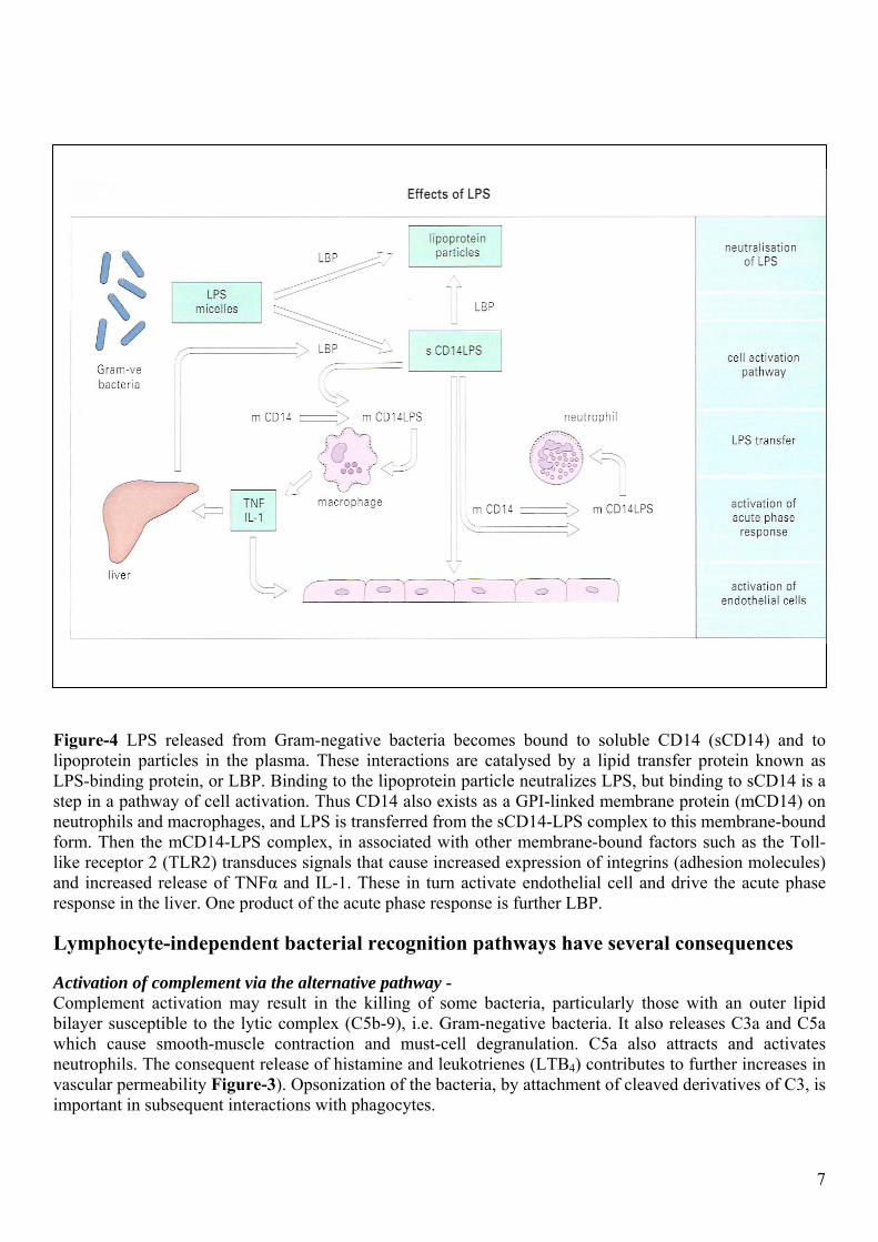
Figure-4 LPS released from Gram-negative bacteria becomes bound to soluble CD14 (sCD14) and to lipoprotein particles in the plasma. These interactions are catalysed by a lipid transfer protein known as LPS-binding protein, or LBP. Binding to the lipoprotein particle neutralizes LPS, but binding to sCD14 is a step in a pathway of cell activation. Thus CD14 also exists as a GPI-linked membrane protein (mCD14) on neutrophils and macrophages, and LPS is transferred from the sCD14-LPS complex to this membrane-bound form. Then the mCD14-LPS complex, in associated with other membrane-bound factors such as the Toll-like receptor 2 (TLR2) transduces signals that cause increased expression of integrins (adhesion molecules) and increased release of TNFα and IL-1. These in turn activate endothelial cell and drive the acute phase response in the liver. One product of the acute phase response is further LBP.
Lymphocyte-independent bacterial recognition pathways have several consequences
Activation of complement via the alternative pathway - Complement activation may result in the killing of some bacteria, particularly those with an outer lipid bilayer susceptible to the lytic complex (C5b-9), i.e. Gram-negative bacteria. It also releases C3a and C5a which cause smooth-muscle contraction and must-cell degranulation. C5a also attracts and activates neutrophils. The consequent release of histamine and leukotrienes (LTB4) contributes to further increases in vascular permeability Figure-3). Opsonization of the bacteria, by attachment of cleaved derivatives of C3, is important in subsequent interactions with phagocytes.
7
8
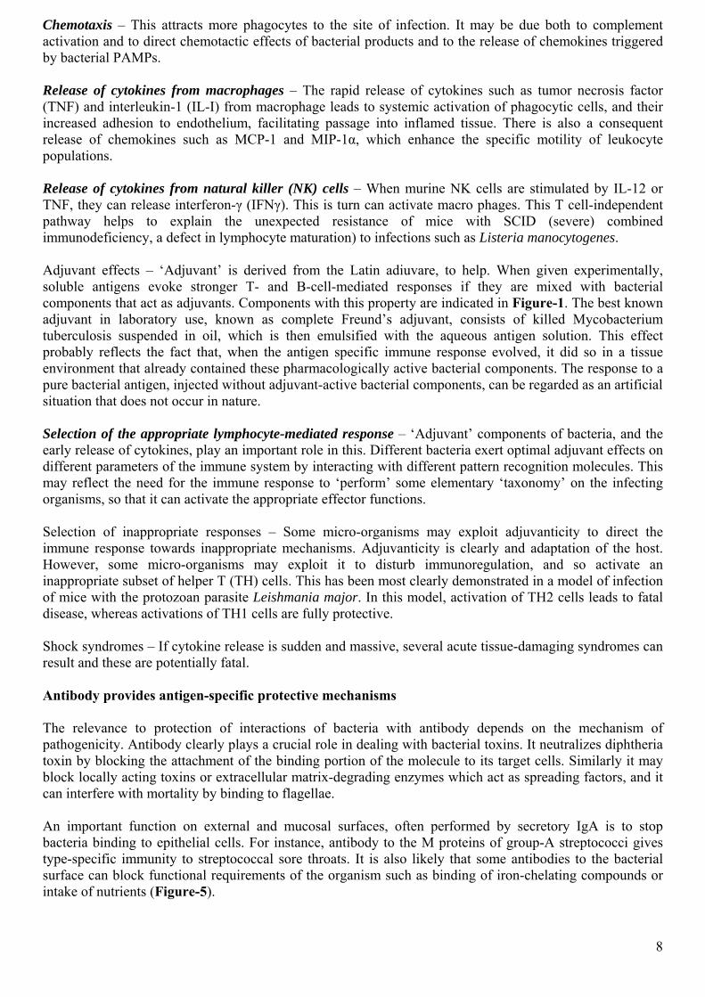
Chemotaxis – This attracts more phagocytes to the site of infection. It may be due both to complement activation and to direct chemotactic effects of bacterial products and to the release of chemokines triggered by bacterial PAMPs. Release of cytokines from macrophages – The rapid release of cytokines such as tumor necrosis factor (TNF) and interleukin-1 (IL-I) from macrophage leads to systemic activation of phagocytic cells, and their increased adhesion to endothelium, facilitating passage into inflamed tissue. There is also a consequent release of chemokines such as MCP-1 and MIP-1α, which enhance the specific motility of leukocyte populations. Release of cytokines from natural killer (NK) cells – When murine NK cells are stimulated by IL-12 or TNF, they can release interferon-γ (IFNγ). This is turn can activate macro phages. This T cell-independent pathway helps to explain the unexpected resistance of mice with SCID (severe) combined immunodeficiency, a defect in lymphocyte maturation) to infections such as Listeria manocytogenes. Adjuvant effects – ‘Adjuvant’ is derived from the Latin adiuvare, to help. When given experimentally, soluble antigens evoke stronger T- and B-cell-mediated responses if they are mixed with bacterial components that act as adjuvants. Components with this property are indicated in Figure-1. The best known adjuvant in laboratory use, known as complete Freund’s adjuvant, consists of killed Mycobacterium tuberculosis suspended in oil, which is then emulsified with the aqueous antigen solution. This effect probably reflects the fact that, when the antigen specific immune response evolved, it did so in a tissue environment that already contained these pharmacologically active bacterial components. The response to a pure bacterial antigen, injected without adjuvant-active bacterial components, can be regarded as an artificial situation that does not occur in nature. Selection of the appropriate lymphocyte-mediated response – ‘Adjuvant’ components of bacteria, and the early release of cytokines, play an important role in this. Different bacteria exert optimal adjuvant effects on different parameters of the immune system by interacting with different pattern recognition molecules. This may reflect the need for the immune response to ‘perform’ some elementary ‘taxonomy’ on the infecting organisms, so that it can activate the appropriate effector functions. Selection of inappropriate responses – Some micro-organisms may exploit adjuvanticity to direct the immune response towards inappropriate mechanisms. Adjuvanticity is clearly and adaptation of the host. However, some micro-organisms may exploit it to disturb immunoregulation, and so activate an inappropriate subset of helper T (TH) cells. This has been most clearly demonstrated in a model of infection of mice with the protozoan parasite Leishmania major. In this model, activation of TH2 cells leads to fatal disease, whereas activations of TH1 cells are fully protective. Shock syndromes – If cytokine release is sudden and massive, several acute tissue-damaging syndromes can result and these are potentially fatal. Antibody provides antigen-specific protective mechanisms The relevance to protection of interactions of bacteria with antibody depends on the mechanism of pathogenicity. Antibody clearly plays a crucial role in dealing with bacterial toxins. It neutralizes diphtheria toxin by blocking the attachment of the binding portion of the molecule to its target cells. Similarly it may block locally acting toxins or extracellular matrix-degrading enzymes which act as spreading factors, and it can interfere with mortality by binding to flagellae. An important function on external and mucosal surfaces, often performed by secretory IgA is to stop bacteria binding to epithelial cells. For instance, antibody to the M proteins of group-A streptococci gives type-specific immunity to streptococcal sore throats. It is also likely that some antibodies to the bacterial surface can block functional requirements of the organism such as binding of iron-chelating compounds or intake of nutrients (Figure-5).
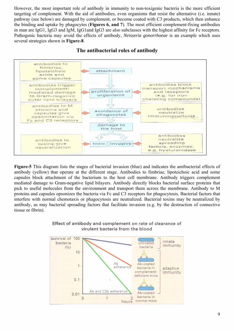
However, the most important role of antibody in immunity to non-toxigenic bacteria is the more efficient targeting of complement. With the aid of antibodies, even organisms that resist the alternative (i.e. innate) pathway (see below) are damaged by complement, or become coated with C3 products, which then enhance the binding and uptake by phagocytes (Figures 6, and 7). The most efficient complement-fixing antibodies in man are IgG1, IgG3 and IgM, IgG1and IgG3 are also subclasses with the highest affinity for Fc receptors. Pathogenic bacteria may avoid the effects of antibody, Neisseria gonorrhoeae is an example which uses several strategies shown in Figure-8.
The antibacterial roles of antibody
Figure-5 This diagram lists the stages of bacterial invasion (blue) and indicates the antibacterial effects of antibody (yellow) that operate at the different stage. Antibodies to fimbriae, lipoteichoic acid and some capsules block attachment of the bacterium to the host cell membrane. Antibody triggers complement mediated damage to Gram-negative lipid bilayers. Antibody directly blocks bacterial surface proteins that pick to useful molecules from the environment and transport them across the membrane. Antibody to M proteins and capsules opsonizes the bacteria via Fc and C3 receptors for phagocytosis. Bacterial factors that interfere with normal chemotaxis or phagocytosis are neutralized. Bacterial toxins may be neutralized by antibody, as may bacterial spreading factors that facilitate invasion (e.g. by the destruction of connective tissue or fibrin).
9
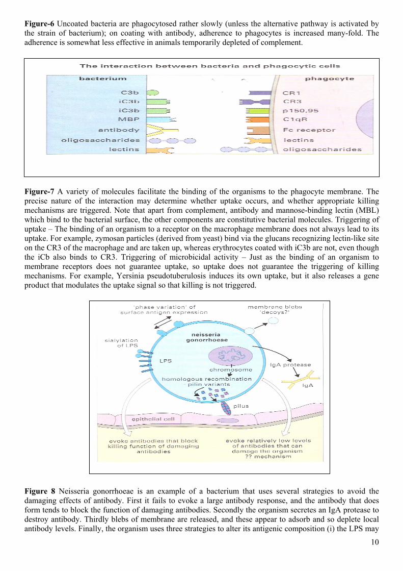
Figure-6 Uncoated bacteria are phagocytosed rather slowly (unless the alternative pathway is activated by the strain of bacterium); on coating with antibody, adherence to phagocytes is increased many-fold. The adherence is somewhat less effective in animals temporarily depleted of complement.
Figure-7 A variety of molecules facilitate the binding of the organisms to the phagocyte membrane. The precise nature of the interaction may determine whether uptake occurs, and whether appropriate killing mechanisms are triggered. Note that apart from complement, antibody and mannose-binding lectin (MBL) which bind to the bacterial surface, the other components are constitutive bacterial molecules. Triggering of uptake – The binding of an organism to a receptor on the macrophage membrane does not always lead to its uptake. For example, zymosan particles (derived from yeast) bind via the glucans recognizing lectin-like site on the CR3 of the macrophage and are taken up, whereas erythrocytes coated with iC3b are not, even though the iCb also binds to CR3. Triggering of microbicidal activity – Just as the binding of an organism to membrane receptors does not guarantee uptake, so uptake does not guarantee the triggering of killing mechanisms. For example, Yersinia pseudotuberulosis induces its own uptake, but it also releases a gene product that modulates the uptake signal so that killing is not triggered.
Figure 8 Neisseria gonorrhoeae is an example of a bacterium that uses several strategies to avoid the damaging effects of antibody. First it fails to evoke a large antibody response, and the antibody that does form tends to block the function of damaging antibodies. Secondly the organism secretes an IgA protease to destroy antibody. Thirdly blebs of membrane are released, and these appear to adsorb and so deplete local antibody levels. Finally, the organism uses three strategies to alter its antigenic composition (i) the LPS may
10
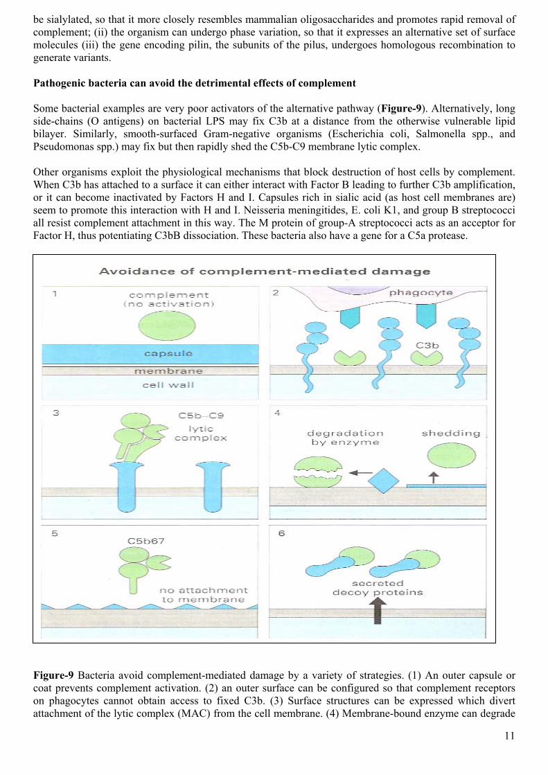
be sialylated, so that it more closely resembles mammalian oligosaccharides and promotes rapid removal of complement; (ii) the organism can undergo phase variation, so that it expresses an alternative set of surface molecules (iii) the gene encoding pilin, the subunits of the pilus, undergoes homologous recombination to generate variants. Pathogenic bacteria can avoid the detrimental effects of complement Some bacterial examples are very poor activators of the alternative pathway (Figure-9). Alternatively, long side-chains (O antigens) on bacterial LPS may fix C3b at a distance from the otherwise vulnerable lipid bilayer. Similarly, smooth-surfaced Gram-negative organisms (Escherichia coli, Salmonella spp., and Pseudomonas spp.) may fix but then rapidly shed the C5b-C9 membrane lytic complex. Other organisms exploit the physiological mechanisms that block destruction of host cells by complement. When C3b has attached to a surface it can either interact with Factor B leading to further C3b amplification, or it can become inactivated by Factors H and I. Capsules rich in sialic acid (as host cell membranes are) seem to promote this interaction with H and I. Neisseria meningitides, E. coli K1, and group B streptococci all resist complement attachment in this way. The M protein of group-A streptococci acts as an acceptor for Factor H, thus potentiating C3bB dissociation. These bacteria also have a gene for a C5a protease.
Figure-9 Bacteria avoid complement-mediated damage by a variety of strategies. (1) An outer capsule or coat prevents complement activation. (2) an outer surface can be configured so that complement receptors on phagocytes cannot obtain access to fixed C3b. (3) Surface structures can be expressed which divert attachment of the lytic complex (MAC) from the cell membrane. (4) Membrane-bound enzyme can degrade
11
12
fixed complement or cause it to be shed. (5) The outer membrane can resist the insertion of the lytic complex. (6) Secreted decoy proteins can cause complement to be deposited on them and not on the bacterium itself. Ultimately most bacteria are killed by phagocytes A few, mostly Gram-negative, bacteria are directly killed by complement, as stated earlier. There are also reports that some organisms, particularly Gram-negative bacteria, can be killed by mere contact with NK cells, or even cytotoxic T (Tc) cells. This probably involves the membrane-lysing mechanism of these cells, acting on the outer lipid bilayer that is characteristic of Gram-negative organisms. However, most bacteria are killed by phagocytes. This process involves steps (Figure-9). Chemotaxis – Bacterial components such as f-Met-Leu-Phe (which is chemotactic for leucocytes), complement products such as C5a, and locally released chemokines and cytokines attract the phagocytes. Attachment of the phagocyte to the organism – This is an important interaction which may determine whether uptake subsequently occurs, and whether killing mechanisms are triggered during uptake. The binding can be mediated by the following entities.
• Lectins on the organism, for example the mannose-binding lectin on the fimbriae of E. coli.
• Lectins on the phagocyte. Of particular interest in this respect are the complement receptors CR3 and p150, 95 and the related molecule LFA-1 (leucocyte functional antigen-1 – a mediator of intercellular adhesion), which have multiple binding sites specific for different carbohydrate moieties. They can bind to β-glucans and to the LPS endotoxin of Gram-negative bacteria. The mannan receptor targets glycoconjugates on the bacterial surface.
• Complement deposited on the organism via the alternative or classic pathways. It has recently been discovered that complement can also be fixed by mannose-binding lectins present in serum, which can itself bind to C1q receptors.
• Fc receptors on the phagocyte, which link to antibody bound to the bacteria (Figure-7).
Phagocytic cells have many killing methods
The killing pathways can be oxygen dependent and oxygen independent. Briefly one oxygen-dependent pathway involves the reduction of oxygen to superoxide anion (which is molecular oxygen to which a single unpaired electron has been added). This then interacts with numerous other molecules to give rise to a series of free radicals and other toxic derivatives. A second oxygen-dependent pathway involves the creation of nitric oxide (NO•) from the guanidine nitrogen of L-arginine. This in turn leads to further toxic substances such as the peroxynitrites, which result from interaction of NO• with the products of the oxygen reduction pathway.
Oxygen-independent killing mechanisms may be important These mechanisms may be more important than was previously thought. Many organisms can be killed by cells from patients with chronic granulomatous disease, which cannot produce reactive oxygen intermediates, or from patients with myeloperoxidase deficiency, which cannot produce hypo-halous acids. Some of this killing may be due to NO, but many organisms can be killed anaerobically, so other mechanisms must exist. Some have been identified. Cationic proteins with antibiotic-like properties – The defensins are cysteine- and arginine-rich cationic peptides of 30-33 amino acids, found in rabbit macrophages and human neutrophil polymorphs, where they comprise 30-50% of the granule proteins. They evolved early in evolution and similar molecules are found
in insects. They form ion-permeable channels in lipid bilayer and probably act early after Phagolysome formation, before acidification takes place. Defensins can kill organisms as diverse as Staphylococcus aureus, Pseudomonas aeruginosa, E. coli, Cryptococcus neoformans and the enveloped virus Herpes simplex. There are also cationic proteins with different pH optima, including cathapsin G and azurocidin, both of which are related to elastase but which have activity against Gram-negative bacteria; this is unrelated to their enzyme activity. Other antimicrobial mechanisms – Following lysosome fusion there is a transient rise in pH before acidification (a fall in pH) of the phago-lysosome takes place. This occurs within 10-15 minutes. Killing of some organisms may be due to the acidification itself, though it is more likely to be related to the low pH optima of lysosomal enzymes. Certain Gram-positive organisms may be killed by lysozyme, which is active against their readily exposed peptidoglycan layer. A variety of other substance, such as lactoferrin (produced by neutrophil polymorphs), have also been implicated in killing. Lactoferrin can bind iron and render it unavailable to bacteria even at an acid pH (thus, the ability of polymorphs to kill some bacteria is lost if they are loaded with iron). These mechanisms may all require phagolysosome fusion (Figure-10).
1 2
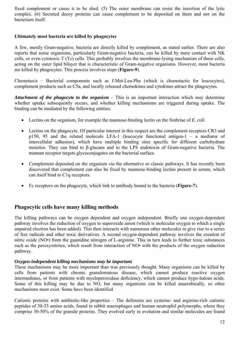
Figure-10 Inhibition or fusion of secondary lysosomes with yeast-containing phagosomes by the addition of ammonium chloride. Mouse peritoneal macrophages were incubated in acridine orange, which concentrates in secondary lysosomes. Live baker's yeast was then added – these assume the appearance of 'holes' in the cell. Normally, the secondary lysosomes fues with the phagosomes, and the acridine orange enters them, fluorescing green, yellow or orange depending on the concentration (1). However, in the 'holes' remain dark (2). Such blocking of lysosomal fusion may be employed by M. tuberculosis and some leishmania, which secrete ammonia. Some polyanions such as polyglutamic acid or suramin may also do this. (Courtesy of Mr. R.Young and Dr. P.D. Hart). Resting macrophages can kill, but killing can be enhanced, and new mechanisms can be expressed on activation Activation occurs through exposure to microbial products, and to lymphokines derived from T cells. Similarly the decline that usually occurs if the cells are kept in culture for a week can be reversed by treatment with suitable activating stimuli. Some microbial products can activate macrophages in the absence of lymphocyte recognition
13
14
A number of microbial products cause direct activation of monocytes and macrophages, or indirect activation by triggering cytokine release from them or from NK cells. The cytokines then activate the phagocytes. This was discussed earlier in relation to the non-lymphocyte-dependent recognition of bacteria. Further activation of macrophages is mediated by lymphokines In vitro, lymphokines released during T-cell-mediated responses are often required for phagocytes to become fully activated. The lymphokine most often implicated is IFNγ, which enhances both oxygen-dependent and oxygen-independent killing mechanisms. There are also reports implicating IL-2, granulocyte-macrophage colony stimulating factor (GM-CSF), TNF, and other cytokines. As discussed in greater detail in Chapter 7, activation of some functions requires combinations of cytokines. Lymphokines in vivo have two effects on phagocytes – attracting them and activating them – and the relative importance of these two components differs for different organisms. Thus for immunity to L. manocytogenes, which can be killed by the baseline levels of oxygen-dependent mechanisms in both monocytes and neutrophils, it is the attraction of the cells to the lesion which is most important. In contrast, for M. tuberculosis, which thrives inside neutrophils and monocytes, it is the activation of the cells which is critical. Human and murine macrophages are different This is important because much experimental work is based on murine macrophages. Mycobacteria illustrate the complexity of this topic. IFNγ can activate murine macrophages to destroy mycobacteria completely. This appears to be due to the nitric oxide pathway. However, IFNγ acting on human macrophages causes, at least, feeble inhibition of M. tuberculosis or, at worst, significantly increased growth. This may relate to species differences in NO• production. On the other hand, human cells do something which has not been reported in murine cells. IFNγ causes human macrophages to express a 1-hydroxylase enzyme that converts the circulating inactive form of 25-hydroxycholecalciferol (vitamin D3) into an active metabolite, 1,25-dihydroxcholecalciferol. This metabolite activates antimycobacterial mechanisms in the macrophages rather more efficiently than INFγ itself, through the mechanism of the enhanced antimicrobial effect is not known. Successful pathogens have evolved mechanisms for the avoidance of phagocyte-mediated killing Since most organisms are ultimately killed by phagocytes, it is not surprising that successful pathogens have evolved an array of mechanisms to counteract this risk (Figure-11). Intracellular pathogens may ‘hide’ in cells that have little antimicrobial potential Infected cells can be killed by Tc cells Some organisms may thrive inside damaged or metabolically deranged host phagocytes, or escape killing by moving out of phagosomes into the cytoplasm. Listeria manocytogenes achieves this by releasing enzymes that lyse the phagosomes membrane. This organism also illustrates the point that bacteria are not just inert particles. They have evolved strategies for taking control of functions of the host cell Figure-12). Other organisms, such as Mycobacterium leprae, can cause themselves to be taken up by cells that are not normally considered phagocytic, and have little antibacterial potential. Before they can be taken up by activated phagocytes, or exposed to other killing mechanisms, the organisms may need to be released from such cells. Tc cells may perform this function by killing the infected cell. For instance mice become strikingly susceptible to M. tuberculosis if the β2-micfroglobulin gene is knocked out so that class I MHC cannot be recognized by the Tc cells. This is consistence with an essential note for cytotoxic T cells.
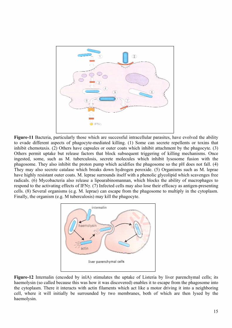
Figure-11 Bacteria, particularly those which are successful intracellular parasites, have evolved the ability to evade different aspects of phagocyte-mediated killing. (1) Some can secrete repellents or toxins that inhibit chemotaxis. (2) Others have capsules or outer coats which inhibit attachment by the phagocyte. (3) Others permit uptake but release factors that block subsequent triggering of killing mechanisms. Once ingested, some, such as M. tuberculosis, secrete molecules which inhibit lysosome fusion with the phagosome. They also inhibit the proton pump which acidifies the phagosome so the pH does not fall. (4) They may also secrete catalase which breaks down hydrogen peroxide. (5) Organisms such as M. leprae have highly resistant outer coats. M. leprae surrounds itself with a phenolic glycolipid which scavenges free radicals. (6) Mycobacteria also release a lipoarabinomannan, which blocks the ability of macrophages to respond to the activating effects of IFNγ. (7) Infected cells may also lose their efficacy as antigen-presenting cells. (8) Several organisms (e.g. M. leprae) can escape from the phagosome to multiply in the cytoplasm. Finally, the organism (e.g. M tuberculosis) may kill the phagocyte.
Figure-12 Internalin (encoded by inlA) stimulates the uptake of Listeria by liver parenchymal cells; its haemolysin (so called because this was how it was discovered) enables it to escape from the phagosome into the cytoplasm. There it interacts with actin filaments which act like a motor driving it into a neighboring cell, where it will initially be surrounded by two membranes, both of which are then lysed by the haemolysin. 15
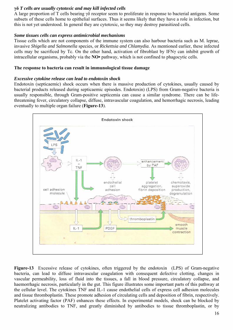
γδ T cells are usually cytotoxic and may kill infected cells A large proportion of T cells bearing γδ receptor seem to proliferate in response to bacterial antigens. Some subsets of these cells home to epithelial surfaces. Thus it seems likely that they have a role in infection, but this is not yet understood. In general they are cytotoxic, so they may destroy parasitized cells. Some tissues cells can express antimicrobial mechanisms Tissue cells which are not components of the immune system can also harbour bacteria such as M. leprae, invasive Shigella and Salmonella species, or Rickettsia and Chlamydia. As mentioned earlier, these infected cells may be sacrificed by Tc. On the other hand, activation of fibroblast by IFNγ can inhibit growth of intracellular organisms, probably via the NO• pathway, which is not confined to phagocytic cells. The response to bacteria can result in immunological tissue damage Excessive cytokine release can lead to endotoxin shock Endotoxin (septicaemic) shock occurs when there is massive production of cytokines, usually caused by bacterial products released during septicaemic episodes. Endotoxin) (LPS) from Gram-negative bacteria is usually responsible, through Gram-positive septicemia can cause a similar syndrome. There can be life-threatening fever, circulatory collapse, diffuse, intravascular coagulation, and hemorrhagic necrosis, leading eventually to multiple organ failure (Figure-13).
Figure-13 Excessive release of cytokines, often triggered by the endotoxin (LPS) of Gram-negative bacteria, can lead to diffuse intravascular coagulation with consequent defective clotting, changes in vascular permeability, loss of fluid into the tissues, a fall in blood pressure, circulatory collapse, and haemorrhagic necrosis, particularly in the gut. This figure illustrates some important parts of this pathway at the cellular level. The cytokines TNF and IL-1 cause endothelial cells of express cell adhesion molecules and tissue thromboplastin. These promote adhesion of circulating cells and deposition of fibrin, respectively. Platelet activating factor (PAF) enhances these effects. In experimental models, shock can be blocked by neutralizing antibodies to TNF, and greatly diminished by antibodies to tissue thromboplastin, or by 16
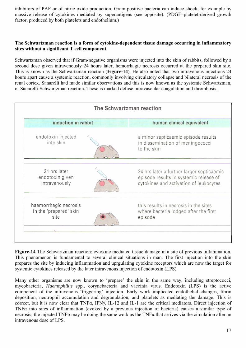
inhibitors of PAF or of nitric oxide production. Gram-positive bacteria can induce shock, for example by massive release of cytokines mediated by superantigens (see opposite). (PDGF=platelet-derived growth factor, produced by both platelets and endothelium.) The Schwartzman reaction is a form of cytokine-dependent tissue damage occurring in inflammatory sites without a significant T cell component Schwartzman observed that if Gram-negative organisms were injected into the skin of rabbits, followed by a second dose given intravenously 24 hours later, hemorrhagic necrosis occurred at the prepared skin site. This is known as the Schwartzman reaction (Figure-14). He also noted that two intravenous injections 24 hours apart cause a systemic reaction, commonly involving circulatory collapse and bilateral necrosis of the renal cortex. Sanarelli had made similar observations and this is now known as the systemic Schwartzman, or Sanarelli-Schwartzman reaction. These is marked defuse intravascular coagulation and thrombosis.
Figure-14 The Schwartzman reaction: cytokine mediated tissue damage in a site of previous inflammation. This phenomenon is fundamental to several clinical situations in man. The first injection into the skin prepares the site by inducing inflammation and uprgulating cytokine receptors which are now the target for systemic cytokines released by the later intravenous injection of endotoxin (LPS). Many other organisms are now known to ‘prepare’ the skin in the same way, including streptococci, mycobacteria, Haemophilus spp., corynebacteria and vaccinia virus. Endotoxin (LPS) is the active component of the intravenous ‘triggering’ injection. Early work implicated endothelial changes, fibrin deposition, neutrophil accumulation and degranulation, and platelets as mediating the damage. This is correct, but it is now clear that TNFα, IFNγ, IL-12 and IL-1 are the critical mediators. Direct injection of TNFα into sites of inflammation (evoked by a previous injection of bacteria) causes a similar type of necrosis; the injected TNFα may be doing the same work as the TNFα that arrives via the circulation after an intravenous dose of LPS.
17
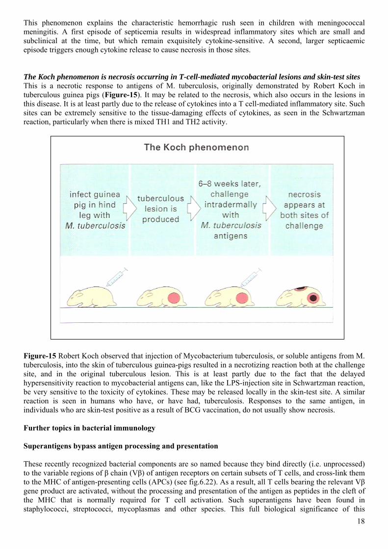
This phenomenon explains the characteristic hemorrhagic rush seen in children with meningococcal meningitis. A first episode of septicemia results in widespread inflammatory sites which are small and subclinical at the time, but which remain exquisitely cytokine-sensitive. A second, larger septicaemic episode triggers enough cytokine release to cause necrosis in those sites. The Koch phenomenon is necrosis occurring in T-cell-mediated mycobacterial lesions and skin-test sites This is a necrotic response to antigens of M. tuberculosis, originally demonstrated by Robert Koch in tuberculous guinea pigs (Figure-15). It may be related to the necrosis, which also occurs in the lesions in this disease. It is at least partly due to the release of cytokines into a T cell-mediated inflammatory site. Such sites can be extremely sensitive to the tissue-damaging effects of cytokines, as seen in the Schwartzman reaction, particularly when there is mixed TH1 and TH2 activity.
Figure-15 Robert Koch observed that injection of Mycobacterium tuberculosis, or soluble antigens from M. tuberculosis, into the skin of tuberculous guinea-pigs resulted in a necrotizing reaction both at the challenge site, and in the original tuberculous lesion. This is at least partly due to the fact that the delayed hypersensitivity reaction to mycobacterial antigens can, like the LPS-injection site in Schwartzman reaction, be very sensitive to the toxicity of cytokines. These may be released locally in the skin-test site. A similar reaction is seen in humans who have, or have had, tuberculosis. Responses to the same antigen, in individuals who are skin-test positive as a result of BCG vaccination, do not usually show necrosis. Further topics in bacterial immunology Superantigens bypass antigen processing and presentation These recently recognized bacterial components are so named because they bind directly (i.e. unprocessed) to the variable regions of β chain (Vβ) of antigen receptors on certain subsets of T cells, and cross-link them to the MHC of antigen-presenting cells (APCs) (see fig.6.22). As a result, all T cells bearing the relevant Vβ gene product are activated, without the processing and presentation of the antigen as peptides in the cleft of the MHC that is normally required for T cell activation. Such superantigens have been found in staphylococci, streptococci, mycoplasmas and other species. This full biological significance of this 18

bacterial adaptation is not yet clear, but one obvious effect can be the toxicity of the massive cytokine/lymphokine release that results from the simultaneous stimulation of a large subset of T cells. The staphylococcal toxins responsible for the toxin shock syndrome (TSST-1, etc.) appear to operate in this way. Figure-16 uses examples to illustrate the relationship between the nature of the organism, the disease and immunopathology caused, and the mechanism of immune response which leads to protection.
Figure-16 This table provides examples to illustrate the relationship between the nature of the organism, the disease and immunopathology caused, and the mechanism of immune response which leads to protection. Heat-shock proteins (stress proteins) are highly conserved and prominent targets of immune responses These proteins are found in all eukaryotic and prokaryotic cells, where they have essential roles in the assembly, folding and transport of other molecules. Cells exposed to abnormally elevated temperatures (or to other stresses) express higher levels of these proteins, which reflects their role sequences are very highly conserved and there is currently much speculation that, because bacterial heat shock proteins are so similar to human ones, they may be involved in the initiation of autoimmunity. Paradoxically, in spite of their similarity to host proteins, they seem to be target antigens in the protective response against many infectious organisms. This makes sense form an evolutionary point of view, because if the host possesses T cells that recognize a range of conserved heat-shock protein epitopes, these T cells are likely to recognize any pathogen encountered. Thus peptides derived from microbial heat-shock proteins (hsp) and presented in the conventional manner by antigen-presenting cells, can be targets of CD4+ T cells or of CD8+ cytotoxic T cells. Infection can also stress a host cell, so that it ‘flags’ its distress by overexpressing its own hsp. There is some evidence that
19
20
peptides derived from host hsp can under these circumstances be T cell targets, so that host hsp may become associated with the cell membrane and act as signaling molecules that alert the system to ‘danger’. The ‘hygiene hypotheses" Several groups of disease, all characterized by defects in the regulation of the immune system, are becoming more common, particularly in developed countries. These diseases include allergies, inflammatory bowel disease (Crohn’s disease and ulcerative colitis) and autoimmune problems such as multiple sclerosis. In Europe increased incidences are seen first in the more developed northern states, and in Western Europe rather than in the old communist block. Epidemiological data are compatible with the hypothesis that increasing immunological dysregulation correlates with decreasing exposure to harmless environmental bacteria. Decreased exposure could be due to hygiene, vaccines and antibiotic use. The hypothesis is rational because environmental inputs that have always been present throughout evolution may have become genetically encoded necessities to the developing immune system. If proved correct the solution will clearly not be the abandonment of the most important achievements of medicates (hygiene, vaccines, and antibiotics) but rather the identification of the environment factors that are lacking from the modern lifestyle, so they can be replaced as vaccines or probiotics. Probiotics are even to the environment in order to exert a therapeutic effect. There is now also talk of ‘probiotics’ which are dietary components intended to influence the bowed flora. Whatever their nature, effective products will need to regulate the balance of TH1/TH2 response, and promote the maturation of the regulatory T cell circuits from that control autoimmunity to host antigen such as heat-shock proteins. There are experimental and clinical trial data to suggest that some bacterial vaccines can achieve both objectives. IMMUNITY TO FUNGI Fungi infection is a growing problem because of the markedly increased numbers of immunologically compromised hosts. Thus it is regularly seen in HIV-infected patients, and in cancer patients undergoing chemotherapy, in transplant patients on immunosuppressive agents and in some patients taking long term corticosteroids. There are four categories of fungal infection Little is known of the precise mechanisms involved in immunity to fungal infections, but it is thought they are essentially similar to those involved in resistance to bacterial infections. The fungal infections of humans fall into four major categories.
• Superficial mycoses: These are caused by fungi known as dermatophytes, and are usually restricted to the nonliving keratinized components of skin, hair and nails.
• Subcutaneous mycoses: Saprophytic fungi can cause chronic nodules or ulcers in subcutaneous tissues following trauma, for example chromomycosis, sporotrichosis and mycetoma.
• Respiratory mycoses: Soil saprophytes produce subclinical or acute lung infections (rarely disseminated), or granulomatous lesions, e.g. histoplasmosis and coccidioido-mycosis.
• Candidiasis: Candida albicans (a ubiquitous commensal) causes superficial (rarely systemic) infections of skin and mucous membranes.
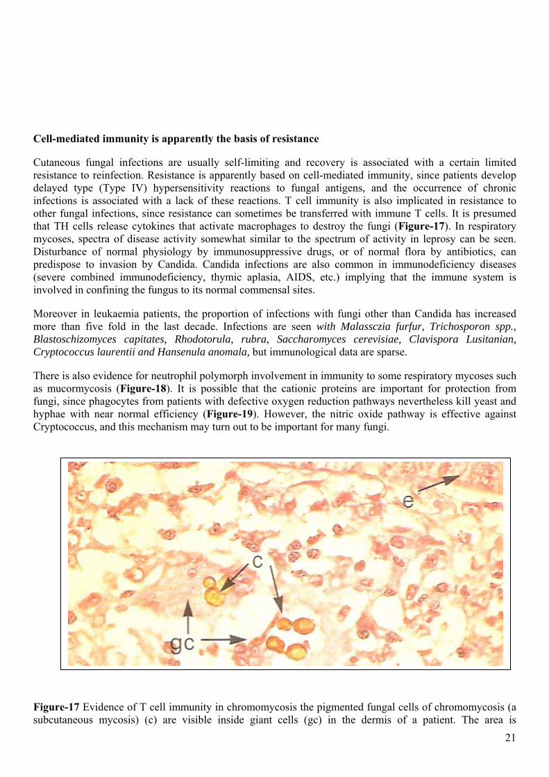
Cell-mediated immunity is apparently the basis of resistance
Cutaneous fungal infections are usually self-limiting and recovery is associated with a certain limited resistance to reinfection. Resistance is apparently based on cell-mediated immunity, since patients develop delayed type (Type IV) hypersensitivity reactions to fungal antigens, and the occurrence of chronic infections is associated with a lack of these reactions. T cell immunity is also implicated in resistance to other fungal infections, since resistance can sometimes be transferred with immune T cells. It is presumed that TH cells release cytokines that activate macrophages to destroy the fungi (Figure-17). In respiratory mycoses, spectra of disease activity somewhat similar to the spectrum of activity in leprosy can be seen. Disturbance of normal physiology by immunosuppressive drugs, or of normal flora by antibiotics, can predispose to invasion by Candida. Candida infections are also common in immunodeficiency diseases (severe combined immunodeficiency, thymic aplasia, AIDS, etc.) implying that the immune system is involved in confining the fungus to its normal commensal sites.
Moreover in leukaemia patients, the proportion of infections with fungi other than Candida has increased more than five fold in the last decade. Infections are seen with Malassczia furfur, Trichosporon spp., Blastoschizomyces capitates, Rhodotorula, rubra, Saccharomyces cerevisiae, Clavispora Lusitanian, Cryptococcus laurentii and Hansenula anomala, but immunological data are sparse.
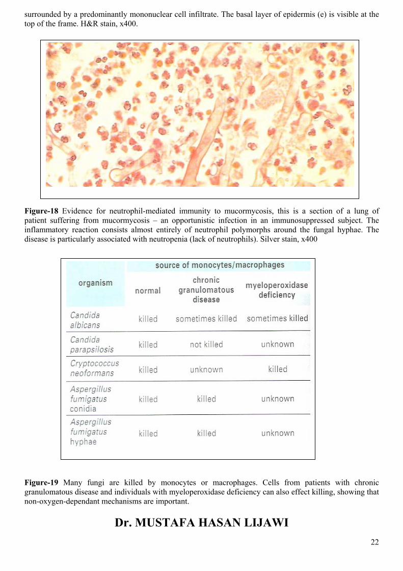
There is also evidence for neutrophil polymorph involvement in immunity to some respiratory mycoses such as mucormycosis (Figure-18). It is possible that the cationic proteins are important for protection from fungi, since phagocytes from patients with defective oxygen reduction pathways nevertheless kill yeast and hyphae with near normal efficiency (Figure-19). However, the nitric oxide pathway is effective against Cryptococcus, and this mechanism may turn out to be important for many fungi.
Figure-17 Evidence of T cell immunity in chromomycosis the pigmented fungal cells of chromomycosis (a subcutaneous mycosis) (c) are visible inside giant cells (gc) in the dermis of a patient. The area is 21
surrounded by a predominantly mononuclear cell infiltrate. The basal layer of epidermis (e) is visible at the top of the frame. H&R stain, x400.
Figure-18 Evidence for neutrophil-mediated immunity to mucormycosis, this is a section of a lung of patient suffering from mucormycosis – an opportunistic infection in an immunosuppressed subject. The inflammatory reaction consists almost entirely of neutrophil polymorphs around the fungal hyphae. The disease is particularly associated with neutropenia (lack of neutrophils). Silver stain, x400
Figure-19 Many fungi are killed by monocytes or macrophages. Cells from patients with chronic granulomatous disease and individuals with myeloperoxidase deficiency can also effect killing, showing that non-oxygen-dependant mechanisms are important.
Dr. MUSTAFA HASAN LIJAWI 22


![No Slide Title · 2017-01-24 · • [3] Forrest Mims, Getting Started in Electronics, 1983 . 57 . Title: No Slide Title Author: Chuck Schuler Created Date: 12/20/2013 2:28:12 PM](https://static.fdocument.org/doc/165x107/5ec5871852e4935eab58019c/no-slide-2017-01-24-a-3-forrest-mims-getting-started-in-electronics-1983.jpg)