The Crystal Structure of the Platelet Activator Aggretin Reveals a Novel (αβ) 2 ...
7
The Crystal Structure of the Platelet Activator Aggretin Reveals a Novel (R) 2 Dimeric Structure †,‡ Elizabeth Hooley, § Evangelos Papagrigoriou, § Alexei Navdaev, |,⊥ Amit V. Pandey, |,@ Jeannine M. Clemetson, | Kenneth J. Clemetson, | and Jonas Emsley* ,§ Centre for Biomolecular Sciences, School of Pharmacy, UniVersity of Nottingham, Nottingham NG7 2RD, U.K., and Theodor Kocher Institute, UniVersity of Berne, Freiestrasse 1, CH-3012 Berne, Switzerland ReceiVed March 27, 2008; ReVised Manuscript ReceiVed June 3, 2008 ABSTRACT: Aggretin is a C-type lectin purified from Calloselasma rhodostoma snake venom. It is a potent activator of platelets, resulting in a collagen-like response by binding and clustering platelet receptor CLEC-2. We present here the crystal structure of aggretin at 1.7 Å which reveals a unique tetrameric quaternary structure. The two R heterodimers are arranged through 2-fold rotational symmetry, resulting in an antiparallel side-by-side arrangement. Aggretin thus presents two ligand binding sites on one surface and can therefore cluster ligands in a manner reminiscent of convulxin and flavocetin. To examine the molecular basis of the interaction with CLEC-2, we used a molecular modeling approach of docking the aggretin R structure with the CLEC-2 N-terminal domain (CLEC-2N). This model positions the CLEC- 2N structure face down in the “saddle”-shaped binding site which lies between the aggretin R and lectin-like domains. A 2-fold rotation of this complex to generate the aggretin tetramer reveals dimer contacts for CLEC-2N which bring the N- and C-termini into the proximity of each other, and a series of contacts involving two interlocking -strands close to the N-terminus are described. A comparison with homologous lectin-like domains from the immunoreceptor family reveals a similar but not identical dimerization mode, suggesting this structure may represent the clustered form of CLEC-2 capable of signaling across the platelet membrane. The first steps of platelet activation are critical to our understanding of hemostasis and thrombosis (1). Exposed matrix components from the subendothelium at sites of vascular damage interact with specific receptors on the surface of platelets, causing platelet activation and spreading to cover the injured site until it has regenerated. Snake venom proteins have been shown to bind a number of platelet receptors involved in platelet function (1–3). In many cases, they bind specifically to platelet surface glycoprotein recep- tors and either block function or activate and thus induce platelet activation via clustering of receptors. Hence, they are frequently used to study mechanisms of platelet activation and aggregation and belong to two major protein families, the C-type lectin family and the metalloproteinase disintegrin cysteine-rich protein family (4). Crystal structures of the snake venom C-type lectins reveal these are R heterodimers (5–10). The dimers are formed by a domain-swapping long loop, which leads to the disruption of the carbohydrate binding site and therefore loss of lectin activity. Aggretin, also known as rhodocytin, has been shown to bind to platelets (11, 12), causing a collagen- like response, which is characterized by a distinct lag phase before initiation of a signaling cascade involving activation of Syk and PLCγ2 leading to platelet aggregation (11–14). This signaling response is also similar to that seen in the autosomal recessive disorder Chediak-Higashi syndrome (CHS) (which is a granule release defect) (15). Aggretin has been shown to bind CLEC-2, 1 GPIbR, and integrin R21; however, knockout studies have disputed a role for the latter two receptors in aggretin activity. Navdaev et al. (14) used antibodies against the thrombin binding site of GPIbR to inhibit the aggretin response in platelets. Chung et al. (16) used the GPIbR agonist agkistin to inhibit the aggretin response, whereas Bergmeier et al. (17) and Shin and Morita (12) showed that platelets without the 45 kDa N-terminal domain of GPIbR respond to aggretin and that echicetin did not inhibit the aggretin response. Navdaev et al. (14) and Chung et al. (16) also used antibodies against R21 to inhibit the aggretin response. Suzuki-Inoue et al. (18) showed that aggretin-Sepharose bound R21-loaded liposomes. Eble et † E.H. is funded by the British Heart Foundation. K.J.C. thanks the Swiss National Science Foundation for support by Grants 31-063868.00 and 31-107754.04. ‡ Coordinates and structure factors have been submitted to the Protein Data Bank (PDB) as entry 3BX4. * To whom correspondence should be addressed. E-mail: [email protected]. Telephone: +44 1158467092. Fax: +44 1158468002. § University of Nottingham. | University of Berne. ⊥ Current address: Institute for Physiological Chemistry, Muenster Hospital, D-48149 Muenster, Germany. @ Current address: Department Klinische Forschung, Tiefenaulabors- DKF-Spital Bern, University of Berne, Tiefenaulabors 120, CH-3004 Berne, Switzerland. 1 Abbreviations: CLEC-2, C-type lectin-like receptor 2; GpIb, platelet glycoprotein Ib; NKG2D, natural killer cell receptor G2D; HIV, human immunodeficiency virus. Biochemistry 2008, 47, 7831–7837 7831 10.1021/bi800528t CCC: $40.75 2008 American Chemical Society Published on Web 07/03/2008
Transcript of The Crystal Structure of the Platelet Activator Aggretin Reveals a Novel (αβ) 2 ...
The Crystal Structure of the Platelet Activator Aggretin Reveals a Novel (R�)2
Dimeric Structure†,‡
Elizabeth Hooley,§ Evangelos Papagrigoriou,§ Alexei Navdaev,|,⊥ Amit V. Pandey,|,@ Jeannine M. Clemetson,|
Kenneth J. Clemetson,| and Jonas Emsley*,§
Centre for Biomolecular Sciences, School of Pharmacy, UniVersity of Nottingham, Nottingham NG7 2RD, U.K., andTheodor Kocher Institute, UniVersity of Berne, Freiestrasse 1, CH-3012 Berne, Switzerland
ReceiVed March 27, 2008; ReVised Manuscript ReceiVed June 3, 2008
ABSTRACT: Aggretin is a C-type lectin purified from Calloselasma rhodostoma snake venom. It is a potentactivator of platelets, resulting in a collagen-like response by binding and clustering platelet receptorCLEC-2. We present here the crystal structure of aggretin at 1.7 Å which reveals a unique tetramericquaternary structure. The two R� heterodimers are arranged through 2-fold rotational symmetry, resultingin an antiparallel side-by-side arrangement. Aggretin thus presents two ligand binding sites on one surfaceand can therefore cluster ligands in a manner reminiscent of convulxin and flavocetin. To examine themolecular basis of the interaction with CLEC-2, we used a molecular modeling approach of docking theaggretin R� structure with the CLEC-2 N-terminal domain (CLEC-2N). This model positions the CLEC-2N structure face down in the “saddle”-shaped binding site which lies between the aggretin R and �lectin-like domains. A 2-fold rotation of this complex to generate the aggretin tetramer reveals dimercontacts for CLEC-2N which bring the N- and C-termini into the proximity of each other, and a series ofcontacts involving two interlocking �-strands close to the N-terminus are described. A comparison withhomologous lectin-like domains from the immunoreceptor family reveals a similar but not identicaldimerization mode, suggesting this structure may represent the clustered form of CLEC-2 capable ofsignaling across the platelet membrane.
The first steps of platelet activation are critical to ourunderstanding of hemostasis and thrombosis (1). Exposedmatrix components from the subendothelium at sites ofvascular damage interact with specific receptors on thesurface of platelets, causing platelet activation and spreadingto cover the injured site until it has regenerated. Snake venomproteins have been shown to bind a number of plateletreceptors involved in platelet function (1–3). In many cases,they bind specifically to platelet surface glycoprotein recep-tors and either block function or activate and thus induceplatelet activation via clustering of receptors. Hence, theyare frequently used to study mechanisms of platelet activationand aggregation and belong to two major protein families,the C-type lectin family and the metalloproteinase disintegrincysteine-rich protein family (4).
Crystal structures of the snake venom C-type lectins revealthese are R� heterodimers (5–10). The dimers are formedby a domain-swapping long loop, which leads to thedisruption of the carbohydrate binding site and therefore lossof lectin activity. Aggretin, also known as rhodocytin, hasbeen shown to bind to platelets (11, 12), causing a collagen-like response, which is characterized by a distinct lag phasebefore initiation of a signaling cascade involving activationof Syk and PLCγ2 leading to platelet aggregation (11–14).This signaling response is also similar to that seen in theautosomal recessive disorder Chediak-Higashi syndrome(CHS) (which is a granule release defect) (15). Aggretin hasbeen shown to bind CLEC-2,1 GPIbR, and integrin R2�1;however, knockout studies have disputed a role for the lattertwo receptors in aggretin activity. Navdaev et al. (14) usedantibodies against the thrombin binding site of GPIbR toinhibit the aggretin response in platelets. Chung et al. (16)used the GPIbR agonist agkistin to inhibit the aggretinresponse, whereas Bergmeier et al. (17) and Shin and Morita(12) showed that platelets without the 45 kDa N-terminaldomain of GPIbR respond to aggretin and that echicetin didnot inhibit the aggretin response. Navdaev et al. (14) andChung et al. (16) also used antibodies against R2�1 to inhibitthe aggretin response. Suzuki-Inoue et al. (18) showed thataggretin-Sepharose bound R2�1-loaded liposomes. Eble et
† E.H. is funded by the British Heart Foundation. K.J.C. thanks theSwiss National Science Foundation for support by Grants 31-063868.00and 31-107754.04.
‡ Coordinates and structure factors have been submitted to the ProteinData Bank (PDB) as entry 3BX4.
* To whom correspondence should be addressed. E-mail:[email protected]. Telephone: +44 1158467092. Fax:+44 1158468002.
§ University of Nottingham.| University of Berne.⊥ Current address: Institute for Physiological Chemistry, Muenster
Hospital, D-48149 Muenster, Germany.@ Current address: Department Klinische Forschung, Tiefenaulabors-
DKF-Spital Bern, University of Berne, Tiefenaulabors 120, CH-3004Berne, Switzerland.
1 Abbreviations: CLEC-2, C-type lectin-like receptor 2; GpIb, plateletglycoprotein Ib; NKG2D, natural killer cell receptor G2D; HIV, humanimmunodeficiency virus.
Biochemistry 2008, 47, 7831–7837 7831
10.1021/bi800528t CCC: $40.75 2008 American Chemical SocietyPublished on Web 07/03/2008
al., (19) however, showed that aggretin did not bind torecombinant R2�1.
CLEC-2 is a type II transmembrane receptor found onplatelets, monocytes, dendritic cells, and granulocytes andin the liver (20). The interaction with aggretin has beencharacterized in some biochemical detail, and mutagenesisof the recombinant CLEC-2 N-terminal domain revealed thatthe long loop region contained residues important for aggretinbinding (21). The physiological ligand for CLEC-2 has notyet been identified, although pathological ligands includeHIV-1 and podoplanin (22–24). Podoplanin is a transmem-brane sialoglycoprotein found on various cancer cells,including squamous cell carcinomas (25), brain tumors (26),and testicular seminoma (27). Tumor-cell induced plateletaggregation by podoplanin is also involved in metastasis andlymphatic vessel formation. The interaction of podoplaninwith CLEC-2 is dependent on the O-linked sialic acid groupson Thr52 (28). The interaction between podoplanin andCLEC-2 causes the same response in platelets as aggretin-CLEC-2 binding, indicating possible similarities in bindingmode. Here we report the high-resolution structure ofaggretin and show that it has a novel quaternary structurenot previously seen in C-type lectin-like snake venomproteins. Modeling studies support a mechanism in whichaggretin dimerizes CLEC-2 and activates platelets througha receptor clustering mechanism.
EXPERIMENTAL PROCEDURES
Crystallization. Aggretin was purified from Calloselasmarhodostoma crude snake venom by gel filtration and ionexchange chromatography as previously described (14). Thepurified protein was concentrated to 12 mg/mL in 50 mMammonium acetate buffer (pH 5.0). Crystals were grown bythe sitting drop vapor diffusion method at 20 °C in 2.4 Mammonium sulfate, 25 mM ammonium acetate (pH 5.0), and2% 2-propanol after 24 h. The crystals belonged to orthor-hombic space group P21212 with the following unit celldimensions: a ) 64.3 Å, b ) 91.3 Å, and c ) 118.9 Å.
Data Collection and Structure Solution. Data were col-lected at the European Synchrotron Radiation Facility(ESRF), beamline ID14-2, using an ADSC CCD detectorand a wavelength of 0.933 Å with a crystal-to-detectordistance of 130 mm. All diffraction data were processed withDenzo and scaled with Scalepack (29). The asymmetric unitcontained two heterodimeric molecules (Table 1). Thestructure was determined by molecular replacement usingAmoRe (30) implemented in the Collaborative Computa-tional Project Number 4 (CCP4) suite of programs. TheR-chain of botrocetin [PDB entry 1FVU (10)] was used asa search model. For the rotation and translation searches,data in the range of 7-2 Å were used. A total number offour solutions were found which were inspected graphically.The correlation coefficient of the solutions was 51.9 and theRfactor 56.6. The phases were significantly improved byperforming noncrystallographic symmetry using the densitymodification package (DM). This resulted in the productionof a high-quality electron density map allowing constructionof the aggretin model.
Refinement. All crystallographic refinements were carriedout using REFMAC 5 (31). Several cycles of model buildingand refinement caused Rfactor and Rfree to drop below 30%.
Water molecules were then located using ArpWarp (32), andtwo sulfate molecules were also added. Further refinementcycles yielded an Rfactor of 20.2% and an Rfree of 23.6%. Thefinal model of a single aggretin heterodimer is comprised oftwo R- and �-subunits. The R-chain contains 132 amino acidsand the �-chain 121. In total, six amino acids could not beidentified, all of which are part of the termini of the twochains. The stereochemistry of the final model was validatedusing Procheck (33).
Analysis of the Interaction of Aggretin with PlateletSurface Receptor CLEC-2 by Docking. Crystal structures ofCLEC-2 (PDB entry 2C6U) and aggretin were prepared fordocking with WHATIF (34). Patchdock (35) was used fordocking calculations for the interaction of aggretin andCLEC-2. Site-directed mutagenesis studies of CLEC-2 haveindicated that electrostatic interactions are involved in itsinteraction with aggretin, and these were taken into consid-eration while calculating the docking interaction (21). Amolecular shape representation was performed by computingthe molecular surface of the molecule. This was followedby a segmentation algorithm which detected geometricpatches; these were filtered, and then a geometric hashingand pose-clustering matching was performed to match thepatches on two structures. The top 10 solutions from eachround of calculations were manually checked for interactionsites and orientation of the molecules. Models were subjectedto a 500 ps refinement followed by energy minimization.
Molecular Dynamics Simulation for Refinement of DockedStructures. The MD simulations were performed usingYASARA dynamics (34). A simulation cell was constructedaround the docked structures with a 7.9 Å cutoff for theelectrostatic forces, which were calculated using the particlemesh Ewald method. The pKa values of the ionizable groupsin the docked structure were predicted and assigned theprotonation states based on pH 7.2. The cell was filled withwater, and the AMBER99 (35) electrostatic potential wasevaluated at all water molecules; the one with the lowest orhighest potential was turned into a sodium or chloride
Table 1: Summary of Crystallographic Analysisa
space group P21212unit cell dimensions 64.3 Å, 91.3 Å, 118.9 Å, 90°,
90°, 90°resolution range (Å) 40.0-1.7no. of unique reflections 93101completeness (%) 99.9 (100)I/σ(I) 42.55 (2.59)Rmerge 0.056 (0.460)redundancy 4.3no. of reflections for refinement 87812final Rfree (%) 23.6final R (%) 20.2rmsd for bonds (Å) 0.20rmsd for angles (deg) 1.876mean B value for the main chain (Å2) 22.31mean B value for the side chain (Å2) 24.33mean B value for the whole chain (Å2) 23.35mean B value for water (Å2) 32.56Ramachandran plot
most favored and additional regions 99.8%generous and disallowed regions 0.2% (one residue, Lys60)a Rmerge ) ∑|I - ⟨I⟩|∑I, where I is the observed intensity and ⟨I⟩ is
the average intensity of multiple observations of symmetry-relatedreflections. R ) ∑||Fo| - |Fc||/∑|Fo|. Rfree is calculated for a randomlyselected 5% of the reflections. Rfactor is calculated for the remaining 95%of the reflections used in refinement.
7832 Biochemistry, Vol. 47, No. 30, 2008 Hooley et al.
counterion until the cell was neutral. A short steepest descentminimization of all atoms removed severe bumps followedby simulated annealing minimizations at 298 K. Velocitieswere scaled down every 10 steps for a total time of 5 ps in500 steps. A start-up simulation was then run for 5 ps, usinga multiple time step of 1 fs for intramolecular and 2 fs forintermolecular forces, with all heavy protein atoms fixed,so that the solvent molecules could smoothly cover theprotein surface. Simulated annealing minimizations werestarted at 298 K, and velocities were scaled down every 10steps for a total time of 5 ps in 500 steps. Moleculardynamics simulations were run with the AMBER99 forcefield at 298 K and 0.9% NaCl in the simulation cell for 500ps to refine the docked structures.
RESULTS
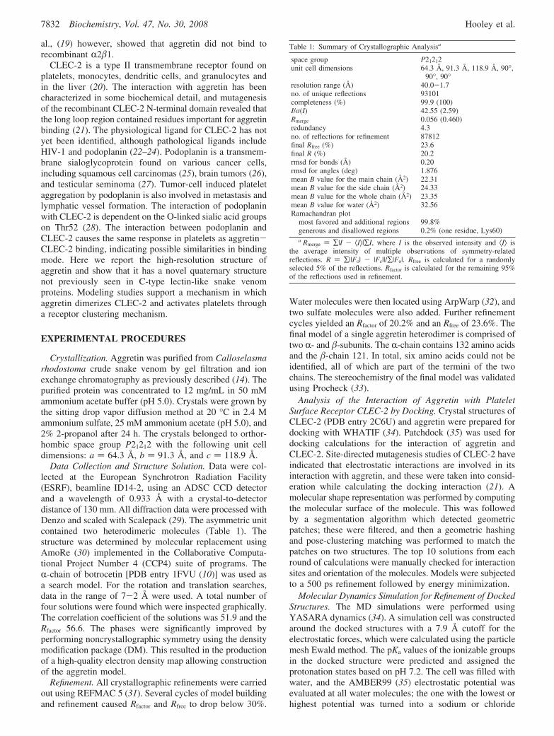
Aggretin Structure. Aggretin, like other members of theC-type lectin-like family, is composed of two structurally
homologous subunits, R and �, that form a disulfide-linkedheterodimer with each subunit containing a compact lectin-like globular domain and a long extended loop region (Figure1A). Superposition of the R- and �-subunits gives a rmsd of1.7 Å. The R- and �-subunits of aggretin have a low degreeof sequence homology of 38%, and the globular domainshave a predominantly hydrophobic core, which includes aperpendicular π-stacking network between the Phe20-Phe32-Phe129 motif in subunit R and the Phe17-Phe29-Phe117motif in subunit � (Figure 1B). The R-subunit has twodisulfide bonds (Cys33-Cys131 and Cys106-Cys123),whereas the �-subunit has three disulfide bonds (Cys2-Cys13,Cys30-Cys119, and Cys96-Cys111) which are conservedin other members of the C-type lectin-like family of snakevenom proteins (Figure 1B). Aggretin is highly homologousin sequence (∼50%) and structure with other members ofthe C-type lectin-like protein superfamily (Figure 2B andFigure S1 of the Supporting Information). These all [botro-
FIGURE 1: (A) Stereoview of the topology of the structure of the aggretin R�-heterodimer. The R-subunit is colored green and the �-subunityellow. Residues that form hydrogen bonds or hydrophobic interactions across the dimer interface are highlighted. Hydrogen bonds betweenside chains are shown as black dashed lines. (B) Stereoview of the topology of the quaternary structure of (R�)2 aggretin. The disulfidebonds of the R�-heterodimer are colored orange. The phenylalanine residues which form a π-stacking network in the globular domains arecolored cyan. (C) Surface representation of the side view of (R�)2 aggretin (same orientation as in panel A). (D) Surface representation of(R�)2 aggretin (same orientation as in panel B). The concave receptor binding site is shown with a black line.
Crystal Structure of Aggretin Biochemistry, Vol. 47, No. 30, 2008 7833
cetin (10), IX/X-bp (7), convulxin (5), flavocetin (8), echi-cetin (6), and aggretin] form R�-heterodimers throughdomain swapping of the extended loop region, and complexcrystal structures have revealed that ligand binding usuallyoccurs in a centrally located concave surface involvingcontacts with the extended loops.
The quaternary structure of aggretin is a novel (R�)2
tetramer which forms into an antiparallel structure due to a2-fold rotation (Figure 1B). The interface buried uponformation of the tetramer has a large surface area (11710Å2, 35% of the total surface area), and it remains a tetramerin gel filtration experiments (14). The side-by-side arrange-ment causes the concave surface of the R�-heterodimersaddle shape to be oriented on the same face (Figure 1C,D).Interactions between the two dimers are primarily betweenthe R-subunits with helix R1 interacting with the opposingR-chain extended loop. There are main chain-side chaininteractions between Glu28 and Ile99, Lys31 and Leu101,Lys31 and Leu98, and Arg34 and Ser94 and side chain-sidechain interactions between Arg43 and Glu80. The majorityof these side chains are not conserved in other C-type lectin-like snake venom proteins, including purpureotin and ag-glucetin which have both been shown to be tetrameric (Figure1B). The crystal structures have yet to be determined forthese proteins although, purpureotin has been modeled as aparallel dimer (36, 37).
Several snake venom C-type lectin-like proteins havedivalent cation binding sites within their globular domains.Factor IX/X-binding protein (FIX/X-bp) has Ca2+ bindingsites in both R- and �-subunits (7); botrocetin has one Ca2+
binding site, its �-subunit, but convulxin, flavocetin, andaggretin lack divalent cation binding sites (5, 8, 10). Assaysof aggretin activity in the presence of EDTA suggested thatits function was independent of divalent cations, and theaggretin structure reveals no bound cations (14). The cationbinding site in the R-subunit of FIX/X-bp consists of Ser41,
Glu43, Glu47, and Glu128 and in the �-subunit Ser41, Glu43,Glu47, and Glu122 which forms a negatively charged cationbinding pocket in IX/X-bp. In aggretin, only homologousresidues Ser44, Glu46, and Glu50 are conserved in both R-and �-subunits whereas Glu128 and Glu122 of IX/X-bp arereplaced with Lys132 and Lys120, respectively. These lattersubstitutions alter the charge of the pocket, and the terminalnitrogen atom of the lysine residues stabilizes both pocketsin the absence of divalent cation.
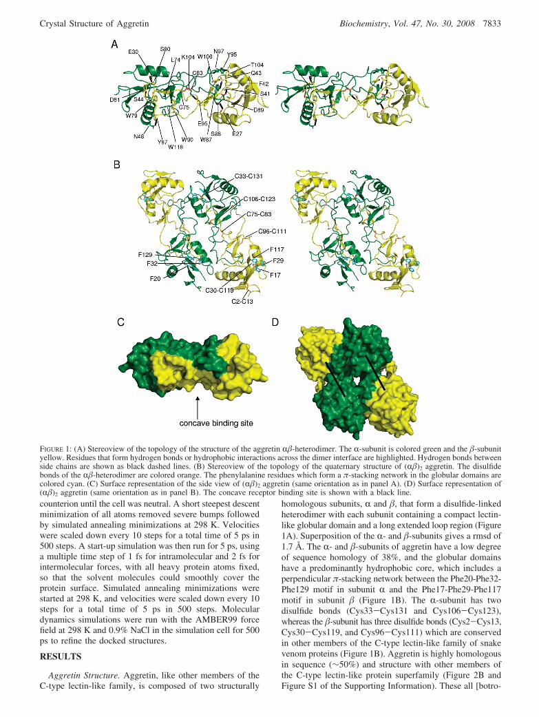
Docking of Aggretin and the CLEC-2 N-Terminal Domain.Aggretin is thought to activate platelets by binding andclustering receptor CLEC-2. The interaction between aggretinand the recombinant N-terminal domain of CLEC-2 (CLEC-2N) has been shown to have a dissociation constant of 1µM (21). To investigate the basis of this interaction, weperformed molecular docking with the aggretin R�-het-erodimer and the CLEC-2N crystal structure (21). Patchdock(35) was used for molecular docking calculations, and thecomplex with the best fit to the available biochemical datawas selected from the top three scoring structures. In thiscomplex, the CLEC-2N domain is positioned covering theconcave surface between the aggretin R�-lectin-like domainsand is slightly off center positioned closer to the �-subunitthan R. Principal contacts are formed between CLEC-2N andboth the R- and �-subunit lectin-like domains and long loopswith a series of salt bridges defining the key interactions(Figure 2A). From the interface, a short helix from CLEC-2N contributes residue Glu184 which contacts the aggretin�-subunit His103; interactions extend from here along thesemihelical long loop region, including CLEC-2N Glu187forming a salt bridge with �-subunit Arg100, and at theopposing end, CLEC-2N Lys190 interacts with the R-subunitside chain Glu110. Further contacts with the aggretinR-subunit are made through CLEC-2N Phe207 and Asn210contacting Leu112 and Asp68, respectively, and at theopposite end of the CLEC-2 sheet, residues Asn200 and
FIGURE 2: (A) Model of CLEC-2N docked with an aggretin R�-heterodimer. Residues involved in the interaction between CLEC-2N andaggretin are highlighted in stick format. CLEC-2 is colored blue, the aggretin R-subunit green, and the aggretin �-subunit yellow. (B)Superposition of the R�-heterodimers of the structurally homologous C-type lectin-like snake venom proteins aggretin (green), EMS16(blue), botrocetin (yellow), and flavocetin (red). (C) Diagram showing the positions of binding of different ligands to C-type lectin-likesnake venom proteins depicted in panel B: CLEC-2 (blue), the Gla domain of factor IX (orange), the integrin R2-I domain (purple), andthe A1 domain of von Willebrand factor (turquoise). These ligands bind aggretin (gray), IX-bp, EMS16, and botrocetin, respectively.
7834 Biochemistry, Vol. 47, No. 30, 2008 Hooley et al.

Lys202 pack against Asp100 from the R-subunit and Glu105from the �-subunit.
Mutagenic studies of CLEC-2N implicate the long loop,and more specifically residues Lys150, Lys171, Glu184,Asp188, Lys190, and Asn192, as being involved in aggretinbinding, and this is in broad agreement with the model (21).One discrepancy is that CLEC-2N residue Asp188 ispredicted to contribute to binding, but in the model, this onlymakes an incidental contact with Met109 from the �-subunit.The CLEC-2N and aggretin structures are essentially treatedas rigid bodies in this docking experiment, and one area ofthe structure which can potentially undergo conformationalchange upon formation of the complex is the CLEC-2N longloop (21). This could undergo a change in conformation toform a better fit into the aggretin groove, and as a result,Asp188 may form more direct interactions. In addition tothe residues previously identified, the model of CLEC-2Ndocked with aggretin also predicts that Asn210, His199,Lys202, and His204 may be important in CLEC-2-aggretinbinding.
The model of the aggretin-CLEC-2N complex is similarto the ligand complex crystal structures of snake venomC-type lectins. Figure 2C shows structures for the EMS16-integrin R2-I domain, botrocetin-von Willebrand factor A1domain, and factor IX binding protein-factor IX Gla domaincomplexes superposed onto the aggretin-/CLEC-2N model,and the alignment of the sequences illustrates the overlap inthe binding footprint (Figure S1 of the Supporting Informa-tion). Differences between the mode of action exist as bothEMS16 and factor IX binding protein have a functionblocking role and structurally exist as R�-dimers, whereas
aggretin activates platelets and has the (R�)2 structure witha clustering function.
Dimerization of CLEC-2N by Aggretin. Dimerization ofCLEC-2 would be an expected consequence of aggretinbinding as it is thought to activate platelets by clusteringthe receptor, thus bringing the cytoplasmic domains closertogether, enabling efficient phosphorylation of the YXXLmotif (24, 38). Figure 3A illustrates the CLEC-2N-aggretincomplex superposed onto the aggretin tetramer. Here the twoCLEC-2N domains are brought into proximity and “fittogether” forming a small interface, effectively bringing theN- and C-termini together through contact of residues101-133 (two �-strands, helix R1, and loop regions).Hydrophobic contacts are formed by Phe117 side chains atthe center of the interface close to the 2-fold axis with furtherside chain-main chain contacts occurring on either side, withthe His119 NE2 atom forming a hydrogen bond to theAsn105 carbonyl oxygen and the Tyr129 OH group forminga hydrogen bond to the main chain nitrogen of Trp106.Above this, as depicted in Figure 3B, the side chain fromThr104 packs against Met133 and Asn105 packs againstTrp106. Below this, Tyr213 hydrogen bonds to the mainchain nitrogen of His154 from helix R1.
Further evidence to support the CLEC-2N-aggretin modelof CLEC-2 dimerization comes from the structurally ho-mologous lectin-like proteins with immune functions, suchas receptors NKG2D, LOX-1, and Ly49C (39–41). Herecrystal structures are available for dimers of both unligandedand ligand complexed crystal structures, and in the case ofNKG2D, the structure of the dimer is remarkably similar tothat of CLEC-2N. The difference is shown in Figure 3C as
FIGURE 3: (A) CLEC-2N-aggretin complex superimposed onto the aggretin (R�)2 tetramer. CLEC-2N is colored blue, the aggretin R-subunitgreen, and the aggretin �-subunit yellow. The R�-heterodimers and two CLEC-2N molecules are differentiated by darker and lighter shading.The N- and C-termini are labeled. (B) Dimer interface of CLEC-2N as determined by superimposing the CLEC-2N-aggretin complex ontothe aggretin tetramer. The interface is predominantly hydrophobic, and those residues involved in dimer formation are highlighted. (C)Comparison of the CLEC-2N (blue) dimer of the CLEC-2N-aggretin complex and the structure of immune receptor NGK2D (red) of theNKG2D-ULBP3 complex (PDB entry 1KCG). The left-hand monomer of each dimer is in the same orientation, thus showing the 45°rotation of the domains.
Crystal Structure of Aggretin Biochemistry, Vol. 47, No. 30, 2008 7835
a 45° rotation of the domains such that instead of the helixR2 forming contacts on the 2-fold axis it is the adjacent�-sheet which interlocks. We next examined whether theCLEC-2N domain could form a dimer structure similar tothe immune receptors by superposing the CLEC-2N structureonto the dimer structures of NKG2D, LOX-1, and Ly49C,and in each case, there were steric clashes indicating thesedimerization modes are unlikely for CLEC-2N.
DISCUSSION
The structure of aggretin reveals a novel tetrameric (R�)2
quaternary structure in which aggretin presents two adjacentreceptor binding sites supporting a receptor clusteringmechanism of action. This is similar to the clustering ofGPVI and GPIbR by convulxin and flavocetin A, respec-tively. The R�-heterodimers of convulxin and flavocetin Aare structurally homologous to that of aggretin; however,their quaternary structure is quite different. Convulxin andflavocetin A are both disulfide-linked cyclic tetramers of(R�)4 structure with the disulfide bond between the C-terminus of the R-subunit and the N-terminus of the�-subunit (8, 42). This allows for the binding and clusteringof four ligand (GPVI) molecules. Aggretin on the other handhas no free cysteine residues and therefore cannot form acyclic structure similar to convulxin and flavocetin A andinstead forms a noncovalent (R�)2 dimer. A series of reportssuggest that CLEC-2 is the principal platelet receptor ligandfor aggretin but that GPIbR and R2�1 may have supple-mentary roles in the same way as in other snake venomC-type lectin-like proteins (12). Podoplanin, which is in-volved in pathological tumor cell-induced platelet aggrega-tion, has also been shown to bind CLEC-2 (23, 24). Theinteraction of podoplanin with CLEC-2 results in a signalingresponse, which subsequently leads to platelet aggregation,similar to that induced by aggretin-CLEC-2 binding. In spiteof this similarity, it appears that aggretin and podoplanininteract with CLEC-2 in a different manner. The interactionof aggretin and CLEC-2 is restricted to the concave surfaceof aggretin and the C-terminal loop of CLEC-2. Thisinteraction mainly involves hydrogen bonds between the sidechains of the two proteins and has been identified bymutagenic studies. The structure of aggretin shows that thereare no glycosylation sites, or other post-translational modi-fications, which could be involved in CLEC-2 binding. Thebinding site for CLEC-2 in podoplanin has been shown toinvolve the sialic acid group of O-glycan on Thr52 andresidues 38-54 which surround it. Podoplanin has beenshown to bind to an area which is predominantly positivelycharged on the surface of CLEC-2N (residues 87-147) (24).Sialic acid has several hydroxyl groups, all of which arepotential hydrogen bond donors, and sialic acid also causesa slight negative charge on the surface of the protein. Thesedata show that aggretin and podoplanin have differentbinding sites on CLEC-2, suggesting that it is the clusteringof the ligand which determines the downstream effects, notthe isolated binding of a ligand to the extracellular domain(24).
In the absence of a high-resolution complex crystalstructure, the aggretin-CLEC-2N model provides predictionswhich can be tested through functional analysis and con-tributes to our understanding of platelet activation and
aggregation and may also help further improve our under-standing of the basis of the “platelet cloak” which contributesto tumor cell survival and metastasis mediated by receptorpodoplanin.
ACKNOWLEDGMENT
We thank the staff at the European Synchrotron ResearchFacility beamline ID 14-2 for their assistance with datacollection and David Leys for his advice on molecularreplacement calculations.
SUPPORTING INFORMATION AVAILABLE
Sequence alignment of the R- and �-chains of aggretin,IX-bp, EMS16, and botrocetin showing conserved residuesand residues involved in ligand binding (Figure S1). Thismaterial is available free of charge via the Internet at http://pubs.acs.org.
REFERENCES
1. Andrews, R. K., and Berndt, M. C. (2000) Snake venom modulatorsof platelet adhesion receptors and their ligands. Toxicon 38, 775–791.
2. Morita, T. (2005) Structures and functions of snake venom CLPs(C-type lectin-like proteins) with anticoagulant-, procoagulant-, andplatelet-modulating activities. Toxicon 45, 1099–1114.
3. Matsui, T., and Hamako, J. (2005) Structure and function of snakevenom toxins interacting with human von Willebrand factor.Toxicon 45, 1075–1087.
4. Braud, S., Bon, C., and Wisner, A. (2000) Snake venom proteinsacting on hemostasis. Biochimie 82, 851–859.
5. Batuwangala, T., Leduc, M., Gibbins, J. M., Bon, C., and Jones,E. Y. (2004) Structure of the snake-venom toxin convulxin. ActaCrystallogr. D60, 46–53.
6. Jasti, J., Paramasivam, M., Srinivasan, A., and Singh, T. P. (2004)Crystal structure of echicetin from Echis carinatus (Indian saw-scaled viper) at 2.4 Å resolution. J. Mol. Biol. 335, 167–176.
7. Mizuno, H., Fujimoto, Z., Koizumi, M., Kano, H., Atoda, H., andMorita, T. (1997) Structure of coagulation factors IX/X-bindingprotein, a heterodimer of C-type lectin domains. Nat. Struct. Biol.4, 438–441.
8. Fukuda, K., Mizuno, H., Atoda, H., and Morita, T. (2000) Crystalstructure of flavocetin-A, a platelet glycoprotein Ib-binding protein,reveals a novel cyclic tetramer of C-type lectin-like heterodimers.Biochemistry 39, 1915–1923.
9. Hirotsu, S., Mizuno, H., Fukuda, K., Qi, M. C., Matsui, T., Hamako,J., Morita, T., and Titani, K. (2001) Crystal structure of bitiscetin,a von Willebrand factor-dependent platelet aggregation inducer.Biochemistry 40, 13592–13597.
10. Sen, U., Vasudevan, S., Subbarao, G., McClintock, R. A., Celikel,R., Ruggeri, Z. M., and Varughese, K. I. (2001) Crystal structureof the von Willebrand factor modulator botrocetin. Biochemistry40, 345–352.
11. Huang, T. F., Liu, C. Z., and Yang, S. H. (1995) Aggretin, a novelplatelet-aggregation inducer from snake (Calloselasma rhodostoma)venom, activates phospholipase C by acting as a glycoprotein Ia/IIa agonist. Biochem. J. 309 (Part 3), 1021–1027.
12. Shin, Y., and Morita, T. (1998) Rhodocytin, a functional novelplatelet agonist belonging to the heterodimeric C-type lectin family,induces platelet aggregation independently of glycoprotein Ib.Biochem. Biophys. Res. Commun. 245, 741–745.
13. Chung, C. H., Au, L. C., and Huang, T. F. (1999) Molecular cloningand sequence analysis of aggretin, a collagen-like platelet aggrega-tion inducer. Biochem. Biophys. Res. Commun. 263, 723–727.
14. Navdaev, A., Clemetson, J. M., Polgar, J., Kehrel, B. E., Glauner,M., Magnenat, E., Wells, T. N., and Clemetson, K. J. (2001)Aggretin, a heterodimeric C-type lectin from Calloselasma rho-dostoma (malayan pit viper), stimulates platelets by binding toR2�1 integrin and glycoprotein Ib, activating Syk and phospho-lipase Cγ2, but does not involve the glycoprotein VI/Fc receptorγ chain collagen receptor. J. Biol. Chem. 276, 20882–20889.
7836 Biochemistry, Vol. 47, No. 30, 2008 Hooley et al.
15. Shiraishi, M., Ogawa, H., Ikeda, M., Kawashima, S., and Ito, K.(2002) Platelet dysfunction in Chediak-Higashi syndrome-affectedcattle. J. Vet. Med. Sci. 64, 751–760.
16. Chung, C. H., Peng, H. C., and Huang, T. F. (2001) Aggretin, aC-type lectin protein, induces platelet aggregation via integrin R2�1
and GPIb in a phosphatidylinositol 3-kinase independent pathway.Biochem. Biophys. Res. Commun. 285, 689–695.
17. Bergmeier, W., Bouvard, D., Eble, J. A., Mokhtari-Nejad, R.,Schulte, V., Zirngibl, H., Brakebusch, C., Fassler, R., andNieswandt, B. (2001) Rhodocytin (aggretin) activates plateletslacking R2�1 integrin, glycoprotein VI, and the ligand-bindingdomain of glycoprotein IbR. J. Biol. Chem. 276, 25121–25126.
18. Suzuki-Inoue, K., Ozaki, Y., Kainoh, M., Shin, Y., Wu, Y., Yatomi,Y., Ohmori, T., Tanaka, T., Satoh, K., and Morita, T. (2001)Rhodocytin induces platelet aggregation by interacting with gly-coprotein Ia/IIa (GPIa/IIa, Integrin R2�1). Involvement of GPIa/IIa-associated src and protein tyrosine phosphorylation. J. Biol.Chem. 276, 1643–1652.
19. Eble, J. A., Beermann, B., Hinz, H. J., and Schmidt-Hederich, A.(2001) R2�1 integrin is not recognized by rhodocytin but is thespecific, high affinity target of rhodocetin, an RGD-independentdisintegrin and potent inhibitor of cell adhesion to collagen. J. Biol.Chem. 276, 12274–12284.
20. Suzuki-Inoue, K., Fuller, G. L., Garcia, A., Eble, J. A., Pohlmann,S., Inoue, O., Gartner, T. K., Hughan, S. C., Pearce, A. C., Laing,G. D., Theakston, R. D., Schweighoffer, E., Zitzmann, N., Morita,T., Tybulewicz, V. L., Ozaki, Y., and Watson, S. P. (2006) A novelSyk-dependent mechanism of platelet activation by the C-type lectinreceptor CLEC-2. Blood 107, 542–549.
21. Watson, A. A., Brown, J., Harlos, K., Eble, J. A., Walter, T. S.,and O’Callaghan, C. A. (2007) The crystal structure and mutationalbinding analysis of the extracellular domain of the platelet-activating receptor CLEC-2. J. Biol. Chem. 282, 3165–3172.
22. Chaipan, C., Soilleux, E. J., Simpson, P., Hofmann, H., Gramberg,T., Marzi, A., Geier, M., Stewart, E. A., Eisemann, J., Steinkasserer,A., Suzuki-Inoue, K., Fuller, G. L., Pearce, A. C., Watson, S. P.,Hoxie, J. A., Baribaud, F., and Pohlmann, S. (2006) DC-SIGNand CLEC-2 mediate human immunodeficiency virus type 1 captureby platelets. J. Virol. 80, 8951–8960.
23. Suzuki-Inoue, K., Kato, Y., Inoue, O., Kaneko, M. K., Mishima,K., Yatomi, Y., Yamazaki, Y., Narimatsu, H., and Ozaki, Y. (2007)Involvement of the snake toxin receptor CLEC-2, in podoplanin-mediated platelet activation, by cancer cells. J. Biol. Chem. 282,25993–26001.
24. Kato, Y., Kaneko, M. K., Kunita, A., Ito, H., Kameyama, A.,Ogasawara, S., Matsuura, N., Hasegawa, Y., Suzuki-Inoue, K.,Inoue, O., Ozaki, Y., and Narimatsu, H. (2008) Molecular analysisof the pathophysiological binding of the platelet aggregation-inducing factor podoplanin to the C-type lectin-like receptor CLEC-2. Cancer Sci. 99, 54–61.
25. Wicki, A., Lehembre, F., Wick, N., Hantusch, B., Kerjaschki, D.,and Christofori, G. (2006) Tumor invasion in the absence ofepithelial-mesenchymal transition: Podoplanin-mediated remodelingof the actin cytoskeleton. Cancer Cell 9, 261–272.
26. Mishima, K., Kato, Y., Kaneko, M. K., Nakazawa, Y., Kunita,A., Fujita, N., Tsuruo, T., Nishikawa, R., Hirose, T., and Matsutani,M. (2006) Podoplanin expression in primary central nervous systemgerm cell tumors: a useful histological marker for the diagnosis ofgerminoma. Acta Neuropathol. 111, 563–568.
27. Kato, Y., Sasagawa, I., Kaneko, M., Osawa, M., Fujita, N., andTsuruo, T. (2004) Aggrus: A diagnostic marker that distinguishes
seminoma from embryonal carcinoma in testicular germ celltumors. Oncogene 23, 8552–8556.
28. Kaneko, M. K., Kato, Y., Kameyama, A., Ito, H., Kuno, A.,Hirabayashi, J., Kubota, T., Amano, K., Chiba, Y., Hasegawa, Y.,Sasagawa, I., Mishima, K., and Narimatsu, H. (2007) Functionalglycosylation of human podoplanin: Glycan structure of plateletaggregation-inducing factor. FEBS Lett. 581, 331–336.
29. Otwinowski, Z., and Minor, W. (1997) Processing of X-raydiffraction data collected in oscillation mode. Methods Enzymol.276, 307–326.
30. Navaza, J. (1994) AMoRE: An automated package for molecularreplacement. Acta Crystallogr. A50, 157–163.
31. Murshudov, G., Vagin, A., and Dodson, E. (1997) Refinement ofMacromolecular Structures by the Maximum-Likelihood Method.Acta Crystallogr. D53, 240–255.
32. Perrakis, A., Morris, R. M., and Lamzin, V. S. (1999) Automatedprotein model building combined with iterative structure refinement.Nat. Struct. Biol. 6, 458–463.
33. Laskowski, R. A., Moss, D. S., and Thornton, J. M. (1993) Main-chain bond lengths and bond angles in protein structures. J. Mol.Biol. 231, 1049–1067.
34. Krieger, E., Darden, T., Nabuurs, S. B., Finkelstein, A., and Vriend,G. (2004) Making optimal use of empirical energy functions: Force-field parameterization in crystal space. Proteins 57, 678–683.
35. Liu, H., Elstner, M., Kaxiras, E., Frauenheim, T., Hermans, J., andYang, W. (2001) Quantum mechanics simulation of proteindynamics on long timescale. Proteins 44, 484–489.
36. Li, X., Zheng, L., Kong, C., Kolatkar, P. R., and Chung, M. C.(2004) Purpureotin: A novel di-dimeric C-type lectin-like proteinfrom Trimeresurus purpureomaculatus venom is stabilized bynoncovalent interactions. Arch. Biochem. Biophys. 424, 53–62.
37. Wang, W. J., and Huang, T. F. (2001) A novel tetrameric venomprotein, agglucetin from Agkistrodon acutus, acts as a glycoproteinIb agonist. Thromb. Haemostasis 86, 1077–1086.
38. Fuller, G. L., Williams, J. A., Tomlinson, M. G., Eble, J. A., Hanna,S. L., Pohlmann, S., Suzuki-Inoue, K., Ozaki, Y., Watson, S. P.,and Pearce, A. C. (2007) The C-type lectin receptors CLEC-2 andDectin-1, but not DC-SIGN, signal via a novel YXXL-dependentsignaling cascade. J. Biol. Chem. 282, 12397–12409.
39. Radaev, S., Rostro, B., Brooks, A. G., Colonna, M., and Sun, P. D.(2001) Conformational plasticity revealed by the cocrystal structureof NKG2D and its class I MHC-like ligand ULBP3. Immunity 15,1039–1049.
40. Ohki, I., Ishigaki, T., Oyama, T., Matsunaga, S., Xie, Q., Ohnishi-Kameyama, M., Murata, T., Tsuchiya, D., Machida, S., Morikawa,K., and Tate, S. (2005) Crystal structure of human lectin-like,oxidized low-density lipoprotein receptor 1 ligand binding domainand its ligand recognition mode to OxLDL. Structure 13, 905–917.
41. Dam, J., Guan, R., Natarajan, K., Dimasi, N., Chlewicki, L. K.,Kranz, D. M., Schuck, P., Margulies, D. H., and Mariuzza, R. A.(2003) Variable MHC class I engagement by Ly49 natural killercell receptors demonstrated by the crystal structure of Ly49C boundto H-2K(b). Nat. Immunol. 4, 1213–1222.
42. Murakami, M. T., Zela, S. P., Gava, L. M., Michelan-Duarte, S.,Cintra, A. C., and Arni, R. K. (2003) Crystal structure of the plateletactivator convulxin, a disulfide-linked R4�4 cyclic tetramer fromthe venom of Crotalus durissus terrificus. Biochem. Biophys. Res.Commun. 310, 478–482.
BI800528T
Crystal Structure of Aggretin Biochemistry, Vol. 47, No. 30, 2008 7837