Bacterial Morphology and Structure Bacterial Morphology and Structure.
55
Bacterial Morphology Bacterial Morphology and Structure and Structure
-
Upload
berenice-annice-jennings -
Category
Documents
-
view
273 -
download
0
Transcript of Bacterial Morphology and Structure Bacterial Morphology and Structure.
Bacterial Morphology and Bacterial Morphology and StructureStructure
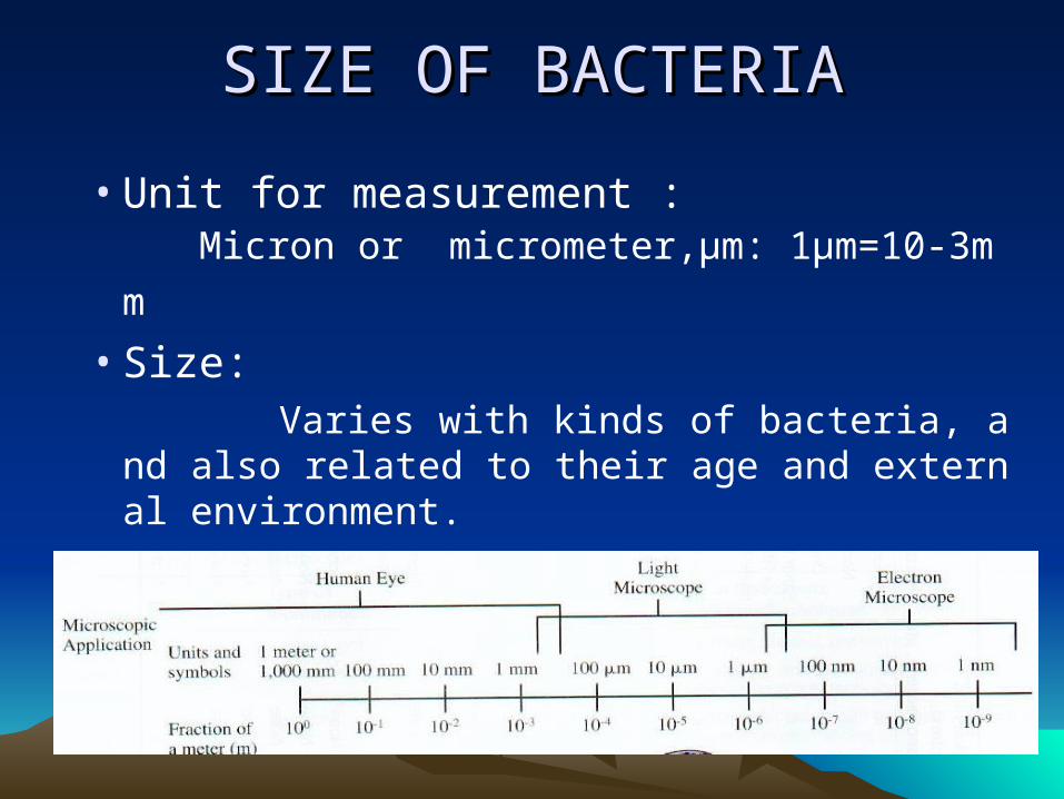
SIZE OF BACTERIASIZE OF BACTERIA
• Unit for measurement : Micr
on or micrometer,μm: 1μm=10-3mm • Size: Varies with kinds of bacteria, and also r
elated to their age and external environment.
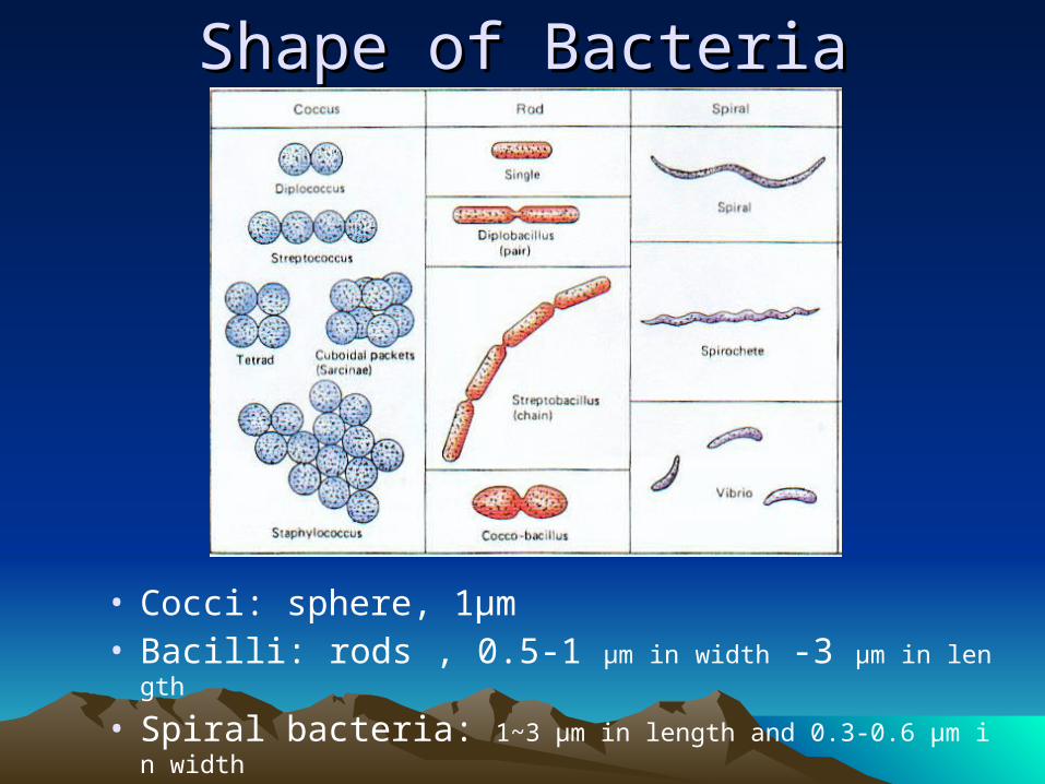
Shape of BacteriaShape of Bacteria
• Cocci: sphere, 1μm • Bacilli: rods , 0.5-1 μm in width -3 μm in length
• Spiral bacteria: 1~3 μm in length and 0.3-0.6 μm in width
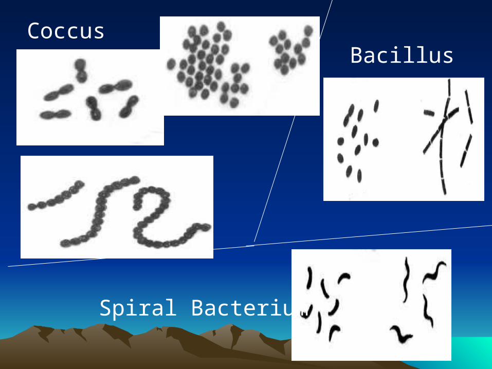
CoccusBacillus
Spiral Bacterium
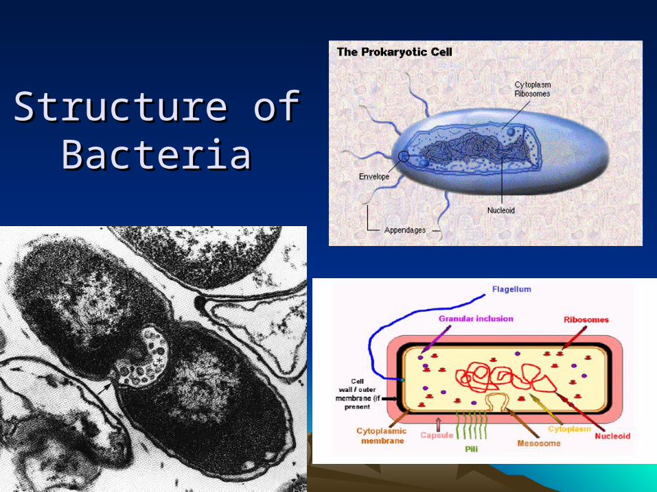
Structure of Structure of BacteriaBacteria
Essential structures Essential structures 基本结构基本结构 cell wall cell wall 细胞壁细胞壁
cell membrane cell membrane 细胞膜细胞膜Cytoplasm Cytoplasm 细胞质细胞质
nuclear materialnuclear material 核质核质
Particular structures 特殊结构capsule 荚膜flagella 鞭毛
pili 菌毛spore 芽胞
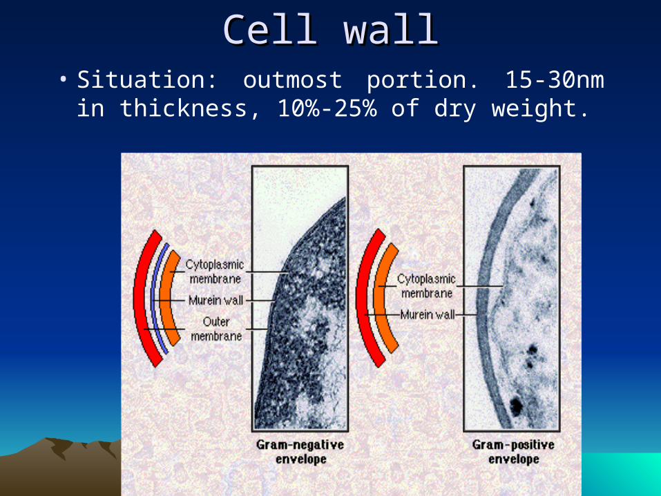
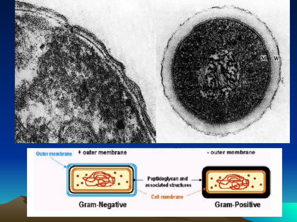
Cell wallCell wall• Situation: outmost portion. 15-30nm in
thickness, 10%-25% of dry weight.

1884: 1884: Christian GramChristian Gram: First publication for the Gram stain method) : First publication for the Gram stain method) Editor's note: I would like to testify that I have found the Gram method to be one ofEditor's note: I would like to testify that I have found the Gram method to be one ofthe best and for many cases the best method which I have ever used for stainingthe best and for many cases the best method which I have ever used for staining Schizomycetes.Schizomycetes.
Gram, C. 1884. Ueber die iGram, C. 1884. Ueber die isolirte Farbung der Schizosolirte Farbung der Schizomyceten in SchnittÄund Tromyceten in SchnittÄund Trockenpraparaten. ckenpraparaten. FortschrittFortschritte der Medicine der Medicin, Vol. 2, page, Vol. 2, pages 185-189.s 185-189.
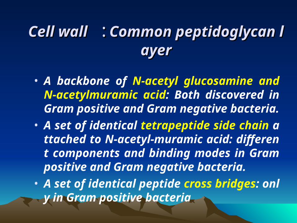
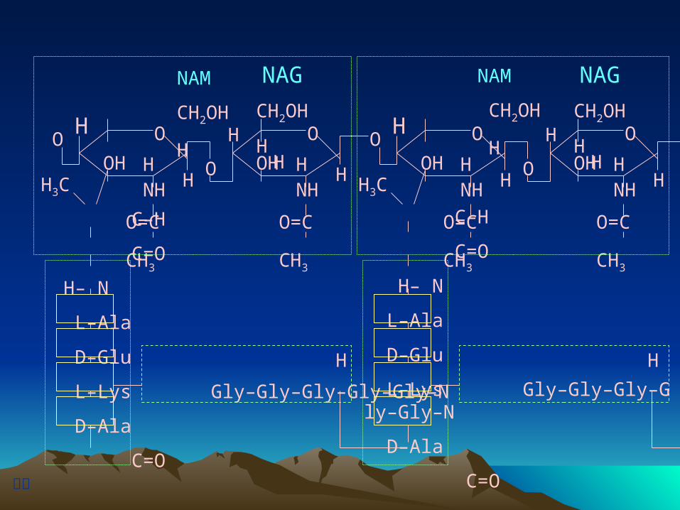
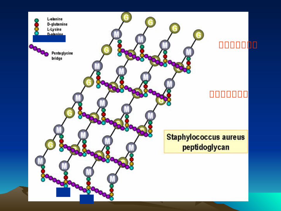
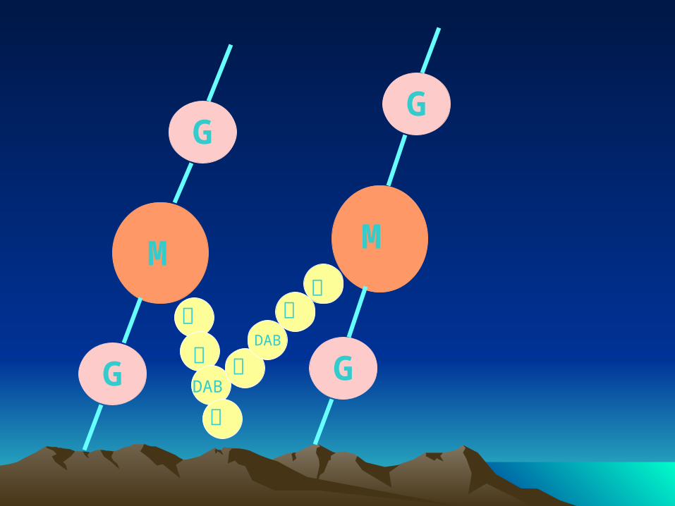
Cell wallCell wall : :Common peptidoglycan lCommon peptidoglycan layerayer
• A backbone of N-acetyl glucosamine and N-acetylmuramic acid: Both discovered in Gram positive and Gram negative bacteria.
• A set of identical tetrapeptide side chain attached to N-acetyl-muramic acid: different components and binding modes in Gram positive and Gram negative bacteria.
• A set of identical peptide cross bridges: only in Gram positive bacteria
图附
NAM
CH2OH
H
C–H
C=O
H– N
L–Ala
D–Glu
L–Lys Gly–Gly–Gly–Gly–Gly–N
D–Ala
C=O
O
OH
HNHH3C
HO
HO
H
O=C CH3
O
OH
HNH
H
O=C CH3
CH2OH
HH
H
NAG NAM
CH2OH
H
C–H
C=O
H– N
L–Ala
D–Glu
L–Lys Gly–Gly–Gly–Gly–Gly–N
D–Ala
C=O
O
OH
HNHH3C
HO
HO
H
O=C CH3
O
OH
HNH
CH2OH
HH
H
NAG
O=C CH3
H
溶菌酶作用位点
青霉素作用位点
G
G
G
G
M M
丙谷DAB
丙
丙DAB
谷丙
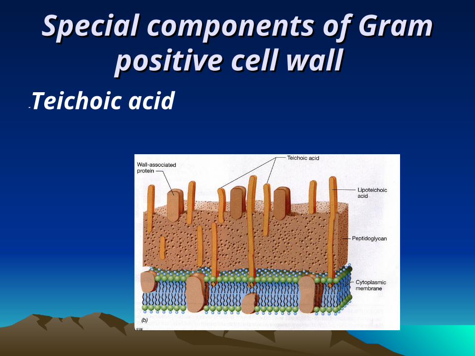
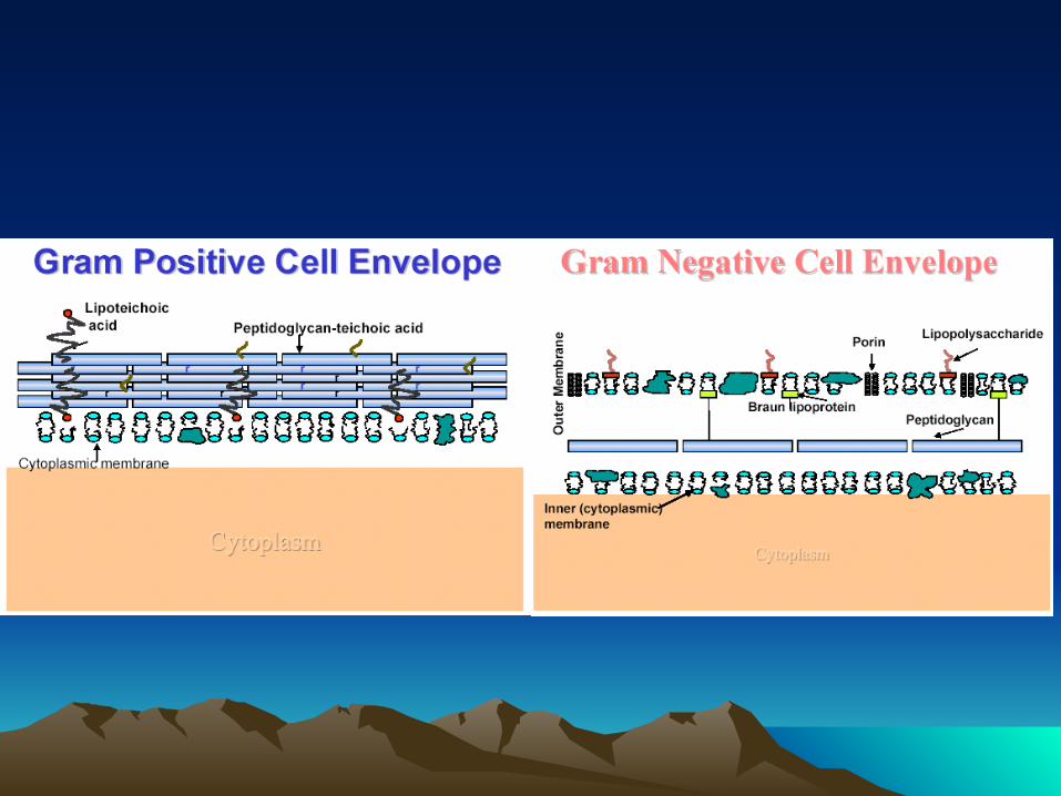
Special components of Gram Special components of Gram positive cell wallpositive cell wall
-Teichoic acid
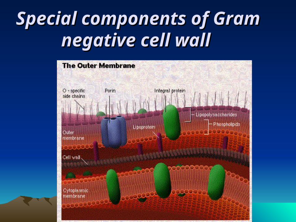
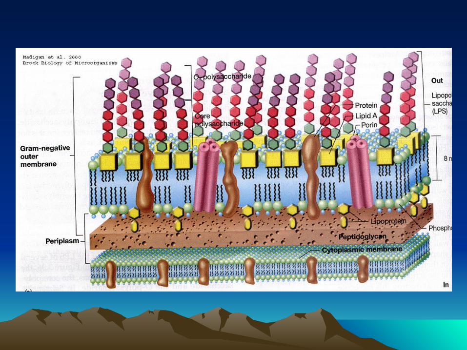
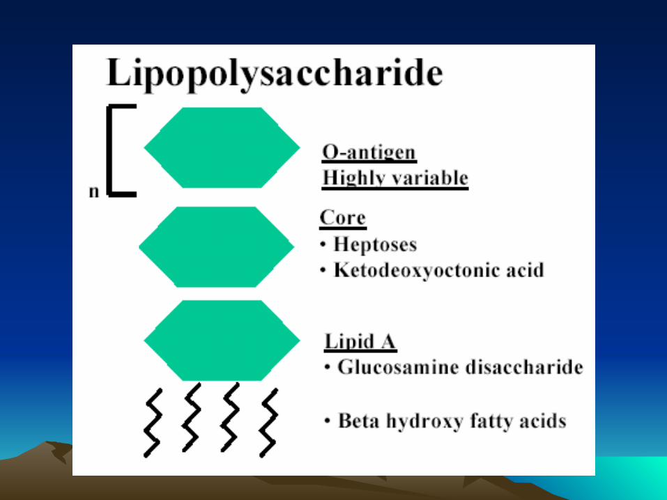
Special components of Special components of Gram negative cell wall Gram negative cell wall
FunctionsFunctions of Cell Wall of Cell Wall
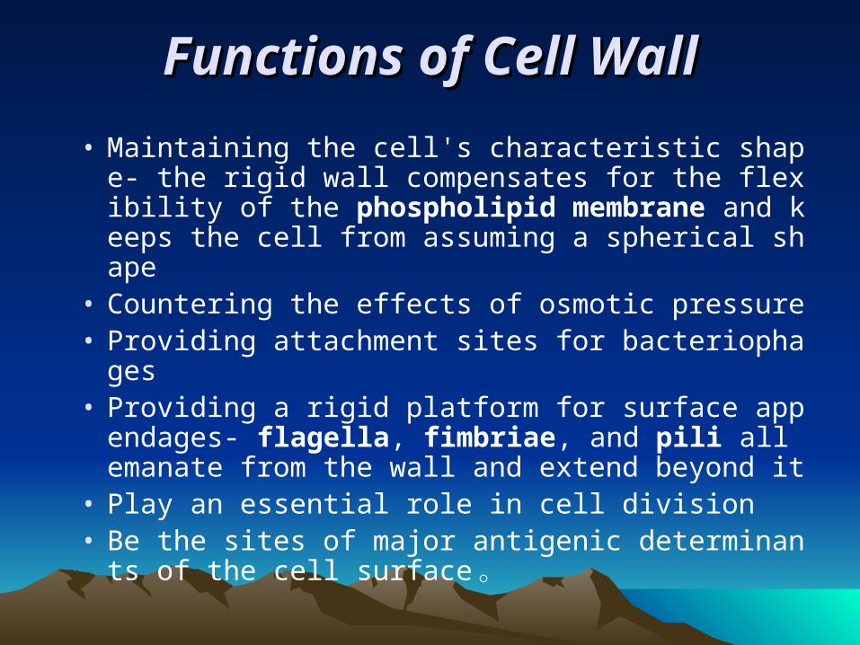
• Maintaining the cell's characteristic shape- the rigid wall compensates for the flexibility of the phospholipid membrane and keeps the cell from assuming a spherical shape
• Countering the effects of osmotic pressure• Providing attachment sites for bacteriophages• Providing a rigid platform for surface appenda
ges- flagella, fimbriae, and pili all emanate from the wall and extend beyond it
• Play an essential role in cell division• Be the sites of major antigenic determinants of
the cell surface 。
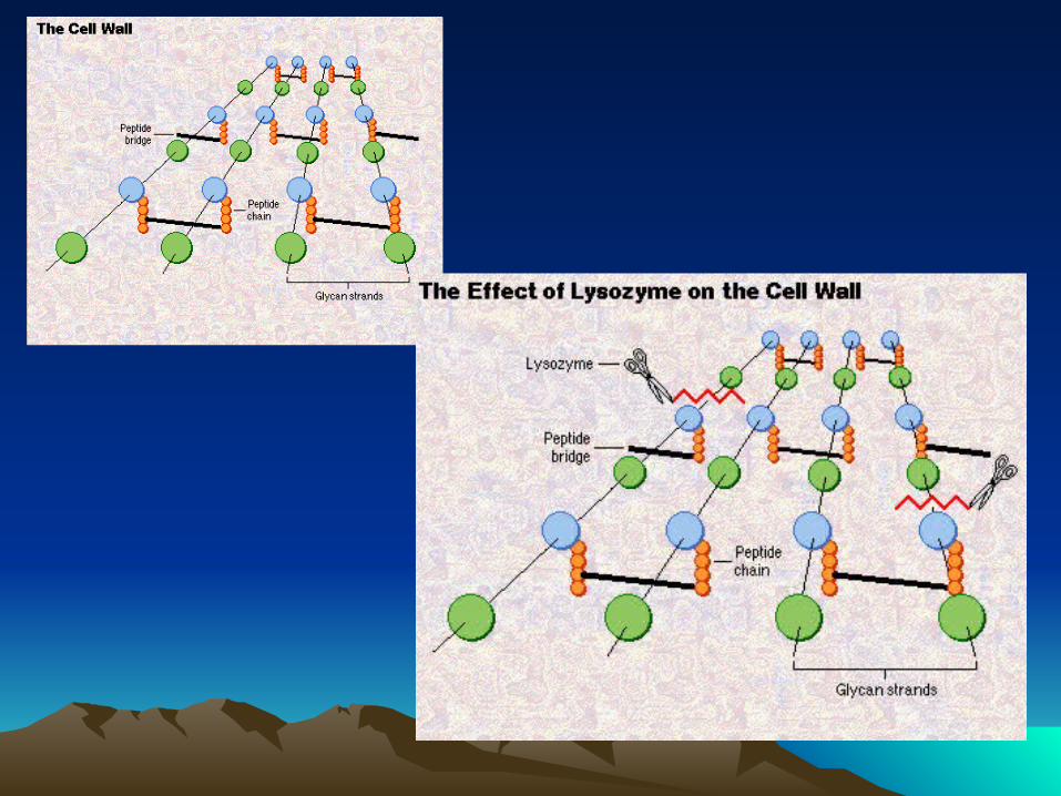
Wall-less forms of BacteriaWall-less forms of Bacteria..• When bacteria are treated with 1) enzymes that are
lytic for the cell wall e.g. lysozyme or 2) antibiotics that interfere with biosynthesis of peptidoglycan, wall-less bacteria are often produced.
• Usually these treatments generate non-viable organisms. Wall-less bacteria that can not replicate are referred to as spheroplasts (when an outer membrane is present) or protoplasts (if an outer membrane is not present).
• Occasionally wall-less bacteria that can replicate are generated by these treatments (L forms).
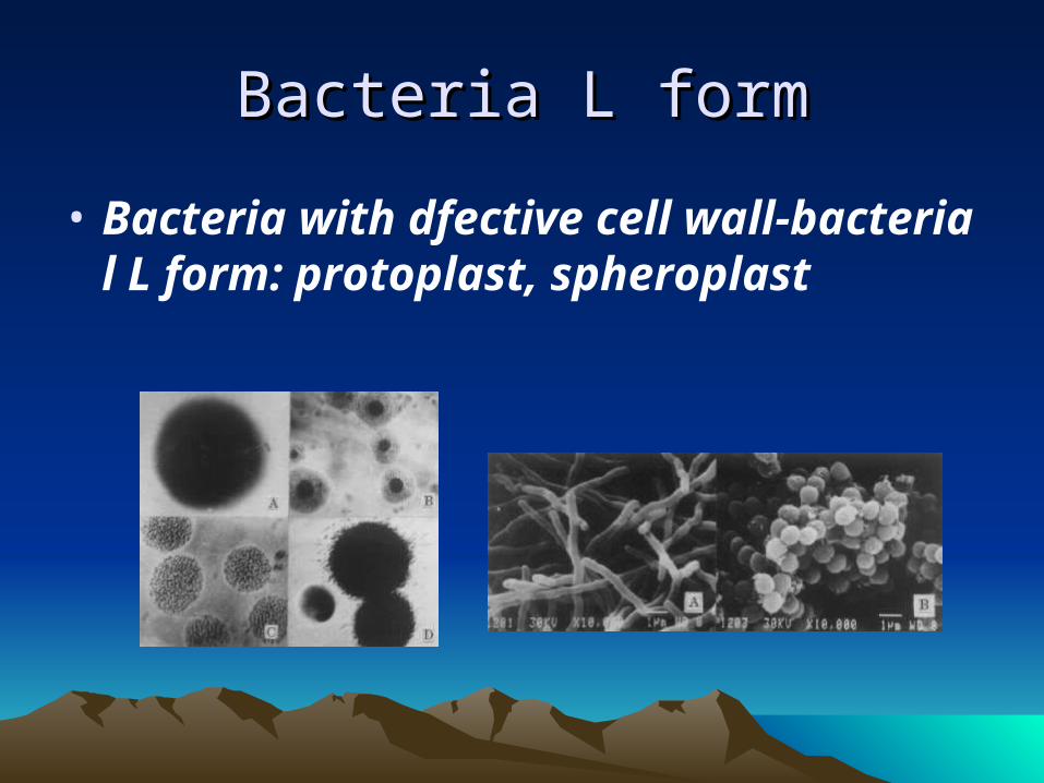
Bacteria L formBacteria L form
• Bacteria with dfective cell wall-bacterial L form: protoplast, spheroplast
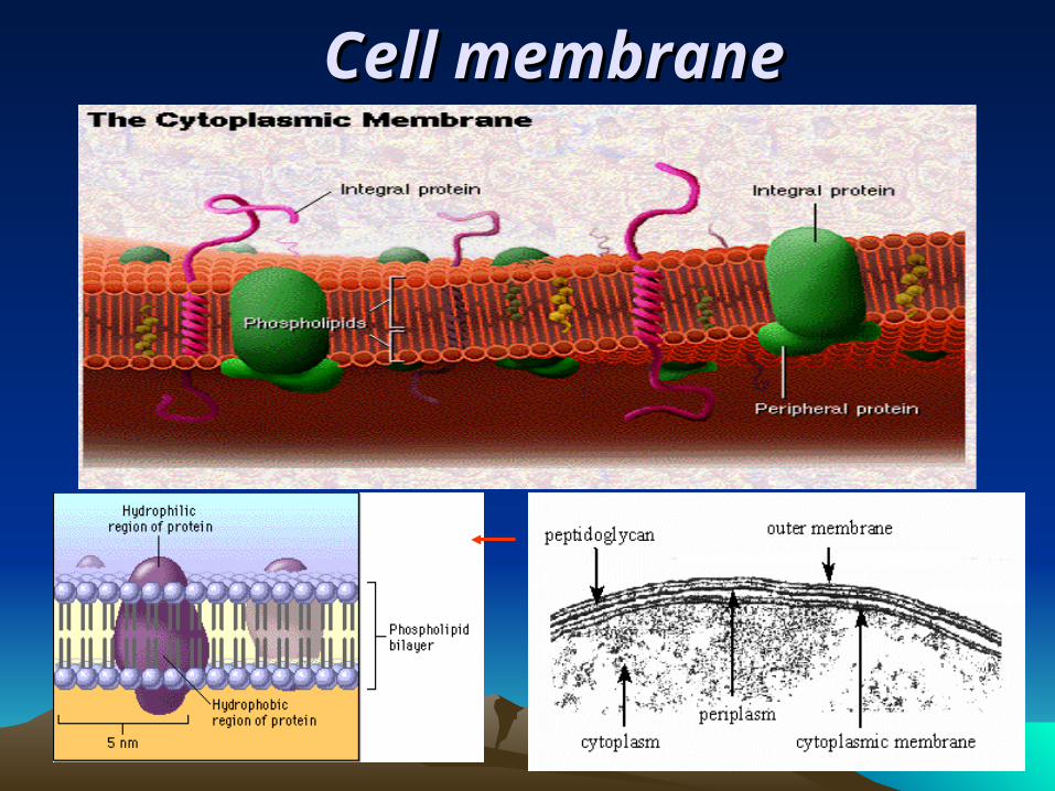
Cell membraneCell membrane
Function of Cell membraneFunction of Cell membrane
a. Selective permeability and transport of solutes into cells
b. Electron transport and oxidative phosphorylation
c. Excretion of hydrolytic exoenzymes
d. Site of biosynthesis of DNA, cell wall polymers and membrane lipids.
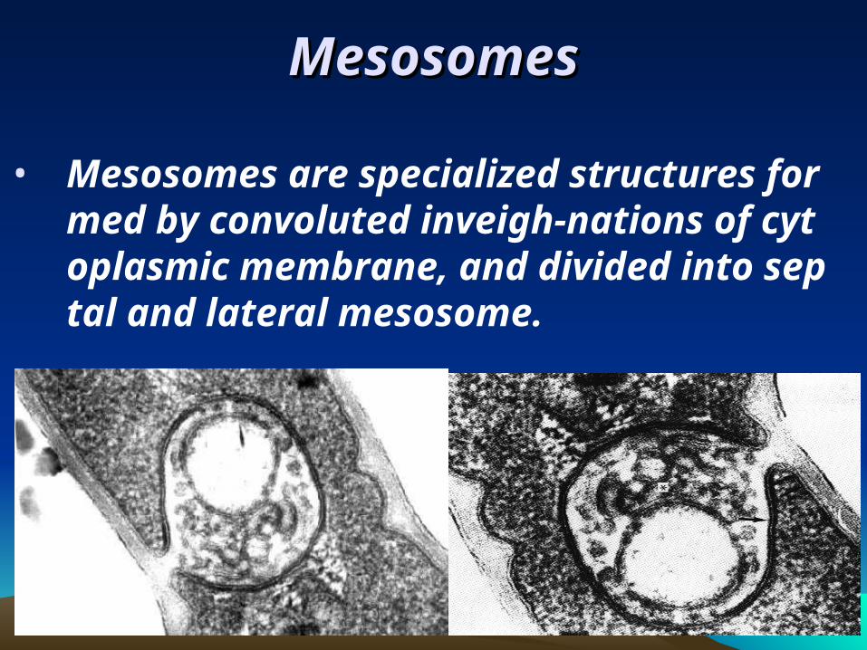
MesosomesMesosomes
• Mesosomes are specialized structures formed by convoluted inveigh-nations of cytoplasmic membrane, and divided into septal and lateral mesosome.
CytoplasmCytoplasm
• Composed largely of water, together with proteins, nucleic acid, lipids and small amount of sugars and salts
• Ribosomes: numerous, 15-20nm in diameter with 70S; distributed throughout the cytoplasm; sensitive to streptomycin and erythromycin site of protein synthesis
• Plasmids: extrachromosomal genetic elements• Inclusions: sources of stored energy, e,g voluti
n
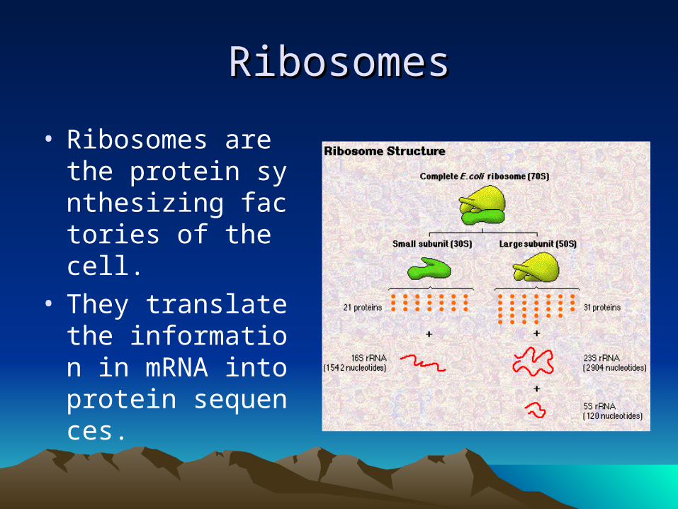
RibosomesRibosomes
• Ribosomes are the protein synthesizing factories of the cell.
• They translate the information in mRNA into protein sequences.
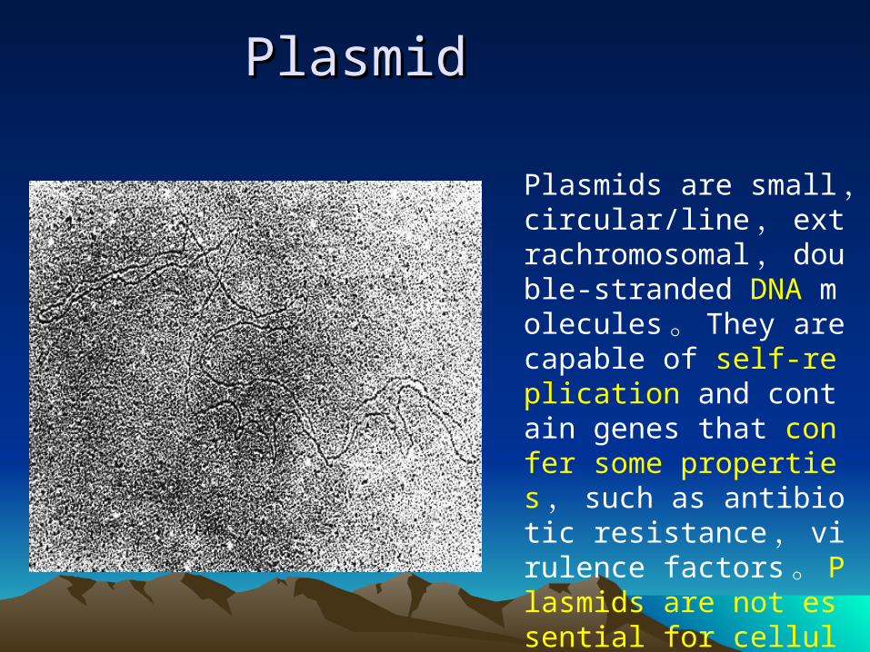
PlasmidPlasmid
Plasmids are small , circular/line , extrachromosomal , double-stranded DNA molecules 。 They are capable of self-replication and contain genes that confer some properties , such as antibiotic resistance , virulence factors 。 Plasmids are not essential for cellular survival.
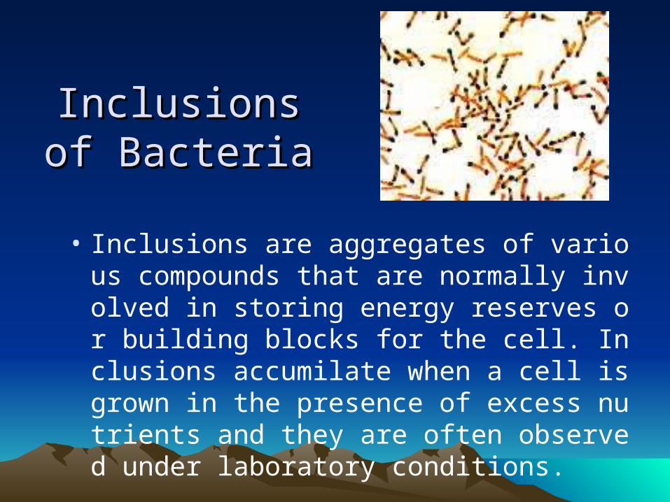
Inclusions of Inclusions of BacteriaBacteria
• Inclusions are aggregates of various compounds that are normally involved in storing energy reserves or building blocks for the cell. Inclusions accumilate when a cell is grown in the presence of excess nutrients and they are often observed under laboratory conditions.
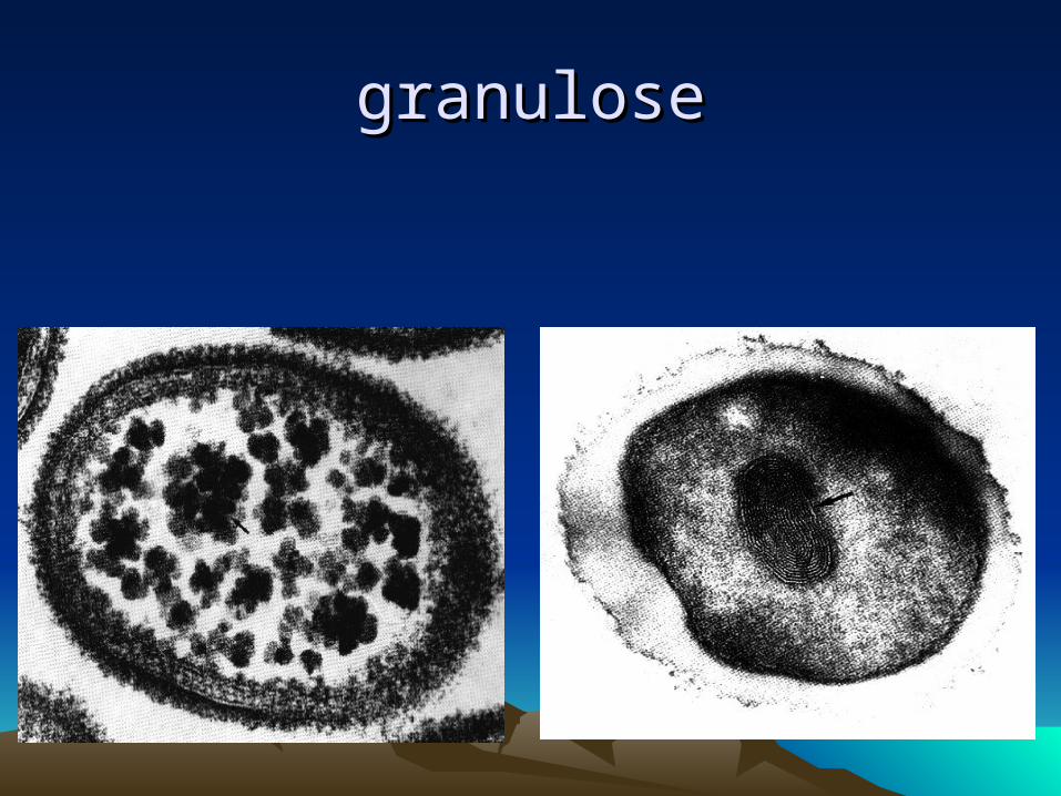
granulosegranulose
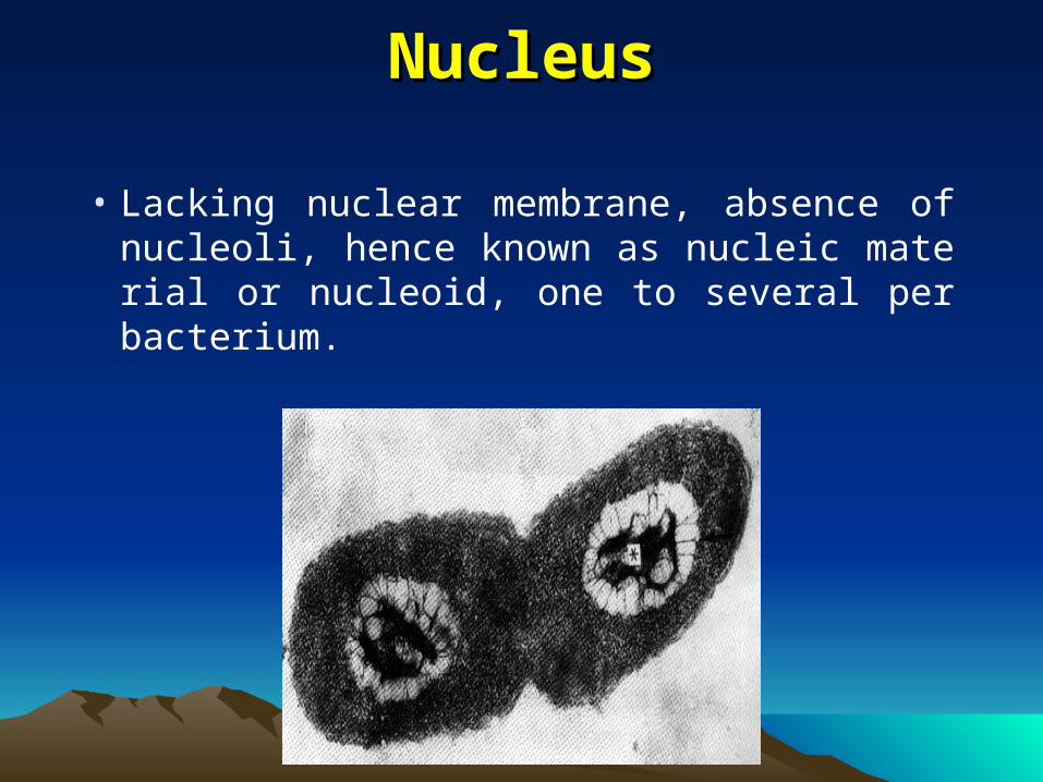
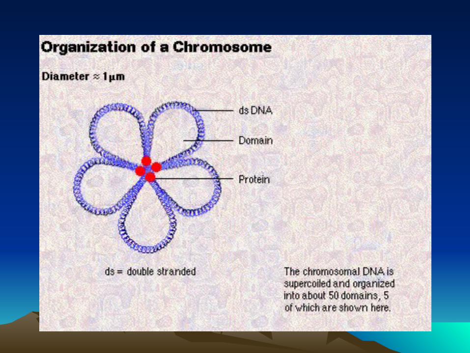
NucleusNucleus
• Lacking nuclear membrane, absence of nucleoli, hence known as nucleic material or nucleoid, one to several per bacterium.
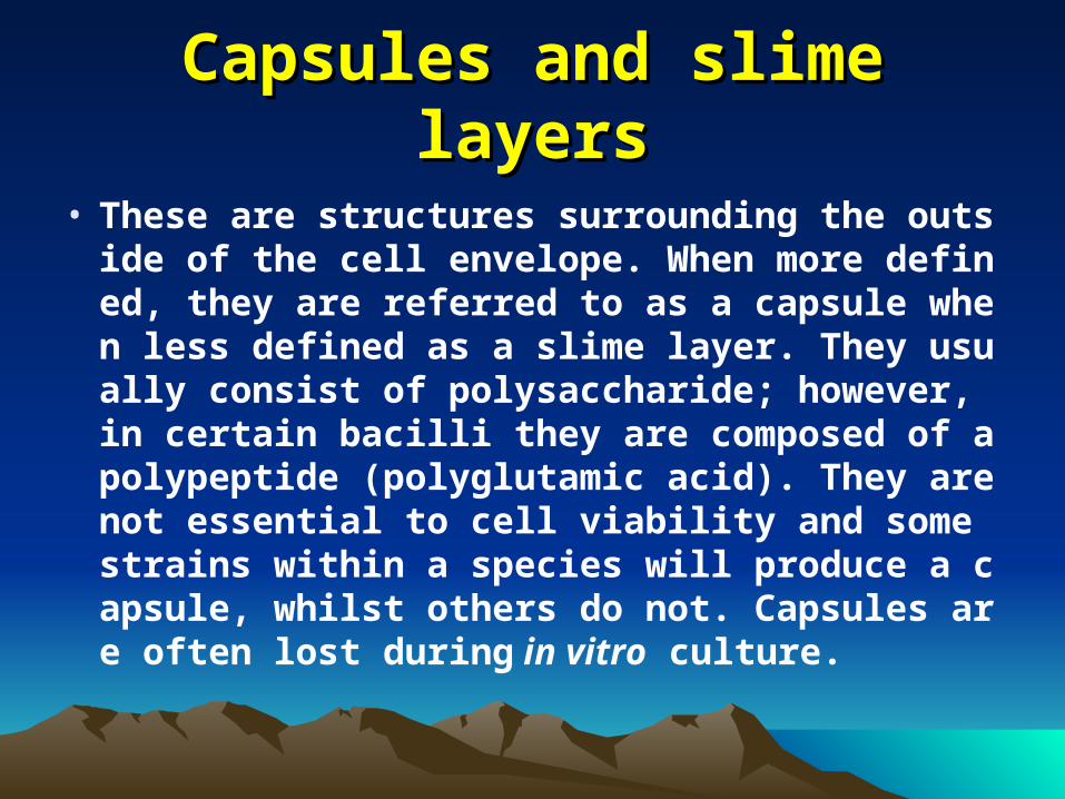
Capsules and slime layersCapsules and slime layers
• These are structures surrounding the outside of the cell envelope. When more defined, they are referred to as a capsule when less defined as a slime layer. They usually consist of polysaccharide; however, in certain bacilli they are composed of a polypeptide (polyglutamic acid). They are not essential to cell viability and some strains within a species will produce a capsule, whilst others do not. Capsules are often lost during in vitro culture.
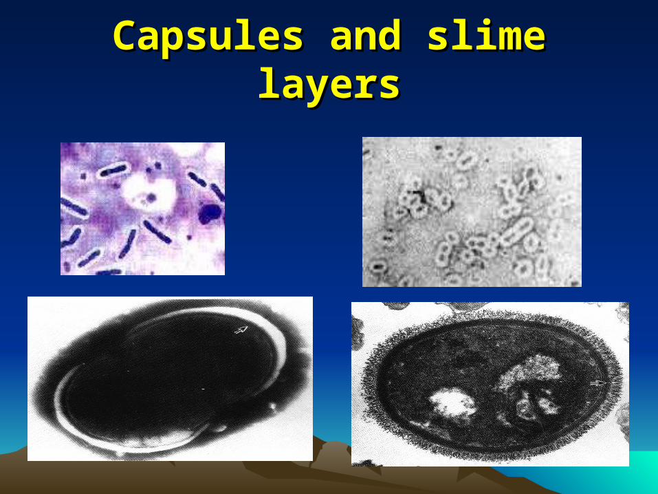
Capsules and slime layersCapsules and slime layers
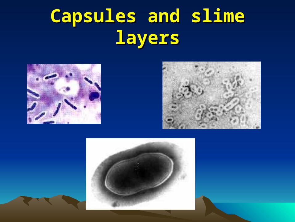
Capsules and slime layersCapsules and slime layers
Function of Function of Capsules and slime layersCapsules and slime layers(1)(1)
• Attachment :These structures are thought to help cells attach to their target environment. Streptococcus mutans produces a slime layer in the presence of sucrose. This results in dental plaque and many bacteria can stick to tooth surfaces and cause decay once S. mutans forms a slime layer. Vibrio cholerae, the cause of cholera, also produces a glycocalyx which helps it attach to the intestinal villi of the host.
Function of Function of Capsules and slime layersCapsules and slime layers(2)(2)
• Protection from phagocytic engulfment. Bacterial pathogens are always in danger of being "eaten" by phagocytes. (Host cells that protect you from invaders.) Streptococcus pneumoniae, when encapsulated is able to kill 90% of infected animals, when non-encapsulated no animals die. The capsule has been found to protect the bacteria by making it difficult for the phagocyte to engulf the microbe.
Function of Function of Capsules and slime layersCapsules and slime layers(3)(3)• Resistance to drying. Capsules and slime
layers inhibit water from escaping into the environment.
Function of Function of Capsules and slime layersCapsules and slime layers(4)(4)• Reservoir for certain nutrients. Glycocalyx
will bind certain ions and molecules. These can then be made available to the cell.
Function of Function of Capsules and slime layersCapsules and slime layers(5)(5)• Depot for waste products. Waste products
of metabolism are excreted from the cell, and will accumulate in the capsule. This binds them up, and prevents the waste from interfering with cell metabolism.
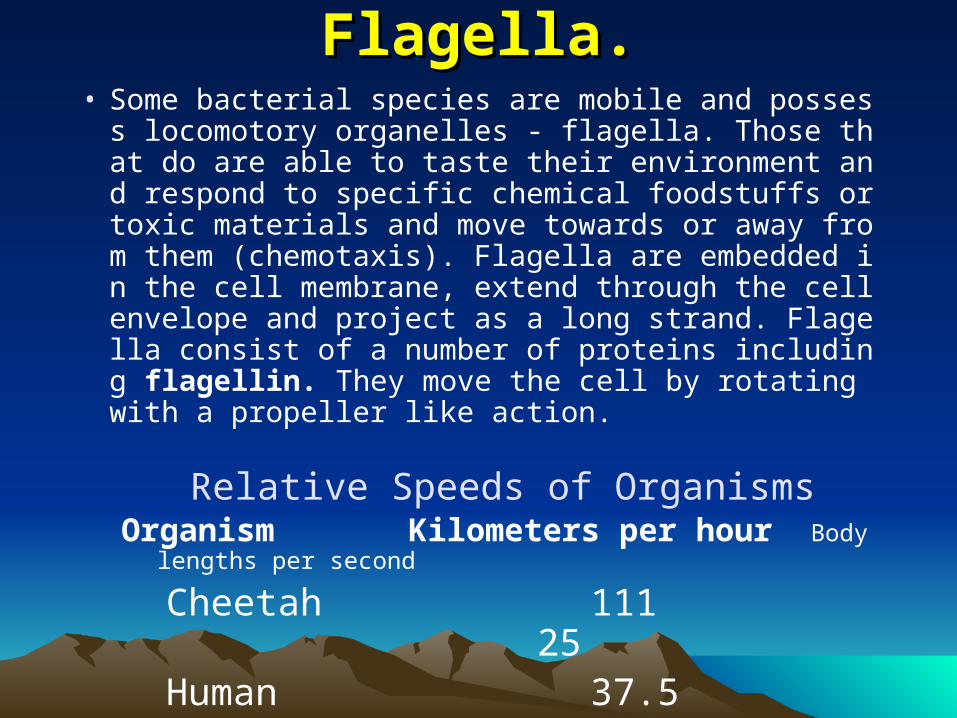
Flagella.Flagella.• Some bacterial species are mobile and possess locomo
tory organelles - flagella. Those that do are able to taste their environment and respond to specific chemical foodstuffs or toxic materials and move towards or away from them (chemotaxis). Flagella are embedded in the cell membrane, extend through the cell envelope and project as a long strand. Flagella consist of a number of proteins including flagellin. They move the cell by rotating with a propeller like action.
Relative Speeds of OrganismsOrganism Kilometers per hour Body lengths per sec
ond
Cheetah 111 25 Human 37.5 5.4 Bacteria 0.00015 10
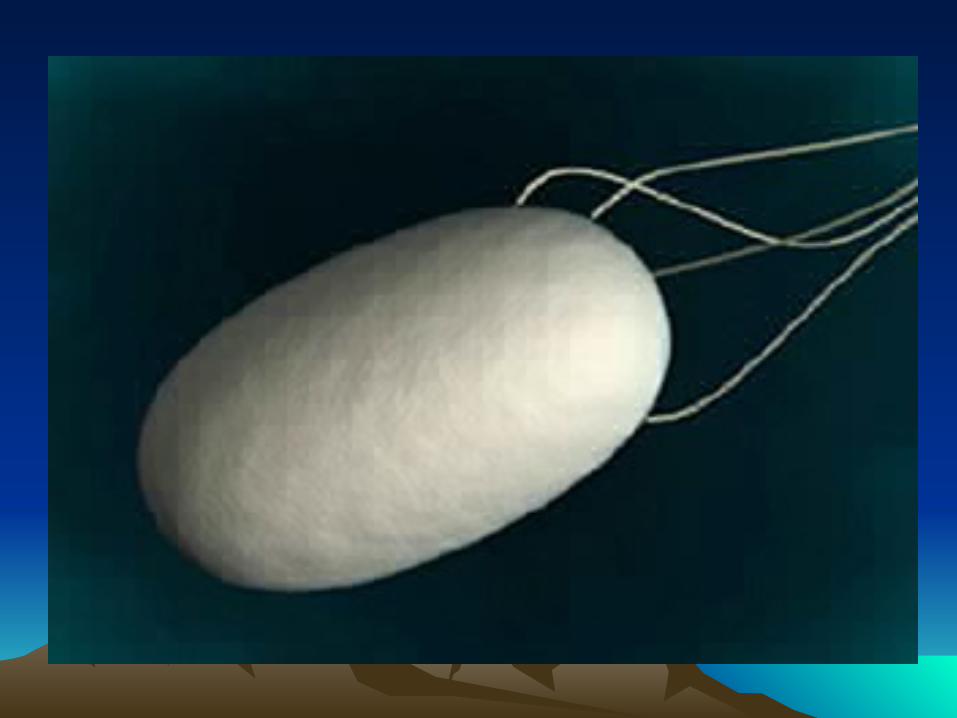
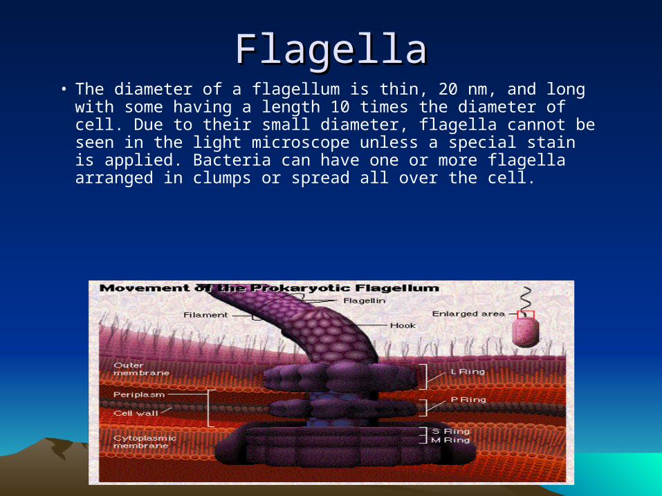
FlagellaFlagella• The diameter of a flagellum is thin, 20 nm, and long with
some having a length 10 times the diameter of cell. Due to their small diameter, flagella cannot be seen in the light microscope unless a special stain is applied. Bacteria can have one or more flagella arranged in clumps or spread all over the cell.
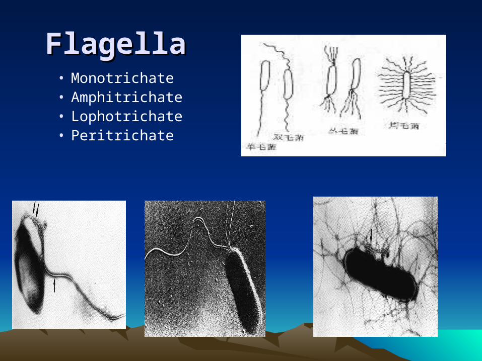
FlagellaFlagella • Monotrichate • Amphitrichate• Lophotrichate• Peritrichate
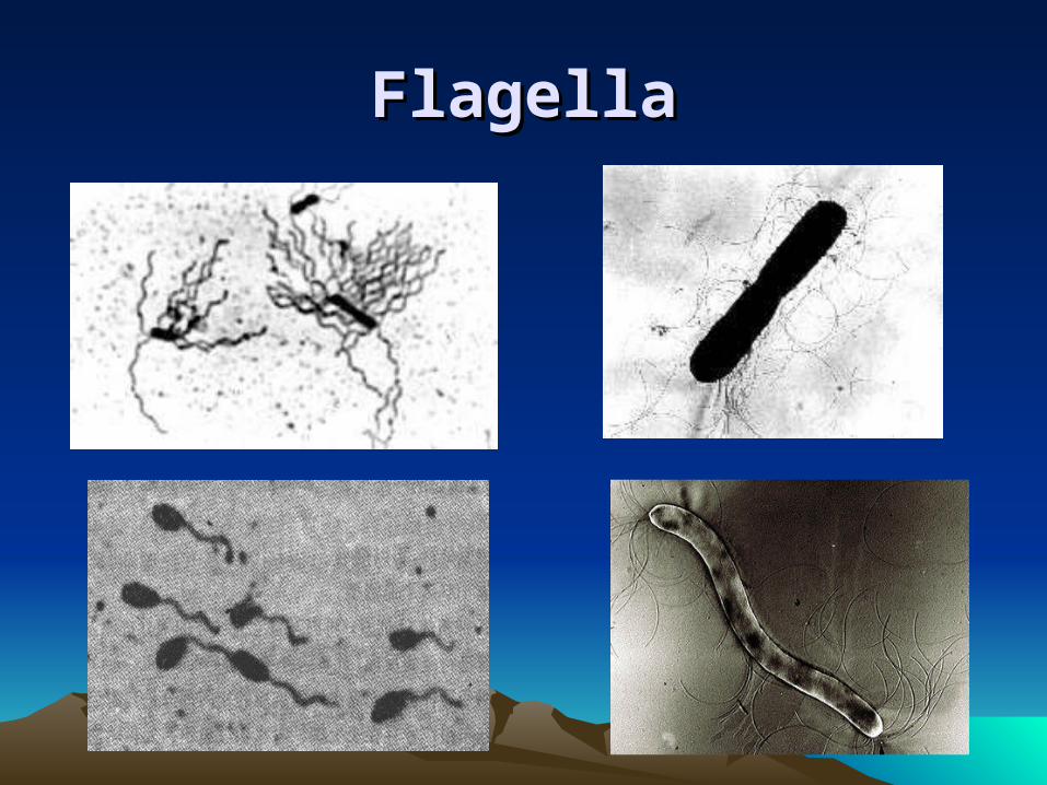
FlagellaFlagella
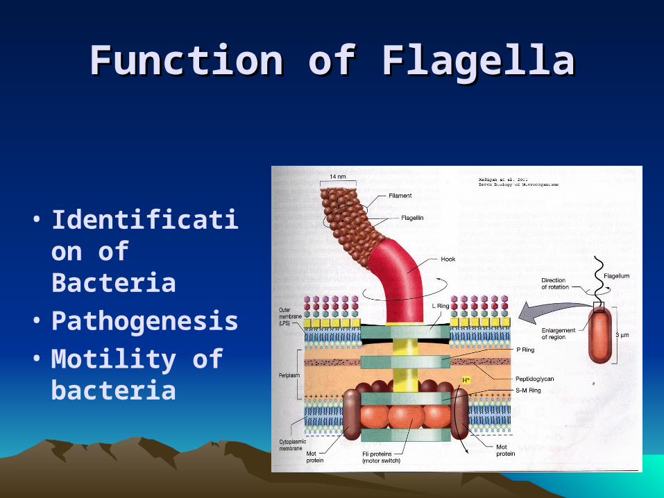
Function ofFunction of FlagellaFlagella
• Identification of Bacteria
• Pathogenesis
• Motility of bacteria
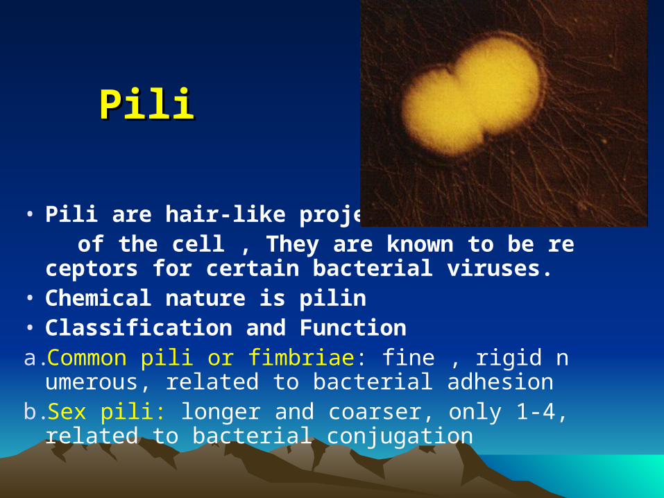
PiliPili
• Pili are hair-like projections of the cell , They are known to be receptors for certa
in bacterial viruses.• Chemical nature is pilin• Classification and Functiona.Common pili or fimbriae: fine , rigid numerous,
related to bacterial adhesionb.Sex pili: longer and coarser, only 1-4, related
to bacterial conjugation
Sex piliSex pili
• A donor bacteria will attach to a recipient via the sex pilus. Then a copy of part of the donor bacterium's genome passes through the sex pilus into the recipient.
• Conjugation, as it is called, is one explanation for the rapid spread of drug resistance in many different species of bacteria.
Common pili or fimbriaeCommon pili or fimbriae
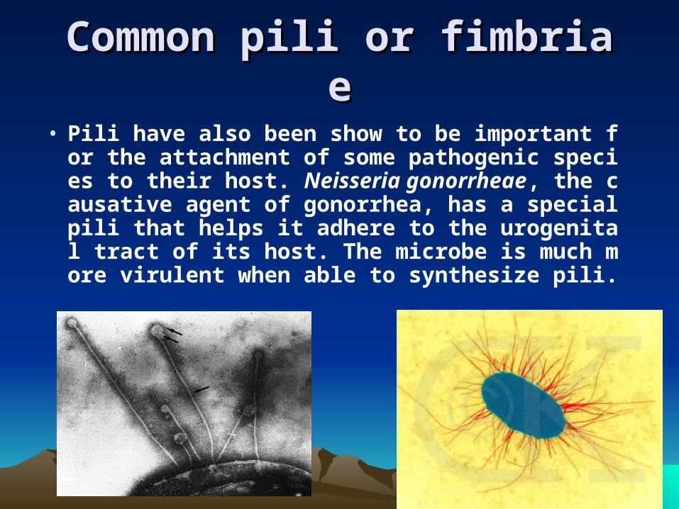
• Pili have also been show to be important for the attachment of some pathogenic species to their host. Neisseria gonorrheae, the causative agent of gonorrhea, has a special pili that helps it adhere to the urogenital tract of its host. The microbe is much more virulent when able to synthesize pili.
EndosporesEndospores
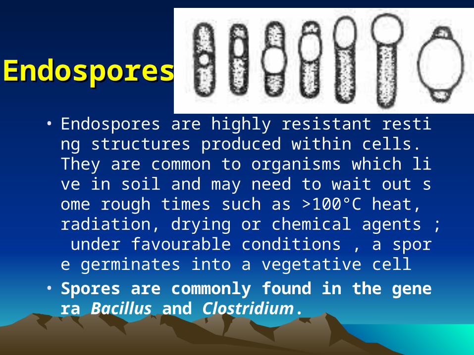
• Endospores are highly resistant resting structures produced within cells. They are common to organisms which live in soil and may need to wait out some rough times such as >100°C heat, radiation, drying or chemical agents ; under favourable conditions , a spore germinates into a vegetative cell
• Spores are commonly found in the genera Bacillus and Clostridium.
DPADPA and and survivesurvive
•Dipicolinic acid,DPA.
• Spores can survive for a very long time, and then regerminate. Spores that were dormant for thousands of years in the great tomes of the Egyption Pharohs were able to germinate and grow when placed in appropriate medium. There are even claims of spores that are over 250 million years old being able to germiinate when placed in appropriate medium. These results have yet to be validated.
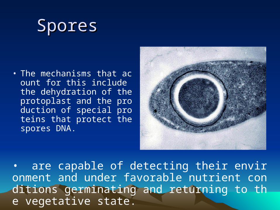
SporesSpores
• The mechanisms that acount for this include the dehydration of the protoplast and the production of special proteins that protect the spores DNA.
• are capable of detecting their environment and under favorable nutrient conditions germinating and returning to the vegetative state.
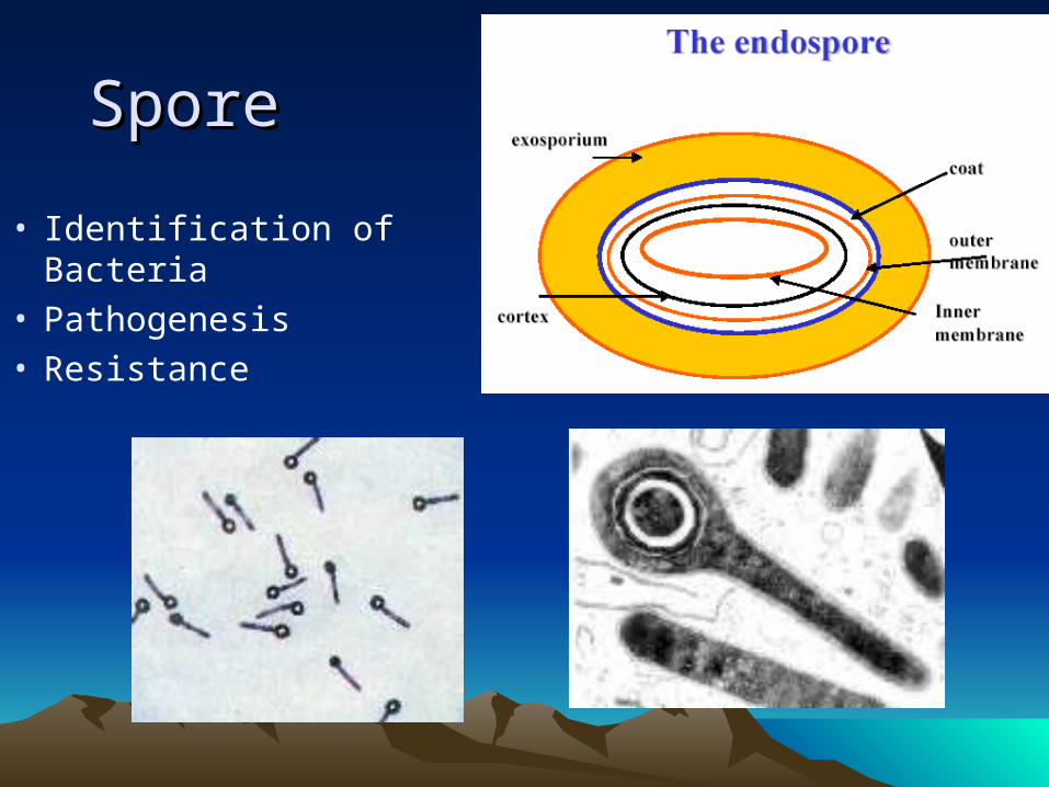
SporeSpore
• Identification of Bacteria• Pathogenesis• Resistance

MicroscopeyMicroscopey
• Light Microscope• Electron Microscope• Darkfield Microscope• Phase Contrast Microsco
pe• Fluorescence Microscop
e• Cofocal Microscope )
Methods
Staining MethodsStaining Methods• Simple staining;• Differential staining ( Gram
stain, Acid-fast stain), • Special staining( Negative stain,
Spore stain, Flagella stain)
Methods