Activator of G-protein signaling 8 is involved in VEGF ...For example, dexamethasone-induced...
13
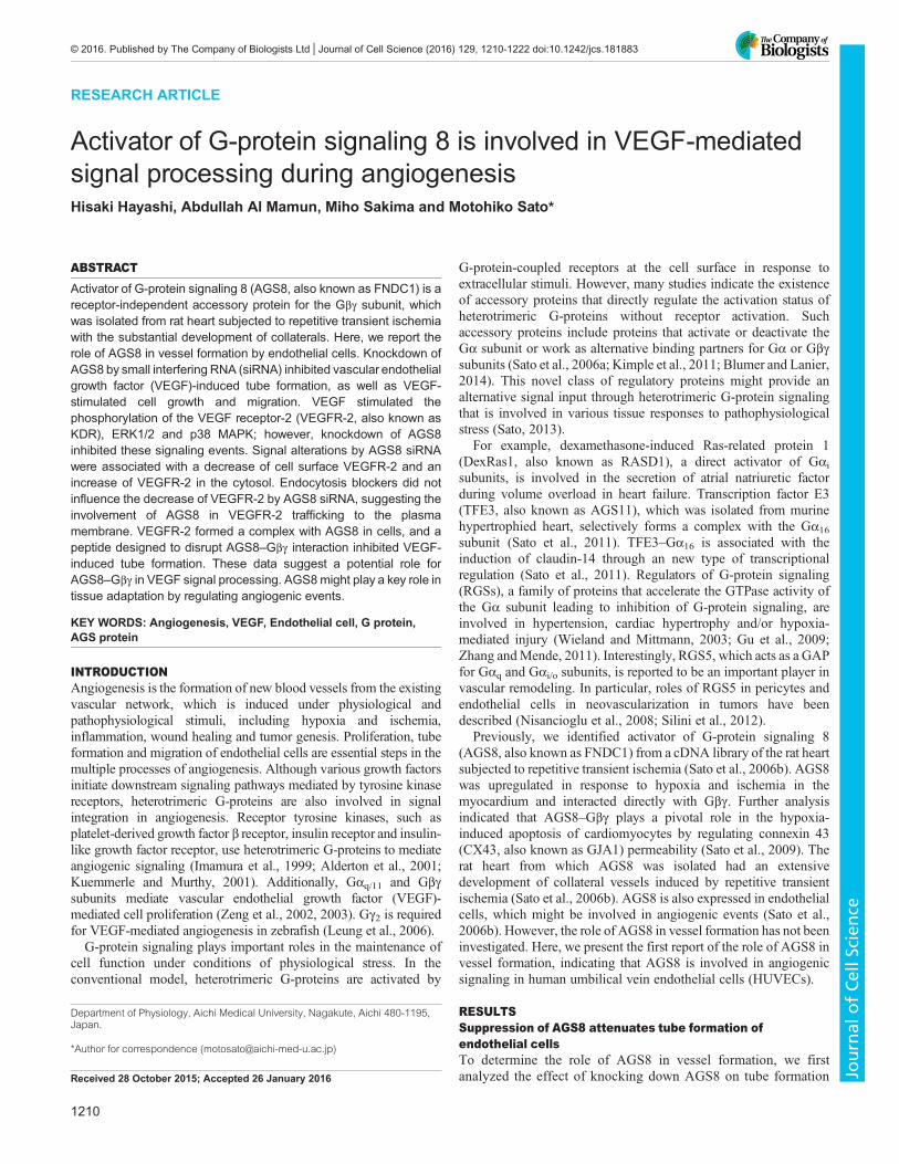
RESEARCH ARTICLE Activator of G-protein signaling 8 is involved in VEGF-mediated signal processing during angiogenesis Hisaki Hayashi, Abdullah Al Mamun, Miho Sakima and Motohiko Sato* ABSTRACT Activator of G-protein signaling 8 (AGS8, also known as FNDC1) is a receptor-independent accessory protein for the Gβγ subunit, which was isolated from rat heart subjected to repetitive transient ischemia with the substantial development of collaterals. Here, we report the role of AGS8 in vessel formation by endothelial cells. Knockdown of AGS8 by small interfering RNA (siRNA) inhibited vascular endothelial growth factor (VEGF)-induced tube formation, as well as VEGF- stimulated cell growth and migration. VEGF stimulated the phosphorylation of the VEGF receptor-2 (VEGFR-2, also known as KDR), ERK1/2 and p38 MAPK; however, knockdown of AGS8 inhibited these signaling events. Signal alterations by AGS8 siRNA were associated with a decrease of cell surface VEGFR-2 and an increase of VEGFR-2 in the cytosol. Endocytosis blockers did not influence the decrease of VEGFR-2 by AGS8 siRNA, suggesting the involvement of AGS8 in VEGFR-2 trafficking to the plasma membrane. VEGFR-2 formed a complex with AGS8 in cells, and a peptide designed to disrupt AGS8–Gβγ interaction inhibited VEGF- induced tube formation. These data suggest a potential role for AGS8–Gβγ in VEGF signal processing. AGS8 might play a key role in tissue adaptation by regulating angiogenic events. KEY WORDS: Angiogenesis, VEGF, Endothelial cell, G protein, AGS protein INTRODUCTION Angiogenesis is the formation of new blood vessels from the existing vascular network, which is induced under physiological and pathophysiological stimuli, including hypoxia and ischemia, inflammation, wound healing and tumor genesis. Proliferation, tube formation and migration of endothelial cells are essential steps in the multiple processes of angiogenesis. Although various growth factors initiate downstream signaling pathways mediated by tyrosine kinase receptors, heterotrimeric G-proteins are also involved in signal integration in angiogenesis. Receptor tyrosine kinases, such as platelet-derived growth factor β receptor, insulin receptor and insulin- like growth factor receptor, use heterotrimeric G-proteins to mediate angiogenic signaling (Imamura et al., 1999; Alderton et al., 2001; Kuemmerle and Murthy, 2001). Additionally, Gα q/11 and Gβγ subunits mediate vascular endothelial growth factor (VEGF)- mediated cell proliferation (Zeng et al., 2002, 2003). Gγ 2 is required for VEGF-mediated angiogenesis in zebrafish (Leung et al., 2006). G-protein signaling plays important roles in the maintenance of cell function under conditions of physiological stress. In the conventional model, heterotrimeric G-proteins are activated by G-protein-coupled receptors at the cell surface in response to extracellular stimuli. However, many studies indicate the existence of accessory proteins that directly regulate the activation status of heterotrimeric G-proteins without receptor activation. Such accessory proteins include proteins that activate or deactivate the Gα subunit or work as alternative binding partners for Gα or Gβγ subunits (Sato et al., 2006a; Kimple et al., 2011; Blumer and Lanier, 2014). This novel class of regulatory proteins might provide an alternative signal input through heterotrimeric G-protein signaling that is involved in various tissue responses to pathophysiological stress (Sato, 2013). For example, dexamethasone-induced Ras-related protein 1 (DexRas1, also known as RASD1), a direct activator of Gα i subunits, is involved in the secretion of atrial natriuretic factor during volume overload in heart failure. Transcription factor E3 (TFE3, also known as AGS11), which was isolated from murine hypertrophied heart, selectively forms a complex with the Gα 16 subunit (Sato et al., 2011). TFE3–Gα 16 is associated with the induction of claudin-14 through an new type of transcriptional regulation (Sato et al., 2011). Regulators of G-protein signaling (RGSs), a family of proteins that accelerate the GTPase activity of the Gα subunit leading to inhibition of G-protein signaling, are involved in hypertension, cardiac hypertrophy and/or hypoxia- mediated injury (Wieland and Mittmann, 2003; Gu et al., 2009; Zhang and Mende, 2011). Interestingly, RGS5, which acts as a GAP for Gα q and Gα i/o subunits, is reported to be an important player in vascular remodeling. In particular, roles of RGS5 in pericytes and endothelial cells in neovascularization in tumors have been described (Nisancioglu et al., 2008; Silini et al., 2012). Previously, we identified activator of G-protein signaling 8 (AGS8, also known as FNDC1) from a cDNA library of the rat heart subjected to repetitive transient ischemia (Sato et al., 2006b). AGS8 was upregulated in response to hypoxia and ischemia in the myocardium and interacted directly with Gβγ. Further analysis indicated that AGS8–Gβγ plays a pivotal role in the hypoxia- induced apoptosis of cardiomyocytes by regulating connexin 43 (CX43, also known as GJA1) permeability (Sato et al., 2009). The rat heart from which AGS8 was isolated had an extensive development of collateral vessels induced by repetitive transient ischemia (Sato et al., 2006b). AGS8 is also expressed in endothelial cells, which might be involved in angiogenic events (Sato et al., 2006b). However, the role of AGS8 in vessel formation has not been investigated. Here, we present the first report of the role of AGS8 in vessel formation, indicating that AGS8 is involved in angiogenic signaling in human umbilical vein endothelial cells (HUVECs). RESULTS Suppression of AGS8 attenuates tube formation of endothelial cells To determine the role of AGS8 in vessel formation, we first analyzed the effect of knocking down AGS8 on tube formation Received 28 October 2015; Accepted 26 January 2016 Department of Physiology, Aichi Medical University, Nagakute, Aichi 480-1195, Japan. *Author for correspondence ([email protected]) 1210 © 2016. Published by The Company of Biologists Ltd | Journal of Cell Science (2016) 129, 1210-1222 doi:10.1242/jcs.181883 Journal of Cell Science
Transcript of Activator of G-protein signaling 8 is involved in VEGF ...For example, dexamethasone-induced...
RESEARCH ARTICLE
Activator of G-protein signaling 8 is involved in VEGF-mediatedsignal processing during angiogenesisHisaki Hayashi, Abdullah Al Mamun, Miho Sakima and Motohiko Sato*
ABSTRACTActivator of G-protein signaling 8 (AGS8, also known as FNDC1) is areceptor-independent accessory protein for the Gβγ subunit, whichwas isolated from rat heart subjected to repetitive transient ischemiawith the substantial development of collaterals. Here, we report therole of AGS8 in vessel formation by endothelial cells. Knockdown ofAGS8 by small interfering RNA (siRNA) inhibited vascular endothelialgrowth factor (VEGF)-induced tube formation, as well as VEGF-stimulated cell growth and migration. VEGF stimulated thephosphorylation of the VEGF receptor-2 (VEGFR-2, also known asKDR), ERK1/2 and p38 MAPK; however, knockdown of AGS8inhibited these signaling events. Signal alterations by AGS8 siRNAwere associated with a decrease of cell surface VEGFR-2 and anincrease of VEGFR-2 in the cytosol. Endocytosis blockers did notinfluence the decrease of VEGFR-2 by AGS8 siRNA, suggesting theinvolvement of AGS8 in VEGFR-2 trafficking to the plasmamembrane. VEGFR-2 formed a complex with AGS8 in cells, and apeptide designed to disrupt AGS8–Gβγ interaction inhibited VEGF-induced tube formation. These data suggest a potential role forAGS8–Gβγ in VEGF signal processing. AGS8might play a key role intissue adaptation by regulating angiogenic events.
KEY WORDS: Angiogenesis, VEGF, Endothelial cell, G protein,AGS protein
INTRODUCTIONAngiogenesis is the formation of new blood vessels from the existingvascular network, which is induced under physiological andpathophysiological stimuli, including hypoxia and ischemia,inflammation, wound healing and tumor genesis. Proliferation, tubeformation and migration of endothelial cells are essential steps in themultiple processes of angiogenesis. Although various growth factorsinitiate downstream signaling pathways mediated by tyrosine kinasereceptors, heterotrimeric G-proteins are also involved in signalintegration in angiogenesis. Receptor tyrosine kinases, such asplatelet-derived growth factor β receptor, insulin receptor and insulin-like growth factor receptor, use heterotrimeric G-proteins to mediateangiogenic signaling (Imamura et al., 1999; Alderton et al., 2001;Kuemmerle and Murthy, 2001). Additionally, Gαq/11 and Gβγsubunits mediate vascular endothelial growth factor (VEGF)-mediated cell proliferation (Zeng et al., 2002, 2003). Gγ2 is requiredfor VEGF-mediated angiogenesis in zebrafish (Leung et al., 2006).G-protein signaling plays important roles in the maintenance of
cell function under conditions of physiological stress. In theconventional model, heterotrimeric G-proteins are activated by
G-protein-coupled receptors at the cell surface in response toextracellular stimuli. However, many studies indicate the existenceof accessory proteins that directly regulate the activation status ofheterotrimeric G-proteins without receptor activation. Suchaccessory proteins include proteins that activate or deactivate theGα subunit or work as alternative binding partners for Gα or Gβγsubunits (Sato et al., 2006a; Kimple et al., 2011; Blumer and Lanier,2014). This novel class of regulatory proteins might provide analternative signal input through heterotrimeric G-protein signalingthat is involved in various tissue responses to pathophysiologicalstress (Sato, 2013).
For example, dexamethasone-induced Ras-related protein 1(DexRas1, also known as RASD1), a direct activator of Gαisubunits, is involved in the secretion of atrial natriuretic factorduring volume overload in heart failure. Transcription factor E3(TFE3, also known as AGS11), which was isolated from murinehypertrophied heart, selectively forms a complex with the Gα16subunit (Sato et al., 2011). TFE3–Gα16 is associated with theinduction of claudin-14 through an new type of transcriptionalregulation (Sato et al., 2011). Regulators of G-protein signaling(RGSs), a family of proteins that accelerate the GTPase activity ofthe Gα subunit leading to inhibition of G-protein signaling, areinvolved in hypertension, cardiac hypertrophy and/or hypoxia-mediated injury (Wieland and Mittmann, 2003; Gu et al., 2009;Zhang andMende, 2011). Interestingly, RGS5, which acts as a GAPfor Gαq and Gαi/o subunits, is reported to be an important player invascular remodeling. In particular, roles of RGS5 in pericytes andendothelial cells in neovascularization in tumors have beendescribed (Nisancioglu et al., 2008; Silini et al., 2012).
Previously, we identified activator of G-protein signaling 8(AGS8, also known as FNDC1) from a cDNA library of the rat heartsubjected to repetitive transient ischemia (Sato et al., 2006b). AGS8was upregulated in response to hypoxia and ischemia in themyocardium and interacted directly with Gβγ. Further analysisindicated that AGS8–Gβγ plays a pivotal role in the hypoxia-induced apoptosis of cardiomyocytes by regulating connexin 43(CX43, also known as GJA1) permeability (Sato et al., 2009). Therat heart from which AGS8 was isolated had an extensivedevelopment of collateral vessels induced by repetitive transientischemia (Sato et al., 2006b). AGS8 is also expressed in endothelialcells, which might be involved in angiogenic events (Sato et al.,2006b). However, the role of AGS8 in vessel formation has not beeninvestigated. Here, we present the first report of the role of AGS8 invessel formation, indicating that AGS8 is involved in angiogenicsignaling in human umbilical vein endothelial cells (HUVECs).
RESULTSSuppression of AGS8 attenuates tube formation ofendothelial cellsTo determine the role of AGS8 in vessel formation, we firstanalyzed the effect of knocking down AGS8 on tube formationReceived 28 October 2015; Accepted 26 January 2016
Department of Physiology, Aichi Medical University, Nagakute, Aichi 480-1195,Japan.
*Author for correspondence ([email protected])
1210
© 2016. Published by The Company of Biologists Ltd | Journal of Cell Science (2016) 129, 1210-1222 doi:10.1242/jcs.181883
Journal
ofCe
llScience
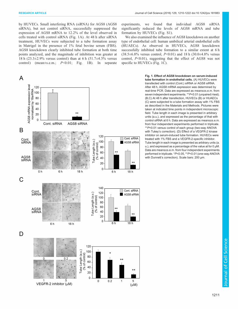
by HUVECs. Small interfering RNA (siRNA) for AGS8 (AGS8siRNA), but not control siRNA, successfully suppressed theexpression of AGS8 mRNA to 12.2% of the level observed incells treated with control siRNA (Fig. 1A). At 48 h after siRNAtreatment, HUVECs were subjected to a tube formation assayin Matrigel in the presence of 1% fetal bovine serum (FBS).AGS8 knockdown clearly inhibited tube formation at both timepoints analyzed, and the magnitude of inhibition was greater at18 h (23.3±2.9% versus control) than at 6 h (51.7±4.3% versuscontrol) (mean±s.e.m.; P<0.01; Fig. 1B). In separate
experiments, we found that individual AGS8 siRNAsignificantly reduced the levels of AGS8 mRNA and tubeformation by HUVECs (Fig. S1).
We also examined the influence of AGS8 knockdown on anothertype of endothelial cell: human umbilical arterial endothelial cells(HUAECs). As observed in HUVECs, AGS8 knockdownsuccessfully inhibited tube formation to a similar extent at 6 h(38.5±6.8% versus control, P<0.01) and 18 h (30.0±4.8% versuscontrol, P<0.01), suggesting that the effect of AGS8 was notspecific to HUVECs (Fig. 1C).
Fig. 1. Effect of AGS8 knockdown on serum-inducedtube formation in endothelial cells. (A) HUVECs weretransfected with control (Cont.) siRNA or AGS8 siRNA.After 48 h, AGS8 mRNA expression was determined byreal-time PCR. Data are expressed as means±s.e.m. fromseven independent experiments. **P<0.01 (unpaired t-test).(B,C) At 48 h after transfection, HUVECs (B) or HUAECs(C) were subjected to a tube formation assay with 1% FBSas described in the Materials and Methods. Pictures weretaken at indicated time points in independent microscopicfield. Tube length in each image is presented in arbitraryunits (a.u.), and expressed as the percentage of that withcontrol siRNA at 6 h. Data are expressed as means±s.e.m.from four independent experiments performed in triplicate.**P<0.01 versus control of each group (two-way ANOVAwith Tukey’s correction). (D) Effect of a VEGFR-2 kinaseinhibitor on serum-induced tube formation. HUVECs weretreated with 1% FBS and a VEGFR-2-specific inhibitor.Tube length in each image is presented as arbitrary units (a.u.), and expressed as a percentage of the value at for 0 μM.Data are means±s.e.m. from four independent experimentsperformed in triplicate. *P<0.05, **P<0.01 (one-way ANOVAwith Dunnett’s correction). Scale bars: 200 μm.
1211
RESEARCH ARTICLE Journal of Cell Science (2016) 129, 1210-1222 doi:10.1242/jcs.181883
Journal
ofCe
llScience
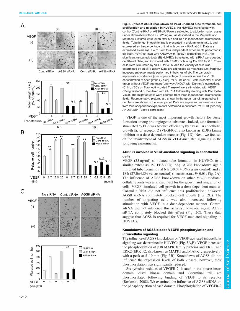
VEGF is one of the most important growth factors for vesselformation among pro-angiogenic substrates. Indeed, tube formationstimulated by FBS was blocked efficiently by a vascular endothelialgrowth factor receptor 2 (VEGFR-2, also known as KDR) kinaseinhibitor in a dose-dependent manner (Fig. 1D). Next, we focusedon the involvement of AGS8 in VEGF-mediated signaling in thefollowing experiments.
AGS8 is involved in VEGF-mediated signaling in endothelialcellsVEGF (25 ng/ml) stimulated tube formation in HUVECs to asimilar extent as 1% FBS (Fig. 2A). AGS8 knockdown againinhibited tube formation at 6 h (30.0±4.0% versus control) and at18 h (27.0±4.8% versus control) (mean±s.e.m.; P<0.01; Fig. 2A).The influence of AGS8 knockdown on other VEGF-mediatedcellular events was analyzed next for the growth and migration ofcells. VEGF simulated cell growth in a dose-dependent manner.Control siRNA did not influence this proliferation; however,AGS8 siRNA completely blocked cell growth (Fig. 2B). Thenumber of migrating cells was also increased followingstimulation with VEGF in a dose-dependent manner. ControlsiRNA did not influence this activity; however, again, AGS8siRNA completely blocked this effect (Fig. 2C). These datasuggest that AGS8 is required for VEGF-mediated signaling inHUVECs.
Knockdown of AGS8 blocks VEGFR phosphorylation andintracellular signalingThe influence of AGS8 knockdown onVEGF-activated intracellularsignaling was determined inHUVECs (Fig. 3A,B). VEGF increasedthe phosphorylation of p38 MAPK family proteins and ERK1 andERK2 (ERK1/2, also known asMAPK3 andMAPK1, respectively)with a peak at 5–10 min (Fig. 3B). Knockdown of AGS8 did notinfluence the expression levels of both kinases; however, theirphosphorylation was significantly reduced.
Six tyrosine residues of VEGFR-2, located in the kinase insertdomain, distal kinase domain and C-terminal tail, arephosphorylated following binding of VEGF to its receptor(Roskoski, 2008). We examined the influence of AGS8 siRNA onthe phosphorylation of each domain. Phosphorylation of VEGFR-2
Fig. 2. Effect of AGS8 knockdown on VEGF-induced tube formation, cellproliferation and migration in HUVECs. (A) HUVECs transfected withcontrol (Cont.) siRNA or AGS8 siRNAwere subjected to a tube formation assayunder stimulation with VEGF (25 ng/ml) as described in the Materials andMethods. Pictures were taken after 6 h and 18 h in independent microscopicfields. Tube length in each image is presented in arbitrary units (a.u.), andexpressed as the percentage of that with control siRNA at 6 h. Data areexpressed as means±s.e.m. from four independent experiments performed intriplicate. **P<0.01 (two-way ANOVA with Tukey’s correction). N.S., notsignificant (unpaired t-test). (B) HUVECs transfected with siRNAwere seededon 96-well plate, and incubated with EBM2 containing 1% FBS for 6 h. Then,cells were stimulated by VEGF for 48 h, and the viability of cells wasdetermined by an MTT assay. Data are expressed as means±s.e.m. from fourindependent experiments performed in batches of six. The bar graphrepresents absorbance (x-axis, percentage of control) versus the VEGFconcentration of each group (y-axis). **P<0.01 or N.S. versus control of eachgroup without VEGF treatment (one-way ANOVA with Dunnett’s correction).(C) HUVECs on fibronectin-coated Transwell were stimulated with VEGF(25 ng/ml) for 4 h, then fixed with 4% PFA followed by staining with 1% CrystalViolet. The migrated cells were counted from three independent microscopicfields. Representative pictures are shown in the upper panel; migrated cellnumbers are shown in the lower panel. Data are expressed as means±s.e.m.from four independent experiments performed in duplicate. **P<0.01 (two-wayANOVA with Tukey’s correction).
1212
RESEARCH ARTICLE Journal of Cell Science (2016) 129, 1210-1222 doi:10.1242/jcs.181883
Journal
ofCe
llScience
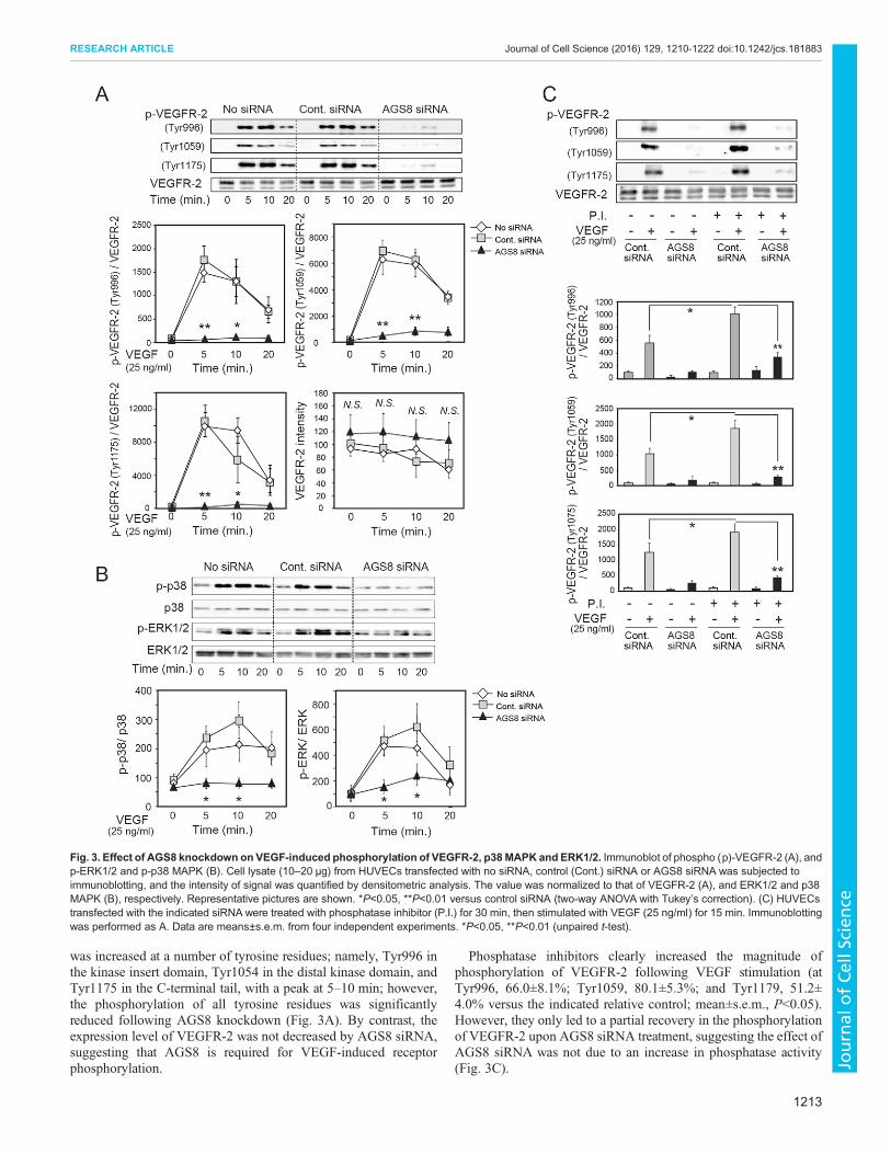
was increased at a number of tyrosine residues; namely, Tyr996 inthe kinase insert domain, Tyr1054 in the distal kinase domain, andTyr1175 in the C-terminal tail, with a peak at 5–10 min; however,the phosphorylation of all tyrosine residues was significantlyreduced following AGS8 knockdown (Fig. 3A). By contrast, theexpression level of VEGFR-2 was not decreased by AGS8 siRNA,suggesting that AGS8 is required for VEGF-induced receptorphosphorylation.
Phosphatase inhibitors clearly increased the magnitude ofphosphorylation of VEGFR-2 following VEGF stimulation (atTyr996, 66.0±8.1%; Tyr1059, 80.1±5.3%; and Tyr1179, 51.2±4.0% versus the indicated relative control; mean±s.e.m., P<0.05).However, they only led to a partial recovery in the phosphorylationof VEGFR-2 upon AGS8 siRNA treatment, suggesting the effect ofAGS8 siRNA was not due to an increase in phosphatase activity(Fig. 3C).
Fig. 3. Effect of AGS8 knockdown on VEGF-induced phosphorylation of VEGFR-2, p38 MAPK and ERK1/2. Immunoblot of phospho (p)-VEGFR-2 (A), andp-ERK1/2 and p-p38 MAPK (B). Cell lysate (10–20 μg) from HUVECs transfected with no siRNA, control (Cont.) siRNA or AGS8 siRNA was subjected toimmunoblotting, and the intensity of signal was quantified by densitometric analysis. The value was normalized to that of VEGFR-2 (A), and ERK1/2 and p38MAPK (B), respectively. Representative pictures are shown. *P<0.05, **P<0.01 versus control siRNA (two-way ANOVA with Tukey’s correction). (C) HUVECstransfected with the indicated siRNA were treated with phosphatase inhibitor (P.I.) for 30 min, then stimulated with VEGF (25 ng/ml) for 15 min. Immunoblottingwas performed as A. Data are means±s.e.m. from four independent experiments. *P<0.05, **P<0.01 (unpaired t-test).
1213
RESEARCH ARTICLE Journal of Cell Science (2016) 129, 1210-1222 doi:10.1242/jcs.181883
Journal
ofCe
llScience
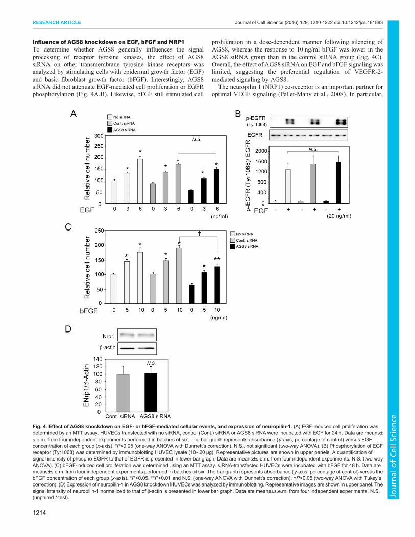
Influence of AGS8 knockdown on EGF, bFGF and NRP1To determine whether AGS8 generally influences the signalprocessing of receptor tyrosine kinases, the effect of AGS8siRNA on other transmembrane tyrosine kinase receptors wasanalyzed by stimulating cells with epidermal growth factor (EGF)and basic fibroblast growth factor (bFGF). Interestingly, AGS8siRNA did not attenuate EGF-mediated cell proliferation or EGFRphosphorylation (Fig. 4A,B). Likewise, bFGF still stimulated cell
proliferation in a dose-dependent manner following silencing ofAGS8, whereas the response to 10 ng/ml bFGF was lower in theAGS8 siRNA group than in the control siRNA group (Fig. 4C).Overall, the effect of AGS8 siRNA on EGF and bFGF signaling waslimited, suggesting the preferential regulation of VEGFR-2-mediated signaling by AGS8.
The neuropilin 1 (NRP1) co-receptor is an important partner foroptimal VEGF signaling (Pellet-Many et al., 2008). In particular,
Fig. 4. Effect of AGS8 knockdown on EGF- or bFGF-mediated cellular events, and expression of neuropilin-1. (A) EGF-induced cell proliferation wasdetermined by an MTT assay. HUVECs transfected with no siRNA, control (Cont.) siRNA or AGS8 siRNA were incubated with EGF for 24 h. Data are means±s.e.m. from four independent experiments performed in batches of six. The bar graph represents absorbance (y-axis, percentage of control) versus EGFconcentration of each group (x-axis). *P<0.05 (one-way ANOVA with Dunnett’s correction). N.S., not significant (two-way ANOVA). (B) Phosphorylation of EGFreceptor (Tyr1068) was determined by immunoblotting HUVEC lysate (10∼20 μg). Representative pictures are shown in upper panels. A quantification ofsignal intensity of phospho-EGFR to that of EGFR is presented in lower bar graph. Data are means±s.e.m. from four independent experiments. N.S. (two-wayANOVA). (C) bFGF-induced cell proliferation was determined using an MTT assay. siRNA-transfected HUVECs were incubated with bFGF for 48 h. Data aremeans±s.e.m. from four independent experiments performed in batches of six. The bar graph represents absorbance (y-axis, percentage of control) versus thebFGF concentration of each group (x-axis). *P<0.05, **P<0.01 and N.S. (one-way ANOVA with Dunnett’s correction); †P<0.05 (two-way ANOVA with Tukey’scorrection). (D) Expression of neuropilin-1 in AGS8 knockdownHUVECswas analyzed by immunoblotting. Representative images are shown in upper panel. Thesignal intensity of neuropilin-1 normalized to that of β-actin is presented in lower bar graph. Data are means±s.e.m. from four independent experiments. N.S.(unpaired t-test).
1214
RESEARCH ARTICLE Journal of Cell Science (2016) 129, 1210-1222 doi:10.1242/jcs.181883
Journal
ofCe
llScience
the NRP1–VEGFR-2 complex plays an important role in vascularformation and cardiovascular development (Pellet-Many et al.,2008). Knockdown of NRP1 reduced the expression of VEGFR-2in endothelial cells (Holmes and Zachary, 2008); however, AGS8siRNA did not influence the protein expression of NRP1 (Fig. 4D).
AGS8 mediates the cell surface expression of VEGFRTo investigate the mechanism by which AGS8 siRNA inhibitedVEGFR signaling, we analyzed the level of cell surface VEGFR-2using FACS analysis with an FITC-conjugated anti-VEGFR-2antibody. Interestingly, VEGFR-2 was significantly decreased at thecell surface following treatment with AGS8 siRNA (37.9±4.8%versus control siRNA), whereas the level of VEGFR-2 wasincreased slightly in pearmeabilized cells (125.5±2.7% versuscontrol siRNA) (mean±s.e.m., P<0.01; Fig. 5A). This was incontrast to the cell surface level of VEGFR-1 (also known as FLT1),which was increased following treatment with AGS8 siRNA (217±2.8% versus control siRNA, P<0.01), without changing the totalexpression level in cell (Fig. 5B).Cell surface VEGFR-2 is known to be internalized upon its
interaction with VEGF, which was detected by FACS analysis(45.0±4.4% versus control) (mean±s.e.m., P<0.01; Fig. 5C). Theendocytosis blockers Pitstop and Dynasore successfullyprevented the VEGF-induced decrease of cell surface VEGFR-2 (Fig. 5C). VEGFR-2 is also constitutively internalized andtransferred to early endosomes (Jopling et al., 2011). Theendocytosis blockers increased cell surface VEGFR-2 in theresting state without VEGF stimulation. However, they failed tolead to a recovery in the amount of VEGFR-2 upon AGS8siRNA treatment, suggesting that the loss of cell surfacereceptors caused by AGS8 siRNA was not due to theacceleration of endocytosis (Fig. 5D).Then, we examined the subcellular localization of VEGFR-2
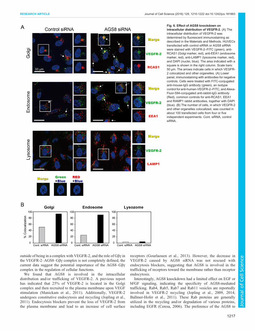
using fluorescent immunostaining (Fig. 6). A proportion ofVEGFR-2 was found in the Golgi during the resting state, asreported previously (Manickam et al., 2011). VEGFR-2 was alsofound in early endosomes, but was not detected in lysosomes.Interestingly, AGS8 knockdown increased the intracellular pool ofVEGFR-2 in the Golgi and early endosomes, but not in lysosomes(Fig. 6). These data suggest that AGS8 is involved in VEGFR-2trafficking from the Golgi to the cell surface, and in VEGFR-2recycling through early endosomes.
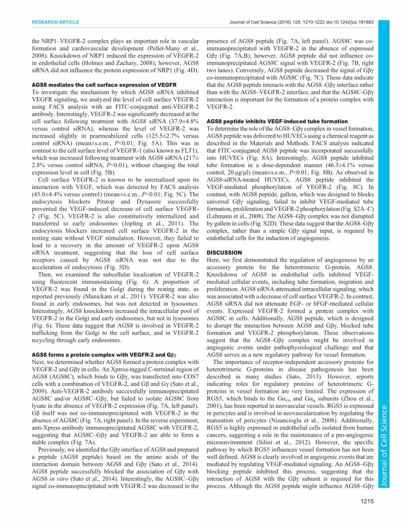
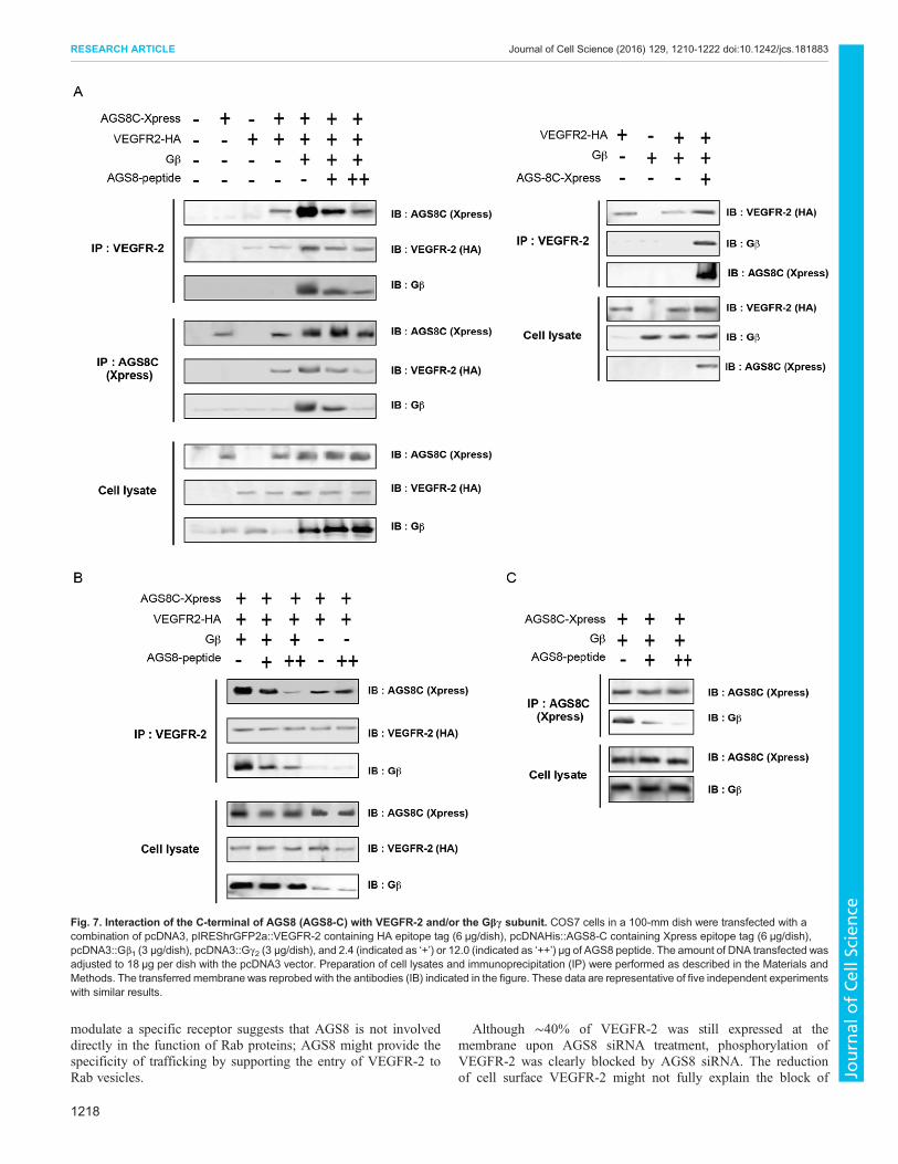
AGS8 forms a protein complex with VEGFR-2 and GβγNext, we determined whether AGS8 formed a protein complex withVEGFR-2 and Gβγ in cells. An Xpress-tagged C-terminal region ofAGS8 (AGS8C), which binds to Gβγ, was transfected into COS7cells with a combination of VEGFR-2, and Gβ and Gγ (Sato et al.,2009). Anti-VEGFR-2 antibody successfully immunoprecipitatedAGS8C and/or AGS8C–Gβγ, but failed to isolate AGS8C fromlysate in the absence of VEGFR-2 expression (Fig. 7A, left panel).Gβ itself was not co-immunoprecipitated with VEGFR-2 in theabsence of AGS8C (Fig. 7A, right panel). In the reverse experiment,anti-Xpress antibody immunoprecipitated AGS8C with VEGFR-2,suggesting that AGS8C–Gβγ and VEGFR-2 are able to form astable complex (Fig. 7A).Previously, we identified the Gβγ interface of AGS8 and prepared
a peptide (AGS8 peptide) based on the amino acids of theinteraction domain between AGS8 and Gβγ (Sato et al., 2014).AGS8 peptide successfully blocked the association of Gβγ withAGS8 in vitro (Sato et al., 2014). Interestingly, the AGS8C–Gβγsignal co-immunoprecipitated with VEGFR-2 was decreased in the
presence of AGS8 peptide (Fig. 7A, left panel). AGS8C was co-immunoprecipitated with VEGFR-2 in the absence of expressedGβγ (Fig. 7A,B); however, AGS8 peptide did not influence co-immunoprecipitated AGS8C signal with VEGFR-2 (Fig. 7B, righttwo lanes). Conversely, AGS8 peptide decreased the signal of Gβγco-immunoprecipitated with AGS8C (Fig. 7C). These data indicatethat the AGS8 peptide interacts with the AGS8–Gβγ interface ratherthan with the AGS8–VEGFR-2 interface, and that the AGS8C-Gβγinteraction is important for the formation of a protein complex withVEGFR-2.
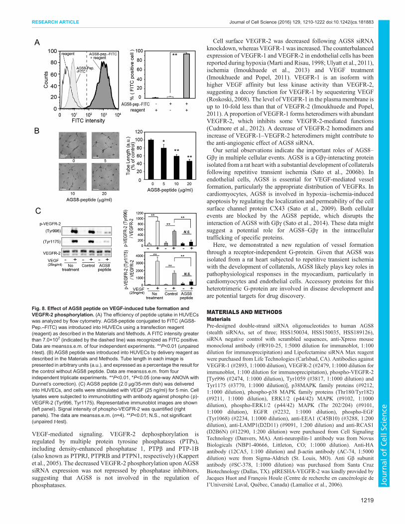
AGS8 peptide inhibits VEGF-induced tube formationTo determine the role of the AGS8–Gβγ complex in vessel formation,AGS8 peptide was delivered to HUVECs using a chemical reagent asdescribed in the Materials and Methods. FACS analysis indicatedthat FITC-conjugated AGS8 peptide was incorporated successfullyinto HUVECs (Fig. 8A). Interestingly, AGS8 peptide inhibitedtube formation in a dose-dependent manner (46.3±4.1% versuscontrol, 20 μg/µl) (mean±s.e.m., P<0.01; Fig. 8B). As observed inAGS8-siRNA-treated HUVECs, AGS8 peptide inhibited theVEGF-mediated phosphorylation of VEGFR-2 (Fig. 8C). Incontrast, with AGS8 peptide, gallein, which was designed to blocksuniversal Gβγ signaling, failed to inhibit VEGF-mediated tubeformation, proliferation andVEGFR-2 phosphorylation (Fig. S2A–C)(Lehmann et al., 2008). The AGS8–Gβγ complex was not disruptedby gallein in cells (Fig. S2D). These data suggest that the AGS8–Gβγcomplex, rather than a simple Gβγ signal input, is required byendothelial cells for the induction of angiogenesis.
DISCUSSIONHere, we first demonstrated the regulation of angiogenesis by anaccessory protein for the heterotrimeric G-protein, AGS8.Knockdown of AGS8 in endothelial cells inhibited VEGF-mediated cellular events, including tube formation, migration andproliferation. AGS8 siRNA attenuated intracellular signaling, whichwas associated with a decrease of cell surface VEGFR-2. In contrast,AGS8 siRNA did not attenuate EGF- or bFGF-mediated cellularevents. Expressed VEGFR-2 formed a protein complex withAGS8C in cells. Additionally, AGS8 peptide, which is designedto disrupt the interaction between AGS8 and Gβγ, blocked tubeformation and VEGFR-2 phosphorylation. These observationssuggest that the AGS8–Gβγ complex might be involved inangiogenic events under pathophysiological challenge and thatAGS8 serves as a new regulatory pathway for vessel formation.
The importance of receptor-independent accessory proteins forheterotrimeric G-proteins in disease pathogenesis has beendescribed in many studies (Sato, 2013). However, reportsindicating roles for regulatory proteins of heterotrimeric G-proteins in vessel formation are very limited. The expression ofRGS5, which binds to the Gαi/o and Gαq subunits (Zhou et al.,2001), has been reported in neovascular vessels. RGS5 is expressedin pericytes and is involved in neovascularization by regulating thematuration of pericytes (Nisancioglu et al., 2008). Additionally,RGS5 is highly expressed in endothelial cells isolated from humancancers, suggesting a role in the maintenance of a pro-angiogenicmicroenvironment (Silini et al., 2012). However, the specificpathway by which RGS5 influences vessel formation has not beenwell defined. AGS8 is clearly involved in angiogenic events that aremediated by regulating VEGF-mediated signaling. An AGS8–Gβγblocking peptide inhibited this process, suggesting that theinteraction of AGS8 with the Gβγ subunit is required for thisprocess. Although the AGS8 peptide might influence AGS8–Gβγ
1215
RESEARCH ARTICLE Journal of Cell Science (2016) 129, 1210-1222 doi:10.1242/jcs.181883
Journal
ofCe
llScience
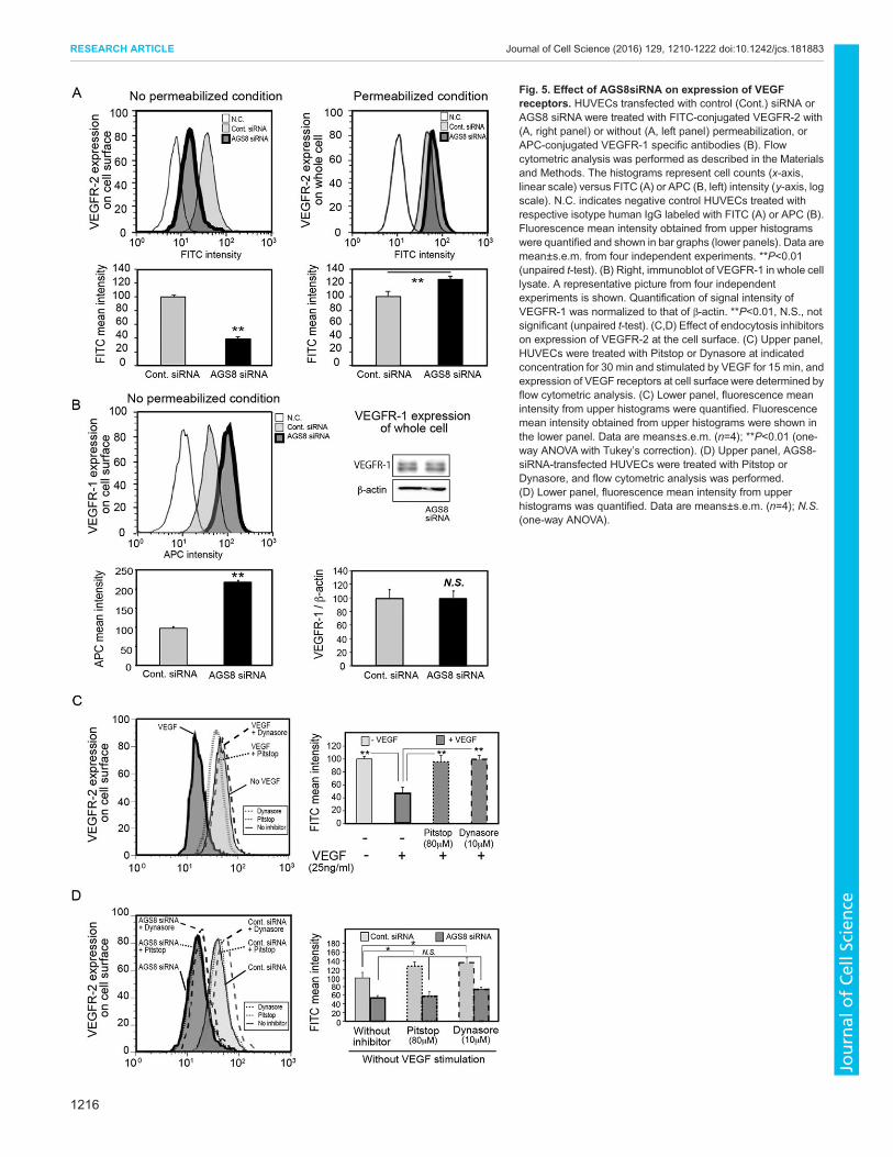
Fig. 5. Effect of AGS8siRNA on expression of VEGFreceptors. HUVECs transfected with control (Cont.) siRNA orAGS8 siRNA were treated with FITC-conjugated VEGFR-2 with(A, right panel) or without (A, left panel) permeabilization, orAPC-conjugated VEGFR-1 specific antibodies (B). Flowcytometric analysis was performed as described in the Materialsand Methods. The histograms represent cell counts (x-axis,linear scale) versus FITC (A) or APC (B, left) intensity (y-axis, logscale). N.C. indicates negative control HUVECs treated withrespective isotype human IgG labeled with FITC (A) or APC (B).Fluorescence mean intensity obtained from upper histogramswere quantified and shown in bar graphs (lower panels). Data aremean±s.e.m. from four independent experiments. **P<0.01(unpaired t-test). (B) Right, immunoblot of VEGFR-1 in whole celllysate. A representative picture from four independentexperiments is shown. Quantification of signal intensity ofVEGFR-1 was normalized to that of β-actin. **P<0.01, N.S., notsignificant (unpaired t-test). (C,D) Effect of endocytosis inhibitorson expression of VEGFR-2 at the cell surface. (C) Upper panel,HUVECs were treated with Pitstop or Dynasore at indicatedconcentration for 30 min and stimulated by VEGF for 15 min, andexpression of VEGF receptors at cell surfacewere determined byflow cytometric analysis. (C) Lower panel, fluorescence meanintensity from upper histograms were quantified. Fluorescencemean intensity obtained from upper histograms were shown inthe lower panel. Data are means±s.e.m. (n=4); **P<0.01 (one-way ANOVA with Tukey’s correction). (D) Upper panel, AGS8-siRNA-transfected HUVECs were treated with Pitstop orDynasore, and flow cytometric analysis was performed.(D) Lower panel, fluorescence mean intensity from upperhistograms was quantified. Data are means±s.e.m. (n=4); N.S.(one-way ANOVA).
1216
RESEARCH ARTICLE Journal of Cell Science (2016) 129, 1210-1222 doi:10.1242/jcs.181883
Journal
ofCe
llScience
outside of being in a complex with VEGFR-2, and the role of Gβγ inthe VEGFR-2–AGS8–Gβγ complex is not completely defined, thecurrent data suggest the potential importance of the AGS8–Gβγcomplex in the regulation of cellular functions.We found that AGS8 is involved in the intracellular
distribution and/or trafficking of VEGFR-2. A previous reporthas indicated that 25% of VEGFR-2 is located in the Golgicomplex and then recruited to the plasma membrane upon VEGFstimulation (Manickam et al., 2011). Additionally, VEGFR-2undergoes constitutive endocytosis and recycling (Jopling et al.,2011). Endocytosis blockers prevent the loss of VEGFR-2 fromthe plasma membrane and lead to an increase of cell surface
receptors (Gourlaouen et al., 2013). However, the decrease inVEGFR-2 caused by AGS8 siRNA was not rescued withendocytosis blockers, suggesting that AGS8 is involved in thetrafficking of receptors toward the membrane rather than receptorendocytosis.
Interestingly, AGS8 knockdown had a limited effect on EGF orbFGF signaling, indicating the specificity of AGS8-mediatedtrafficking. Rab4, Rab5, Rab7 and Rab11 vesicles are reportedlyinvolved in VEGFR-2 recycling (Jopling et al., 2009, 2014;Ballmer-Hofer et al., 2011). These Rab proteins are generallyutilized in the recycling and/or degradation of various proteins,including EGFR (Ceresa, 2006). The preference of the AGS8 to
Fig. 6. Effect of AGS8 knockdown onintracellular distribution of VEGFR-2. (A) Theintracellular distribution of VEGFR-2 wasdetermined by fluorescent immunostaining asdescribed in the Materials and Methods. HUVECstransfected with control siRNA or AGS8 siRNAwere stained with VEGFR-2–FITC (green), anti-RCAS1 (Golgi marker, red), anti-EEA1 (endosomemarker, red), anti-LAMP1 (lysosome marker, red),and DAPI (nuclei, blue). The area indicated with asquare is shown in the right column. Scale bars:50 μm. The arrows indicate cells in which VEGFR-2 colocalized and other organelles. (A) Lowerpanel, immunostaining with antibodies for negativecontrols. Cells were treated with FITC-conjugatedanti-mouse-IgG antibody (green), an isotypecontrol for anti-human-VEGFR-2–FITC, and Alexa-Fluor-594-conjugated anti-rabbit-IgG antibody(Red), common controls for anti-RCAS1, EEA1and RAMP1 rabbit antibodies, together with DAPI(blue). (B) The number of cells, in which VEGFR-2and other organelles colocalized, was counted inabout 100 transfected cells from four or fiveindependent experiments. Cont. siRNA, controlsiRNA.
1217
RESEARCH ARTICLE Journal of Cell Science (2016) 129, 1210-1222 doi:10.1242/jcs.181883
Journal
ofCe
llScience
modulate a specific receptor suggests that AGS8 is not involveddirectly in the function of Rab proteins; AGS8 might provide thespecificity of trafficking by supporting the entry of VEGFR-2 toRab vesicles.
Although ∼40% of VEGFR-2 was still expressed at themembrane upon AGS8 siRNA treatment, phosphorylation ofVEGFR-2 was clearly blocked by AGS8 siRNA. The reductionof cell surface VEGFR-2 might not fully explain the block of
Fig. 7. Interaction of the C-terminal of AGS8 (AGS8-C) with VEGFR-2 and/or the Gβγ subunit. COS7 cells in a 100-mm dish were transfected with acombination of pcDNA3, pIREShrGFP2a::VEGFR-2 containing HA epitope tag (6 μg/dish), pcDNAHis::AGS8-C containing Xpress epitope tag (6 μg/dish),pcDNA3::Gβ1 (3 μg/dish), pcDNA3::Gγ2 (3 μg/dish), and 2.4 (indicated as ‘+’) or 12.0 (indicated as ‘++’) μg of AGS8 peptide. The amount of DNA transfected wasadjusted to 18 μg per dish with the pcDNA3 vector. Preparation of cell lysates and immunoprecipitation (IP) were performed as described in the Materials andMethods. The transferred membrane was reprobed with the antibodies (IB) indicated in the figure. These data are representative of five independent experimentswith similar results.
1218
RESEARCH ARTICLE Journal of Cell Science (2016) 129, 1210-1222 doi:10.1242/jcs.181883
Journal
ofCe
llScience
VEGF-mediated signaling. VEGFR-2 dephosphorylation isregulated by multiple protein tyrosine phosphatases (PTPs),including density-enhanced phosphatase 1, PTPβ and PTP-1B(also known as PTPRJ, PTPRB and PTPN1, respectively) (Kappertet al., 2005). The decreased VEGFR-2 phosphorylation upon AGS8siRNA expression was not repressed by phosphatase inhibitors,suggesting that AGS8 is not involved in the regulation ofphosphatases.
Cell surface VEGFR-2 was decreased following AGS8 siRNAknockdown,whereasVEGFR-1was increased. The counterbalancedexpression of VEGFR-1 and VEGFR-2 in endothelial cells has beenreported during hypoxia (Marti and Risau, 1998; Ulyatt et al., 2011),ischemia (Imoukhuede et al., 2013) and VEGF treatment(Imoukhuede and Popel, 2011). VEGFR-1 is an isoform withhigher VEGF affinity but less kinase activity than VEGFR-2,suggesting a decoy function for VEGFR-1 by sequestering VEGF(Roskoski, 2008). The level of VEGFR-1 in the plasmamembrane isup to 10-fold less than that of VEGFR-2 (Imoukhuede and Popel,2011). A proportion of VEGFR-1 forms heterodimers with abundantVEGFR-2, which inhibits some VEGFR-2-mediated functions(Cudmore et al., 2012). A decrease of VEGFR-2 homodimers andincrease of VEGFR-1–VEGFR-2 heterodimers might contribute tothe anti-angiogenic effect of AGS8 siRNA.
Our serial observations indicate the important roles of AGS8–Gβγ in multiple cellular events. AGS8 is a Gβγ-interacting proteinisolated from a rat heart with a substantial development of collateralsfollowing repetitive transient ischemia (Sato et al., 2006b). Inendothelial cells, AGS8 is essential for VEGF-mediated vesselformation, particularly the appropriate distribution of VEGFRs. Incardiomyocytes, AGS8 is involved in hypoxia–ischemia-inducedapoptosis by regulating the localization and permeability of the cellsurface channel protein CX43 (Sato et al., 2009). Both cellularevents are blocked by the AGS8 peptide, which disrupts theinteraction of AGS8 with Gβγ (Sato et al., 2014). These data mightsuggest a potential role for AGS8–Gβγ in the intracellulartrafficking of specific proteins.
Here, we demonstrated a new regulation of vessel formationthrough a receptor-independent G-protein. Given that AGS8 wasisolated from a rat heart subjected to repetitive transient ischemiawith the development of collaterals, AGS8 likely plays key roles inpathophysiological responses in the myocardium, particularly incardiomyocytes and endothelial cells. Accessory proteins for thisheterotrimeric G-protein are involved in disease development andare potential targets for drug discovery.
MATERIALS AND METHODSMaterialsPre-designed double-strand siRNA oligonucleotides to human AGS8(stealth siRNAs, set of three; HSS150034, HSS150035, HSS189126),siRNA negative control with scrambled sequences, anti-Xpress mousemonoclonal antibody (#R910-25, 1:5000 dilution for immunoblot, 1:100dilution for immunoprecipitation) and Lipofectamine siRNA Max reagentwere purchased from Life Technologies (Carlsbad, CA). Antibodies againstVEGFR-1 (#2893, 1:1000 dilution), VEGFR-2 (#2479, 1:1000 dilution forimmunoblot, 1:100 dilution for immunoprecipitation), phospho-VEGFR-2[Tyr996 (#2474, 1:1000 dilution), Tyr1059 (#3817, 1:1000 dilution) andTyr1175 (#3770, 1:1000 dilution)], p38MAPK family proteins (#9212,1:1000 dilution), phospho-p38 MAPK family proteins (Thr180/Tyr182)(#9211, 1:1000 dilution), ERK1/2 (p44/42) MAPK (#9102, 1:1000dilution), phospho-ERK1/2 (p44/42) MAPK (Thr 202/204) (#9101,1:1000 dilution), EGFR (#2232, 1:1000 dilution), phospho-EGF(Tyr1068) (#2234, 1:1000 dilution), anti-EEA1 (C45B10) (#3288, 1:200dilution), anti-LAMP1(D2D11) (#9091, 1:200 dilution) and anti-RCAS1(D2B6N) (#12290, 1:200 dilution) were purchased from Cell SignalingTechnology (Danvers, MA). Anti-neuropilin-1 antibody was from NovusBiologicals (NBP1-40666, Littleton, CO; 1:1000 dilution). Anti-HAantibody (12CA5, 1:100 dilution) and β-actin antibody (AC-74, 1:5000dilution) were from Sigma-Aldrich (St. Louis, MO). Anti Gβ subunitantibody (#SC-378, 1:1000 dilution) was purchased from Santa CruzBiotechnology (Dallas, TX). pIRESHA-VEGFR-2 was kindly provided byJacques Huot and François Houle (Centre de recherche en cancérologie deI’Université Laval, Québec, Canada) (Lamalice et al., 2006).
Fig. 8. Effect of AGS8 peptide on VEGF-induced tube formation andVEGFR-2 phosphorylation. (A) The efficiency of peptide uptake in HUVECswas analyzed by flow cytometry. AGS8-peptide conjugated to FITC (AGS8-Pep.–FITC) was introduced into HUVECs using a transfection reagent(reagent) as described in the Materials and Methods. A FITC intensity greaterthan 7.0×10² (indicated by the dashed line) was recognized as FITC positive.Data are means±s.e.m. of four independent experiments. **P<0.01 (unpairedt-test). (B) AGS8 peptide was introduced into HUVECs by delivery reagent asdescribed in the Materials and Methods. Tube length in each image ispresented in arbitrary units (a.u.), and expressed as a percentage the result forthe control without AGS8 peptide. Data are means±s.e.m. from fourindependent triplicate experiments. **P<0.01, *P<0.05 (one-way ANOVA withDunnet’s correction). (C) AGS8 peptide (2.0 μg/35-mm dish) was deliveredinto HUVECs, and cells were stimulated with VEGF (25 ng/ml) for 5 min. Celllysates were subjected to immunoblotting with antibody against phospho (p)-VEGFR-2 (Tyr996, Tyr1175). Representative immunoblot images are shown(left panel). Signal intensity of phospho-VEGFR-2 was quantified (rightpanels). The data are means±s.e.m. (n=4). **P<0.01; N.S., not significant(unpaired t-test).
1219
RESEARCH ARTICLE Journal of Cell Science (2016) 129, 1210-1222 doi:10.1242/jcs.181883
Journal
ofCe
llScience
Cell culture and siRNA transfectionHuman umbilical vein endothelial cells (HUVECs) and human umbilicalartery endothelial cells (HUAECs), were cultured in endothelial cell growthmedium (EGM)-2 (Lonza, Basel, Switzerland) or endothelial cell growthmedium on fibronectin-coated (Sigma-Aldrich) culture dish or plate(Corning, Schiphol, The Netherlands). For siRNA transfection, cells wereincubated in six-well plates and treated with three sets of each 40 nM StealthsiRNAs or negative control with Lipofectamine siRNA Max according tothe manufacturer’s instruction. AGS8mRNA expression was determined byreal-time PCR (Sato et al., 2009). The primers for RT-PCR were as follows:human AGS8, forward, 5′-TTCCGTAACCCTCTCTCCCG-3′; reverse,5′-AACCCACGATCAAGGTCCAC-3′. Each AGS8 siRNA significantlyreduced the level of AGS8 mRNA (H.H. and M.S., unpublishedobservation).
COS-7 were cultured and transfected as described previously (Sato et al.,2011). In brief, cells were suspended at 0.5–1.0×10⁵ cells/ml, and 10 ml ofsuspension was plated in 100-mm dishes. After 18 h, cells were transfectedwith 18 μg of cDNA with Lipofectamine 3000 (Life Technologies)according to the manufacturer’s instruction. Cell lysis and fractionationwere performed as described previously (Sato et al., 1995, 2004).
Tube formation assayGrowth-factor-reduced Matrigel (BD Biosciences, San Jose, CA) was usedto coat the 96-well plates (50 μl/well), which was then allowed topolymerize at 37°C for 1 h. Cells were incubated with endothelial cellbasal medium (EBM)-2 (Lonza) containing 1% fetal bovine serum (FBS)for 6 h, and were then trypsinized and suspended in 250 μl of EBM-2containing 1% FBS. Cells were seeded at 1×10⁴ cells/well ontoMatrigel andfurther incubated. In some experiments, cells were treated with VEGF at25 ng/ml or VEGFR-2 inhibitor (R&D Systems, Minneapolis, MN) at 0.1 to5 μM, a Gβγ inhibitor gallein (Millipore, Billerica, MA) at 3.3 to 30 μM, orAGS8-blocking peptide (Sato et al., 2014) at 100 μg/ml. Pictures ofcapillary-like structures were taken at 6-h and 18-h time points. Tube lengthwas determined by tracing lines along the cells using ImageJ software(Schneider et al., 2012). The total tube length of each image was determinedby an observer blinded to the conditions. The data are presented in arbitraryunits (a.u.).
MTT assayHuman recombinant VEGF, epidermal growth factor (EGF) and basicfibroblast growth factor (bFGF) were purchased from Wako Chemicals(Osaka, Japan). The cell proliferation was analyzed using a Vybrant MTTcell proliferation assay kit (Life Technologies), according to the productinstructions. Briefly, cells in six-well plates were transfected with siRNAsand incubated for 24 h. Next, cells were transferred to 96-well plates at5×103 cells/well. After 24 h, cells were incubated with EBM-2 containing1% FBS for 6 h, and treated with VEGF for 48 h, EGF for 24 h or bFGF for48 h. Then, cells were incubated with MTT at 37°C for 4 h. Medium wasremoved, and SDS-HCl solution (10% SDS, 0.01 M HCl) was added toeach well. After incubation for 18 h at 37°C, the absorbance was measuredwith a SpectraMax (Molecular Devices) at a wavelength of 570 nm.
Cells migration assayHUVECs suspended at 5×10⁴ cells/200 μl were seeded on fibronectin-coated Transwell inserts with 8 μm pores (BD Biosciences) that hadbeen pretreated with 3% BSA. Cell migration was stimulated by VEGF(25 ng/ml) in the lower chamber of the 24-well plate. After 4 h, theTranswell inserts were fixed in 4% PFA, stained with 1% Crystal Violet.Digital microscopic images of the underside of each Transwell insert atindependent microscopic fields were taken, and cells stained were counted.
Peptide deliveryDelivery of peptide to cells was essentially performed as describedpreviously (Sato et al., 2014). Briefly, peptide was incubated with 4 μl ofPLUSin (Polyplus, Illkirch, France) in 100 μl of EBM-2 for 15 min, andthen the mixture was added to each well of cultured HUVECs in 12-wellplates. Cells were then incubated for 4 h before stimulation by VEGF(25 ng/ml) for tube formation assay or immunoblotting. The incorporation
of the peptide by cells was determined with fluorescein isothiocyanate(FITC)-conjugated AGS8 peptide by flow cytometric analysis (Sato et al.,2014). In some immunoprecipitation experiments, 2.4–12.0 μg of AGS8peptide was incubated with 12 μl of Lipofectamine 3000 (LifeTechnologies) for 5 min, and then added the mixture to COS7 cells in a10-cm dish 4 h before harvesting.
ImmunoblottingHUVECs were cultured in six-well plates were washed with ice-cold PBS,and collected in SDS-PAGE sample buffer (65.8 mM Tris-HCl, pH 6.8,2.1% SDS, 26.3% glycerol, 0.01% Bromophenol Blue) with proteaseinhibitor cocktail (Roche, Indianapolis, IN) and 4% 2-mercaptoethanol. Thelysates were separated by SDS-PAGE, and the gel was transferred onto thePVDF membrane (Millipore, Billerica, MA). The membrane was blockedwith Tris-buffered saline containing 5% bovine serum albumin (Sigma-Aldrich) and incubated with primary antibodies. The membranes were thentreated with horseradish-peroxidase-conjugated secondary antibodies (GEHealthcare, Munich, Germany), and signals were detected usingImmunostar LD western blotting detection reagents (Wako Chemicals,Osaka, Japan) and a LAS4000 image analyzer (GE Healthcare). Forquantification, the band intensity was analyzed by the ImageJ software.
ImmunocytochemistryHUVECs were cultured on the 13-mm poly-L-lysine-coated coverslips(Matsunami Glass, Osaka, Japan). Cells were washed twice with PBS, andthen fixed with 4% paraformaldehyde containing 4% sucrose in PBS for15 min followed by incubation with 0.2% Triton X-100 in PBS for 5 min.After three washes with PBS, cells were incubated with 5% normal donkeyserum in PBS for 1 h to block the nonspecific staining, incubated withfluorescein-conjugated anti-human VEGFR-2 antibody (1:200 dilution;FAB357F, R&D Systems, Minneapolis, MN) and antibodies for organellesfor 18 h at 4°C. Cells were incubated with DAPI (Life Technologies) in PBSfor 5 min. Slides were then mounted with glass coverslips with FluoroKeeper antifade reagent (Nacalai Tesque, Kyoto, Japan) or ProLongantifade Gold (Life Technologies). Images were obtained with a laserconfocal microscope, LSM710 (Carl Zeiss, Jena, Germany).
Flow cytometric analysisFluorescence-activated cell sorting (FACS) was performed using a FACSCanto II (BDBiosciences, San Jose, CA). Cells werewashed with PBS anddetached from culture plate with Accutase (Innovative Cell Technologies,San Diego, CA), then fixed with 2% PFA for 30 min. In the experimentsusing permeabilization, the cells were incubated with 90% methanol onice for 30 min. Cells were washed with PBS containing 1% FBS, andlabeled with fluorescein-conjugated anti-human VEGFR-2 antibody(1:100 dilution) and allophycocyanin (APC)-conjugated anti-VEGFR-1antibody (1:100 dilution; FAB321A, R&D Systems, Minneapolis, MN),fluorescein-conjugated IgG1 isotype control (1:100 dilution; IC002F,R&D Systems, Minneapolis, MN) or IgG1 APC-conjugated isotypecontrol (1:100 dilution; IC002A, R & D Systems, Minneapolis, MN) for1 h in the dark on ice. In some experiments, cells were treated with Pitstop(Abcam, Cambridge, MA, USA) or Dynasore (Sigma-Aldrich) for 30 min,then stimulated with VEGF (25 ng/ml) for 15 min before collecting cells.FACS Calibur and CellQuest software (BD Biosciences) was used for flowcytometric analysis and for data acquisition, respectively. Flow Jo(TreeStar, Ashland, OR, USA) was used for data analysis.
ImmunoprecipitationCell lysates were prepared in 500 µl immune precipitation buffer [50 mMTris-HCl pH 7.4, 70 mM NaCl, 5 mM EDTA, 1% IGEPAL CA-630(Sigma), protease inhibitor cocktail (Complete Mini; Roche AppliedScience)]. At total of 500–800 μg of lysate were incubated with 1.0–6.0 µg antibody for 18 h after pre-clearing with 25 µl of 50% Sepharose-Gfor 1 h at 4°C. The samples were incubated with 25 µl of 50% Sepharose-Gfor 1 h at 4°C, and the pellets were washed three times with the immuneprecipitation buffer. Proteins were eluted in 30 µl of 2× Laemmli buffer andresolved by SDS-PAGE (Sato et al., 2009).
1220
RESEARCH ARTICLE Journal of Cell Science (2016) 129, 1210-1222 doi:10.1242/jcs.181883
Journal
ofCe
llScience
Other procedures and data analysisOther procedures were as previously described (Sato et al., 2009, 2011).Data are expressed as means±s.e.m. from independent experiments.Statistics were performed using a unpaired t-test or one-way or two-wayANOVA with Dunnett’s or Tukey’s correction as indicated in figurelegends. All statistical analyses were performed with IBM SPSS software.
AcknowledgementsWe thank Jacques Huot and Dr François Houle (Centre de recherche encancerologie de I’Universite Laval, Quebec, Canada) for providing pIRESHA-VEGFR-2. We are also grateful to Hiroko Suzuki and Rie Takahashi for theirtechnical assistance.
Competing interestsThe authors declare no competing or financial interests.
Author contributionsM.S. conceived and coordinated the study and wrote the paper. H.H. designed andperformed the study, and wrote the paper. A.A.M. and M.S. provided substantialtechnical assistance and contributed to the preparation of the figures. All authorsanalyzed the results and approved the final version of the manuscript.
FundingThis study was supported by a Grant-in-Aid for Scientific Research on InnovativeAreas from Japan Society for Promotion of Science (JSPS) [grant number 25136721toM.S.]; and aGrant-in-Aid for Scientific Research from Japan Society for Promotionof Science (JSPS) [grant number 24590280 to M.S.]; The Naito Foundation (toM.S.); DAIKO Foundation (to M.S.); the Takeda Science Foundation (to H.H.); and aStrategic Research Foundation Grant-aided Project for Private Universities from theMinistry of Education, Culture, Sports, Science, and Technology, Japan, 2011–2015[grant number S1101027].
Supplementary informationSupplementary information available online athttp://jcs.biologists.org/lookup/suppl/doi:10.1242/jcs.181883/-/DC1
ReferencesAlderton, F., Rakhit, S., Kong, K. C., Palmer, T., Sambi, B., Pyne, S. and Pyne,N. J. (2001). Tethering of the platelet-derived growth factor beta receptor to G-protein-coupled receptors. A novel platform for integrative signaling by thesereceptor classes in mammalian cells. J. Biol. Chem. 276, 28578-28585.
Ballmer-Hofer, K., Andersson, A. E., Ratcliffe, L. E. and Berger, P. (2011).Neuropilin-1 promotes VEGFR-2 trafficking through Rab11 vesicles therebyspecifying signal output. Blood 118, 816-826.
Blumer, J. B. and Lanier, S. M. (2014). Activators of G protein signaling exhibitbroad functionality and define a distinct core signaling triad. Mol. Pharmacol. 85,388-396.
Ceresa, B. P. (2006). Regulation of EGFR endocytic trafficking by rab proteins.Histol. Histopathol. 21, 987-993.
Cudmore, M. J., Hewett, P. W., Ahmad, S., Wang, K.-Q., Cai, M., Al-Ani, B.,Fujisawa, T., Ma, B., Sissaoui, S., Ramma, W. et al. (2012). The role ofheterodimerization between VEGFR-1 and VEGFR-2 in the regulation ofendothelial cell homeostasis. Nat. Commun. 3, 972.
Gourlaouen, M., Welti, J. C., Vasudev, N. S. and Reynolds, A. R. (2013).Essential role for endocytosis in the growth factor-stimulated activation of ERK1/2in endothelial cells. J. Biol. Chem. 288, 7467-7480.
Gu, S., Cifelli, C., Wang, S. and Heximer, S. P. (2009). RGS proteins: identifyingnew GAPs in the understanding of blood pressure regulation and cardiovascularfunction. Clin. Sci. 116, 391-399.
Holmes, D. I. R. and Zachary, I. C. (2008). Vascular endothelial growth factorregulates stanniocalcin-1 expression via neuropilin-1-dependent regulation ofKDR and synergism with fibroblast growth factor-2. Cell. Signal. 20, 569-579.
Imamura, T., Vollenweider, P., Egawa, K., Clodi, M., Ishibashi, K., Nakashima,N., Ugi, S., Adams, J. W., Brown, J. H. and Olefsky, J. M. (1999). G alpha-q/11protein plays a key role in insulin-induced glucose transport in 3T3-L1 adipocytes.Mol. Cell. Biol. 19, 6765-6774.
Imoukhuede, P. I. and Popel, A. S. (2011). Quantification and cell-to-cell variationof vascular endothelial growth factor receptors. Exp. Cell Res. 317, 955-965.
Imoukhuede, P. I., Dokun, A. O., Annex, B. H. andPopel, A. S. (2013). Endothelialcell-by-cell profiling reveals the temporal dynamics of VEGFR1 and VEGFR2membrane localization after murine hindlimb ischemia. Am. J. Physiol. Heart Circ.Physiol. 304, H1085-H1093.
Jopling, H. M., Odell, A. F., Hooper, N. M., Zachary, I. C., Walker, J. H. andPonnambalam, S. (2009). Rab GTPase regulation of VEGFR2 trafficking andsignaling in endothelial cells. Arterioscler. Thromb. Vasc. Biol. 29, 1119-1124.
Jopling, H. M., Howell, G. J., Gamper, N. and Ponnambalam, S. (2011). TheVEGFR2 receptor tyrosine kinase undergoes constitutive endosome-to-plasmamembrane recycling. Biochem. Biophys. Res. Commun. 410, 170-176.
Jopling, H. M., Odell, A. F., Pellet-Many, C., Latham, A. M., Frankel, P.,Sivaprasadarao, A., Walker, J. H., Zachary, I. C. and Ponnambalam, S. (2014).Endosome-to-plasma membrane recycling of VEGFR2 receptor tyrosine kinaseregulates endothelial function and blood vessel formation. Cells 3, 363-385.
Kappert, K., Peters, K. G., Bohmer, F. D. and Ostman, A. (2005). Tyrosinephosphatases in vessel wall signaling. Cardiovasc. Res. 65, 587-598.
Kimple, A. J., Bosch, D. E., Giguere, P. M. and Siderovski, D. P. (2011).Regulators of G-protein signaling and their Galpha substrates: promises andchallenges in their use as drug discovery targets. Pharmacol. Rev. 63,728-749.
Kuemmerle, J. F. and Murthy, K. S. (2001). Coupling of the insulin-like growthfactor-I receptor tyrosine kinase to Gi2 in human intestinal smooth muscle:Gbetagamma-dependent mitogen-activated protein kinase activation and growth.J. Biol. Chem. 276, 7187-7194.
Lamalice, L., Houle, F. and Huot, J. (2006). Phosphorylation of Tyr 1214 withinVEGFR-2 triggers the recruitment of Nck and activation of Fyn leading to SAPK2/p38 activation and endothelial cell migration in response to VEGF. J. Biol. Chem.281, 34009-34020.
Lehmann, D. M., Seneviratne, A. M. P. B. and Smrcka, A. V. (2008). Smallmolecule disruption of G protein beta gamma subunit signaling inhibits neutrophilchemotaxis and inflammation. Mol. Pharmacol. 73, 410-418.
Leung, T., Chen, H., Stauffer, A. M., Giger, K. E., Sinha, S., Horstick, E. J.,Humbert, J. E., Hansen, C. A. and Robishaw, J. D. (2006). Zebrafish G proteingamma2 is required for VEGF signaling during angiogenesis. Blood 108,160-166.
Manickam, V., Tiwari, A., Jung, J.-J., Bhattacharya, R., Goel, A.,Mukhopadhyay, D. and Choudhury, A. (2011). Regulation of vascularendothelial growth factor receptor 2 trafficking and angiogenesis by Golgilocalized t-SNARE syntaxin 6. Blood 117, 1425-1435.
Marti, H. H. and Risau, W. (1998). Systemic hypoxia changes the organ-specificdistribution of vascular endothelial growth factor and its receptors. Proc. Natl.Acad. Sci. USA 95, 15809-15814.
Nisancioglu, M. H., Mahoney, W. M., Jr, Kimmel, D. D., Schwartz, S. M.,Betsholtz, C. and Genove, G. (2008). Generation and characterization of rgs5mutant mice. Mol. Cell. Biol. 28, 2324-2331.
Pellet-Many, C., Frankel, P., Jia, H. and Zachary, I. (2008). Neuropilins: structure,function and role in disease. Biochem. J. 411, 211-226.
Roskoski, R. (2008). VEGF receptor protein–tyrosine kinases: structure andregulation. Biochem. Biophys. Res. Commun. 375, 287-291.
Sato, M. (2013). Roles of accessory proteins for heterotrimeric g-protein in thedevelopment of cardiovascular diseases. Circ. J. 77, 2455-2461.
Sato, M., Kataoka, R., Dingus, J., Wilcox, M., Hildebrandt, J. D. and Lanier, S. M.(1995). Factors determining specificity of signal transduction by G-protein-coupled receptors: regulation of signal transfer from receptor to G-protein. J. Biol.Chem. 270, 15269-15276.
Sato, M., Gettys, T. W. and Lanier, S. M. (2004). AGS3 and signal integration byGalpha(s)- and Galpha(i)-coupled receptors: AGS3 blocks the sensitization ofadenylyl cyclase following prolonged stimulation of a Galpha(i)-coupled receptorby influencing processing of Galpha(i). J. Biol. Chem. 279, 13375-13382.
Sato, M., Blumer, J. B., Simon, V. and Lanier, S. M. (2006a). Accessory proteinsfor G proteins: partners in signaling. Annu. Rev. Pharmacol. Toxicol. 46, 151-187.
Sato, M., Cismowski, M. J., Toyota, E., Smrcka, A. V., Lucchesi, P. A., Chilian,W.M. and Lanier, S.M. (2006b). Identification of a receptor-independent activatorof G protein signaling (AGS8) in ischemic heart and its interaction withGbetagamma. Proc. Natl. Acad. Sci. USA 103, 797-802.
Sato, M., Jiao, Q., Honda, T., Kurotani, R., Toyota, E., Okumura, S., Takeya, T.,Minamisawa, S., Lanier, S. M. and Ishikawa, Y. (2009). Activator of G proteinsignaling 8 (AGS8) is required for hypoxia-induced apoptosis of cardiomyocytes:role of G betagamma and connexin 43 (CX43). J. Biol. Chem. 284, 31431-31440.
Sato, M., Hiraoka, M., Suzuki, H., Bai, Y., Kurotani, R., Yokoyama, U., Okumura,S., Cismowski, M. J., Lanier, S. M. and Ishikawa, Y. (2011). Identification oftranscription factor E3 (TFE3) as a receptor-independent activator of Galpha16:gene regulation by nuclear Galpha subunit and its activator. J. Biol. Chem. 286,17766-17776.
Sato, M., Hiraoka, M., Suzuki, H., Sakima, M., Mamun, A. A., Yamane, Y., Fujita,T., Yokoyama, U., Okumura, S. and Ishikawa, Y. (2014). Protection ofcardiomyocytes from the hypoxia-mediated injury by a peptide targeting theactivator of G-protein signaling 8. PLoS ONE 9, e91980.
Schneider, C. A., Rasband,W. S. and Eliceiri, K. W. (2012). NIH Image to ImageJ:25 years of image analysis. Nat. Methods 9, 671-675.
Silini, A., Ghilardi, C., Figini, S., Sangalli, F., Fruscio, R., Dahse, R., Pedley,R. B., Giavazzi, R. and Bani, M. (2012). Regulator of G-protein signaling 5(RGS5) protein: a novel marker of cancer vasculature elicited and sustained by thetumor’s proangiogenic microenvironment. Cell. Mol. Life Sci. 69, 1167-1178.
Ulyatt, C., Walker, J. and Ponnambalam, S. (2011). Hypoxia differentiallyregulates VEGFR1 and VEGFR2 levels and alters intracellular signaling and
1221
RESEARCH ARTICLE Journal of Cell Science (2016) 129, 1210-1222 doi:10.1242/jcs.181883
Journal
ofCe
llScience
cell migration in endothelial cells. Biochem. Biophys. Res. Commun. 404,774-779.
Wieland, T. and Mittmann, C. (2003). Regulators of G-protein signalling:multifunctional proteins with impact on signalling in the cardiovascular system.Pharmacol. Ther. 97, 95-115.
Zeng, H., Zhao, D. andMukhopadhyay, D. (2002). Flt-1-mediated down-regulationof endothelial cell proliferation through pertussis toxin-sensitive G proteins, betagamma subunits, small GTPase CDC42, and partly by Rac-1. J. Biol. Chem. 277,4003-4009.
Zeng, H., Zhao, D., Yang, S., Datta, K. and Mukhopadhyay, D. (2003).Heterotrimeric G alpha q/G alpha 11 proteins function upstream of vascularendothelial growth factor (VEGF) receptor-2 (KDR) phosphorylation in vascularpermeability factor/VEGF signaling. J. Biol. Chem. 278, 20738-20745.
Zhang, P. and Mende, U. (2011). Regulators of G-protein signaling in the heart andtheir potential as therapeutic targets. Circ. Res. 109, 320-333.
Zhou, J., Moroi, K., Nishiyama, M., Usui, H., Seki, N., Ishida, J., Fukamizu, A.and Kimura, S. (2001). Characterization of RGS5 in regulation of G protein-coupled receptor signaling. Life Sci. 68, 1457-1469.
1222
RESEARCH ARTICLE Journal of Cell Science (2016) 129, 1210-1222 doi:10.1242/jcs.181883
Journal
ofCe
llScience