Short Cationic Peptide Derived from Archaea with Dual ...
6
Short Cationic Peptide Derived from Archaea with Dual Antibacterial Properties and Anti-Infective Potential Elizabete S. Câ ndido, †,‡,# Marlon H. Cardoso, †,‡,§,∥,# Lai Y. Chan, ∥ Marcelo D. T. Torres, ⊥,¶,∇,α,β Karen G. N. Oshiro, ‡,§ William F. Porto, ‡,○ Suzana M. Ribeiro, ‡ Evan F. Haney, ◆ Robert E. W. Hancock, ◆ Timothy K. Lu, ⊥,¶ Cesar de la Fuente-Nunez, ⊥,¶,α,β David J. Craik, ∥ and Octa ́ vio L. Franco* ,†,‡,§ † Centro de Aná lises Proteô micas e Bioquímicas, Pó s-Graduaç ã o em Ciê ncias Genô micas e Biotecnologia, Universidade Cató lica de Brasília, SGAN 916 Mó dulo B, Asa Norte, Brasília, Distrito Federal 70790160, Brazil ‡ S-Inova Biotech, Programa de Pó s-Graduaç ã o em Biotecnologia, Universidade Cató lica Dom Bosco, Avenida Tamandaré 6000, Campo Grande, Mato Grosso do Sul 79117900, Brazil § Programa de Pó s-Graduaç ã o em Patologia Molecular, Faculdade de Medicina, Universidade de Brasília, Campus Darcy Ribeiro, Asa Norte, Brasília, Distrito Federal 70910900, Brazil ∥ Institute for Molecular Bioscience, The University of Queensland, 306 Carmody Road, Brisbane, Queensland 4072, Australia ⊥ Synthetic Biology Group, MIT Synthetic Biology Center; The Center for Microbiome Informatics and Therapeutics; Research Laboratory of Electronics, Department of Biological Engineering, and Department of Electrical Engineering and Computer Science, Massachusetts Institute of Technology, Cambridge, Massachusetts 02139, United States of America ¶ Broad Institute of MIT and Harvard, Cambridge, Massachusetts 02139, United States of America ∇ Centro de Ciê ncias Naturais e Humanas, Universidade Federal do ABC, Santo André , Sã o Paulo 09210170, Brazil ○ Porto Reports, Brasília, Distrito Federal 70790160, Brazil ◆ Centre for Microbial Diseases and Immunity Research, University of British Columbia, Vancouver, British Columbia V6T 1Z4, Canada ABSTRACT: Bacterial biofilms and associated infections represent one of the biggest challenges in the clinic, and as an alternative to counter bacterial infections, antimicrobial peptides have attracted great attention in the past decade. Here, ten short cationic antimicrobial peptides were generated through a sliding-window strategy on the basis of the 19- amino acid residue peptide, derived from a Pyrobaculum aerophilum ribosomal protein. PaDBS1R6F10 exhibited anti- infective potential as it decreased the bacterial burden in murine Pseudomonas aeruginosa cutaneous infections by more than 1000-fold. Adverse cytotoxic and hemolytic effects were not detected against mammalian cells. The peptide demon- strated structural plasticity in terms of its secondary structure in the different environments tested. PaDBS1R6F10 represents a promising antimicrobial agent against bacteria infections, without harming human cells. KEYWORDS: antimicrobial peptide, biofilm, cutaneous infection, CD spectroscopy T he high incidence of drug-resistant bacterial and biofilm- related infections currently represents a global health concern, demanding an urgent search for new antimicrobial strategies. 1 Interestingly, despite recent efforts aimed at eradicating biofilm-related infections, only a few new antimicrobial drugs are specially aimed at biofilms. 2 Moreover, the resistance associated with bacterial biofilms imposes numerous challenges for the use of conventional antimicrobials to treat these infections. Natural antimicrobial peptides (AMPs) represent a promis- ing alternative therapy for the treatment of drug-resistant infections. These molecules present high structural diversity and broad-spectrum antimicrobial activity. 3 Natural AMPs have been explored with the aim of making a new generation of synthetic bioinspired molecules, which are a promising option for the engineering of more active and multifunctional drugs. 4 The use of rational design approaches has enabled the generation of improved AMP synthetic analogues, reducing the limitations and increasing the advantages of these natural molecules. 5 In particular, short AMPs represent attractive Received: February 20, 2019 Published: April 24, 2019 Letter pubs.acs.org/journal/aidcbc Cite This: ACS Infect. Dis. XXXX, XXX, XXX-XXX © XXXX American Chemical Society A DOI: 10.1021/acsinfecdis.9b00073 ACS Infect. Dis. XXXX, XXX, XXX−XXX Downloaded by UNIV OF BRITISH COLUMBIA at 10:36:27:111 on May 23, 2019 from https://pubs.acs.org/doi/10.1021/acsinfecdis.9b00073.
Transcript of Short Cationic Peptide Derived from Archaea with Dual ...
Short Cationic Peptide Derived from Archaea with DualAntibacterial Properties and Anti-Infective PotentialElizabete S. Candido,†,‡,# Marlon H. Cardoso,†,‡,§,∥,# Lai Y. Chan,∥ Marcelo D. T. Torres,⊥,¶,∇,α,β
Karen G. N. Oshiro,‡,§ William F. Porto,‡,○ Suzana M. Ribeiro,‡ Evan F. Haney,◆
Robert E. W. Hancock,◆ Timothy K. Lu,⊥,¶ Cesar de la Fuente-Nunez,⊥,¶,α,β David J. Craik,∥
and Octavio L. Franco*,†,‡,§
†Centro de Analises Proteomicas e Bioquímicas, Pos-Graduacao em Ciencias Genomicas e Biotecnologia, Universidade Catolica deBrasília, SGAN 916 Modulo B, Asa Norte, Brasília, Distrito Federal 70790160, Brazil‡S-Inova Biotech, Programa de Pos-Graduacao em Biotecnologia, Universidade Catolica Dom Bosco, Avenida Tamandare 6000,Campo Grande, Mato Grosso do Sul 79117900, Brazil§Programa de Pos-Graduacao em Patologia Molecular, Faculdade de Medicina, Universidade de Brasília, Campus Darcy Ribeiro, AsaNorte, Brasília, Distrito Federal 70910900, Brazil∥Institute for Molecular Bioscience, The University of Queensland, 306 Carmody Road, Brisbane, Queensland 4072, Australia⊥Synthetic Biology Group, MIT Synthetic Biology Center; The Center for Microbiome Informatics and Therapeutics; ResearchLaboratory of Electronics, Department of Biological Engineering, and Department of Electrical Engineering and Computer Science,Massachusetts Institute of Technology, Cambridge, Massachusetts 02139, United States of America¶Broad Institute of MIT and Harvard, Cambridge, Massachusetts 02139, United States of America∇Centro de Ciencias Naturais e Humanas, Universidade Federal do ABC, Santo Andre, Sao Paulo 09210170, Brazil○Porto Reports, Brasília, Distrito Federal 70790160, Brazil◆Centre for Microbial Diseases and Immunity Research, University of British Columbia, Vancouver, British Columbia V6T 1Z4,Canada
ABSTRACT: Bacterial biofilms and associated infectionsrepresent one of the biggest challenges in the clinic, and asan alternative to counter bacterial infections, antimicrobialpeptides have attracted great attention in the past decade.Here, ten short cationic antimicrobial peptides were generatedthrough a sliding-window strategy on the basis of the 19-amino acid residue peptide, derived from a Pyrobaculumaerophilum ribosomal protein. PaDBS1R6F10 exhibited anti-infective potential as it decreased the bacterial burden inmurine Pseudomonas aeruginosa cutaneous infections by morethan 1000-fold. Adverse cytotoxic and hemolytic effects werenot detected against mammalian cells. The peptide demon-strated structural plasticity in terms of its secondary structure in the different environments tested. PaDBS1R6F10 represents apromising antimicrobial agent against bacteria infections, without harming human cells.
KEYWORDS: antimicrobial peptide, biofilm, cutaneous infection, CD spectroscopy
The high incidence of drug-resistant bacterial and biofilm-related infections currently represents a global health
concern, demanding an urgent search for new antimicrobialstrategies.1 Interestingly, despite recent efforts aimed ateradicating biofilm-related infections, only a few newantimicrobial drugs are specially aimed at biofilms.2 Moreover,the resistance associated with bacterial biofilms imposesnumerous challenges for the use of conventional antimicrobialsto treat these infections.Natural antimicrobial peptides (AMPs) represent a promis-
ing alternative therapy for the treatment of drug-resistantinfections. These molecules present high structural diversity
and broad-spectrum antimicrobial activity.3 Natural AMPshave been explored with the aim of making a new generation ofsynthetic bioinspired molecules, which are a promising optionfor the engineering of more active and multifunctional drugs.4
The use of rational design approaches has enabled thegeneration of improved AMP synthetic analogues, reducingthe limitations and increasing the advantages of these naturalmolecules.5 In particular, short AMPs represent attractive
Received: February 20, 2019Published: April 24, 2019
Letter
pubs.acs.org/journal/aidcbcCite This: ACS Infect. Dis. XXXX, XXX, XXX−XXX
© XXXX American Chemical Society A DOI: 10.1021/acsinfecdis.9b00073ACS Infect. Dis. XXXX, XXX, XXX−XXX
Dow
nloa
ded
by U
NIV
OF
BR
ITIS
H C
OL
UM
BIA
at 1
0:36
:27:
111
on M
ay 2
3, 2
019
from
http
s://p
ubs.
acs.
org/
doi/1
0.10
21/a
csin
fecd
is.9
b000
73.
targets for drug development as their size makes them lessexpensive to synthesize compared to larger peptides.In a previous work, our research group described a novel
computational tool to insert patterns into primary sequencesfor designing AMPs, denominated the Joker algorithm.6 In thatwork, Porto et al.6 identified the α-helical pattern (KK[ILV]-x(3)[AILV]) from 248 helical AMPs deposited in theAntimicrobial Peptide Database (APD).7 Further, peptidesequences matching this α-helical pattern were obtained fromthe National Center for Biotechnology Information (NCBI)non-redundant (NR) protein database. As a result, aPyrobaculum aerophilum ribosomal protein fragment (L39e,MARNKPLGKKLRLAAAFK) was identified and used astemplate sequence for Joker, aiming at sequence optimization.A total of nine variants were generated (PaDBS1R1−R9), andthe highest antibacterial activity was reported for PaDBS1R6,which was recently characterized as a selective antibacterialpeptide against Gram-negative bacteria.8 Thus, considering thepromising antibacterial potential of PaDBS1R6 and thepharmaceutical interest in short bioactive peptides, here, weapplied a sliding-window strategy to generate fragmentscontaining 10-amino acid residues, named PaDBS1R6F1−F10. On the basis of our initial analyses, the fragmentPaDBS1R6F10 was the most effective peptide at inhibitingbacterial growth. We therefore selected this short cationicpeptide for detailed antibacterial, antibiofilm, anti-infective,hemolytic, and cytotoxic assays, as well as secondary structurecharacterization.The results for the initial screening for antibacterial activities
against a P. aeruginosa bioluminescent strain using thefragments PaDBS1R6F1 to F10 are summarized in Table 1,revealing the highest activity for PaDBS1R6F10 (minimalinhibitory concentration (MIC) = 20 μM).
This peptide was tested further against Gram-positive and-negative bacteria commonly reported in nosocomial in-fections,9 using the broth dilution method for MICdetermination.10 PaDBS1R6F10 was more effective atinhibiting Escherichia coli (ATCC 25922 and the clinical strainKpC+ 001812446; KpC is Klebsiella pneumoniae carbapene-mase) and Enterococcus faecalis (ATCC19433) strains (Table2). At the maximum concentration tested (32 μM),PaDBS1R6F10 was active against P. aeruginosa and Staph-ylococcus aureus.In the literature, P. aeruginosa strains have been highlighted
as opportunistic nosocomial pathogens in immunocompro-
mised patients.11 Therefore, P. aeruginosa PAO1 was chosenhere to perform the antibiofilm assays.12 Two-day-oldP. aeruginosa PAO1 biofilms were grown in BM2 mediumand subsequently treated with PaDBS1R6F10. PaDBS1R6F10killed P. aeruginosa biofilm-constituting cells at a dose of 16μM (Figure 1A). However, complete biofilm eradication wasnot observed (Figure 1B,C). Combinatorial approaches maybe required to fully eliminate biofilms and prevent persister cellformation. A potential strategy may involve the use of AMPs incombination with antibiotics, which are known to syner-gize.13,14 Additional strategies may include using AMPs inconjunction with molecules that degrade the extracellularpolymeric substance (EPS) of biofilms and target both residentand persister microorganisms.2
Prior to in vivo studies, the cytotoxicity potential ofPaDBS1R6F10 was evaluated against mammalian cells,15
including mouse adipocyte (3T3-L1) and human umbilicalvein endothelial cells (HUVEC) (non-cancerous cells lines)and human prostate cancer cells (PC-3), human breast cancercells (MCF-7), and human colon adenocarcinoma cells (HT-29) (cancerous cell lines) (Table 2). PaDBS1R6F10 was nottoxic against 3T3-L1, HUVEC, and PC-3 cell lines atconcentrations up to 100 μM; however, a decrease of 25% incell viability was observed against MCF-7 and HT-29 (Table2). In addition, hemolytic assays showed that PaDBS1R6F10was not hemolytic against healthy human erythrocytes atconcentrations corresponding to 3.125−25-fold higher thanthe MIC, demonstrating the safety of the peptide andhighlighting its potential for clinical development.P. aeruginosa has been reported as a pathogen of major
concern in skin injury infections, causing high mortality ratesin care units.16 In addition, despite the use of top-of-the-lineantibiotics, little success has been achieved thus far in terms ofpatient recovery. We therefore performed bacterial killing
Table 1. Antibacterial Properties of 10 Sliding-WindowFragments from PaDBS1R6 against BioluminescentP. aeruginosa H1001
peptides sequence MICa (μM)
PaDBS1R6F1 PMARNKKLLK >100PaDBS1R6F2 MARNKKLLKK >100PaDBS1R6F3 ARNKKLLKKL >100PaDBS1R6F4 RNKKLLKKLR >100PaDBS1R6F5 NKKLLKKLRL >100PaDBS1R6F6 KKLLKKLRLK >100PaDBS1R6F7 KLLKKLRLKI >100PaDBS1R6F8 LLKKLRLKIA 80PaDBS1R6F9 LKKLRLKIAF 40PaDBS1R6F10 KKLRLKIAFK 20
aMIC: minimal inhibitory concentration.
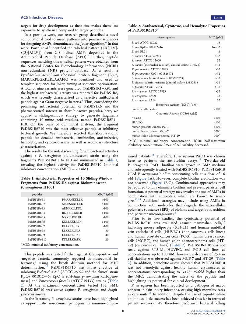
Table 2. Antibacterial, Cytotoxic, and Hemolytic Propertiesof PaDBS1R6F10a
microorganism MIC (μM)
E. coli ATCC 25922 16E. coli KpC+ 001812446 16−32E. coli BL21 4S. aureus ATCC 25923 >32S. aureus ATCC 12600 32S. aureus (methicillin resistant, clinical isolate 713623) >32K. pneumoniae ATCC 13883 >32K. pneumoniae KpC+ 001825971 >32A. baumannii (clinical isolate 003326263) >32E. cloacae colistin resistant (clinical isolate 1383251) >32E. faecalis ATCC 19433 4−8P. aeruginosa ATCC 27853 >32P. aeruginosa PAO1 32P. aeruginosa PA14 32
Hemolytic Activity (IC50) (μM)human erythrocytes >100
Cytotoxic Activity (IC50) (μM)3T3-L1 >100HUVECs >100human prostate cancer, PC-3 >100human breast cancer, MCF-7 100b
human colon adenocarcinoma, HT-29 100b
aMIC: minimal inhibitory concentration. IC50: half-maximuminhibitory concentration. b25% of cell viability decreased.
ACS Infectious Diseases Letter
DOI: 10.1021/acsinfecdis.9b00073ACS Infect. Dis. XXXX, XXX, XXX−XXX
B
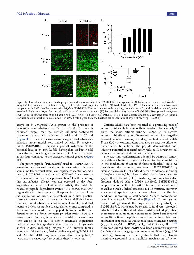
assays on P. aeruginosa PA14 grown in the presence ofincreasing concentrations of PaDBS1R6F10. The resultsobtained suggest that the peptide exhibited bactericidalproperties against this particular bacterial strain at 32 μM(Figure 1D). Further, in vivo assays using a scarification skininfection mouse model were carried out with P. aeruginosaPA14. PaDBS1R6F10 caused a gradual reduction of thebacterial load at 64 μM (2-fold higher than its bactericidalconcentration), reaching a maximum 103 CFU·mL−1 decreaseat day four, compared to the untreated control groups (Figure1E).The parent peptide (PaDBS1R6)8 used for PaDBS1R6F10
generation was recently evaluated in vivo using this sameanimal model, bacterial strain, and peptide concentration. As aresult, PaDBS1R6 caused a 103 CFU·mL−1 decrease inP. aeruginosa counts 2 days post-infection.8 On the contrary,this anti-infective efficacy was not observed at day four,suggesting a time-dependent in vivo activity that might berelated to peptide degradation events.8 It is known that AMPdegradation in animal models still represents a bottleneck forthe application of these antimicrobials in clinical practice.Here, we present a short, cationic, and linear AMP that has nochemical modifications to assist structural stability and thatseems to be less susceptible to degradation events compared toits 19-amino acid residue parent peptide (considering the time-dependent in vivo data). Interestingly, other studies have alsoshown similar findings, in which shorter AMPs present long-term effects in vivo due to higher enzymatic degradationresistance (less cleavage sites) when compared to larger well-known AMPs, including magainin and buforin familymembers.17 Nevertheless, further studies regarding PaDBS1R6and PaDBS1R6F10 enzymatic degradation susceptibility/resistance are encouraged to confirm these hypotheses.
Cationic AMPs have been reported as a promising class ofantimicrobial agents because of their broad-spectrum activity.18
Here, the short, cationic peptide PaDBS1R6F10 showedantimicrobial effects against Gram-positive and Gram-negativebacterial strains, including the drug-resistant clinical isolateE. coli KpC+ at concentrations that have no negative effects onhuman cells. In addition, the peptide demonstrated anti-infective potential as it significantly reduced P. aeruginosa cellcounts in a murine model of skin infections.The structural conformations adopted by AMPs in contact
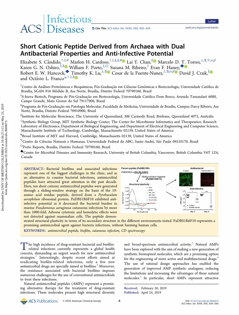
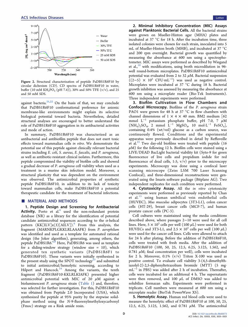
with different bacterial targets are known to play a crucial rolein the mechanism of action of these molecules.3 Here, weinvestigated the secondary structure of PaDBS1R6F10 bycircular dichroism (CD) under different conditions, includinghydrophilic (water/phosphate buffer), hydrophobic (water/2,2,2-trifluoroethanol (TFE) mixtures), and membrane-like(sodium dodecyl sulfate (SDS) micelles). PaDBS1R6F10adopted random coil conformations in both water and buffer,as well as a weak α-helical structure in TFE mixtures. However,a canonical spectral signature was observed in the lastcondition, indicating a well-defined β-sheet conformationwhen in contact with SDS micelles (Figure 2). Taken together,these findings reveal the high structural plasticity ofPaDBS1R6F10, which may be related to its diverse biologicalactivities. Indeed, other short cationic AMPs that adopt β-sheetconformations in an anionic environment have been reportedas multifunctional peptides, presenting antimicrobial andantibiofilm properties, as well as endotoxin neutralizing activity(e.g., (IRIK)2-NH2, (IRVK)3-NH2, and (VRVK)3-NH2).
19,20
Moreover, short β-sheet AMPs have been commonly reportedfor their ability to aggregate in anionic conditions (e.g., SDSmicelles), forming extended β-sheets that may triggermembrane-associated or intracellular mechanisms of action
Figure 1. Flow cell analysis, bactericidal properties, and in vivo activity of PaDBS1R6F10. P. aeruginosa PAO1 biofilms were stained and visualizedusing SYTO-9 to stain live biofilm cells (green, live cells) and propidium iodide (PI) (red, dead cells). PAO1 biofilm untreated controls werecompared with PAO1 biofilm treated with 16 μM of PaDBS1R6F10, and the dead cells only (A), live cells only (B), and dead/live cells (C) werevisualized. Scale bar = 20 μm for controls; scale bar = 30 μm for treatments. (D) Bactericidal activity in vitro of PaDBS1R6F10 against P. aeruginosaPA14 at doses ranging from 0 to 64 μM (*p > 0.01 for 64 to 8 μM). (E) PaDBS1R6F10 in vivo activity against P. aeruginosa PA14 using ascarification skin infection mouse model (64 μM, 2-fold higher than the bactericidal concentration) (*p > 0.01; ***p > 0.0001).
ACS Infectious Diseases Letter
DOI: 10.1021/acsinfecdis.9b00073ACS Infect. Dis. XXXX, XXX, XXX−XXX
C
against bacteria.21,22 On the basis of that, we may concludethat PaDBS1R6F10 conformational preference for anionicmembrane-like environments might explain its selectivebiological potential toward bacteria. Nevertheless, detailedstructural analyses are encouraged to better understand therole of PaDBS1R6F10 aggregation in its antibacterial activitiesand mode of action.In summary, PaDBS1R6F10 was characterized as an
antibacterial and antibiofilm peptide that does not exert toxiceffects toward mammalian cells in vitro. We demonstrate thepotential use of this peptide against clinically relevant bacterialstrains including E. coli, S. aureus, E. faecalis, and P. aeruginosa,as well as antibiotic-resistant clinical isolates. Furthermore, thispeptide compromised the viability of biofilm cells and showeda gradual decrease of P. aeruginosa cell viability when used as atreatment in a murine skin infection model. Moreover, astructural plasticity that was dependent on the environmentwas observed. The antimicrobial properties of syntheticpeptide PaDBS1R6F10, in addition to its lack of toxicitytoward mammalian cells, make PaDBS1R6F10 a potentialtherapeutic candidate for the treatment of bacterial infections.
■ MATERIAL AND METHODS1. Peptide Design and Screening for Antibacterial
Activity. Porto et al.6 used the non-redundant proteindatabase (NR) as a library for the identification of potentialcandidate antimicrobial sequences according to the α-helicalpattern (KK[ILV]x(3)[AILV]).6 From this analysis, thefragment (MARNKPLGKKLRLAAAFK) from P. aerophilumwas identified and used as a template for automated rationaldesign (the Joker algorithm), generating, among others, thepeptide PaDBS1R6.6,8 Here, PaDBS1R6 was used as templatefor a sliding-window strategy (window size = 10), whichgenerated ten synthetic variants (PaDBS1R6F1 toPaDBS1R6F10). These variants were initially synthesized inthe present study using the SPOT technology23 and submittedto initial antimicrobial/hemolytic screening, according toHilpert and Hancock.24 Among the variants, the tenthfragment (PaDBS1R6F10-KKLRLKIAFK) presented higherantimicrobial potential with MIC of 20 μM against abioluminescent P. aeruginosa strain (Table 1) and, therefore,was selected for further investigation. For this, PaDBS1R6F10was obtained from Peptide 2.0 Incorporated (USA), whichsynthesized the peptide at 95% purity by the stepwise solid-phase method using the N-9-fluorenylmethyloxycarbonyl(Fmoc) strategy on a Rink amide resin.
2. Minimal Inhibitory Concentration (MIC) Assaysagainst Planktonic Bacterial Cells. All the bacterial strainswere grown on Mueller-Hinton agar (MHA) plates andincubated at 37 °C for 18 h. After the incubation time, threeisolated colonies were chosen for each strain, inoculated into 5mL of Mueller-Hinton broth (MHB), and incubated at 37 °Cand 200 rpm overnight. Bacterial growth was quantified bymeasuring the absorbance at 600 nm using a spectropho-tometer. MIC assays were performed as described by Wiegandet al.,10 with modifications, using broth microdilution in 96-well round-bottom microplates. PaDBS1R6F10 antimicrobialpotential was evaluated from 2 to 32 μM. Bacterial suspension((2−5) × 105 CFU·mL−1) was used as negative control.Microplates were incubated at 37 °C during 18 h. Bacterialgrowth inhibition was assessed by measuring the absorbance at600 nm using a microplate reader (Bio-Tek Instruments).Three independent experiments were performed.
3. Biofilm Cultivation in Flow Chambers andConfocal Microscopy. Biofilms of the P. aeruginosa strainPAO1 were grown for 48 h at 37 °C in flow chambers withchannel dimensions of 1 × 4 × 40 mm. BM2 medium [62mmol L−1 potassium phosphate buffer, pH 7.0, 7 μM(NH4)2SO4, 2 mmol L−1 MgSO4, 10 mol·L−1 FeSO4],containing 0.4% (wt/vol) glucose as a carbon source, wascontinuously flowed. Conditions and the experimentalapparatus were previously described in detail by Reffuveilleet al.25 Two day-old biofilms were treated with peptide (16μM) for the following 12 h. Biofilm cells were stained using aLIVE/DEAD BacLight bacterial viability kit (Syto-9 for greenfluorescence of live cells and propidium iodide for redfluorescence of dead cells, 1:5, v/v) prior to the microscopyexperiments. Microscopy was done using a confocal laserscanning microscope (Zeiss LSM 700 Laser ScanningConfocal), and three-dimensional reconstructions were gen-erated using the Imaris software package (Bitplane AG). Twoindependent replicates for each condition were performed.
4. Cytotoxicity Assay. All the in vitro cytotoxicityexperiments were performed as previously reported by Chanet al.15 using human umbilical vein endothelial cells(HUVEC), Mus musculus adipocytes (3T3-L1), colon cancercells (HT-29), breast cancer cells (MCF-7), and humanprostate cancer cells (PC-3).Cell cultures were maintained using the media conditions
described above, where passages 2−10 were used for all celllines. Here, 5 × 10³ cells per well (100 μL) were used for bothHUVECs and 3T3-L1, and 2.5 × 10³ cells per well (100 μL)were used for the cancer cell lines. Cells were allowed to attachfor 24 h after plating. Before the addition of PaDBS1R6F10,cells were treated with fresh media. After the addition ofPaDBS1R6F10 (100, 50, 25, 12.5, 6.25, 3.125, 1.562, and0.781 μM; final concentration per well), cells were incubatedfor 2 h. Moreover, 0.1% (v/v) Triton X-100 was used aspositive control. To evaluate cell viability 3-(4,5-dimethylth-iazolyl-2)-2,5-diphenyltetrazolium bromide (MTT) (5 mg·mL−1 in PBS) was added after 2 h of incubation. Thereafter,cells were incubated for an additional 4 h. The supernatantswere then removed, and 100 μL of DMSO was added tosolubilize formazan salts. Experiments were performed intriplicate. Cell numbers were measured at 600 nm using amicroplate reader (BioTek PowerWave XS).
5. Hemolytic Assay. Human red blood cells were used tomeasure the hemolytic effect of PaDBS1R6F10 at 100, 50, 25,12.5, 6.25, 3.125, 1.562, and 0.781 μM. The antimicrobial
Figure 2. Structural characterization of peptide PaDBS1R6F10 bycircular dichroism (CD). CD spectra of PaDBS1R6F10 in water,buffer (10 mM KH2PO4 (pH 7.4)), 30% and 50% TFE (v/v), and 25and 50 mM SDS.
ACS Infectious Diseases Letter
DOI: 10.1021/acsinfecdis.9b00073ACS Infect. Dis. XXXX, XXX, XXX−XXX
D
peptide melittin was used as positive control following 2-folddilutions starting from 20 μM (final concentration per well). Inaddition, 1% (v/v) Triton X-100 and erythrocytes in PBS wereused as positive and negative controls, respectively. Sampleabsorbance was measured at 415 nm using a microplate reader(BioTek PowerWave XS). Experiments were performed intriplicates.6. Bacterial Killing Experiments. A killing experiment
was performed with 1:100 dilutions of overnight cultures ofP. aeruginosa PA14 in the absence or presence of increasingconcentrations of PaDBS1R6F10 (0−64 μM). After 24 h oftreatment, 10-fold serial dilutions were performed; bacteriawere plated on Pseudomonas Isolation Agar and allowed togrow overnight at 37 °C, after which colony-forming unit(CFU) counts were recorded. Three independent experimentswere performed.7. Scarification Skin Infection Mouse Model. P. aerugi-
nosa strain PA14 was grown to an optical density at 600 nm of0.5 in tryptic soy broth (TSB) medium. Subsequently, cellswere washed twice with sterile PBS (pH 7.4, 13 000 rpm for 1min) and resuspended to a final concentration of 5 × 106 CFU·20 μL−1. Skin infection was established according to Cardosoet al.26 One day after the infection, 64 μM of PaDBS1R6F10was administered to the infected area. Animals wereeuthanized 2 or 4 days postinfection, and the area of scarifiedskin (∼1 cm2) was excised and suspended in 1 mL of PBS. Theexcised skin sample was then homogenized using a bead beaterfor 20 min (25 Hz) and serially diluted for CFU quantification.Two independent experiments were performed with 4 mice pergroup in each condition. Statistical significance was assessedusing a one-way ANOVA, followed by Dunnett’s test. Animalswere maintained in accordance with the Guide for the Careand Use of Laboratory Animals in an AAALAC-accreditedfacility. All procedures were approved by the MIT’s Institu-tional Animal Care and Use Committee (IACUC), protocolnumber 1016-064-19.8. Structural Analysis. The PaDBS1R6F10 secondary
structure was characterized as described by Cardoso et al.,27
using a Jasco J-810 spectropolarimeter. PaDBS1R6F10 wasprepared at 50 μM in different solutions, including ultrapurewater, 10 mM KH2PO4 (pH 7.4), 2,2,2-trifluoroethanol (TFE)mixtures (30% and 50% (v/v) in water), and sodium dodecylsulfate (SDS) micelles (25 and 50 mM). Spectra wererecorded at room temperature in 0.1 cm path length quartzcells. Five scans were accumulated for each sample from 185 to260 nm at a scan speed of 50 nm·min−1.
■ AUTHOR INFORMATIONCorresponding Author*Phone: +55 061 34487120. E-mail: [email protected] H. Cardoso: 0000-0001-6676-5362Lai Y. Chan: 0000-0002-9346-2487Karen G. N. Oshiro: 0000-0002-8813-6433Evan F. Haney: 0000-0003-3645-770XTimothy K. Lu: 0000-0002-3918-8923Cesar de la Fuente-Nunez: 0000-0002-2005-5629David J. Craik: 0000-0003-0007-6796Octavio L. Franco: 0000-0001-9546-0525Present AddressαM.D.T.T. and C.F.-N.: Department of Psychiatry, andDepartment of Microbiology, Perelman School of Medicine,
University of Pennsylvania, Philadelphia, PA 19104, UnitedStatesAuthor Contributions#E.S.C. and M.H.C. contributed equally to this work. W.F.P.performed the in silico selection of peptides. S.R.M., E.F.H.,and R.E.W.H. performed the preliminary experiments withluminescent P. aeruginosa. E.S.C., M.H.C., and K.G.N.O.performed the antibacterial experiments. M.D.T.T., C.F.-N.,and T.K.L. performed the antibiofilm and in vivo scarificationexperiments. E.S.C., M.H.C., and L.Y.C. performed thestructural analyses. E.S.C. and M.H.C. wrote and organizedthe paper with input from all authors. D.J.C. and O.L.F.supervised and reviewed this work. All authors reviewed thispaper.NotesThe authors declare no competing financial interest.βM.D.T.T.: Department of Bioengineering, University ofPennsylvania, Philadelphia, PA 19104, US, United States
■ ACKNOWLEDGMENTSThis work was supported by grants from Coordenacao deAperfeicoamento de Pessoal de Nivel Superior (CAPES) (toM.H.C. 88881.134423/2016-01), Conselho Nacional deDesenvolvimento e Tecnolo gico (CNPq) (to M.H.C.141518/2015-4), Fundacao de Apoio ao Desenvolvimentodo Ensino, Ciencia e Tecnologia do Estado de Mato Grosso doSul (FUNDECT), Brazil, Fundacao de Apoio a Pesquisa doDistrito Federal (FAPDF), Ramon Areces Foundation (toC.F.-N.), DTRA (DTRA HDTRA1-15-1-0050) (to T.K.L.),and Fundacao de Amparo a Pesquisa do Estado de Sao Paulo(M.D.T.T. # 2014/04507-5 and 2016/24413-0). D.J.C. is anARC Australian Laureate Fellow (FL150100146). L.Y.C. wassupported by the Advance Queensland Women’s AcademicFund (WAF-6884942288). R.E.W.H. holds a Canada ResearchChair. We thank Robin Kramer for her assistance with the skinscarification mouse model.
■ ABBREVIATIONSAMPs, antimicrobial peptides; KpC, Klebsiella pneumoniaecarbapenemase; TFE, 2,2,2-trifluoroethanol; SDS, sodiumdodecyl sulfate; MIC, minimal inhibitory concentration;HUVEC, human umbilical vein endothelial cells; 3T3-L1,mouse adipocyte; PC-3, human prostate cancer cells; MCF-7,human breast cancer cells; HT-29, human colon adenocarci-noma cells
■ REFERENCES(1) Haney, E. F., Brito-Sanchez, Y., Trimble, M. J., Mansour, S. C.,Cherkasov, A., and Hancock, R. E. W. (2018) Computer-aideddiscovery of peptides that specifically attack bacterial biofilms. Sci.Rep. 8, 1871.(2) Koo, H., Allan, R. N., Howlin, R. P., Stoodley, P., and Hall-Stoodley, L. (2017) Targeting microbial biofilms: current andprospective therapeutic strategies. Nat. Rev. Microbiol. 15, 740−755.(3) Lee, T. H., Hall, K. N., and Aguilar, M. I. (2015) Antimicrobialpeptide structure and mechanism of action: A focus on the role ofmembrane structure. Curr. Top. Med. Chem. 16, 25−39.(4) Wiradharma, N., Sng, M. Y., Khan, M., Ong, Z. Y., and Yang, Y.Y. (2013) Rationally designed α-helical broad-spectrum antimicrobialpeptides with idealized facial amphiphilicity. Macromol. RapidCommun. 34, 74−80.(5) Fjell, C. D., Hiss, J. A., Hancock, R. E., and Schneider, G. (2012)Designing antimicrobial peptides: form follows function. Nat. Rev.Drug Discovery 11, 37−51.
ACS Infectious Diseases Letter
DOI: 10.1021/acsinfecdis.9b00073ACS Infect. Dis. XXXX, XXX, XXX−XXX
E
(6) Porto, W. F., Fensterseifer, I. C. M., Ribeiro, S. M., and Franco,O. L. (2018) Joker: An algorithm to insert patterns into sequences fordesigning antimicrobial peptides. Biochim. Biophys. Acta, Gen. Subj.1862, 2043−2052.(7) Wang, G., Li, X., and Wang, Z. (2016) APD3: the antimicrobialpeptide database as a tool for research and education. Nucleic AcidsRes. 44, D1087−1093.(8) Fensterseifer, I. C. M., Felicio, M. R., Alves, E. S. F., Cardoso, M.H., Torres, M. D. T., Matos, C. O., Silva, O. N., Lu, T. K., Freire, M.V., Neves, N. C., Goncalves, S., Liao, L. M., Santos, N. C., Porto, W.F., de la Fuente-Nunez, C., and Franco, O. L. (2019) Selectiveantibacterial activity of the cationic peptide PaDBS1R6 against Gram-negative bacteria. Biochim. Biophys. Acta, Biomembr., DOI: 10.1016/j.bbamem.2019.03.016.(9) Micek, S. T., Hampton, N., and Kollef, M. (2018) Risk factorsand outcomes for ineffective empiric treatment of sepsis caused bygram-negative pathogens: Stratification by onset of infection.Antimicrob. Agents Chemother. 62, e01577−01517.(10) Wiegand, I., Hilpert, K., and Hancock, R. E. (2008) Agar andbroth dilution methods to determine the minimal inhibitoryconcentration (MIC) of antimicrobial substances. Nat. Protoc. 3,163−175.(11) El Chakhtoura, N. G., Saade, E., Iovleva, A., Yasmin, M.,Wilson, B., Perez, F., and Bonomo, R. A. (2018) Therapies formultidrug resistant and extensively drug-resistant non-fermentinggram-negative bacteria causing nosocomial infections: a perilousjourney toward ’molecularly targeted’ therapy. Expert Rev. Anti-Infect.Ther. 16, 89−110.(12) de la Fuente-Nunez, C., Korolik, V., Bains, M., Nguyen, U.,Breidenstein, E. B., Horsman, S., Lewenza, S., Burrows, L., andHancock, R. E. (2012) Inhibition of bacterial biofilm formation andswarming motility by a small synthetic cationic peptide. Antimicrob.Agents Chemother. 56, 2696−2704.(13) Ribeiro, S. M., de la Fuente-Nunez, C., Baquir, B., Faria-Junior,C., Franco, O. L., and Hancock, R. E. (2015) Antibiofilm peptidesincrease the susceptibility of carbapenemase-producing Klebsiellapneumoniae clinical isolates to β-lactam antibiotics. Antimicrob. AgentsChemother. 59, 3906−3912.(14) de la Fuente-Nunez, C., Cardoso, M. H., de Souza Candido, E.,Franco, O. L., and Hancock, R. E. (2016) Synthetic antibiofilmpeptides. Biochim. Biophys. Acta, Biomembr. 1858, 1061−1069.(15) Chan, L. Y., Zhang, V. M., Huang, Y. H., Waters, N. C., Bansal,P. S., Craik, D. J., and Daly, N. L. (2013) Cyclization of theantimicrobial peptide gomesin with native chemical ligation:influences on stability and bioactivity. ChemBioChem 14, 617−624.(16) Sousa Dominguez, A., Perez-Rodriguez, M. T., Nodar, A.,Martinez-Lamas, L., Perez-Landeiro, A., and Crespo Casal, M. (2016)Successful treatment of MDR Pseudomonas aeruginosa skin and soft-tissue infection with ceftolozane/tazobactam. J. Antimicrob. Chemo-ther. 72, 1262−1263.(17) Kim, H., Jang, J. H., Kim, S. C., and Cho, J. H. (2014) De novogeneration of short antimicrobial peptides with enhanced stability andcell specificity. J. Antimicrob. Chemother. 69, 121−132.(18) Bormann, N., Koliszak, A., Kasper, S., Schoen, L., Hilpert, K.,Volkmer, R., Kikhney, J., and Wildemann, B. (2017) A short artificialantimicrobial peptide shows potential to prevent or treat boneinfections. Sci. Rep. 7, 1506.(19) Ong, Z. Y., Gao, S. J., and Yang, Y. Y. (2013) Short synthetic β-sheet forming peptide amphiphiles as broad spectrum antimicrobialswith antibiofilm and endotoxin neutralizing capabilities. Adv. Funct.Mater. 23, 3682−3692.(20) Zhong, G., Cheng, J., Liang, Z. C., Xu, L., Lou, W., Bao, C.,Ong, Z. Y., Dong, H., Yang, Y. Y., and Fan, W. (2017) Short syntheticβ-sheet antimicrobial peptides for the treatment of multidrug-resistantPseudomonas aeruginosa burn wound infections. Adv. HealthcareMater. 6, 1601134.(21) Manzo, G., Scorciapino, M. A., Wadhwani, P., Burck, J.,Montaldo, N. P., Pintus, M., Sanna, R., Casu, M., Giuliani, A., Pirri,G., Luca, V., Ulrich, A. S., and Rinaldi, A. C. (2015) Enhanced
amphiphilic profile of a short β-stranded peptide improves itsantimicrobial activity. PLoS One 10, No. e0116379.(22) Phambu, N., Almarwani, B., Garcia, A. M., Hamza, N. S.,Muhsen, A., Baidoo, J. E., and Sunda-Meya, A. (2017) Chain lengtheffect on the structure and stability of antimicrobial peptides of the(RW)n series. Biophys. Chem. 227, 8−13.(23) Winkler, D. F., Hilpert, K., Brandt, O., and Hancock, R. E.(2009) Synthesis of peptide arrays using SPOT-technology and theCelluSpots-method. Methods Mol. Biol. 570, 157−174.(24) Hilpert, K., and Hancock, R. E. (2007) Use of luminescentbacteria for rapid screening and characterization of short cationicantimicrobial peptides synthesized on cellulose using peptide arraytechnology. Nat. Protoc. 2, 1652−1660.(25) Reffuveille, F., de la Fuente-Nunez, C., Mansour, S., andHancock, R. E. (2014) A broad-spectrum antibiofilm peptideenhances antibiotic action against bacterial biofilms. Antimicrob.Agents Chemother. 58, 5363−5371.(26) Cardoso, M. H., Candido, E. S., Chan, L. Y., Torres, M. T.,Oshiro, K. G. N., Rezende, S. B., Porto, W. F., Lu, T. K., de la Fuente-Nunez, C., Craik, D. J., and Franco, O. L. (2018) A computationallydesigned peptide derived from Escherichia coli as a potential drugtemplate for antibacterial and antibiofilm therapies. ACS Infect. Dis. 4,1727−1736.(27) Cardoso, M. H., Ribeiro, S. M., Nolasco, D. O., de la Fuente-Nunez, C., Felicio, M. R., Goncalves, S., Matos, C. O., Liao, L. M.,Santos, N. C., Hancock, R. E., Franco, O. L., and Migliolo, L. (2016)A polyalanine peptide derived from polar fish with anti-infectiousactivities. Sci. Rep. 6, 21385.
ACS Infectious Diseases Letter
DOI: 10.1021/acsinfecdis.9b00073ACS Infect. Dis. XXXX, XXX, XXX−XXX
F