Novel Nuclear Factor-KappaB Targeting Peptide Suppresses β ...
22
RESEARCH ARTICLE Novel Nuclear Factor-KappaB Targeting Peptide Suppresses β-Amyloid Induced Inflammatory and Apoptotic Responses in Neuronal Cells Mythily Srinivasan 1 *, Baindu Bayon 2 , Nipun Chopra 2 , Debomoy K. Lahiri 2 1 School of Dentistry, Indiana University–Purdue University Indianapolis, Indianapolis, Indiana, United States of America, 2 Institute of Psychiatry Research, Department of Psychiatry and Medical & Molecular Genetics, School of Medicine, Indiana University–Purdue University Indianapolis, Indianapolis, Indiana, United States of America * [email protected] Abstract In the central nervous system (CNS), activation of the transcription factor nuclear factor- kappa B (NF-κβ) is associated with both neuronal survival and increased vulnerability to apoptosis. The mechanisms underlying these dichotomous effects are attributed to the composition of NF-κΒ dimers. In Alzheimer’s disease (AD), β-amyloid (Aβ) and other aggregates upregulate activation of p65:p50 dimers in CNS cells and enhance transactiva- tion of pathological mediators that cause neuroinflammation and neurodegeneration. Hence selective targeting of activated p65 is an attractive therapeutic strategy for AD. Here we report the design, structural and functional characterization of peptide analogs of a p65 interacting protein, the glucocorticoid induced leucine zipper (GILZ). By virtue of binding the transactivation domain of p65 exposed after release from the inhibitory IκΒ proteins in activated cells, the GILZ analogs can act as highly selective inhibitors of activated p65 with minimal potential for off-target effects. 1. Introduction Anaccumulatingbodyofevidencesuggeststhatacombinationofagerelatedchangesinthe centralnervoussystem(CNS)withexcessiveorprolongedinflammatoryresponsescontribute tothepathophysiologyofneurodegeneration,synapticdysfunctionandhippocampalbehavior deficitsinconditionssuchasAlzheimer'sdisease(AD)[1, 2].Thepleiotropictranscriptionfac- tor,nuclearfactor-kappaB(NF-κΒ)isinducedbymanyphysiologicalandpathologicalstimuli intheCNS[2–4].TheNF-κΒfamilyconsistsoffivemembers,p50,c-rel,p65,RelBandp52 thatcandiverselycombinetoformtranscriptionallyactivedimers.Ithasbeensuggestedthat thenatureofthedimersdeterminetheeffectsofactivatedNF-κΒ.Whilec-relcontaining dimerspreferentiallypromotetransactivationofanti-apoptoticfactors,activationofp65/p50 PLOS ONE | DOI:10.1371/journal.pone.0160314 October 20, 2016 1 / 22 a11111 OPEN ACCESS Citation: Srinivasan M, Bayon B, Chopra N, Lahiri DK (2016) Novel Nuclear Factor-KappaB Targeting Peptide Suppresses β-Amyloid Induced Inflammatory and Apoptotic Responses in Neuronal Cells. PLoS ONE 11(10): e0160314. doi:10.1371/journal.pone.0160314 Editor: Xing-Ming Shi, Georgia Regents University, UNITED STATES Received: January 11, 2016 Accepted: July 18, 2016 Published: October 20, 2016 Copyright: © 2016 Srinivasan et al. This is an open access article distributed under the terms of the Creative Commons Attribution License, which permits unrestricted use, distribution, and reproduction in any medium, provided the original author and source are credited. Data Availability Statement: All relevant data are within the paper and its Supporting Information files. Funding: We sincerely appreciate the grant support from the Alzheimer’s Association (IIRG-11- 206418) and the National Institute on Aging, NIH (5R21AG042804 and 1R21AG047) to DKL. We sincerely appreciate the grant supports from the Indiana CTSI to MS and DKL. Debomoy Lahiri received additional support from the following sources: Scientific advisory board- QR Pharma, Yuma Therapeutics, Entia Biosci-ences;
Transcript of Novel Nuclear Factor-KappaB Targeting Peptide Suppresses β ...
RESEARCH ARTICLE
Novel Nuclear Factor-KappaB Targeting
Peptide Suppresses β-Amyloid Induced
Inflammatory and Apoptotic Responses in
Neuronal Cells
Mythily Srinivasan1*, Baindu Bayon2, Nipun Chopra2, Debomoy K. Lahiri2
1 School of Dentistry, Indiana University–Purdue University Indianapolis, Indianapolis, Indiana, United
States of America, 2 Institute of Psychiatry Research, Department of Psychiatry and Medical & Molecular
Genetics, School of Medicine, Indiana University–Purdue University Indianapolis, Indianapolis, Indiana,
United States of America
Abstract
In the central nervous system (CNS), activation of the transcription factor nuclear factor-
kappa B (NF-κβ) is associated with both neuronal survival and increased vulnerability to
apoptosis. The mechanisms underlying these dichotomous effects are attributed to the
composition of NF-κΒ dimers. In Alzheimer’s disease (AD), β-amyloid (Aβ) and other
aggregates upregulate activation of p65:p50 dimers in CNS cells and enhance transactiva-
tion of pathological mediators that cause neuroinflammation and neurodegeneration.
Hence selective targeting of activated p65 is an attractive therapeutic strategy for AD. Here
we report the design, structural and functional characterization of peptide analogs of a p65
interacting protein, the glucocorticoid induced leucine zipper (GILZ). By virtue of binding
the transactivation domain of p65 exposed after release from the inhibitory IκΒ proteins in
activated cells, the GILZ analogs can act as highly selective inhibitors of activated p65 with
minimal potential for off-target effects.
1. Introduction
An accumulating body of evidence suggests that a combination of age related changes in thecentral nervous system (CNS) with excessive or prolonged inflammatory responses contributeto the pathophysiology of neurodegeneration, synaptic dysfunction and hippocampal behaviordeficits in conditions such as Alzheimer's disease (AD) [1, 2]. The pleiotropic transcription fac-tor, nuclear factor-kappa B (NF-κΒ) is induced by many physiological and pathological stimuliin the CNS [2–4]. The NF-κΒ family consists of five members, p50, c-rel, p65, RelB and p52that can diversely combine to form transcriptionally active dimers. It has been suggested thatthe nature of the dimers determine the effects of activated NF-κΒ. While c-rel containingdimers preferentially promote transactivation of anti-apoptotic factors, activation of p65/p50
PLOS ONE | DOI:10.1371/journal.pone.0160314 October 20, 2016 1 / 22
a11111
OPENACCESS
Citation: Srinivasan M, Bayon B, Chopra N, Lahiri
DK (2016) Novel Nuclear Factor-KappaB Targeting
Peptide Suppresses β-Amyloid Induced
Inflammatory and Apoptotic Responses in
Neuronal Cells. PLoS ONE 11(10): e0160314.
doi:10.1371/journal.pone.0160314
Editor: Xing-Ming Shi, Georgia Regents University,
UNITED STATES
Received: January 11, 2016
Accepted: July 18, 2016
Published: October 20, 2016
Copyright: © 2016 Srinivasan et al. This is an open
access article distributed under the terms of the
Creative Commons Attribution License, which
permits unrestricted use, distribution, and
reproduction in any medium, provided the original
author and source are credited.
Data Availability Statement: All relevant data are
within the paper and its Supporting Information
files.
Funding: We sincerely appreciate the grant support
from the Alzheimer’s Association (IIRG-11-
206418) and the National Institute on Aging, NIH
(5R21AG042804 and 1R21AG047) to DKL. We
sincerely appreciate the grant supports from the
Indiana CTSI to MS and DKL. Debomoy Lahiri
received additional support from the following
sources: Scientific advisory board- QR Pharma,
Yuma Therapeutics, Entia Biosci-ences;

dimers primarily enhance inflammatory and pro-apoptotic gene transcription. Positive andnegative regulatorymechanisms maintain a balance between the neuroprotective c-rel dimersand the predominantly deleterious p65:p50 dimers in healthy CNS [2, 5, 6].In AD, secondary stimuli such as accumulating beta amyloid (Aβ) and oxidative stress
increase activation of p65:p50 dimers in glial cells [7]. Cleavage of amyloid precursor protein(APP) by beta site amyloid precursor protein cleaving enzyme-1 (BACE-1) is essential for Aβgeneration. The promoter region of human BACE-1 gene exhibits κΒ binding elements thatphysically interact with NF-κΒ p65 [8, 9]. Activation of NF-κΒ p65 increases endogenous BACE-1 transcription and consequent Aβ production [8, 10]. Increased presence of activated p65 andBACE-1 has been observedaround Aβ plaques in postmortemAD tissues [11–13]. ExtracellularAβpeptides predominantly activate p65:p50 dimers in glia and post-mitotic neurons and enhancetransactivation of inflammatory and pro-apoptotic genes [13–15]. Increased presence of IL-1β,IL-6, and TNF-α have been reported in the affected tissues, serum and CSF of AD patients [16,17]. Elevated Bax (proapoptotic) to Bcl-2 (anti-apoptotic) ratio have been observed in Aβ stimu-lated neuronal cells [18, 19]. A feed-back loop of excessive Aβ accumulation, NF-κΒ activation,cytotoxicity and more Aβ production culminate in neurodegeneration [20]. Conditional knockout of p65 has been shown to attenuate BACE-1 transcription and Aβ genesis in ADmice [10].Absence of p65 co-factors such as p300/CREB binding associated factor has been shown to medi-ate resistance to Aβ induced toxicity [21]. Thus, although neuronal p65 has been shown to con-tribute to the physiological functions of synapse formation and transmission, considerableevidence suggest that excessive activated p65 in the CNS lead to neurodegenerative pathology.Hence selective inhibition of activated p65 could suppress AD [2, 16].Structurally p65 has an amino terminal rel homology domain (RHD), a nuclear localization
sequence (NLS) masked by the κΒ inhibitory complex and a carboxy terminal transactivationdomain (TAD). The transactivation activity of p65 is mediated by interactions of the TADwith co-regulators and the basal transcriptionmachinery [22, 23]. Glucocorticoid induced leu-cine zipper (GILZ) is a p65 binding protein that sequesters activated p65 and inhibits transacti-vation of inflammatory and apoptotic factors [24, 25]. Mutational and binding analyseslocalized the interaction interface to the proline rich carboxy terminus of GILZ and the TAD ofp65 [26]. Molecular modeling suggested that the p65 binding domain of GILZ adopts a flexiblepolyproline type II (PPII) helical conformation that interacts with the highly conservedF534/F542 in p65-TAD [27].In recent years, considerable success has been achieved in the development of structurally
engineered peptide analogs of the binding epitope(s) of a protein as therapeutic leads [28, 29].The strategy is increasingly adopted in the design of mimics of proline rich motif that mediatetransient intermolecular interactions. The specificity of the interaction is determined by thenature of the proline rich binding domain interface [30, 31]. Here we investigated the efficacyof rationally designed peptide analogs of the p65-TAD binding region of GILZ to selectivelysequester activated p65. Structural and functional analyses suggest that select GILZ analog(GA) bind p65-TAD with optimum affinity, exhibit an estimated half minimal lethal dosecomparable to known peptide drugs and suppress Aβ1–42 induced cytotoxicity.
2. Materials and Methods
Peptides and reagents
All GILZ peptides were synthesized as peptide amides with amino-terminal acetylation (Gene-script, Piscataway, NJ) at 95% purity as confirmedby mass spectrometry. To facilitate intracellulardelivery the GA were either co-synthesizedwith the cell penetrating agent, TAT (transactivator oftranscription) peptide or used as covalent mixture with Pep-1 chariot peptide (Anaspec, Fremont,
Inflammation, Neurodegeneration and NF-κB
PLOS ONE | DOI:10.1371/journal.pone.0160314 October 20, 2016 2 / 22
International advisory board- Drug Discovery and
Therapy World Congress; Stock options, QR
Pharma; Patent involving AIT-082, memantine and
pending on acamprosate; Editor in Chief, Current
Alzheimer Research; Research Grant Support from
Baxter Healthcare. Mythily Srinivasan: Co-Founder,
Provaidya LLC., Indianapolis and patent involving
GILZ analogs. The funders had no role in study
design, data collection and analysis, decision to
publish, or preparation of the manuscript.
Competing Interests: The authors have the
following interests: Debomoy Lahiri: Scientific
advisory board- QR Pharma, Yuma Therapeutics,
Entia Biosci-ences; International advisory board-
Drug Discovery and Therapy World Congress;
Stock options, QR Pharma; Patent involving AIT-
082, memantine and pending on acamprosate;
Editor in Chief, Current Alzheimer Research; and
Research Grant Support from Baxter Healthcare.
Mythily Srinivasan: Co-Founder, Provaidya LLC.,
Indianapolis and patent involving GILZ analogs.
This does not alter our adherence to PLOS ONE
policies on sharing data and materials.
CA). Recombinant human p65 protein (r-p65) with DDK tag (catalog number TP320780), puri-fied recombinant human GILZ protein (r-GILZ) with GST tag and biotinylated anti-DDK anti-bodywere fromOri-GeneTechnologies Inc., Rockville,MD. PurifiedAβ1–42 peptidewaspurchased fromAmerican Peptide company (American peptide company, Sunnyvale, CA: Prod-uct # 62-0-80 Lot # 1310160T). Aβ1–42 peptide stock (1 mg/mL) was prepared in cell culturemedium and incubated at 37°C for 24h prior to use in cell cultures [32].
Comparative modeling
Models of human GILZ and its mimics were built by the CPHmodels and Geno3D serversusing delta sleep inducing peptide (DSIP-PDB:1DIP) as template based on> 90% sequencesimilarity[33].While the Geno3D system builds models based on ‘topology mapping, the CPHsystem uses profile-based alignment as seed for developing energy-minimizedhomologymodel [34, 35]. The secondary structure assignment of the GILZmodels was independentlyassessed by the PROSS (Protein dihedral angle-based Secondary Structure assignment) pro-gram[36]. Superimposition of the model of each GILZmimic with experimentally determinedPPII helix and wild type GILZ determined the similarity between the structures in terms of rootmean square deviation (RMSD). Homology models of p65-TAD was developed similarly usingelongation factor eEF3 (PDB:3H7H) as template with which it shares 42% sequence similarity[37].
GILZ:p65-TAD docking
Models of human GILZ or GILZmimic and the p65-TAD were applied as probe and targetrespectively in PatchDock, a geometry based algorithm that yields docked transformationsscored on the basis of molecular shape complementarity and atomic desolvation energy [38–40]. Top one thousand solutions were refined using FireDock (Fast Interaction Refinement inmolecular docking), a program that optimizes binding of the probe by restricting side-chainflexibility to clashing interface residues. The refined docking solutions were scored based onsoftened van derWaals interactions, atomic contact energy, electrostatic and additional bind-ing free energy estimations. The top ranked solutions so obtained were further screened usingChimera for interatomic distance of<5Å between the residues of GILZmimic and the func-tionally critical residues of p65-TAD [41]. The solution with most contacts was further refinedby FlexPepDock using the wild type GILZ:p65-TAD complex with greater than 50% intermo-lecular residue contacts as reference.
Binding of GILZ and human r-p65
Previously we observed that the r-GILZ exhibits ten times higher affinity than a 22 residueGILZ peptide for r-p65 [27]. We used similar method to determine direct binding kinetics ofGA hexapeptides with human r-p65. High binding ELISA plates were coated with increasingconcentrations of r-GILZ (0.5μM to 20μM) or each GA or control peptide (20μM-640μM) andprobed with 80μM r-p65 followed by detectionwith anti-DDK antibody. Absorbance at 650nmwas measured with a mixing time of 30s using BIORADmicroplate reader. Percent bound p65was determined considering the binding response of r-GILZ (20μM) with r-p65 as 100%. Thedissociation constant of the interaction between the GA or the control peptide and r-p65 wasdetermined by the method of Friguet et al., as described [42, 43]. A fraction of the bound r-p65(x) and the ratio of bound r-p65 to the free GA or control peptide (y) was determined by theequations: x = (Ao − A)/(Ao), where Ao is the absorbance of r-p65�anti-p65 complex in theabsence of bound GA or control peptide and y = (Ao−A/Ao)/(ao− io)×(Ao − A/Ao), where ao is
Inflammation, Neurodegeneration and NF-κB
PLOS ONE | DOI:10.1371/journal.pone.0160314 October 20, 2016 3 / 22

the total concentration of GA or control peptide and io is the total concentration of r-p65. KD
for the interaction was determined by the Scatchard equation: x = 1 + KD/y.
Cell Titer-Glo (CTG) luminescent cell viability assay
Human neuroblastoma (SK-N-SH) cells cultured in minimal essential medium (MEM) supple-mented with 1% fetal bovine serum (FBS) and 1% penicillin (100U/ml)/streptomycin (100μg/ml) were differentiated with 10 μM all-trans retinoic acid for 7 days [32, 44]. After resting for24h in the low serummedium, the cells were seeded in 24 well (105cells/well) culture plates infresh medium and incubated with 50μM or 500μM of individual GA or control peptide in non-covalent mixture with Pep-1 or Pep-1 alone for additional 24h. The cultures were then photo-graphed using a phase contrast Leicamicroscope (LeicaMicrosystems Inc, Buffalogrove, IL).Subsequently, the cells were harvested, lysed with lysis buffer (M-PER, Pierce) and the lysatewas assessed for metabolic activity using the luciferase based CTG assay (CTG, Promega,Madison,WI). Briefly, cell lysate (5μL) in 25μL of phosphate buffered saline was transferred toan opaque white 96-well plate, and then 30μL of CTG assay solution was added. The relativeluminescent signal (RLT) was quantified using a Glowmax luminometer (Promega).
Lactate dehydrogenase (LDH) assay
To detect direct GA-induced cell lysis we performed LDH release assays (RocheMolecularDiagnostics).Human glioblastoma (U373) cells were maintained in MEM supplemented with10% FBS and 1% penicillin (100U/ml)/streptomycin (100μg/ml) at 37°C in 5% CO2-humidifiedincubators and sub-cultured once or twice a week [32, 45]. Approximately 5 x 10⁴ U373 cells/well were cultured in 96-well plates in the presence of increasing concentrations of individualGA or control peptide from 0.5μM to 500μM. Cells treated with 2% triton-X 100 (SigmaAldrich, St. Luis, MO) for 10 minutes served as positive control. Untreated cells served as con-trols for spontaneous LDH-release. Specific LDH-release was calculated according to the fol-lowing formula: LDH-release % = 100×(GA or control peptide treated cells—untreated cells)/(positive control-untreated cells). The IC50 values were extrapolated by logarithmic estimation.The LD50 in mg/kg was predicted using the formula, Log (LD50) = 0.435x (log IC50) + 0.625[46].
Detection of apoptosis by flow cytometry
To further assess cell cytotoxicity, the apoptotic effects of individual GA or control peptide wasevaluated by the Annexin-V and propidium iodide (PI) dual staining method (Annexin-V-Fluos staining kit, Roche Diagnositics,Mannheim, Germany) [47]. As opposed to apoptoticcells, necrotic cells with ruptured cell membrane take up PI, the DNA binding dye. Thus, cellswhich take up both fluorochromes are a mixture of apoptotic and necrotic cells, whereas cellsthat exclude PI but bind Annexin V are (early) apoptotic cells. U373 cells cultured with varyingconcentrations of individual GA or control peptide (0.5μM to 50μM) for 24h were centrifugedand suspended in 100μl of Annexin V/PI labelling solution (20μl each of Annexin-V-Fluoslabelling reagent and PI in 1ml of binding buffer) for 15min at room temperature. After wash-ing the cells were resuspended in PBS:1% paraformaldehyde and analyzed using a FACScanflow cytometer (BD Biosciences, San Jose, CA).
Functional assays in human primary mixed brain cultures (HFB)
HFBwere prepared and cultured as described [48] (S1 and S2 Docs). Briefly, cells were culturedin Neurobasal medium (Invitrogen) without phenol red supplemented with 1xB27, 50mM
Inflammation, Neurodegeneration and NF-κB
PLOS ONE | DOI:10.1371/journal.pone.0160314 October 20, 2016 4 / 22
GlutaMAX, 1xantibiotic cocktail, 5ng/mL recombinant fibroblast growth factor 2 (bFGF)(Invitrogen), and 2μL/mLNormocin (InVivoGen, San Diego, CA, USA). Cells were countedand seeded onto poly-D-lysine (Sigma-Aldrich) coated 24-well plates (Corning, Lowell, MA,USA) at 1.5x10⁵ cells per well and maintained at 37°C in a 5% CO2 incubator. Half mediachanges were performed every 4th day of culture and morphologywas monitored via phasecontrast microscopy. Culture mediumwas removed from cells on day 17 (DIV17) and replacedwith Neurobasal mediumwith B27. Appropriate wells were then added vehicle, or individualGA or control peptide (at 40μM or highest LD50 concentration) covalently synthesized withTAT or carrier peptide alone for 30min followed by exposure to Aβ1–42 at a final concentrationof 10μM/ well and incubated for 4h or 48h. Cells and conditionedmedia were harvested andstored for further analysis. Relative ATP concentration was measured using the CTG kit (Pro-mega) [45]. Data is presented as ΔRLU = RLU of Aβ1–42 exposed cells–RLU unexposed cells.Conditionedmedia collectedwere assessed for specific cytokines using the OptEIA kits (BDBiosciences).
NF-κΒ assay
PrimaryHFB exposed to Aβ1–42 and treated with GA or CP as above was harvested at the endof 4h. Nuclear and cytoplasmic fractions were extracted using the CelLytic™ NuCLEAR™ExtractionKit (Sigma) followingmanufacturer’s protocol. Five microgram of nuclear extractswas incubated in a 96-well plate coated with oligonucleotides containing the NF-κΒ consensusnucleotide sequence (5’-GGGACTTTCC-3’).The activated NF-κΒ bound to DNA wasdetected by anti-p65 antibody followed by a peroxidase coupled secondary antibody and sub-strate using the TransAM kit protocol (Active Motif).Nuclear extracts of Raji cells was used asthe positive control [49].
Quantitative real-time polymerase chain reaction (RT-PCR)
PrimaryHFB exposed to Aβ1–42 and treated with GA or CP as above was harvested at the endof 24h. Total cellular RNA was isolated using Qiagen kit (Invitrogen, Carlsbad, CA) followingmanufacturer's protocol. Total RNA (2–4 μg) was reverse transcribedusing iScript cDNA syn-thesis kit (Biorad, CA). The concentration of the cDNA was measured at 260 and 280 nm bythe Gensys5 model UV-visible spectrophotometer (ThermoelectronicCorp.,CA). Real-timePCR was performed by using the SYBR green/ROX qPCRmaster mix (SABiosciences, Freder-ick, MD) according to manufacturer's recommendations on the CFX96 Touch™ Real-TimePCRDetection System (Biorad laboratories, Hercules, California, USA). Each reaction contains2×12.5 μl of SYBR greenMaster Mix, 1 μl of 10 μM of primers and 50 ng of the cDNA, to atotal volume of 25 μl. The thermal cycling conditions included an initial denaturation step at50°C for 2 min, 95°C for 3 min, 39 cycles at 95°C for 30 s, annealing temperature at 54°C for30 s and extension at 72°C for 30s. The primers are F-GAPDH-50-AAGGTGAAGGTCGGAGTC AAC-30; R-GAPDH:50-G GGGTCATTGATGGCAACAATA-3 0(102bp); F-IL-1β:50-AGCTG ATGGCCCTAAACAGA-3 0; R-IL-1β:50GGTCGGAGATTCGTAGCTGG-3 0 (89 bp);F-IL-6:50-AATTCGGTACATCCTCG ACGG-30; R-IL-6:50-CAGCTCTGGCTTGTTCCTCA-30 (260bp); F-Caspase3:50-CATGGAAG CGAATCAATGGACT -30; R-Caspase 3–50 TTCCCTGAGGTTTGCTGCAT-30 (165 bp); F-Caspase 9: 50-CACTTCCCCTGAAGACGAGTC-30 andR-Caspase 9: 50-CTGATGTGCTCACCTGGGAAA-30 (162 bp). The gene-specific thresholdcycle (Ct) for each sample (ΔCt) was corrected by subtracting the Ct for the housekeeping geneglyceraldehyde 3-phosphate dehydrogenase (GAPDH). Untreated controls were chosen as thereference samples, and the ΔCt for all experimental samples was subtracted by the ΔCt for thecontrol samples (ΔΔCt). The difference in each gene-specific threshold between the samples
Inflammation, Neurodegeneration and NF-κB
PLOS ONE | DOI:10.1371/journal.pone.0160314 October 20, 2016 5 / 22
from vehicle treated and GA or CP treated cells was determined to obtain the relative changein the specificmRNA. The magnitude of change in the mRNA was expressed as 2ΔΔCt. Eachmeasurement of a sample was conducted in duplicate.
Statistical analysis
Statistical significancewas assessed by one-way analysis of variance (ANOVA) followed byTuckey post hoc. A value of p< 0.05 was considered significant.
3. Results
Design and modeling of GILZ mimic
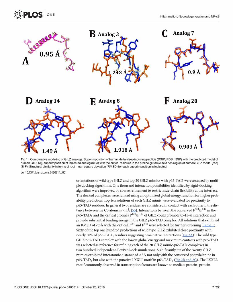
Strategies to determine the smallest biologically active fragment of a lead peptide involves trun-cation, deletion, alanine scanning and substitution of residues[29]. Deletion studies suggestedthat the amino terminal helix of GILZ is critical for dimerization but is not involved in theinteraction with NF-κΒ. Truncation mutants suggested that the residues spanning 120P-P127 ofthe GILZwere critical for GILZmediated inhibition of NF-κΒ transactivation [24, 26]. Molecu-lar modeling showed that the GILZ-COOHor a 22 residue peptide derived from the prolinerich region adopted an extended PPII helical conformation and interacted with the p65-TADexhibiting weak binding kinetics [27]. Alanine scanningmutagenesis suggested that the substi-tution of the 120PXXP123 motif abrogated the ability to inhibit NF-κΒ transactivation poten-tially due to loss of PPII conformation[24, 25]. In human and mouse GILZ, hydrogen bondingbetween the side chain of Ser or Thr and the backbone carbonyl of Glu or Pro respectivelycould contribute to the stability of PPII conformation [50]. We designed 40 GILZmimics byincorporating rational substitutions in the p65 binding motif of GILZ with residues thatincrease the propensity for PPII helical conformation and stabilize it. Comparative modelingwith substituted residues for all 40 GA was performed to obtain structural representation ofeach with reference to the adjacent residues of human GILZ. In addition we introduced confor-mational constraints by superimposing each GILZmimic model on structures with solved orexperimental PPII helix and wild type human GILZ to select for mimics with significant struc-tural homology (Fig 1). The PPII content of GILZmimics as determined by PROSS rangedfrom 14.3%, 28.6% and 42.9%. Since PPII helix formation is a locally driven event with little/noinvolvement of long-range interactions [51, 52], it is logical to presume that the synthetic GILZmimetic peptide with blocked end groups will adopt a similar conformation as in the predictedmodel. Twenty GILZmimics that exhibit near structural congruencewith DSIP (<1Å) or wildtype GILZ or experimentally determined PPII structure (<2Å) were selected for in silicodocking.
Docking of GILZ mimic and p65-TAD
To be of potential therapeutic value, the GILZmimic should adopt PPII helical conformationin the context of the critical binding residues in p65-TAD. The p65-TAD is commonly dividedinto two distinct regions, TAD-1521−551 consisting of 36 amino acids and TAD-2428−520 with 92residues [23]. It has been reported that the TAD1 accounts for nearly 95% of the transactivationpotential of full-length p65 and that the TAD2 alone is less potent mediating about 30% activa-tion [22, 53]. In particular, the highly conserved aromatic residues (F534, F542), acidic residues(D531,D533) and phosphorylation sites (Ser529,Ser536) in p65-TAD1 have been identified as criti-cal for transactivation [54].Homology model of p65-TAD was built using solution structure of the elongation factor
eEF3 (PDB: 2XI3, 2WI3) with which it shares 42% sequence similarity[37]. The spatial
Inflammation, Neurodegeneration and NF-κB
PLOS ONE | DOI:10.1371/journal.pone.0160314 October 20, 2016 6 / 22
orientations of wild type GILZ and top 20 GILZmimics with p65-TAD were assessed by multi-ple docking algorithms. One thousand interaction possibilities identified by rigid-dockingalgorithmwere improved by coarse refinement to restrict side-chain flexibility at the interface.The docked complexes were ranked using an optimized global energy function for higher prob-ability prediction. Top ten solutions of each GILZmimic were evaluated for proximity top65-TAD residues. In general two residues are considered in contact with each other if the dis-tance between the Cβ atoms is<5Å [55]. Interactions between the conservedF534/F542 in thep65-TAD1 and the critical prolines P120/P123 of GILZ could promote C–H��π interaction andprovide substantial binding energy in the GILZ:p65-TAD complex. All solutions that exhibitedan RMSD of<5Å with the critical F534 and F542 were selected for further screening (Table 1).Sixty of the top one hundred predictions of wild type GILZ exhibited close proximity withnearly 50% of p65-TAD1 residues suggesting near-native interactions (Fig 2A). The wild typeGILZ:p65-TAD complex with the lowest global energy and maximum contacts with p65-TADwas selected as reference for refining each of the 20 GILZmimic-p65TAD complexes intwo hundred independent FlexPepDock simulations. Significantly ten of the twenty GILZmimics exhibited interatomic distance of<5Å not only with the conservedphenylalanine inp65-TAD1 but also with the putative LXXLLmotif in p65-TAD2 (Fig 2B and 2C). The LXXLLmotif commonly observed in transcription factors are known to mediate protein–protein
Fig 1. Comparative modeling of GILZ analogs: Superimposition of human delta sleep inducing peptide (DSIP; PDB: 1DIP) with the predicted model of
human GILZ (A), superimposition of indicated analog (blue) with the critical residues in the proline glutamic acid rich region of human GILZ model (red)
(B-F). Structural similarity in terms of root mean square deviation (RMSD) for each superimposition is indicated.
doi:10.1371/journal.pone.0160314.g001
Inflammation, Neurodegeneration and NF-κB
PLOS ONE | DOI:10.1371/journal.pone.0160314 October 20, 2016 7 / 22
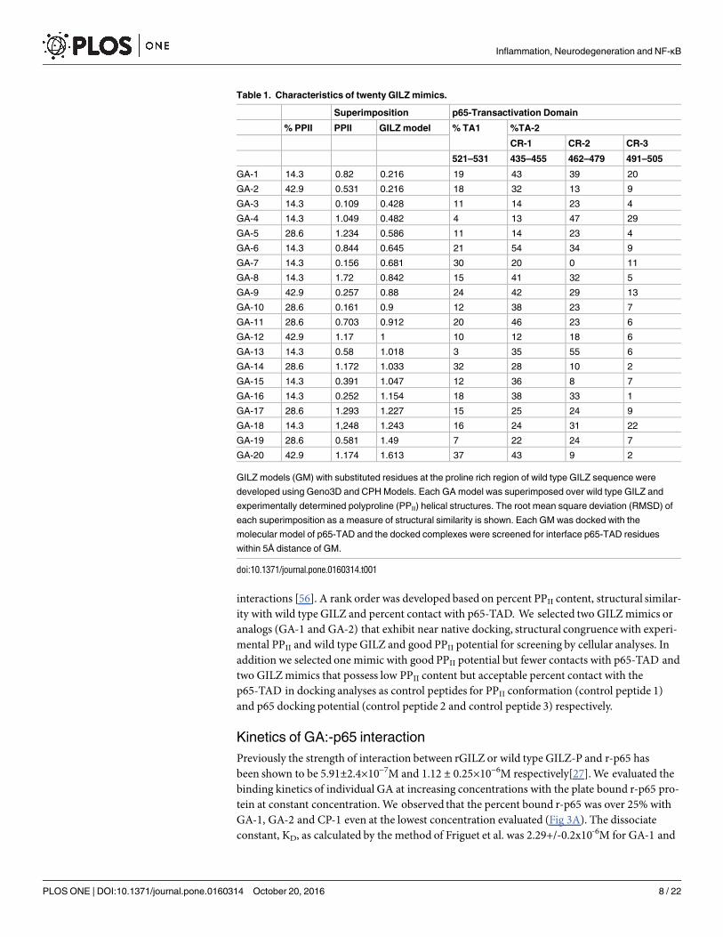
interactions [56]. A rank order was developed based on percent PPII content, structural similar-ity with wild type GILZ and percent contact with p65-TAD. We selected two GILZmimics oranalogs (GA-1 and GA-2) that exhibit near native docking, structural congruencewith experi-mental PPII and wild type GILZ and good PPII potential for screening by cellular analyses. Inaddition we selected one mimic with good PPII potential but fewer contacts with p65-TAD andtwo GILZmimics that possess low PPII content but acceptable percent contact with thep65-TAD in docking analyses as control peptides for PPII conformation (control peptide 1)and p65 docking potential (control peptide 2 and control peptide 3) respectively.
Kinetics of GA:-p65 interaction
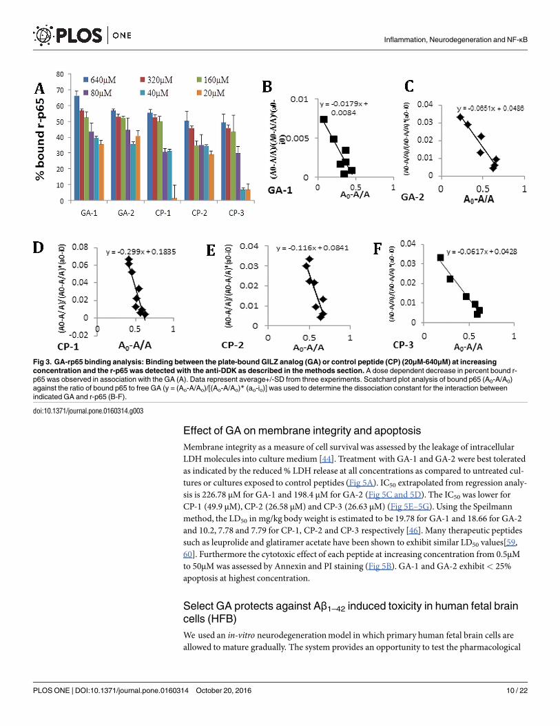
Previously the strength of interaction between rGILZ or wild type GILZ-P and r-p65 hasbeen shown to be 5.91±2.4×10⁻⁷M and 1.12 ± 0.25×10⁻⁶M respectively[27].We evaluated thebinding kinetics of individual GA at increasing concentrations with the plate bound r-p65 pro-tein at constant concentration.We observed that the percent bound r-p65 was over 25% withGA-1, GA-2 and CP-1 even at the lowest concentration evaluated (Fig 3A). The dissociateconstant, KD, as calculated by the method of Friguet et al. was 2.29+/-0.2x10-6M for GA-1 and
Table 1. Characteristics of twenty GILZ mimics.
Superimposition p65-Transactivation Domain
% PPII PPII GILZ model % TA1 %TA-2
CR-1 CR-2 CR-3
521–531 435–455 462–479 491–505
GA-1 14.3 0.82 0.216 19 43 39 20
GA-2 42.9 0.531 0.216 18 32 13 9
GA-3 14.3 0.109 0.428 11 14 23 4
GA-4 14.3 1.049 0.482 4 13 47 29
GA-5 28.6 1.234 0.586 11 14 23 4
GA-6 14.3 0.844 0.645 21 54 34 9
GA-7 14.3 0.156 0.681 30 20 0 11
GA-8 14.3 1.72 0.842 15 41 32 5
GA-9 42.9 0.257 0.88 24 42 29 13
GA-10 28.6 0.161 0.9 12 38 23 7
GA-11 28.6 0.703 0.912 20 46 23 6
GA-12 42.9 1.17 1 10 12 18 6
GA-13 14.3 0.58 1.018 3 35 55 6
GA-14 28.6 1.172 1.033 32 28 10 2
GA-15 14.3 0.391 1.047 12 36 8 7
GA-16 14.3 0.252 1.154 18 38 33 1
GA-17 28.6 1.293 1.227 15 25 24 9
GA-18 14.3 1,248 1.243 16 24 31 22
GA-19 28.6 0.581 1.49 7 22 24 7
GA-20 42.9 1.174 1.613 37 43 9 2
GILZ models (GM) with substituted residues at the proline rich region of wild type GILZ sequence were
developed using Geno3D and CPH Models. Each GA model was superimposed over wild type GILZ and
experimentally determined polyproline (PPII) helical structures. The root mean square deviation (RMSD) of
each superimposition as a measure of structural similarity is shown. Each GM was docked with the
molecular model of p65-TAD and the docked complexes were screened for interface p65-TAD residues
within 5Å distance of GM.
doi:10.1371/journal.pone.0160314.t001
Inflammation, Neurodegeneration and NF-κB
PLOS ONE | DOI:10.1371/journal.pone.0160314 October 20, 2016 8 / 22

3.24+/-0.19x10-6M for GA-2. The strength of interaction of the GA:p65-TAD binding is con-sistent with other transient protein:peptide interactions such as that of peptide inhibitors ofmatrix metalloproteinase and that of SMRT peptides binding the BTB domain of BCL6 [57,58]. The control peptides exhibited higher KD values of 3.27+/-1.8x10-6M, 3.4+/-0.15x10-6Mand 4.28+/-0.5 x10-6M for CP-1, CP-2 and CP-3 respectively (Fig 3B–3F).
Effect of GA on cellular morphology and metabolic activity
Any compound with potential therapeutic effect should be biocompatible and nontoxic. So, wefirst screened the effects of GA on cellular morphology and viability. Cultures of neuroblastsexposed to 50μM or 500μM of individual GA or CP-1 or CP-2 did not show any change in cellmorphology suggesting that the four peptides were not toxic. Exposure to CP-3 at either con-centration showed morphological changes consistent with cell death (Fig 4A). The effect of GAon cellular metabolic activity was assessed by the CTG assay which measures intracellular ATPconcentrations as an indicator of actual cell number. The results of the CTG assay were verysimilar and showed that the two GA and CP-1 and CP-2 did not adversely affect the differenti-ated neuroblastoma cells, but treatment with CP-3 reduced the viability of the cells at both con-centrations tested (Fig 4B).
Fig 2. The docked complex of wild type GILZ-PER (proline glutamic acid rich region) or GILZ analog (GA) with human p65-TAD. Representative
molecular model of p65-TAD docked (blue) with wild type GILZ-PER (A) and indicated analog (B-F) (red) are shown. The residues in each analog <5Ådistance of highly conserved residues in p65-TAD2 (Phe or Phe) and the “LXXLL” motif in p65-TAD1 that suggest proximity with residues critical for
transcriptional activity are shown.
doi:10.1371/journal.pone.0160314.g002
Inflammation, Neurodegeneration and NF-κB
PLOS ONE | DOI:10.1371/journal.pone.0160314 October 20, 2016 9 / 22
Effect of GA on membrane integrity and apoptosis
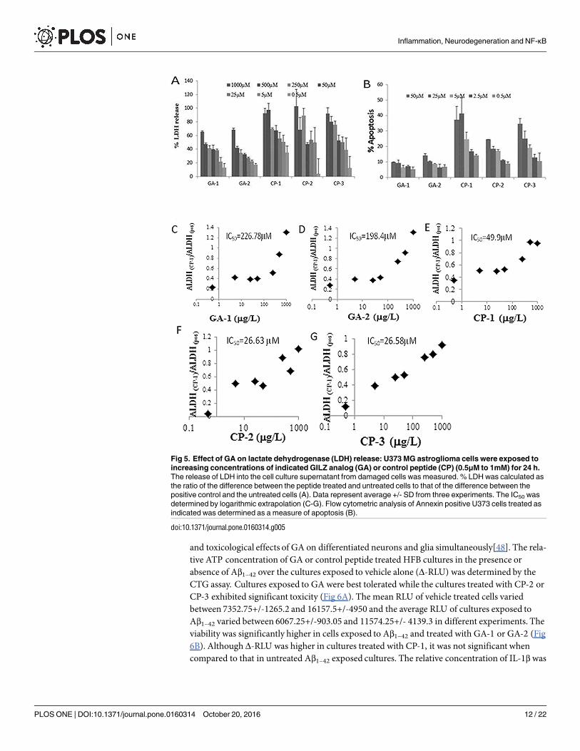
Membrane integrity as a measure of cell survival was assessed by the leakage of intracellularLDHmolecules into culture medium [44]. Treatment with GA-1 and GA-2 were best toleratedas indicated by the reduced% LDH release at all concentrations as compared to untreated cul-tures or cultures exposed to control peptides (Fig 5A). IC50 extrapolated from regression analy-sis is 226.78 μM for GA-1 and 198.4 μM for GA-2 (Fig 5C and 5D). The IC50 was lower forCP-1 (49.9 μM), CP-2 (26.58 μM) and CP-3 (26.63 μM) (Fig 5E–5G). Using the Speilmannmethod, the LD50 in mg/kg body weight is estimated to be 19.78 for GA-1 and 18.66 for GA-2and 10.2, 7.78 and 7.79 for CP-1, CP-2 and CP-3 respectively [46]. Many therapeutic peptidessuch as leuprolide and glatiramer acetate have been shown to exhibit similar LD50 values[59,60]. Furthermore the cytotoxic effect of each peptide at increasing concentration from 0.5μMto 50μM was assessed by Annexin and PI staining (Fig 5B). GA-1 and GA-2 exhibit< 25%apoptosis at highest concentration.
Select GA protects against Aβ1–42 induced toxicity in human fetal brain
cells (HFB)
We used an in-vitro neurodegenerationmodel in which primary human fetal brain cells areallowed to mature gradually. The system provides an opportunity to test the pharmacological
Fig 3. GA-rp65 binding analysis: Binding between the plate-bound GILZ analog (GA) or control peptide (CP) (20μM-640μM) at increasing
concentration and the r-p65 was detected with the anti-DDK as described in the methods section. A dose dependent decrease in percent bound r-
p65 was observed in association with the GA (A). Data represent average+/-SD from three experiments. Scatchard plot analysis of bound p65 (A0-A/A0)
against the ratio of bound p65 to free GA (y = (Ao-A/Ao)/[(Ao-A/Ao)* (ao-io)] was used to determine the dissociation constant for the interaction between
indicated GA and r-p65 (B-F).
doi:10.1371/journal.pone.0160314.g003
Inflammation, Neurodegeneration and NF-κB
PLOS ONE | DOI:10.1371/journal.pone.0160314 October 20, 2016 10 / 22
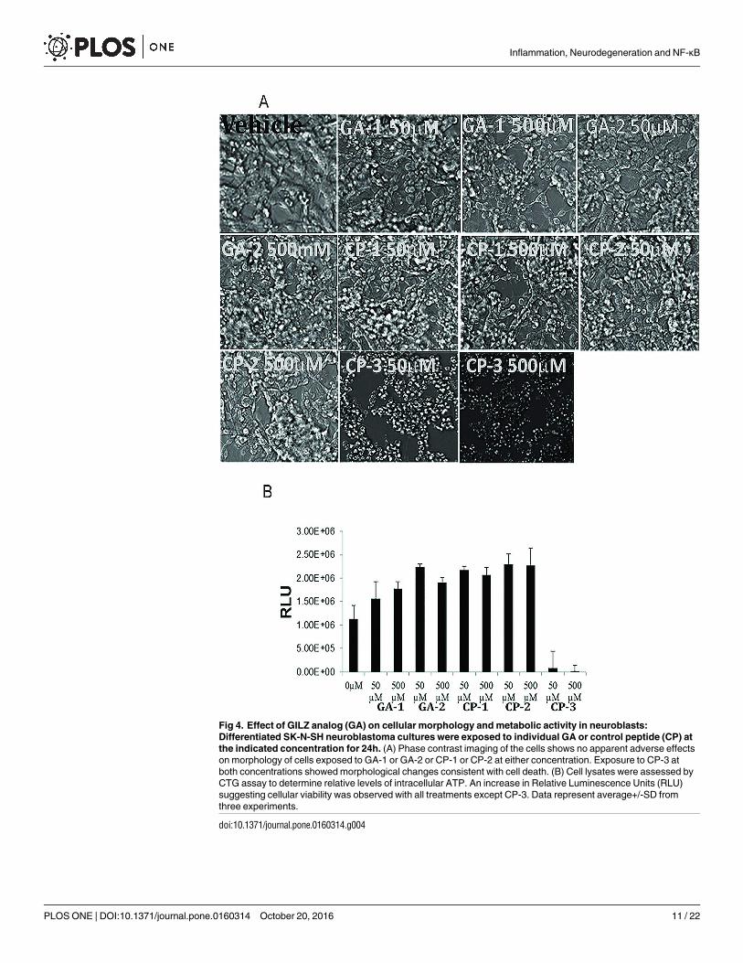
Fig 4. Effect of GILZ analog (GA) on cellular morphology and metabolic activity in neuroblasts:
Differentiated SK-N-SH neuroblastoma cultures were exposed to individual GA or control peptide (CP) at
the indicated concentration for 24h. (A) Phase contrast imaging of the cells shows no apparent adverse effects
on morphology of cells exposed to GA-1 or GA-2 or CP-1 or CP-2 at either concentration. Exposure to CP-3 at
both concentrations showed morphological changes consistent with cell death. (B) Cell lysates were assessed by
CTG assay to determine relative levels of intracellular ATP. An increase in Relative Luminescence Units (RLU)
suggesting cellular viability was observed with all treatments except CP-3. Data represent average+/-SD from
three experiments.
doi:10.1371/journal.pone.0160314.g004
Inflammation, Neurodegeneration and NF-κB
PLOS ONE | DOI:10.1371/journal.pone.0160314 October 20, 2016 11 / 22
and toxicological effects of GA on differentiated neurons and glia simultaneously[48]. The rela-tive ATP concentration of GA or control peptide treated HFB cultures in the presence orabsence of Aβ1–42 over the cultures exposed to vehicle alone (Δ-RLU) was determined by theCTG assay. Cultures exposed to GA were best tolerated while the cultures treated with CP-2 orCP-3 exhibited significant toxicity (Fig 6A). The mean RLU of vehicle treated cells variedbetween 7352.75+/-1265.2 and 16157.5+/-4950 and the average RLU of cultures exposed toAβ1–42 varied between 6067.25+/-903.05 and 11574.25+/- 4139.3 in different experiments. Theviability was significantly higher in cells exposed to Aβ1–42 and treated with GA-1 or GA-2 (Fig6B). Although Δ-RLU was higher in cultures treated with CP-1, it was not significant whencompared to that in untreated Aβ1–42 exposed cultures. The relative concentration of IL-1β was
Fig 5. Effect of GA on lactate dehydrogenase (LDH) release: U373 MG astroglioma cells were exposed to
increasing concentrations of indicated GILZ analog (GA) or control peptide (CP) (0.5μM to 1mM) for 24 h.
The release of LDH into the cell culture supernatant from damaged cells was measured. % LDH was calculated as
the ratio of the difference between the peptide treated and untreated cells to that of the difference between the
positive control and the untreated cells (A). Data represent average +/- SD from three experiments. The IC50 was
determined by logarithmic extrapolation (C-G). Flow cytometric analysis of Annexin positive U373 cells treated as
indicated was determined as a measure of apoptosis (B).
doi:10.1371/journal.pone.0160314.g005
Inflammation, Neurodegeneration and NF-κB
PLOS ONE | DOI:10.1371/journal.pone.0160314 October 20, 2016 12 / 22

lower in culture medium of cells exposed to Aβ1–42 and GA-1 (16.4 +/-3.3pg/ml) or GA-2 (30.5+/-7.9 pg/ml) treated cells as compared with control peptide treated (CP-1:21.7+/-5.03pg/ml,CP-2: 42.1+/-1.3pg/ml,CP-3: 47.8+/-4.4pg/ml) or untreated (36.1+/-5.6pg/ml) cells. SimilarlyIL-6 and IL-12 secretion was lower in GA-1 (0.9+/-1.4pg/ml and 61.6+/-32.2pg/ml respec-tively) or GA-2 (18.4+/-0.1pg/ml and 104.5+/-24.8pg/ml respectively) treated cells as com-pared with CP-1 (19.17+/-12.2pg/ml and 125.7+/-46.3pg/ml respectively), CP-2 (38.6+/-10.2pg/ml and 149.9+/-50.7pg/ml respectively), CP-3 (39.2+/-11.7pg/ml and 239.9+/-19.4pg/ml
Fig 6. Effect of GILZ analog (GA) treatment on human fetal brain cells (HFB): (A,B): Select GA decrease metabolic activity in Aβ1–42
exposed HFB. Primary cultures of HFB (Div 17) were exposed to Aβ1–42 and treated with 50M of indicated GA or control peptide (CP).
Cytoplasmic extracts of cells collected at 24h was assessed for viability by CTG assay. Data are presented as ΔRLU (difference in relative
luminescent units (RLU) between the Aβ1–42 exposed cells and unexposed cells) (A, B). GA suppresses inflammatory cytokines in activated
HFB. HFB cells were cultured as above and culture medium collected at 24hrs was assessed for pro-inflammatory (IL-1β, IL-6 and IL-17) (C,
D, E and F) and anti-inflammatory (TGF-β) (G) cytokines. (H) Effect of GA on NF-κΒ activation. Primary cultures of HFB exposed to Aβ1–42
(10μg/ml) and treated with indicated GA or CP as above were harvested at the end of 4 h. 5μg of nuclear extract was tested for binding of the
activated p65 NF-κΒ subunit to an NF-κΒ consensus sequence using the Trans AM NF-κΒ ELISA kit. The p65 DNA binding activity was
calculated as the ratio of absorbance from Aβ1–42 stimulated cells to that of unstimulated cells. Values are the average/-S.D from two
experiments. * = p<0.05 as compared to Aβ1–42 exposed cells, @ = p<0.05 as compared with Aβ1–42 and CP-1 or CP-2 treated cultures.
doi:10.1371/journal.pone.0160314.g006
Inflammation, Neurodegeneration and NF-κB
PLOS ONE | DOI:10.1371/journal.pone.0160314 October 20, 2016 13 / 22
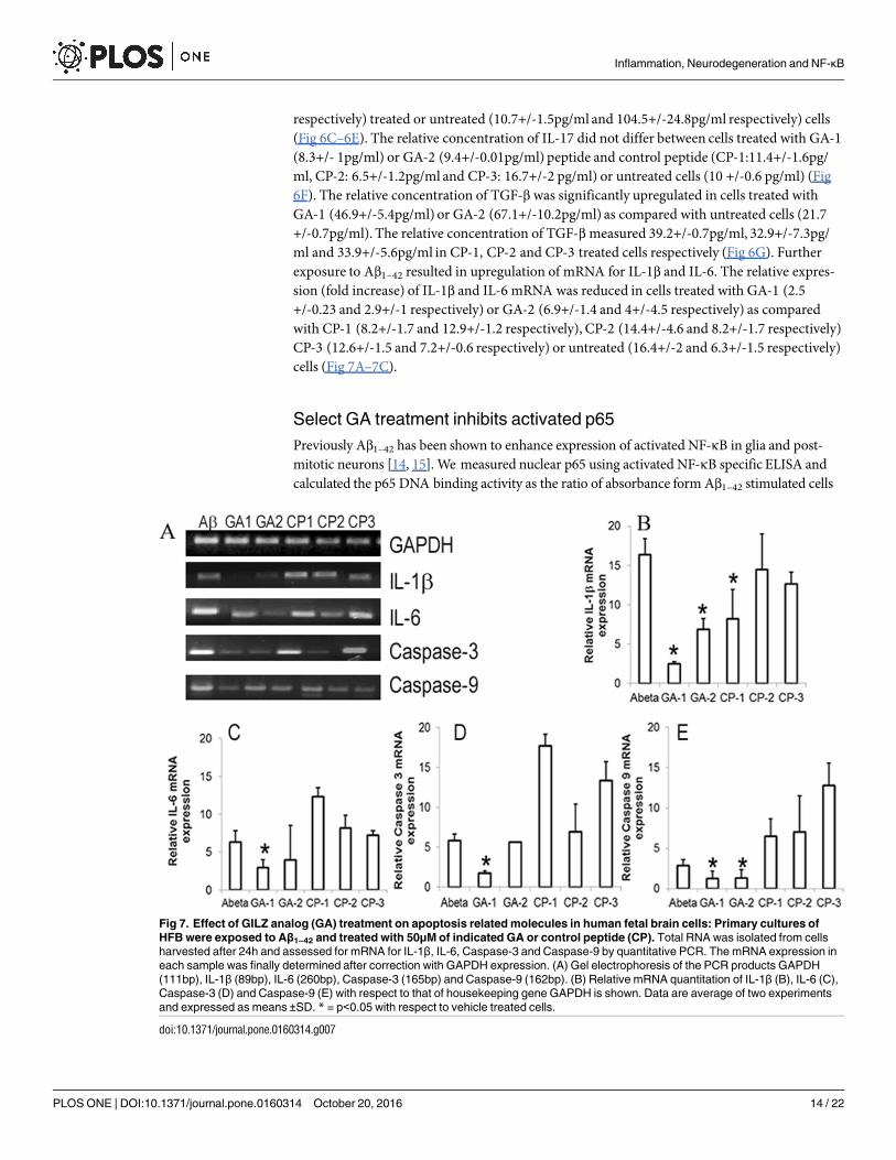
respectively) treated or untreated (10.7+/-1.5pg/ml and 104.5+/-24.8pg/ml respectively) cells(Fig 6C–6E). The relative concentration of IL-17 did not differ between cells treated with GA-1(8.3+/- 1pg/ml) or GA-2 (9.4+/-0.01pg/ml) peptide and control peptide (CP-1:11.4+/-1.6pg/ml, CP-2: 6.5+/-1.2pg/ml and CP-3: 16.7+/-2 pg/ml) or untreated cells (10 +/-0.6 pg/ml) (Fig6F). The relative concentration of TGF-β was significantly upregulated in cells treated withGA-1 (46.9+/-5.4pg/ml) or GA-2 (67.1+/-10.2pg/ml) as compared with untreated cells (21.7+/-0.7pg/ml). The relative concentration of TGF-βmeasured 39.2+/-0.7pg/ml, 32.9+/-7.3pg/ml and 33.9+/-5.6pg/ml in CP-1, CP-2 and CP-3 treated cells respectively (Fig 6G). Furtherexposure to Aβ1–42 resulted in upregulation of mRNA for IL-1β and IL-6. The relative expres-sion (fold increase) of IL-1β and IL-6 mRNA was reduced in cells treated with GA-1 (2.5+/-0.23 and 2.9+/-1 respectively) or GA-2 (6.9+/-1.4 and 4+/-4.5 respectively) as comparedwith CP-1 (8.2+/-1.7 and 12.9+/-1.2 respectively), CP-2 (14.4+/-4.6 and 8.2+/-1.7 respectively)CP-3 (12.6+/-1.5 and 7.2+/-0.6 respectively) or untreated (16.4+/-2 and 6.3+/-1.5 respectively)cells (Fig 7A–7C).
Select GA treatment inhibits activated p65
Previously Aβ1–42 has been shown to enhance expression of activated NF-κΒ in glia and post-mitotic neurons [14, 15]. We measured nuclear p65 using activated NF-κΒ specific ELISA andcalculated the p65 DNA binding activity as the ratio of absorbance form Aβ1–42 stimulated cells
Fig 7. Effect of GILZ analog (GA) treatment on apoptosis related molecules in human fetal brain cells: Primary cultures of
HFB were exposed to Aβ1–42 and treated with 50μM of indicated GA or control peptide (CP). Total RNA was isolated from cells
harvested after 24h and assessed for mRNA for IL-1β, IL-6, Caspase-3 and Caspase-9 by quantitative PCR. The mRNA expression in
each sample was finally determined after correction with GAPDH expression. (A) Gel electrophoresis of the PCR products GAPDH
(111bp), IL-1β (89bp), IL-6 (260bp), Caspase-3 (165bp) and Caspase-9 (162bp). (B) Relative mRNA quantitation of IL-1β (B), IL-6 (C),
Caspase-3 (D) and Caspase-9 (E) with respect to that of housekeeping gene GAPDH is shown. Data are average of two experiments
and expressed as means ±SD. * = p<0.05 with respect to vehicle treated cells.
doi:10.1371/journal.pone.0160314.g007
Inflammation, Neurodegeneration and NF-κB
PLOS ONE | DOI:10.1371/journal.pone.0160314 October 20, 2016 14 / 22
to that of unstimulated cells. Nuclear p65 was significantly higher in cells exposed to Aβ1–42(14.75+/-3.5) than in unexposed cells (2.5+/-1.7). There was a trend towards decreasednuclearp65 in Aβ1–42 exposed cells treated with GA-1 (6.13+/-1.3) and GA-2 (6.7+/-1.5) as compared tountreated cells (14.75+/-3.5). Nuclear p65 of Aβ1–42 exposed cells treated with CP-1, CP-2 andCP-3 measured 6.8+/-3.9, 10.2+/-2.9 and 10.1+/-3.7 respectively (Fig 6H). No significant differ-ence in nuclear p65 was observed in cells treated only with the peptides in the absence of expo-sure to Aβ1–42 (data not shown). Similar assessment showed no significant difference in thecytoplasmicNF-κΒ p65 betweenGA treated and untreated or CP treated cells (data not shown).Although we observed slightly elevated nuclear p50 in Aβ1–42 (6.15+/-1.2) than in unexposedcells (0.5+/-2.7), no differencewas observedbetweenGA peptide of CP treated cells anduntreated cells (data not shown).
Select GA protects against Aβ1–42 induced apoptosis related markers in
human fetal brain cells (HFB)
We next determined the message for apoptosis relevant molecules Caspase 3 and Caspase 9 byquantitative PCR. As shown in Fig 7, exposure to Aβ1–42 resulted in upregulation of mRNA forcaspase 3 and caspase 9 in HFB. The relative expression (fold increase) of IL-1β and caspase 9mRNA was reduced in cells treated with GA-1 (2.5+/-0.23 and 1.3+/-0.9 respectively) or GA-2(6.9+/-1.4 and 1.3+/-1 respectively) as compared with CP-1 (8.2+/-1.7 and 6.5 +/-2.2 respec-tively), CP-2 (14.4+/-4.6 and 7.1+/-4.5 respectively) CP-3 (12.6+/-1.5 and 12.8+/-2.8 respec-tively) or untreated (16.4+/-2 and 2.9+/-0.7 respectively) cells (Fig 7A, 7B and 7E). Further theIL-6 and Caspase 3 mRNA exhibited significant reduction in GA-1 (2.9+/-1 and 1.7+/-0.3respectively) treated cells as compared to that in CP-1 (12.9+/-1.2 and 17.7+/-1.5 respectively)treated or untreated (6.3+/-1.5 and 5.8+/-0.9 respectively) cells (Fig 7A, 7C and 7D).Comparative analysis of physical and functional characteristics of known receptor antago-
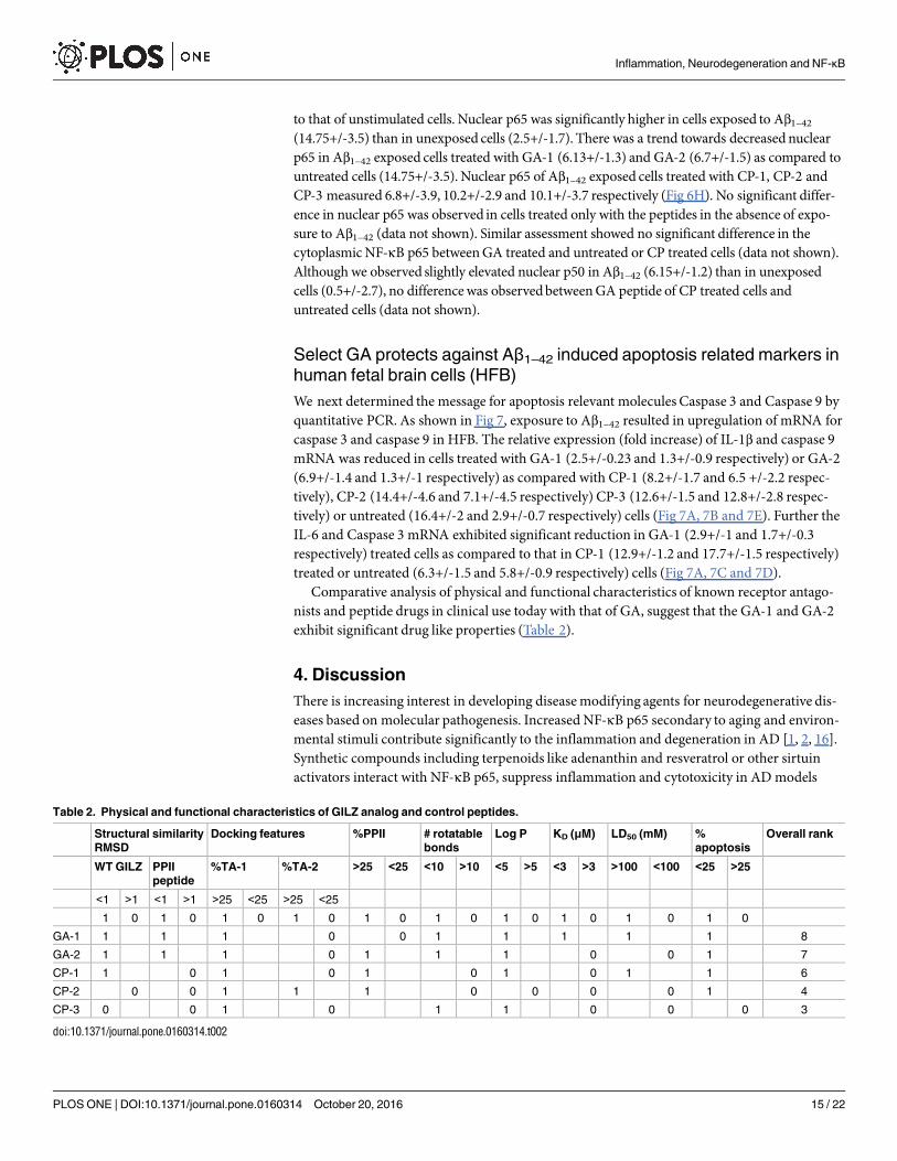
nists and peptide drugs in clinical use today with that of GA, suggest that the GA-1 and GA-2exhibit significant drug like properties (Table 2).
4. Discussion
There is increasing interest in developing diseasemodifying agents for neurodegenerative dis-eases based on molecular pathogenesis. Increased NF-κΒ p65 secondary to aging and environ-mental stimuli contribute significantly to the inflammation and degeneration in AD [1, 2, 16].Synthetic compounds including terpenoids like adenanthin and resveratrol or other sirtuinactivators interact with NF-κΒ p65, suppress inflammation and cytotoxicity in ADmodels
Table 2. Physical and functional characteristics of GILZ analog and control peptides.
Structural similarity
RMSD
Docking features %PPII # rotatable
bonds
Log P KD (μM) LD50 (mM) %
apoptosis
Overall rank
WT GILZ PPII
peptide
%TA-1 %TA-2 >25 <25 <10 >10 <5 >5 <3 >3 >100 <100 <25 >25
<1 >1 <1 >1 >25 <25 >25 <25
1 0 1 0 1 0 1 0 1 0 1 0 1 0 1 0 1 0 1 0
GA-1 1 1 1 0 0 1 1 1 1 1 8
GA-2 1 1 1 0 1 1 1 0 0 1 7
CP-1 1 0 1 0 1 0 1 0 1 1 6
CP-2 0 0 1 1 1 0 0 0 0 1 4
CP-3 0 0 1 0 1 1 0 0 0 3
doi:10.1371/journal.pone.0160314.t002
Inflammation, Neurodegeneration and NF-κB
PLOS ONE | DOI:10.1371/journal.pone.0160314 October 20, 2016 15 / 22
[61]. As opposed to high throughput screening, computational design of interface peptidemimics followed by functional evaluation represents an efficientmethod in the drug designand discovery process [28, 29]. Here we report the design and physicochemical characteristicsof peptide analogs of the GILZ:p65 interface, show that select analogs bind p65-TAD and sup-press Aβ induced toxicity in human fetal brain cells exhibiting potential therapeutic value forAD.In human interactome, preponderant transient intermolecular interactions are mediated by
proline rich epitope of one protein binding the aromatic residue rich flat interface of the secondprotein. Often the proline rich epitope adopts an extended PPII helical conformation that behavesas an adaptable glove in obtaining the correct binding orientation [31, 52]. Here it is pertinent tonote that the template basedmodeling suggested that the p65 binding domain of GILZ exhibits aPPII helical conformation and that the p65-TAD is unstructured or flat. Mutational analysesidentified 120PEA(S)P124 of GILZ as hot spot residues for interacting with NF-κΒ p65[24, 26]. Inthe GILZ:p65-TAD complex, the critical proline of GILZ, 120P exhibits φ and ψ angles of -67° ±5°of 142.5° ± 15° respectively and is in close proximity with the conservedphenylalanines (534F,542F) of p65-TAD. Collectively, these observations suggest that the GILZ:p65-TAD complex rep-resents a druggable target for development of specific therapeutic leads.Incorporating rational substitutions in the polyproline motif of GILZ in the context of the
p65-TAD interface we designedmultiple peptide analogs of GILZ or GA.Molecular superposi-tion is one of the most important means to interpret the relations between three-dimensionalstructures. The low RMSD upon superimpositionwith experimental PPII and wild type GILZsuggests that the select GA represent true structuralmimic of the p65 binding domain of GILZ.Significantly docking analyses showed that the top ranked GA exhibited>20% contact withthe functionally critical p65-TAD residues (F534, F542) in>90% of docked solutions.An important advantage of proline-rich motif at interface of transient intermolecular inter-
actions is the weak binding kinetics without compromising affinity. Furthermore, it allows forintroduction of small changes in the sequence of the motif or its binding domain to mediatelarge changes in the affinity of the interaction [31, 62, 63]. We observed that the GA-1 and GA-2 exhibited greater affinity for binding r-p65 than the full length r-GILZ. Previously, prolinerich peptides that bind Src homology 3 binding domain or the transcription factor humanestrogen receptor alpha or the cell surface CD80 ligand have been shown to exhibit dissociationconstant (KD) in the micromolar range and inhibit protein:protein interactions [64, 65].Functionally, GA-1 and GA-2 inhibitedmetabolic activity and suppressed cytokine
responses in activated human brain cells suggesting a protective potential against Aβ inducedpathology. Further we observed that GA-1 significantly suppressed caspase-3 and caspase-9transcripts in Aβ induced human brain cells. Considerable evidence suggests that the caspasesplay active role in Aβ1–42 induced neurotoxicity. Multiple caspases including caspase-3 and cas-pase-9 are transcriptionally elevated in AD [66, 67]. Caspase-3 has been shown to cleave APP,thereby enhancing Aβ plaque formation and toxicity. In AD transgenic mouse model thatoverexpress human mutant APP, Aβ accumulation has been shown to lead to aberrant cas-pase-3 activation triggering a cascade of down-stream signaling events leading to synaptic lossand behavioral changes [67, 68]. Caspase-9 has been shown to induce apoptosis by directlycleaving APP or indirectly by triggering caspase-3 cleavage in AD [66, 69]. Many of the NF-κΒantagonists including peroxisome proliferator-activated receptor-gamma agonists and anti-oxidants such as melatonin have been shown to suppress Aβ-induced caspase-3 activity [70,71]. Collectively, the ability to suppress Aβ induced inflammatory and cytotoxic responses inhuman fetal brain cells support therapeutic potential for GA-1 and GA-2 in AD.Many peptide drugs including copaxone, leuprolide acetate/goserelin (peptide antagonists
of GnRH receptor), octreotide (a cyclic octapeptidemimicking natural hormone somatostatin)
Inflammation, Neurodegeneration and NF-κB
PLOS ONE | DOI:10.1371/journal.pone.0160314 October 20, 2016 16 / 22

and glucagon like peptide-1(GLP-1) analog have been evaluated in models of AD substantiat-ing the credibility of peptide drugs for AD [59, 60, 66]. Proline rich PPII helical peptides suchas apidaecin, oncocin and drosocin have shown to exhibit significant influx into CNS and dis-tribution within the brain parenchyma [67]. Hence PGA is likely to cross the blood brain
Fig 8. Schematic representation of pathological mechanisms of AD and points of intervention by GA: Increased oxidative stress
and other age related changes upregulate NF-κΒ p65 which in turn increase transcription of beta site amyloid precursor protein
cleaving enzyme-1 (BACE-1) leading to generation and accumulation of Aβ peptides in the CNS parenchyma. Glial cells exposed to
Aβ peptides exhibit increased p65 activation and secrete inflammatory and apoptosis mediators. Affected neurons upregulate Aβ peptides
and the vicious cycle of Aβ deposition, inflammation and neuronal apoptosis leads to AD. GILZ analogs (GA) by virtue of binding activated
NF-κΒ p65 blocks Aβ generation and suppressing inflammation, thereby ameliorating AD pathology.
doi:10.1371/journal.pone.0160314.g008
Inflammation, Neurodegeneration and NF-κB
PLOS ONE | DOI:10.1371/journal.pone.0160314 October 20, 2016 17 / 22

barrier and reach optimal concentration in the brain to be clinically efficient. Significantly, theestimated LD50 for GA-1 and GA-2 as determined by the method of Speilmann et al. is withinthe range of these peptide drugs [46]. Furthermore the ability of GA-1 and GA-2 to suppressAβ induced inflammatory and cytotoxic responses in human fetal brain cells suggests potentialas AD therapeutic agents.
5. Conclusion
The goal of biological therapies is to restore healthy balance by targeting specificmoleculesthat are critical for mediating or perpetuating imbalanced cellular responses. In recent yearspeptide-baseddrugs have gained considerable value in the discovery phase of drug develop-ment, in particular in the design of interface mimotopes [28, 29]. The functionally active pep-tides are amenable to furthermodifications into peptidomimetic compounds or smallmolecules with improved pharmacokinetic properties. In this context, the low molecularweight GA-1 and GA-2 can act as lead compounds in the development of specific small mole-cule inhibitors of NF-κΒ p65 with significant therapeutic potential for chronic neurodegenera-tive diseases including AD (Fig 8). Since the GA peptides exhibit similar hydrophobicity,hydropathicity, hydrophilicty and amphipathicity, the cellular uptake is likely to be equivalent.Evaluation of in-vivo stability and pharmacokinetics will be included in futures studies.
Supporting Information
S1 Doc. IRB: 1106006166- LahiriD: Non-Human Subject Research.(PDF)
S2 Doc. IBC 1623 CR15 Lahiri Final.(PDF)
Acknowledgments
We sincerely appreciate the grant support from the Alzheimer’s Association (IIRG-11-206418)and the National Institute on Aging, NIH (5R21AG042804 and 1R21AG047) to DKL(1R41AG053117-01) to MS and DKL.We sincerely appreciate the grant supports from theIndiana CTSI to MS and DKL. The funders had no role in study design, data collection andanalysis, decision to publish, or preparation of the manuscript.Financial disclosures: Debomoy Lahiri: Scientific advisory board- QR Pharma, Yuma Thera-
peutics, Entia Biosci-ences; International advisory board- DrugDiscovery and Therapy WorldCongress; Stock options, QR Pharma; Patent involving AIT-082, memantine and pending onacamprosate; Editor in Chief, Current Alzheimer Research; Research Grant Support from Bax-ter Healthcare. Mythily Srinivasan: Co-Founder, Provaidya LLC., Indianapolis and patentinvolving GILZ analogs. This does not alter our adherence to PLOS ONE policies on sharingdata and materials.
Author Contributions
Conceived and designed the experiments:MSDKL.
Performed the experiments:NC BB.
Analyzed the data:NC BBMS DKL.
Contributed reagents/materials/analysis tools:MSDKL.
Inflammation, Neurodegeneration and NF-κB
PLOS ONE | DOI:10.1371/journal.pone.0160314 October 20, 2016 18 / 22

Wrote the paper:MSDKL.
References1. Grilli M, Memo M. Nuclear factor-kappaB/Rel proteins: a point of convergence of signalling pathways
relevant in neuronal function and dysfunction. Biochem Pharmacol. 1999; 57(1):1–7. PMID: 9920279.
2. Lukiw WJ. NF-kappaB-regulated, proinflammatory miRNAs in Alzheimer’s disease. Alzheimer’s
research & therapy. 2012; 4(6):47. doi: 10.1186/alzrt150 PMID: 23217212; PubMed Central PMCID:
PMC3580456.
3. Kaltschmidt C, Kaltschmidt B, Neumann H, Wekerle H, Baeuerle PA. Constitutive NF-kappa B activity
in neurons. Mol Cell Biol. 1994; 14(6):3981–92. Epub 1994/06/01. PMID: 8196637; PubMed Central
PMCID: PMC358764.
4. Mattson MP, Camandola S. NF-kappaB in neuronal plasticity and neurodegenerative disorders. J Clin
Invest. 2001; 107(3):247–54. Epub 2001/02/13. doi: 10.1172/JCI11916 PMID: 11160145; PubMed
Central PMCID: PMC199201.
5. Pizzi M, Goffi F, Boroni F, Benarese M, Perkins SE, Liou HC, et al. Opposing roles for NF-kappa B/Rel
factors p65 and c-Rel in the modulation of neuron survival elicited by glutamate and interleukin-1beta.
J Biol Chem. 2002; 277(23):20717–23. doi: 10.1074/jbc.M201014200 PMID: 11912207.
6. Sarnico I, Lanzillotta A, Boroni F, Benarese M, Alghisi M, Schwaninger M, et al. NF-kappaB p50/RelA
and c-Rel-containing dimers: opposite regulators of neuron vulnerability to ischaemia. Journal of neu-
rochemistry. 2009; 108(2):475–85. doi: 10.1111/j.1471-4159.2008.05783.x PMID: 19094066.
7. Bales KR, Du Y, Dodel RC, Yan GM, Hamilton-Byrd E, Paul SM. The NF-kappaB/Rel family of proteins
mediates Abeta-induced neurotoxicity and glial activation. Brain Res Mol Brain Res. 1998; 57(1):63–
72. PMID: 9630519.
8. Buggia-Prevot V, Sevalle J, Rossner S, Checler F. NF-kappaB-dependent control of BACE1 promoter
transactivation by Abeta42. J Biol Chem. 2008; 283(15):10037–47. doi: 10.1074/jbc.M706579200
PMID: 18263584.
9. Sambamurti K, Kinsey R, Maloney B, Ge YW, Lahiri DK. Gene structure and organization of the
human beta-secretase (BACE) promoter. FASEB J. 2004; 18(9):1034–6. doi: 10.1096/fj.03-1378fje
PMID: 15059975.
10. Chen CH, Zhou W, Liu S, Deng Y, Cai F, Tone M, et al. Increased NF-kappaB signalling up-regulates
BACE1 expression and its therapeutic potential in Alzheimer’s disease. The international journal of
neuropsychopharmacology / official scientific journal of the Collegium Internationale Neuropsycho-
pharmacologicum. 2012; 15(1):77–90. doi: 10.1017/S1461145711000149 PMID: 21329555.
11. Boissiere F, Hunot S, Faucheux B, Duyckaerts C, Hauw JJ, Agid Y, et al. Nuclear translocation of NF-
kappaB in cholinergic neurons of patients with Alzheimer’s disease. Neuroreport. 1997; 8(13):2849–
52. PMID: 9376517.
12. Coulson DT, Beyer N, Quinn JG, Brockbank S, Hellemans J, Irvine GB, et al. BACE1 mRNA expres-
sion in Alzheimer’s disease postmortem brain tissue. J Alzheimers Dis. 2010; 22(4):1111–22. doi: 10.
3233/JAD-2010-101254 PMID: 20930286.
13. Terai K, Matsuo A, McGeer PL. Enhancement of immunoreactivity for NF-kappa B in the hippocampal
formation and cerebral cortex of Alzheimer’s disease. Brain research. 1996; 735(1):159–68. PMID:
8905182.
14. Bales KR, Du Y, Holtzman D, Cordell B, Paul SM. Neuroinflammation and Alzheimer’s disease: critical
roles for cytokine/Abeta-induced glial activation, NF-kappaB, and apolipoprotein E. Neurobiology of
aging. 2000; 21(3):427–32; discussion 51–3. PMID: 10858588.
15. Valerio A, Boroni F, Benarese M, Sarnico I, Ghisi V, Bresciani LG, et al. NF-kappaB pathway: a target
for preventing beta-amyloid (Abeta)-induced neuronal damage and Abeta42 production. The Euro-
pean journal of neuroscience. 2006; 23(7):1711–20. doi: 10.1111/j.1460-9568.2006.04722.x PMID:
16623827.
16. Ascolani A, Balestrieri E, Minutolo A, Mosti S, Spalletta G, Bramanti P, et al. Dysregulated NF-kappa B
Pathway in Peripheral Mononuclear Cells of Alzheimer’s Disease Patients. PMID: Current Alzheimer
Research. 2012; 9(1):128–37. PMID: WOS:000300288900011.
17. Dursun E, Gezen-Ak D, Hanagasi H, Bilgic B, Lohmann E, Ertan S, et al. The interleukin 1 alpha, inter-
leukin 1 beta, interleukin 6 and alpha-2-macroglobulin serum levels in patients with early or late onset
Alzheimer’s disease, mild cognitive impairment or Parkinson’s disease. Journal of neuroimmunology.
2015; 283:50–7. doi: 10.1016/j.jneuroim.2015.04.014 PMID: 26004156.
18. Clementi ME, Pezzotti M, Orsini F, Sampaolese B, Mezzogori D, Grassi C, et al. Alzheimer’s amyloid
beta-peptide (1–42) induces cell death in human neuroblastoma via bax/bcl-2 ratio increase: an
Inflammation, Neurodegeneration and NF-κB
PLOS ONE | DOI:10.1371/journal.pone.0160314 October 20, 2016 19 / 22
intriguing role for methionine 35. Biochem Biophys Res Commun. 2006; 342(1):206–13. doi: 10.1016/
j.bbrc.2006.01.137 PMID: 16472763.
19. Qin ZH, Tao LY, Chen X. Dual roles of NF-kappaB in cell survival and implications of NF-kappaB inhibi-
tors in neuroprotective therapy. Acta pharmacologica Sinica. 2007; 28(12):1859–72. doi: 10.1111/j.
1745-7254.2007.00741.x PMID: 18031598.
20. Chami L, Buggia-Prevot V, Duplan E, Delprete D, Chami M, Peyron JF, et al. Nuclear factor-kappaB
regulates betaAPP and beta- and gamma-secretases differently at physiological and supraphysiologi-
cal Abeta concentrations. J Biol Chem. 2012; 287(29):24573–84. doi: 10.1074/jbc.M111.333054
PMID: 22654105; PubMed Central PMCID: PMCPMC3397882.
21. Duclot F, Meffre J, Jacquet C, Gongora C, Maurice T. Mice knock out for the histone acetyltransferase
p300/CREB binding protein-associated factor develop a resistance to amyloid toxicity. Neuroscience.
2010; 167(3):850–63. doi: 10.1016/j.neuroscience.2010.02.055 PMID: 20219649.
22. O’Shea JM, Perkins ND. Regulation of the RelA (p65) transactivation domain. Biochem Soc Trans.
2008; 36(Pt 4):603–8. Epub 2008/07/18. BST0360603 [pii] doi: 10.1042/BST0360603 PMID:
18631125.
23. Schmitz ML, dos Santos Silva MA, Altmann H, Czisch M, Holak TA, Baeuerle PA. Structural and func-
tional analysis of the NF-kappa B p65 C terminus. An acidic and modular transactivation domain with
the potential to adopt an alpha-helical conformation. J Biol Chem. 1994; 269(41):25613–20. Epub
1994/10/14. PMID: 7929265.
24. Ayroldi E, Riccardi C. Glucocorticoid-induced leucine zipper (GILZ): a new important mediator of gluco-
corticoid action. FASEB J. 2009; 23(11):3649–58. Epub 2009/07/02. fj.09-134684 [pii] doi: 10.1096/fj.
09-134684 PMID: 19567371.
25. Riccardi C. [GILZ (glucocorticoid-induced leucine zipper), a mediator of the anti-inflammatory and
immunosuppressive activity of glucocorticoids]. Annali di igiene: medicina preventiva e di comunita.
2010; 22(1 Suppl 1):53–9. Epub 2010/08/13. PMID: 20701225.
26. Di Marco B, Massetti M, Bruscoli S, Macchiarulo A, Di Virgilio R, Velardi E, et al. Glucocorticoid-
induced leucine zipper (GILZ)/NF-kappaB interaction: role of GILZ homo-dimerization and C-terminal
domain. Nucleic Acids Res. 2007; 35(2):517–28. Epub 2006/12/16. gkl1080 [pii] doi: 10.1093/nar/
gkl1080 PMID: 17169985; PubMed Central PMCID: PMC1802615.
27. Srinivasan M, Janardhanam S. Novel p65 Binding Glucocorticoid-induced Leucine Zipper Peptide
Suppresses Experimental Autoimmune Encephalomyelitis. Journal of Biological Chemistry. 2011; 286
(52):44799–810. doi: 10.1074/jbc.M111.279257 PMID: 21965677
28. Edwards PJ, LaPlante SR. Peptides as leads for drug discovery. In: Castanho M, Santos NC, editors.
Peptide Drug Discovery and Development: Translational Research in Academia and Industry,. Part I.
First ed: WILEY-VCH Verlag GmbH & Co. KGaA, Weinheim .; 2011. p. 3–55.
29. Groner B, editor. Peptides as Drugs: Discovery and Development: WILEY-VCH Verlag GmbH & Co.
KGaA, Weinheim; 2009.
30. Kim HO, Kahn M. A merger of rational drug design and combinatorial chemistry: development and
application of peptide secondary structure mimetics. Comb Chem High Throughput Screen. 2000; 3
(3):167–83. Epub 2000/07/21. PMID: 10903377.
31. Srinivasan M, Dunker AK. Proline rich motifs as drug targets in immune mediated disorders. Int J Pept.
2012; 2012:634769. Epub 2012/06/06. doi: 10.1155/2012/634769 PMID: 22666276; PubMed Central
PMCID: PMC3362030.
32. Bailey JA, Ray B, Greig NH, Lahiri DK. Rivastigmine lowers Abeta and increases sAPPalpha levels,
which parallel elevated synaptic markers and metabolic activity in degenerating primary rat neurons.
PLOS ONE. 2011; 6(7):e21954. doi: 10.1371/journal.pone.0021954 PMID: 21799757; PubMed Cen-
tral PMCID: PMC3142110.
33. Seidel G, Adermann K, Schindler T, Ejchart A, Jaenicke R, Forssmann WG, et al. Solution structure of
porcine delta sleep-inducing peptide immunoreactive peptide A homolog of the shortsighted gene
product. J Biol Chem. 1997; 272(49):30918–27. Epub 1998/01/10. PMID: 9388238.
34. Combet C, Jambon M, Deleage G, Geourjon C. Geno3D: automatic comparative molecular modelling
of protein. Bioinformatics. 2002; 18(1):213–4. Epub 2002/02/12. PMID: 11836238.
35. Nielsen M, Lundegaard C, Lund O, Petersen TN. CPHmodels-3.0—remote homology modeling using
structure-guided sequence profiles. Nucleic Acids Res. 2010; 38(Web Server issue):W576–81. doi:
10.1093/nar/gkq535 PMID: 20542909; PubMed Central PMCID: PMCPMC2896139.
36. Srinivasan R, Rose GD. A physical basis for protein secondary structure. Proc Natl Acad Sci U S A.
1999; 96(25):14258–63. Epub 1999/12/10. PMID: 10588693; PubMed Central PMCID: PMC24424.
Inflammation, Neurodegeneration and NF-κB
PLOS ONE | DOI:10.1371/journal.pone.0160314 October 20, 2016 20 / 22
37. Wenzel S, Martins BM, Rosch P, Wohrl BM. Crystal structure of the human transcription elongation
factor DSIF hSpt4 subunit in complex with the hSpt5 dimerization interface. Biochem J. 2010; 425
(2):373–80. doi: 10.1042/BJ20091422 PMID: 19860741.
38. Andrusier N, Nussinov R, Wolfson HJ. FireDock: fast interaction refinement in molecular docking. Pro-
teins. 2007; 69(1):139–59. doi: 10.1002/prot.21495 PMID: 17598144.
39. Schneidman-Duhovny D, Inbar Y, Nussinov R, Wolfson HJ. PatchDock and SymmDock: servers for
rigid and symmetric docking. Nucleic Acids Res. 2005; 33(Web Server issue):W363–7. doi: 10.1093/
nar/gki481 PMID: 15980490; PubMed Central PMCID: PMC1160241.
40. Schneidman-Duhovny D, Inbar Y, Polak V, Shatsky M, Halperin I, Benyamini H, et al. Taking geometry
to its edge: fast unbound rigid (and hinge-bent) docking. Proteins. 2003; 52(1):107–12. doi: 10.1002/
prot.10397 PMID: 12784375.
41. Pettersen EF, Goddard TD, Huang CC, Couch GS, Greenblatt DM, Meng EC, et al. UCSF chimera—A
visualization system for exploratory research and analysis. J Comput Chem. 2004; 25(13):1605–12.
doi: 10.1002/jcc.20084 PMID: WOS:000223379100005.
42. Friguet B, Chaffotte AF, Djavadi-Ohaniance L, Goldberg ME. Measurements of the true affinity con-
stant in solution of antigen-antibody complexes by enzyme-linked immunosorbent assay. J Immunol
Methods. 1985; 77(2):305–19. Epub 1985/03/18. PMID: 3981007.
43. Heinrich L, Tissot N, Hartmann DJ, Cohen R. Comparison of the results obtained by ELISA and sur-
face plasmon resonance for the determination of antibody affinity. J Immunol Methods. 2010; 352(1–
2):13–22. Epub 2009/10/27. S0022-1759(09)00312-3 [pii] doi: 10.1016/j.jim.2009.10.002 PMID:
19854197.
44. Lahiri DK, Ray B. Intravenous immunoglobulin treatment preserves and protects primary rat hippocam-
pal neurons and primary human brain cultures against oxidative insults. Curr Alzheimer Res. 2014; 11
(7):645–54. PMID: 25115544.
45. Long JM, Ray B, Lahiri DK. MicroRNA-153 physiologically inhibits expression of amyloid-beta precur-
sor protein in cultured human fetal brain cells and is dysregulated in a subset of Alzheimer disease
patients. J Biol Chem. 2012. Epub 2012/06/27. M112.366336 [pii] doi: 10.1074/jbc.M112.366336
PMID: 22733824.
46. Spielmann H, Genschow E, Liebsch M, Halle W. Determination of the Starting Dose for Acute Oral
Toxicity (LD50) Testing in the Up and Down Procedure (UDP) From Cytotoxicity Data. Alternatives to
laboratory animals: ATLA. 1999; 27(6):957–66. PMID: 25490464.
47. Kitamura Y, Shimohama S, Kamoshima W, Ota T, Matsuoka Y, Nomura Y, et al. Alteration of proteins
regulating apoptosis, Bcl-2, Bcl-x, Bax, Bak, Bad, ICH-1 and CPP32, in Alzheimer’s disease. Brain
research. 1998; 780(2):260–9. PMID: 9507158.
48. Ray B, Chopra N, Long JM, Lahiri DK. Human primary mixed brain cultures: preparation, long-term
maintenance, characterization and application to neuroscience research. Molecular brain. 2014; 7
(1):63. doi: 10.1186/s13041-014-0063-0 PMID: 25223359.
49. Srinivasan M, Blackburn C, Lahiri D. Functional characterization of a competitive peptide antagonist of
p65 in human macrophage like cells suggests a therapeutic potential for chronic inflammation. Drug
Des Devel Ther. 2014;(Accepted (In press)).
50. Unal EB, Gursoy A, Erman B. Conformational energies and entropies of peptides, and the peptide-pro-
tein binding problem. Phys Biol. 2009; 6(3):036014. Epub 2009/06/25. S1478-3975(09)10557-5 [pii]
doi: 10.1088/1478-3975/6/3/036014 PMID: 19549999.
51. Creamer TP. Left-handed polyproline II helix formation is (very) locally driven. Proteins. 1998; 33
(2):218–26. PMID: 9779789
52. Cubellis MV, Caillez F, Blundell TL, Lovell SC. Properties of polyproline II, a secondary structure ele-
ment implicated in protein-protein interactions. Proteins. 2005; 58(4):880–92. Epub 2005/01/20. doi:
10.1002/prot.20327 PMID: 15657931.
53. Schmitz ML, Stelzer G, Altmann H, Meisterernst M, Baeuerle PA. Interaction of the COOH-terminal
transactivation domain of p65 NF-kappa B with TATA-binding protein, transcription factor IIB, and
coactivators. J Biol Chem. 1995; 270(13):7219–26. Epub 1995/03/31. PMID: 7706261.
54. Blair WS, Bogerd HP, Madore SJ, Cullen BR. Mutational analysis of the transcription activation domain
of RelA: identification of a highly synergistic minimal acidic activation module. Mol Cell Biol. 1994; 14
(11):7226–34. Epub 1994/11/01. PMID: 7935437.
55. Bhattacharyya R, Chakrabarti P. Stereospecific interactions of proline residues in protein structures
and complexes. J Mol Biol. 2003; 331(4):925–40. Epub 2003/08/12. S0022283603007599 [pii]. PMID:
12909019.
Inflammation, Neurodegeneration and NF-κB
PLOS ONE | DOI:10.1371/journal.pone.0160314 October 20, 2016 21 / 22
56. Plevin MJ, Mills MM, Ikura M. The LxxLL motif: a multifunctional binding sequence in transcriptional
regulation. Trends in biochemical sciences. 2005; 30(2):66–9. doi: 10.1016/j.tibs.2004.12.001 PMID:
15691650.
57. Ahmad KF, Melnick A, Lax S, Bouchard D, Liu J, Kiang CL, et al. Mechanism of SMRT corepressor
recruitment by the BCL6 BTB domain. Mol Cell. 2003; 12(6):1551–64. PMID: 14690607.
58. Ndinguri MW, Bhowmick M, Tokmina-Roszyk D, Robichaud TK, Fields GB. Peptide-based selective
inhibitors of matrix metalloproteinase-mediated activities. Molecules. 2012; 17(12):14230–48. doi: 10.
3390/molecules171214230 PMID: 23201642; PubMed Central PMCID: PMCPMC3678729.
59. Leuprolide acetate (USAN:USP). 2015.
60. Teva Pharmaceuticals. Copaxome: product information. 2011.
61. Srinivasan M, Lahiri DK. Significance of NF-kappaB as a pivotal therapeutic target in the neurodegen-
erative pathologies of Alzheimer’s disease and multiple sclerosis. Expert opinion on therapeutic tar-
gets. 2015; 19(4):471–87. doi: 10.1517/14728222.2014.989834 PMID: 25652642.
62. Ball LJ, Kuhne R, Schneider-Mergener J, Oschkinat H. Recognition of proline-rich motifs by protein-
protein-interaction domains. Angew Chem Int Ed Engl. 2005; 44(19):2852–69. Epub 2005/05/10. doi:
10.1002/anie.200400618 PMID: 15880548.
63. Stein A, Aloy P. Contextual specificity in peptide-mediated protein interactions. PLOS ONE. 2008; 3
(7):e2524. doi: 10.1371/journal.pone.0002524 PMID: 18596940; PubMed Central PMCID:
PMCPMC2438476.
64. Byrne C, Miclet E, Broutin I, Gallo D, Pelekanou V, Kampa M, et al. Identification of polyproline II
regions derived from the proline-rich nuclear receptor coactivators PNRC and PNRC2: new insights for
ERalpha coactivator interactions. Chirality. 2013; 25(10):628–42. doi: 10.1002/chir.22188 PMID:
23925889.
65. Srinivasan M, Lu D, Eri R, Brand DD, Haque A, Blum JS. CD80 binding polyproline helical peptide
inhibits T cell activation. J Biol Chem. 2005; 280(11):10149–55. Epub 2004/12/16. M409521200 [pii]
doi: 10.1074/jbc.M409521200 PMID: 15598660.
66. Greig NH, Tweedie D, Rachmany L, Li Y, Rubovitch V, Schreiber S, et al. Incretin mimetics as pharma-
cologic tools to elucidate and as a new drug strategy to treat traumatic brain injury. Alzheimers
Dement. 2014; 10(1 Suppl):S62–75. doi: 10.1016/j.jalz.2013.12.011 PMID: 24529527; PubMed Cen-
tral PMCID: PMCPMC4201593.
67. Stalmans S, Wynendaele E, Bracke N, Knappe D, Hoffmann R, Peremans K, et al. Blood-brain barrier
transport of short proline-rich antimicrobial peptides. Protein Pept Lett. 2014; 21(4):399–406. PMID:
24164260.
68. D’Amelio M, Cavallucci V, Middei S, Marchetti C, Pacioni S, Ferri A, et al. Caspase-3 triggers early syn-
aptic dysfunction in a mouse model of Alzheimer’s disease. Nat Neurosci. 2011; 14(1):69–76. doi: 10.
1038/nn.2709 PMID: 21151119.
69. Lu DC, Soriano S, Bredesen DE, Koo EH. Caspase cleavage of the amyloid precursor protein modu-
lates amyloid beta-protein toxicity. J Neurochem. 2003; 87(3):733–41. PMID: 14535955.
70. Jang MH, Jung SB, Lee MH, Kim CJ, Oh YT, Kang I, et al. Melatonin attenuates amyloid beta25-35-
induced apoptosis in mouse microglial BV2 cells. Neurosci Lett. 2005; 380(1–2):26–31. doi: 10.1016/j.
neulet.2005.01.003 PMID: 15854745.
71. Wang L, Yu CJ, Liu W, Cheng LY, Zhang YN. Rosiglitazone protects neuroblastoma cells against
advanced glycation end products-induced injury. Acta Pharmacol Sin. 2011; 32(8):991–8. doi: 10.
1038/aps.2011.81 PMID: 21765445; PubMed Central PMCID: PMCPMC4002533.
Inflammation, Neurodegeneration and NF-κB
PLOS ONE | DOI:10.1371/journal.pone.0160314 October 20, 2016 22 / 22