Novel Nuclear Factor-KappaB Targeting Peptide Suppresses β ...
Gordy et al. Journal for ImmunoTherapy of Cancer (2016) 4:96 DOI 10.1186/s40425-016-0189-y
RESEARCH ARTICLE Open Access
Fusion of the dendritic cell-targetingchemokine MIP3α to melanoma antigenGp100 in a therapeutic DNA vaccinesignificantly enhances immunogenicity andsurvival in a mouse melanoma model
James T. Gordy1, Kun Luo1, Hong Zhang1, Arya Biragyn2 and Richard B. Markham1*Abstract
Background: Although therapeutic cancer vaccines have been mostly disappointing in the clinic, the advent of novelimmunotherapies and the future promise of neoantigen-based therapies have created the need for new vaccinemodalities that can easily adapt to current and future developments in cancer immunotherapy. One such novelplatform is a DNA vaccine fusing the chemokine Macrophage Inflammatory Protein-3α (MIP-3α) to an antigen, heremelanoma antigen gp100. Previous published work has indicated that MIP-3α targets nascent peptides to immaturedendritic cells, leading to processing by class I and II MHC pathways. This platform has shown enhanced efficacy inprophylactic melanoma and therapeutic lymphoma model systems.
Methods: The B16F10 melanoma syngeneic mouse model system was utilized, with a standard therapeutic protocol:challenge with lethal dose of B16F10 cells (5 × 10⁴) on day 0 and then vaccinate by intramuscular electroporation with50 μg plasmid on days three, 10, and 17. Efficacy was assessed by analysis of tumor burden, tumor growth, and mousesurvival, using the statistical tests ANOVA, mixed effects regression, and log-rank, respectively. Immunogenicity wasassessed by ELISA and flow cytometric methods, including intracellular cytokine staining to assess vaccine-specificT-cell responses, all tested by ANOVA.
Results: We demonstrate that the addition of MIP3α to gp100 significantly enhances systemic anti-gp100immunological parameters. Further, chemokine-fusion vaccine therapy significantly reduces tumor burden,slows tumor growth, and enhances mouse overall survival compared to antigen-only, irrelevant-antigen, and mockvaccines, with efficacy mediated by both CD4+ and CD8+ effector T cells. Antigen-only, irrelevant-antigen, andchemokine-fusion vaccines elicit significantly higher and similar CD4+ and CD8+ tumor-infiltrating lymphocyte (TIL)levels compared to mock vaccine. However, vaccine-specific CD8+ TILs are significantly higher in thechemokine-fusion vaccine group, indicating that the critical step induced by the fusion vaccine construct isthe enhancement of vaccine-specific T-cell effectors.(Continued on next page)
* Correspondence: [email protected] Department of Molecular Microbiology and Immunology, JohnsHopkins Bloomberg School of Public Health, 615 N. Wolfe Street, Baltimore,MD 21205, USAFull list of author information is available at the end of the article
© The Author(s). 2016 Open Access This article is distributed under the terms of the Creative Commons Attribution 4.0International License (http://creativecommons.org/licenses/by/4.0/), which permits unrestricted use, distribution, andreproduction in any medium, provided you give appropriate credit to the original author(s) and the source, provide a link tothe Creative Commons license, and indicate if changes were made. The Creative Commons Public Domain Dedication waiver(http://creativecommons.org/publicdomain/zero/1.0/) applies to the data made available in this article, unless otherwise stated.
Gordy et al. Journal for ImmunoTherapy of Cancer (2016) 4:96 Page 2 of 11
(Continued from previous page)
Conclusions: The current study shows that fusion of MIP3α to melanoma antigen gp100 enhances the immunogenicityand efficacy of a DNA vaccine in a therapeutic B16F10 mouse melanoma model. This study analyzes an adaptable andeasily produced MIP3α-antigen modular vaccine platform that could lend itself to a variety of functionalities, includingcombination treatments and neoantigen vaccination in the pursuit of personalized cancer therapy.
Keywords: DNA Vaccine, MIP3α, MIP3alpha, or CCL20, B16 Melanoma, Gp100, Therapeutic cancer vaccine,Chemokine-antigen fusion, In vivo electroporation
BackgroundThe recent therapeutic success of immunotherapies [1] andthe identification of cancer neoantigens as potential thera-peutic targets [2, 3] have generated renewed interest in thefield of cancer vaccines. Although only one therapeutic can-cer vaccine is currently FDA-approved (Sipuleucel-T [4]),hypothesized synergies between current and future im-munotherapies [5] have increased the need for new vaccineplatforms that can best address the new immunotherapeu-tic opportunities.DNA vaccines offer many advantages as cancer therap-
ies. They generate effector immunity from all three armsof the adaptive immune response, particularly includingCD8+ T-cells [6]. They avoid the inclusion of extraneousand possible deleterious antigens that may be compo-nents of bacterial or viral-based vaccines [6]. Theystimulate innate immunity and avoid issues of safety andpracticality associated with various vectors [6]. They canalso be readily adapted to novel or mutating antigenictargets, are stable at room temperature, and can be con-structed quickly [6]. Clinical trials with a variety of anti-gens have demonstrated safety and immunogenicity ofclinical DNA vaccines [7, 8]. However, initial trials fortherapeutic DNA cancer vaccines have all shown limitedeffectiveness [9]. More recent advances in DNA vaccin-ation modalities have rekindled interest in their potentialefficacy for cancer therapy [10, 11]. Of note, DNA vac-cines have shown efficacy in animals, with three licensedfor veterinary use [12–14].One of the primary hurdles for DNA vaccines has been
their limited potency in the clinical setting [6]. Novel ap-proaches to in vivo DNA delivery are being developed to ad-dress this issue. In vivo electroporation has been shown inanimal models to enhance the breadth and potency of elic-ited immune responses [15–18]. Mechanistic studies haveshown electroporation increases DNA uptake, stimulateslocal inflammation at the vaccination site, and enhancesamount of vaccine antigen produced in situ [19–21]. In vivoelectroporation is currently being utilized in the veterinaryclinic as a mode of introducing a hormone into pregnantsows [22] and is currently undergoing clinical trials [23, 24].Additionally, investigators have been taking advantage
of the inherent flexibility of DNA to add immunomodu-lators to the vaccine construct in order to enhance the
efficiency of initiating a specific immune response. Manystudies have focused on increasing productive contact ofnascent vaccine antigens to antigen presenting cells(APCs), especially dendritic cells (DCs). One approach isto fuse antigens to cytokines such as GM-CSF that canstimulate the development, proliferation, and maturationof DCs and monocytes [25–27] or to chemokines likeCCL5 [28], CCL19 [29], MIP3α (also known as CCL20)[30–35], or other molecules [36–40] that can recruitand/or target nascent peptides to APCs. MIP3α fusion vac-cines have been shown to direct antigen to immature DCsvia CCR6 and mediate antigen uptake in a fusiondependent manner [39], after which, antigens are crosspresented by both MHC class I and II, activating significantresponses from both CD4+ and CD8+ T cells [31–33].In the current studies, a DNA vaccine administered by
intramuscular electroporation with a construct fusingMIP3α to the melanoma tumor-associated antigengp100 has been analyzed in a therapeutic vaccinationprotocol utilizing the B16F10 melanoma mouse modelsystem. MIP3α-antigen fusion DNA vaccine constructshave shown efficacy in a prophylactic melanoma modelagainst gp100 [33], a therapeutic lymphoma modelagainst oncofetal antigen (OFA) [31], and a prophylacticmalaria model against circumsporozoite protein (CSP)[30]. Here we compare therapeutic MIP3α-gp100 vaccin-ation to a construct with a mutated MIP3α sequencethat abrogates its function, effectively providing a gp100antigen-only vaccine, and to a construct fusing the che-mokine to an antigen irrelevant to this system, CSP.These experiments show that inclusion of functionalMIP3α in the vaccine construct used in the therapeuticprotocol enhances immunogenicity, slows tumor growth,and significantly extends survival compared to antigen-only and irrelevant-antigen vaccinations.
MethodsAnimals and tumor modelFive to six week old female C57BL/6 (H-2b) mice werepurchased from Charles River Laboratories (Wilming-ton, MA) and maintained in a pathogen-free micro-isolation facility in accordance with the National Insti-tutes of Health guidelines for the humane use of labora-tory animals. All experimental procedures involving
Gordy et al. Journal for ImmunoTherapy of Cancer (2016) 4:96 Page 3 of 11
mice were approved by the IACUC of the Johns HopkinsUniversity (Protocol number MO13H219 and MO16H85).B16F10 mouse melanoma cells were a generous gift fromDr. Arya Biragyn (NIH, Baltimore, MD). Six to eight weekold mice were challenged in the left flank subcutaneouslywith a lethal dose (5 × 10⁴ cells) of B16F10 melanoma.Tumor size was recorded as square mm, representingtumor length × width (opposing axes) measured by cali-pers every 1–3 days. Mice were kept in the study until oneof the following occurred: mouse death, tumor size eclips-ing 20 mm in any direction, or extensive tumor necrosisresulting in excessive bleeding.
Plasmids and vaccinationVaccine consisted of purified plasmid DNA in endotoxin-free PBS. The plasmid encoded either MIP3α-gp100,MIP3α-CSP as described [30], or dMIP3α-gp100 fusionsequence as described [33]. dMIP3α-gp100 vaccine DNAis identical except for a point mutation in the chemokinechanging a structurally necessary cysteine to serine (C6S),which abrogates chemokine functionality [33]. Vaccinationplasmid was extracted from E. coli using Qiagen®(Germantown, Md) EndoFree® Plasmid Maxi and GigaKits. Vaccine DNA purity, quality, and quantity were veri-fied by gel electrophoresis, restriction enzyme analysis,Nanodrop® spectrophotometry, and full insert sequencing.Mock vaccinations comprised of endotoxin-free PBS only.DNA injections were administered into the hind leg tibi-alis muscle. Immediately following injection, the musclewas pulsed using an ECM 830 Electro Square Porator™with 2-Needle Array™ Electrode (BTX Harvard Appar-atus®; Holliston, MA) under the following parameters:106 V; 20 ms pulse length; 200 ms pulse interval; 8 totalpulses. Vaccinations of 50ug/dose were delivered at daysnoted in figure legends. Prophylactic efficacy of the vac-cine was confirmed, replicating previously reported resultsin which DNA was delivered by gene gun [33] [Additionalfile 1]. Vaccine DNA was also confirmed to express spe-cific protein after transfection into Hek-293 T cells[Additional file 2], as detected by Western blot analysisusing antibodies targeting the myc tag present at the 3′end of the construct. As described by others, vaccinationfor the therapeutic model began on day three [41, 42].
In cell ELISAHumoral immune responses to the vaccine were testedby an In-Cell ELISA assay to detect overall response tonative B16F10 proteins, including gp100. The studiesutilized the standard protocol for In-Cell ELISA fromAbcam® (Cambridge, UK). In brief, wells of tissue-culture treated 96-well plates were seeded with 5 × 10⁴
B16F10 cells and were allowed to adhere for 3–4 h at37 °C. Adherence was verified by microscopy before pro-ceeding. Cells were fixed, incubated with serum or
primary control antibody (rabbit anti-gp100 ab137078[Abcam, Inc.; Cambridge, UK]) at varying dilutionsovernight at 4 °C, blocked with 2% BSA, and then in-cubated with peroxidase-conjugated goat anti-mouseIgG (serum) or goat anti-rabbit IgG(control) (JacksonImmunoResearch Laboratories, West Grove, PA) at adilution of 1:5000. Wells were developed for 1 husing ABTS® ELISA HRP Substrate (KPL, Gaithers-burg, MD). The data were collected using the Syn-ergy™ HT (BioTek Instruments, Inc. Winooski, VT).
Extraction of splenocytes and TILsSpleen and tumor cell suspensions were prepared bygrinding sterile excised tissue between the frosted endsof microscope slides and then passing the tissue througha sterile 60 μM mesh. Splenocytes were processed by lys-ing red blood cells and washing with sterile PBS. Tumorlysate was washed with sterile PBS, and tumor-infiltrating lymphocyte (TIL) fraction was enriched byLympholyte®-M Cell Separation Media (Cedarlane®,Burlington, NC) according to the manufacturer’sprotocol. Prior to use all cells were counted by a Z1™Coulter Counter® (Beckman Coulter, Inc.; Brea, CA)and/or a hemocytometer with Gibco™ Trypan Blue so-lution 0.4% (Life Technologies, Carlsbad, CA).
Intracellular cytokine staining and flow cytometryEnriched splenocytes or TILs were seeded onto Falcon®Multiwell 24-well tissue culture treated plates (Corning,Inc.; Corning, NY) at 1 × 10⁶ cells per well (or all cells iftotal is less) and stimulated for 3–4 h at 37 °C withknown immunodominant gp10025-33 (KVPRNQDWL)peptide or control HA (YPYDVPDYA) peptide (JHUSchool of Medicine Synthesis & Sequencing Facility;Baltimore, MD) combined with Protein TransportInhibitor Cocktail and costimulatory anti-CD28 andanti-CD49d agonizing antibodies (eBioscience, Inc.San Diego, Ca). Cells were collected, washed, fixed,permeabilized, and stained using standard laboratoryprotocols for intracellular staining. Fixation andpermeabilization buffers from Mouse Regulatory TCell Staining Kit #2 (eBioscience, Inc. San Diego, Ca)were used. Stains utilized were the following anti-mouse mAbs: PercPCy5.5 conjugated anti-CD3, APC-conjugated anti-IFNγ, FITC-conjugated anti-CD8, andPE-conjugated anti-CD4 (eBioscience, Inc. San Diego,CA). Utilized FACSCalibur™ and LSRII™ Flow Cytometers(BD Biosciences, San Jose, CA). Flow Data analyzed byFlowJo Software (FlowJo, LLC Ashland, OR).
Lymphocyte depletionTo deplete the CD4+, CD8+, or both T cell subsets, im-munized mice were injected i.p. with anti-CD4 (GK1.5),anti-CD8 (2.43), or both mAbs, which were generous
Gordy et al. Journal for ImmunoTherapy of Cancer (2016) 4:96 Page 4 of 11
gifts from Dr. Fidel Zavala (JHSPH, Baltimore, MD).Negative control vaccinated mice received isotype RatIgG2b antibody against KLH (LTF-2) purchased fromBioXCell (West Lebanon, NH). 100 μg of antibody wasgiven to each mouse i.p. on days -1, 0, and 7 from tumorchallenge. Depletion efficacy was tested on days 0 and 8or 10 by two-color flow cytometry analysis of peripheralblood lymphocytes using a FACSCalibur™ cytometer (BDBiosciences, San Jose, Ca) with FITC conjugated anti-mouse CD4 and APC-conjugated anti-mouse CD8 mAbs(eBioscience, Inc. San Diego, Ca).
Statistics and availability of dataTumor size, immunologic, and flow cytometric analyseswere statistically tested by one-way ANOVA withBonferonni correction and/or Tukey’s multiple compari-sons test. Mouse survival studies were statistically testedby the log-rank test. Tumor time course regressionswere analyzed by mixed effect regression models.STATA v11.2 (StataCorp, College Station, TX) and Prism6 (GraphPad Software, Inc. San Diego, CA) were utilizedfor statistical analyses and figure creation. Significancelevel of α ≤ 0.05 was set for all experiments. The datasetsupporting the conclusions of this article is includedwithin the article’s additional files [Additional file 3].
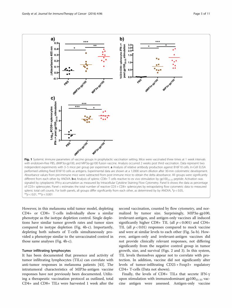
ResultsSystemic immune responseTo initially evaluate the immunogenicity of the DNAconstruct, systemic immune parameters were examined.Mice were vaccinated three times at 1 week intervalsand then analyzed 2 weeks after the third immunization.The MIP3α-gp100 vaccine elicited significantly higherlevels of B16F10-specific antibodies than antigen-onlyvaccine, denoted as dMIP3α-gp100 (p = 0.004), andmock vaccine (p < 0.001; Fig. 1). Interestingly, antigen-only vaccine had significantly higher B16F10-specificantibody levels than mock vaccine (p = 0.044).As was the case for the antibody concentration, the
antigen-only vaccine elicited a moderate vaccine-specificCD8+ T-cell response that significantly differed from themock vaccination by both percentage (p = 0.030; Fig. 1b)and total number (p < 0.001; Fig. 1c) of CD8+ T cells re-active to the immunogenic gp10025-33 peptide. Theaddition of MIP3α to the vaccine significantly increasedthe percentage of (p = 0.049) and total number of (p =0.026) vaccine induced CD8+ T cells compared to theantigen only vaccine, increasing the CD8+ T cell num-bers by 46% (Fig. 1b-c). The MIP3α-gp100 vaccine elic-ited significantly higher percentages and numbers ofvaccine-specific CD8+ T cells compared to mock vaccin-ation (p < 0.001 for both; Fig. 1b-c).
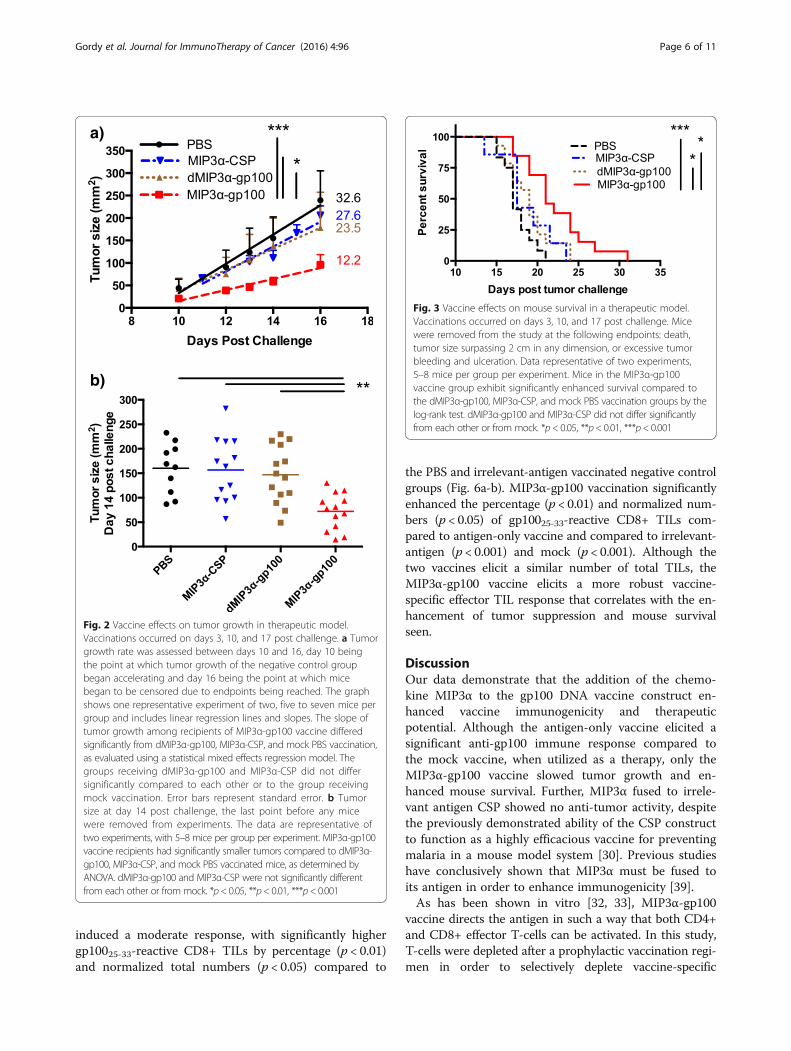
Therapeutic vaccination modelThe potential of this vaccine construct to be utilized in atherapeutic setting against a solid tumor was assessed,comparing MIP3α-gp100 vaccination to dMIP3α-gp100,MIP3α-CSP, and PBS vaccines. A therapeutic regimenwas developed with mice vaccinated on days three, 10,and 17 post challenge with a lethal dose of B16F10 cells.Utilizing statistical mixed effects regression models, itwas determined that the overall slope of the tumorgrowth regression line was reduced in the MIP3α-gp100vaccinated group compared to the antigen-only vacci-nated group by 48% (p = 0.029), to the irrelevant-antigengroup by 56% (p < 0.001), and to the mock vaccine groupby 63% (p < 0.001), whereas mock, antigen-only, andirrelevant-antigen vaccines showed no significant differ-ences to each other in slope (Fig. 2a). Slower overallgrowth also provides evidence that the differences seenin these experiments are not due to blocks to tumortransplantation.In addition to tumor growth, the tumor burden of
MIP3α-gp100 vaccine recipients proved to be signifi-cantly lower than mock vaccination at most time-pointstested and significantly lower than antigen only vaccineon the critical day 14 time-point – the last time pointbefore any mice were removed from the study. On day14 post challenge, the average tumor size was reduced inthe MIP3α-gp100 group by 51% compared to theantigen-only group (p = 0.004), by 54% compared toirrelevant-antigen group (p = 0.001), and by 55% com-pared to the mock group (p = 0.001; Fig. 2b). Survivalanalysis mirrored tumor growth and burden analyses.MIP3α-gp100 vaccination significantly enhanced survivalas compared to antigen-only (p = 0.017), irrelevant-antigen (p = 0.021) and mock (p < 0.001) vaccines.MIP3α-gp100 vaccination enhanced median survival by10%, 24% and 24% compared to antigen-only, irrelevant-antigen, and mock vaccinations, respectively (Fig. 3).Antigen-only, irrelevant-antigen, and mock vaccinationsdid not have significantly different survival curves com-pared to each other (Fig. 3).
T-cell subset depletionTo determine if effector T-cells played a role in mediat-ing this enhanced protection, and, if so, which subsetsmight be involved, groups of mice were vaccinated threetimes over 3 weeks to develop vaccine-specific effectorresponses and then were challenged with tumor underdiffering depletion conditions: depleting CD4+, CD8+,both CD4+ and CD8+, and no depletion of T cells.Figure 4a shows representative flow cytometric analysisof depletion efficacy. In a mouse lymphoma model, asimilar MIP3α-OFA vaccine showed the CD8+ T-cell ef-fector response to be essential for protection with theCD4+ T-cell effector response being expendable [31].
γ
γΔ
a)
c)
b)
Fig. 1 Systemic immune parameters of vaccine groups in prophylactic vaccination setting. Mice were vaccinated three times at 1 week intervalswith endotoxin-free PBS, dMIP3α-gp100, and MIP3α-gp100 fusion vaccine. Analysis occurred 2 weeks post third vaccination. Data represent twoindependent experiments with 3–5 mice per group per experiment. a Analysis of relative antibody production against B16F10 cells. In-Cell ELISAperformed utilizing fixed B16F10 cells as antigens. Experimental data are shown at a 1:2000 serum dilution after 30-min colorimetric development.Absorbance values from pre-immune mice were subtracted from post immune mice to obtain the delta absorbance. All groups were significantlydifferent from each other by ANOVA. b-c Analysis of splenic CD8+ T cells reactive to ex vivo stimulation by gp10025-33 peptide. Activation wassignaled by cytoplasmic IFN-γ accumulation as measured by Intracellular Cytokine Staining Flow Cytometry. Panel b shows the data as percentageof CD3+ splenocytes. Panel c estimates the total number of reactive CD3 + CD8+ splenocytes by extrapolating flow cytometric data to measuredsplenic total cell counts. For both panels, all groups differ significantly from each other, as determined by by ANOVA, *p < 0.05,**p < 0.01, ***p < 0.001
Gordy et al. Journal for ImmunoTherapy of Cancer (2016) 4:96 Page 5 of 11
However, in this melanoma solid tumor model, depletingCD4+ or CD8+ T-cells individually show a similarphenotype as the isotype depletion control. Single deple-tions have similar tumor growth rates and tumor sizescompared to isotype depletion (Fig. 4b-c). Importantly,depleting both subsets of T-cells simultaneously pro-vided a phenotype similar to the unvaccinated control inthose same analyses (Fig. 4b-c).
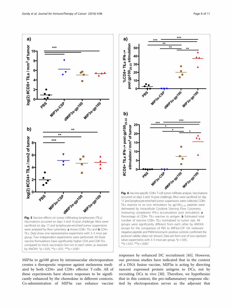
Tumor infiltrating lymphocytesIt has been documented that presence and activity oftumor infiltrating lymphocytes (TILs) can correlate withanti-tumor responses in melanoma patients [43]. Theintratumoral characteristics of MIP3α-antigen vaccineresponses have not previously been documented. Utiliz-ing a therapeutic vaccination protocol as outlined, totalCD4+ and CD8+ TILs were harvested 1 week after the
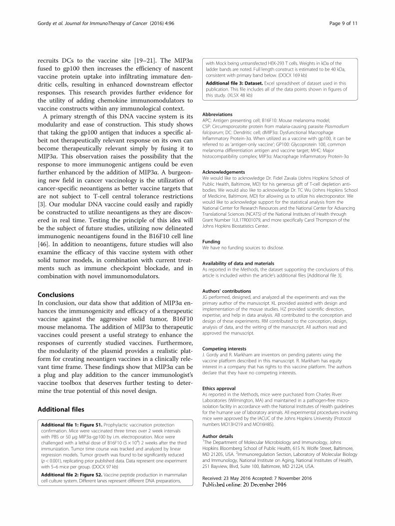
second vaccination, counted by flow cytometry, and nor-malized by tumor size. Surprisingly, MIP3α-gp100,irrelevant-antigen, and antigen-only vaccines all inducedsignificantly higher CD8+ TIL (all p < 0.001) and CD4+TIL (all p < 0.01) responses compared to mock vaccineand were at similar levels to each other (Fig. 5a-b). How-ever, antigen-only and irrelevant-antigen vaccines didnot provide clinically relevant responses, not differingsignificantly from the negative control group in tumorgrowth, size, and survival (Figs. 2 and 3). In this system,TIL levels themselves appear not to correlate with pro-tection. In addition, vaccine did not significantly alterlevels of tumor-infiltrating CD25 + Foxp3+ regulatoryCD4+ T-cells (Data not shown).Finally, the levels of CD8+ TILs that secrete IFN-γ
upon stimulation with immunodominant gp10025-33 vac-cine antigen were assessed. Antigen-only vaccine
a)
b)
Fig. 2 Vaccine effects on tumor growth in therapeutic model.Vaccinations occurred on days 3, 10, and 17 post challenge. a Tumorgrowth rate was assessed between days 10 and 16, day 10 beingthe point at which tumor growth of the negative control groupbegan accelerating and day 16 being the point at which micebegan to be censored due to endpoints being reached. The graphshows one representative experiment of two, five to seven mice pergroup and includes linear regression lines and slopes. The slope oftumor growth among recipients of MIP3α-gp100 vaccine differedsignificantly from dMIP3α-gp100, MIP3α-CSP, and mock PBS vaccination,as evaluated using a statistical mixed effects regression model. Thegroups receiving dMIP3α-gp100 and MIP3α-CSP did not differsignificantly compared to each other or to the group receivingmock vaccination. Error bars represent standard error. b Tumorsize at day 14 post challenge, the last point before any micewere removed from experiments. The data are representative oftwo experiments, with 5–8 mice per group per experiment. MIP3α-gp100vaccine recipients had significantly smaller tumors compared to dMIP3α-gp100, MIP3α-CSP, and mock PBS vaccinated mice, as determined byANOVA. dMIP3α-gp100 and MIP3α-CSP were not significantly differentfrom each other or from mock. *p< 0.05, **p< 0.01, ***p< 0.001
Fig. 3 Vaccine effects on mouse survival in a therapeutic model.Vaccinations occurred on days 3, 10, and 17 post challenge. Micewere removed from the study at the following endpoints: death,tumor size surpassing 2 cm in any dimension, or excessive tumorbleeding and ulceration. Data representative of two experiments,5–8 mice per group per experiment. Mice in the MIP3α-gp100vaccine group exhibit significantly enhanced survival compared tothe dMIP3α-gp100, MIP3α-CSP, and mock PBS vaccination groups by thelog-rank test. dMIP3α-gp100 and MIP3α-CSP did not differ significantlyfrom each other or from mock. *p< 0.05, **p< 0.01, ***p< 0.001
Gordy et al. Journal for ImmunoTherapy of Cancer (2016) 4:96 Page 6 of 11
induced a moderate response, with significantly highergp10025-33-reactive CD8+ TILs by percentage (p < 0.01)and normalized total numbers (p < 0.05) compared to
the PBS and irrelevant-antigen vaccinated negative controlgroups (Fig. 6a-b). MIP3α-gp100 vaccination significantlyenhanced the percentage (p < 0.01) and normalized num-bers (p < 0.05) of gp10025-33-reactive CD8+ TILs com-pared to antigen-only vaccine and compared to irrelevant-antigen (p < 0.001) and mock (p < 0.001). Although thetwo vaccines elicit a similar number of total TILs, theMIP3α-gp100 vaccine elicits a more robust vaccine-specific effector TIL response that correlates with the en-hancement of tumor suppression and mouse survivalseen.
DiscussionOur data demonstrate that the addition of the chemo-kine MIP3α to the gp100 DNA vaccine construct en-hanced vaccine immunogenicity and therapeuticpotential. Although the antigen-only vaccine elicited asignificant anti-gp100 immune response compared tothe mock vaccine, when utilized as a therapy, only theMIP3α-gp100 vaccine slowed tumor growth and en-hanced mouse survival. Further, MIP3α fused to irrele-vant antigen CSP showed no anti-tumor activity, despitethe previously demonstrated ability of the CSP constructto function as a highly efficacious vaccine for preventingmalaria in a mouse model system [30]. Previous studieshave conclusively shown that MIP3α must be fused toits antigen in order to enhance immunogenicity [39].As has been shown in vitro [32, 33], MIP3α-gp100
vaccine directs the antigen in such a way that both CD4+and CD8+ effector T-cells can be activated. In this study,T-cells were depleted after a prophylactic vaccination regi-men in order to selectively deplete vaccine-specific
a)
c)
b)
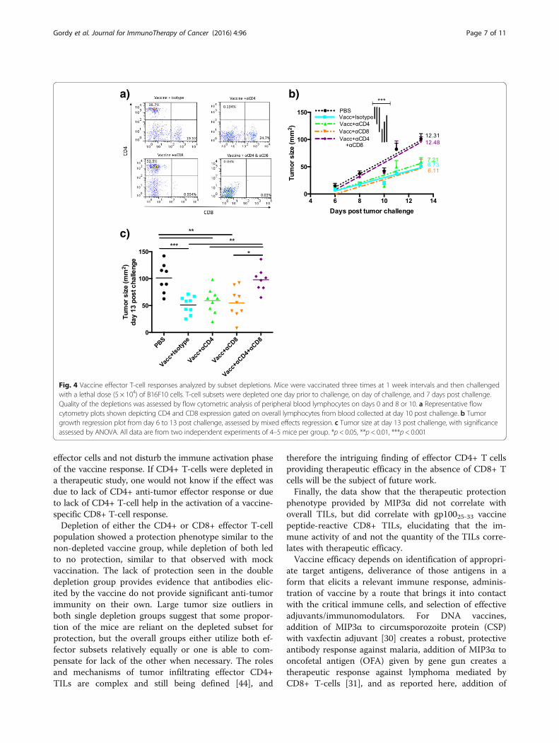
Fig. 4 Vaccine effector T-cell responses analyzed by subset depletions. Mice were vaccinated three times at 1 week intervals and then challengedwith a lethal dose (5 × 10⁴) of B16F10 cells. T-cell subsets were depleted one day prior to challenge, on day of challenge, and 7 days post challenge.Quality of the depletions was assessed by flow cytometric analysis of peripheral blood lymphocytes on days 0 and 8 or 10. a Representative flowcytometry plots shown depicting CD4 and CD8 expression gated on overall lymphocytes from blood collected at day 10 post challenge. b Tumorgrowth regression plot from day 6 to 13 post challenge, assessed by mixed effects regression. c Tumor size at day 13 post challenge, with significanceassessed by ANOVA. All data are from two independent experiments of 4–5 mice per group. *p < 0.05, **p < 0.01, ***p < 0.001
Gordy et al. Journal for ImmunoTherapy of Cancer (2016) 4:96 Page 7 of 11
effector cells and not disturb the immune activation phaseof the vaccine response. If CD4+ T-cells were depleted ina therapeutic study, one would not know if the effect wasdue to lack of CD4+ anti-tumor effector response or dueto lack of CD4+ T-cell help in the activation of a vaccine-specific CD8+ T-cell response.Depletion of either the CD4+ or CD8+ effector T-cell
population showed a protection phenotype similar to thenon-depleted vaccine group, while depletion of both ledto no protection, similar to that observed with mockvaccination. The lack of protection seen in the doubledepletion group provides evidence that antibodies elic-ited by the vaccine do not provide significant anti-tumorimmunity on their own. Large tumor size outliers inboth single depletion groups suggest that some propor-tion of the mice are reliant on the depleted subset forprotection, but the overall groups either utilize both ef-fector subsets relatively equally or one is able to com-pensate for lack of the other when necessary. The rolesand mechanisms of tumor infiltrating effector CD4+TILs are complex and still being defined [44], and
therefore the intriguing finding of effector CD4+ T cellsproviding therapeutic efficacy in the absence of CD8+ Tcells will be the subject of future work.Finally, the data show that the therapeutic protection
phenotype provided by MIP3α did not correlate withoverall TILs, but did correlate with gp10025-33 vaccinepeptide-reactive CD8+ TILs, elucidating that the im-mune activity of and not the quantity of the TILs corre-lates with therapeutic efficacy.Vaccine efficacy depends on identification of appropri-
ate target antigens, deliverance of those antigens in aform that elicits a relevant immune response, adminis-tration of vaccine by a route that brings it into contactwith the critical immune cells, and selection of effectiveadjuvants/immunomodulators. For DNA vaccines,addition of MIP3α to circumsporozoite protein (CSP)with vaxfectin adjuvant [30] creates a robust, protectiveantibody response against malaria, addition of MIP3α tooncofetal antigen (OFA) given by gene gun creates atherapeutic response against lymphoma mediated byCD8+ T-cells [31], and as reported here, addition of
a)
b)
Fig. 5 Vaccine effects on tumor infiltrating lymphocytes (TILs).Vaccinations occurred on days 3 and 10 post challenge. Mice weresacrificed on day 17 and lymphocyte-enriched tumor suspensionswere analyzed by flow cytometry. a shows CD8+ TILs and b CD4+TILs. Data show one representative experiment with 3–5 mice pergroup. Two independent experiments were performed. All threevaccine formulations have significantly higher CD4 and CD8 TILscompared to mock vaccination but not to each other, as assessedby ANOVA. *p < 0.05, **p < 0.01, ***p < 0.001
γγ
a)
b)
Fig. 6 Vaccine-specific CD8+ T-cell tumor infiltrate analysis. Vaccinationsoccurred on days 3 and 10 post challenge. Mice were sacrificed on day17 and lymphocyte-enriched tumor suspensions were collected. CD8+TILs reactive to ex vivo stimulation by gp10025-33 peptide weredelineated by Intracellular Cytokine Staining Flow Cytometrymeasuring cytoplasmic IFN-γ accumulation post stimulation. aPercentage of CD8+ TILs reactive to antigen. b Estimated totalnumber of reactive CD8+ TILs normalized to tumor size. Allgroups were significantly different from each other by ANOVAexcept for the comparison of PBS to MIP3α-CSP. HA irrelevantnegative peptide and PMA/ionomycin positive controls confirmed theprotocol validity (data not shown). Data are from one of two represen-tative experiments with 3–5 mice per group. *p < 0.05,**p < 0.01, ***p < 0.001
Gordy et al. Journal for ImmunoTherapy of Cancer (2016) 4:96 Page 8 of 11
MIP3α to gp100 given by intramuscular electroporationcreates a therapeutic response against melanoma medi-ated by both CD4+ and CD8+ effector T-cells. All ofthese experiments have shown responses to be signifi-cantly enhanced by the chemokine in different contexts.Co-administration of MIP3α can enhance vaccine
responses by enhanced DC recruitment [45]. However,our previous studies have indicated that in the contextof a DNA fusion vaccine, MIP3α is acting by directingnascent expressed protein antigens to DCs, not byrecruiting DCs in vivo [30]. Therefore, we hypothesizethat in this context, the pro-inflammatory response elic-ited by electroporation serves as the adjuvant that
Gordy et al. Journal for ImmunoTherapy of Cancer (2016) 4:96 Page 9 of 11
recruits DCs to the vaccine site [19–21]. The MIP3αfused to gp100 then increases the efficiency of nascentvaccine protein uptake into infiltrating immature den-dritic cells, resulting in enhanced downstream effectorresponses. This research provides further evidence forthe utility of adding chemokine immunomodulators tovaccine constructs within any immunological context.A primary strength of this DNA vaccine system is its
modularity and ease of construction. This study showsthat taking the gp100 antigen that induces a specific al-beit not therapeutically relevant response on its own canbecome therapeutically relevant simply by fusing it toMIP3α. This observation raises the possibility that theresponse to more immunogenic antigens could be evenfurther enhanced by the addition of MIP3α. A burgeon-ing new field in cancer vaccinology is the utilization ofcancer-specific neoantigens as better vaccine targets thatare not subject to T-cell central tolerance restrictions[3]. Our modular DNA vaccine could easily and rapidlybe constructed to utilize neoantigens as they are discov-ered in real time. Testing the principle of this idea willbe the subject of future studies, utilizing now delineatedimmunogenic neoantigens found in the B16F10 cell line[46]. In addition to neoantigens, future studies will alsoexamine the efficacy of this vaccine system with othersolid tumor models, in combination with current treat-ments such as immune checkpoint blockade, and incombination with novel immunomodulators.
ConclusionsIn conclusion, our data show that addition of MIP3α en-hances the immunogenicity and efficacy of a therapeuticvaccine against the aggressive solid tumor, B16F10mouse melanoma. The addition of MIP3α to therapeuticvaccines could present a useful strategy to enhance theresponses of currently studied vaccines. Furthermore,the modularity of the plasmid provides a realistic plat-form for creating neoantigen vaccines in a clinically rele-vant time frame. These findings show that MIP3α can bea plug and play addition to the cancer immunologist’svaccine toolbox that deserves further testing to deter-mine the true potential of this novel design.
Additional files
Additional file 1: Figure S1. Prophylactic vaccination protectionconfirmation. Mice were vaccinated three times over 2 week intervalswith PBS or 50 μg MIP3α-gp100 by i.m. electroporation. Mice werechallenged with a lethal dose of B16F10 (5 × 10⁴) 2 weeks after the thirdimmunization. Tumor time course was tracked and analyzed by linearregression models. Tumor growth was found to be significantly reduced(p < 0.001), replicating prior published data. Data represent one experimentwith 5–6 mice per group. (DOCX 97 kb)
Additional file 2: Figure S2. Vaccine peptide production in mammaliancell culture system. Different lanes represent different DNA preparations,
with Mock being untransfected HEK-293 T cells. Weights in kDa of theladder bands are noted. Full length construct is estimated to be 40 kDa,consistent with primary band below. (DOCX 169 kb)
Additional file 3: Dataset. Excel spreadsheet of dataset used in thispublication. This file includes all of the data points shown in figures ofthis study. (XLSX 48 kb)
AbbreviationsAPC: Antigen presenting cell; B16F10: Mouse melanoma model;CSP: Circumsporozoite protein from malaria-causing parasite Plasmodiumfalciparum; DC: Dendritic cell; dMIP3α: Dysfunctional MacrophageInflammatory Protein-3α. When utilized as a vaccine with gp100, it can bereferred to as ‘antigen-only vaccine’; GP100: Glycoprotein 100, commonmelanoma differentiation antigen and vaccine target; MHC: Majorhistocompatibility complex; MIP3α: Macrophage Inflammatory Protein-3α
AcknowledgementsWe would like to acknowledge Dr. Fidel Zavala (Johns Hopkins School ofPublic Health, Baltimore, MD) for his generous gift of T-cell depletion anti-bodies. We would also like to acknowledge Dr. TC Wu (Johns Hopkins Schoolof Medicine, Baltimore, MD) for allowing us to utilize his electroporator. Wewould like to acknowledge support for the statistical analysis from theNational Center for Research Resources and the National Center for AdvancingTranslational Sciences (NCATS) of the National Institutes of Health throughGrant Number 1UL1TR001079, and more specifically Carol Thompson of theJohns Hopkins Biostatistics Center.
FundingWe have no funding sources to disclose.
Availability of data and materialsAs reported in the Methods, the dataset supporting the conclusions of thisarticle is included within the article’s additional files [Additional file 3].
Authors’ contributionsJG performed, designed, and analyzed all the experiments and was theprimary author of the manuscript. KL provided assisted with design andimplementation of the mouse studies. HZ provided scientific direction,expertise, and help in data analysis. AB contributed to the conception anddesign of these experiments. RM contributed to the conception, design,analysis of data, and the writing of the manuscript. All authors read andapproved the manuscript.
Competing interestsJ. Gordy and R. Markham are inventors on pending patents using thevaccine platform described in this manuscript. R. Markham has equityinterest in a company that has rights to this vaccine platform. The authorsdeclare that they have no competing interests.
Ethics approvalAs reported in the Methods, mice were purchased from Charles RiverLaboratories (Wilmington, MA) and maintained in a pathogen-free micro-isolation facility in accordance with the National Institutes of Health guidelinesfor the humane use of laboratory animals. All experimental procedures involvingmice were approved by the IACUC of the Johns Hopkins University (Protocolnumbers MO13H219 and MO16H85).
Author details1The Department of Molecular Microbiology and Immunology, JohnsHopkins Bloomberg School of Public Health, 615 N. Wolfe Street, Baltimore,MD 21205, USA. 2Immunoregulation Section, Laboratory of Molecular Biologyand Immunology, National Institute on Aging, National Institutes of Health,251 Bayview, Blvd, Suite 100, Baltimore, MD 21224, USA.
Received: 23 May 2016 Accepted: 7 November 2016
Gordy et al. Journal for ImmunoTherapy of Cancer (2016) 4:96 Page 10 of 11
References1. Eggermont AMM, Maio M, Robert C. Immune checkpoint inhibitors in
melanoma provide the cornerstones for curative therapies. Semin Oncol.2015;42:429–35.
2. Overwijk W, Wang E, Marincola F, Rammensee H-G, Restifo N. Mining themutanome: developing highly personalized Immunotherapies based onmutational analysis of tumors. J Immuno Ther Cancer. 2013;1:11.
3. Hacohen N, Fritsch E, Carter T, Lander E. Getting personal with neoantigen-based therapeutic cancer vaccines. Cancer Immunol Res. 2013;1:11–5.
4. Small EJ, Schellhammer PF, Higano CS, Redfern CH, Nemunaitis JJ,Valone FH, Verjee SS, Jones LA, Hershberg RM. Placebo-controlled phase IIItrial of immunologic therapy with sipuleucel-T (APC8015) in patients withmetastatic, asymptomatic hormone refractory prostate cancer. J Clin Oncol.2006;24:3089–94.
5. Ali OA, Lewin SA, Dranoff G, Mooney DJ. Vaccines combined with immunecheckpoint antibodies promote cytotoxic T-cell activity and tumoreradication. Cancer Immunol Res. 2016;4:95–100.
6. Liu M. DNA vaccines: an historical perspective and view to the future.Immunol Rev. 2011;239:62–84.
7. Tagawa S, Lee P, Snively J, Boswell W, Ounpraseuth S, Lee S, Hickingbottom B,Smith J, Johnson D, Weber J. Phase I study of intranodal delivery of a plasmidDNA vaccine for patients with Stage IV melanoma. Cancer. 2003;98:144–54.
8. Conry RM, Curiel DT, Strong TV, Moore SE, Allen KO, Barlow DL, Shaw DR,LoBuglio AF. Safety and immunogenicity of a DNA vaccine encodingcarcinoembryonic antigen and hepatitis B surface antigen in colorectalcarcinoma patients. Clin Cancer Res. 2002;8:2782–7.
9. Liu MA, Ulmer JB. Human clinical trials of plasmid DNA vaccines. Adv Genet.2005;55:25–40.
10. Senovilla L, Vacchelli E, Garcia P, Eggermont A, Fridman W, Galon J, Zitvogel L,Kroemer G, Galluzzi L. Trial watch. OncoImmunology. 2013;2:e23803.
11. Rice J, Ottensmeier C, Stevenson F. DNA vaccines: precision tools foractivating effective immunity against cancer. Nat Rev Cancer. 2008;8:108–20.
12. Davis BS, Chang GJ, Cropp B, Roehrig JT, Martin DA, Mitchell CJ, Bowen R,Bunning ML. West Nile virus recombinant DNA vaccine protects mouse andhorse from virus challenge and expresses in vitro a noninfectiousrecombinant antigen that can be used in enzyme-linked immunosorbentassays. J Virol. 2001;75:4040–7.
13. Anderson ED, Mourich DV, Fahrenkrug SC, LaPatra S, Shepherd J, Leong JA.Genetic immunization of rainbow trout (Oncorhynchus mykiss) againstinfectious hematopoietic necrosis virus. Mol Marine Biol Biotechnol. 1996;5:114–22.
14. Bergman PJ, McKnight J, Novosad A, Charney S, Farrelly J, Craft D, Wulderk M,Jeffers Y, Sadelain M, Hohenhaus AE, Segal N, Gregor P, Engelhorn M, Riviere I,Houghton AN, Wolchok JD. Long-term survival of dogs with advancedmalignant melanoma after DNA vaccination with xenogeneic humantyrosinase: a phase I trial. Clin Cancer Res. 2003;9:1284–90.
15. Chen J, Fang F, Li X, Chang H, Chen Z. Protection against influenza virusinfection in BALB/c mice immunized with a single dose of neuraminidase-expressing DNAs by electroporation. Vaccine. 2005;23:4322–8.
16. Medi BM, Hoselton S, Marepalli RB, Singh J. Skin targeted DNA vaccine deliveryusing electroporation in rabbits. I: efficacy. Int J Pharm. 2005;294:53–63.
17. Otten G, Schaefer M, Doe B, Liu H, Srivastava I, Zur Megede J, O’Hagan D,Donnelly J, Widera G, Rabussay D, Lewis MG, Barnett S, Ulmer JB.Enhancement of DNA vaccine potency in rhesus macaques byelectroporation. Vaccine. 2004;22:2489–93.
18. Tollefsen S, Vordermeier M, Olsen I, Storset AK, Reitan LJ, Clifford D,Lowrie DB, Wiker HG, Huygen K, Hewinson G, Mathiesen I, Tjelle TE. DNAinjection in combination with electroporation: a novel method forvaccination of farmed ruminants. Scand J Immunol. 2003;57:229–38.
19. Ahlén G, Söderholm J, Tjelle T, Kjeken R, Frelin L, Höglund U, Blomberg P,Fons M, Mathiesen I, Sällberg M. In vivo electroporation enhances theimmunogenicity of hepatitis C virus nonstructural 3/4A DNA by increasedlocal DNA uptake, protein expression, inflammation, and infiltration of CD3+T cells. J Immunol. 2007;179:4741–53.
20. Liu J, Kjeken R, Mathiesen I, Barouch D. Recruitment of antigen-presentingcells to the site of inoculation and augmentation of humanimmunodeficiency virus type 1 DNA vaccine immunogenicity by in vivoelectroporation. J Virol. 2008;82:5643–9.
21. Babiuk S, Baca-Estrada ME, Foldvari M, Middleton DM, Rabussay D, Widera G,Babiuk LA. Increased gene expression and inflammatory cell infiltration
caused by electroporation are both important for improving the efficacy ofDNA vaccines. J Biotechnol. 2004;110:1–10.
22. Person R, Bodles-Brakhop AM, Pope MA, Brown PA, Khan AS, Draghia-Akli R.Growth hormone-releasing hormone plasmid treatment by electroporationdecreases offspring mortality over three pregnancies. Mol Ther. 2008;16:1891–7.
23. Vasan S, Hurley A, Schlesinger SJ, Hannaman D. In vivo electroporationenhances the immunogenicity of an HIV-1 DNA vaccine candidate inhealthy volunteers. PLoS One. 2011;6:e19252.
24. Low L, Mander A, McCann K, Dearnaley D. DNA vaccination withelectroporation induces increased antibody responses in patients withprostate cancer. Hum Gene Ther. 2009;20:1269–78.
25. Loudon PT, Yager EJ, Lynch DT, Narendran A, Stagnar C, Franchini AM,Fuller JT, White PA, Nyuandi J, Wiley CA, Murphey-Corb M, Fuller DH. GM-CSF increases mucosal and systemic immunogenicity of an H1N1 influenzaDNA vaccine administered into the epidermis of non-human primates. PLoSOne. 2010;5:e11021.
26. Zheng Q, Fan D, Gao N, Chen H, Wang J, Ming Y, Li J, An J. Evaluation of aDNA vaccine candidate expressing prM-E-NS1 antigens of dengue virusserotype 1 with or without granulocyte-macrophage colony-stimulatingfactor (GM-CSF) in immunogenicity and protection. Vaccine. 2011;29:763–71.
27. Rodríguez AM, Pascutti MFF, Maeto C, Falivene J, Holgado MPP, Turk G,Gherardi MMM. IL-12 and GM-CSF in DNA/MVA immunizations against HIV-1 CRF12_BF Nef induced T-cell responses with an enhanced magnitude,breadth and quality. PLoS One. 2012;7:e37801.
28. Lapteva N, Huang XF. CCL5 as an adjuvant for cancer immunotherapy.Expert Opin Biol Ther. 2010;10:725–33.
29. Nguyen-Hoai T, Hohn O, Vu MD, Baldenhofer G, Sayed Ahmed MS, Dörken B,Norley S, Lipp M, Pezzutto A, Westermann J. CCL19 as an adjuvant forintradermal gene gun immunization in a Her2/neu mouse tumor model:improved vaccine efficacy and a role for B cells as APC. Cancer Gene Ther.2012;19:880–7.
30. Luo K, Zhang H, Zavala F, Biragyn A, Espinosa D, Markham R. Fusion ofantigen to a dendritic cell targeting chemokine combined with adjuvantyields a malaria DNA vaccine with enhanced protective capabilities. PLoSOne. 2014;9:e90413.
31. Biragyn A, Schiavo R, Olkhanud P, Sumitomo K, King A, McCain M, Indig FE,Almanzar G, Baatar D. Tumor-associated embryonic antigen-expressingvaccines that target CCR6 elicit potent CD8+ T cell-mediated protective andtherapeutic antitumor immunity. J Immunol. 2007;179:1381–8.
32. Biragyn A, Ruffini P, Coscia M, Harvey L, Neelapu S, Baskar S, Wang J-M,Kwak L. Chemokine receptor-mediated delivery directs self-tumor antigenefficiently into the class II processing pathway in vitro and inducesprotective immunity in vivo. Blood. 2004;104:1961–9.
33. Schiavo R, Baatar D, Olkhanud P, Indig F, Restifo N, Taub D, Biragyn A.Chemokine receptor targeting efficiently directs antigens to MHC class Ipathways and elicits antigen-specific CD8+ T-cell responses. Blood. 2006;107:4597–605.
34. Guo J, Fan M, Sun J, Jia R. Fusion of antigen to chemokine CCL20 orCXCL13 strategy to enhance DNA vaccine potency. Int Immunopharmacol.2009;9:925930.
35. Song R, Liu S, Leong KW. Effects of MIP-1 alpha, MIP-3 alpha, and MIP-3beta on the induction of HIV Gag-specific immune response with DNAvaccines. Mol Ther. 2007;15:1007–15.
36. Oppenheim JJ, Biragyn A, Kwak LW, Yang D. Roles of antimicrobial peptidessuch as defensins in innate and adaptive immunity. Ann Rheum Dis. 2003;62 Suppl 2:ii17–21.
37. Coscia M, Biragyn A. Cancer immunotherapy with chemoattractantpeptides. Semin Cancer Biol. 2004;14:209218.
38. Biragyn A, Tani K, Grimm M, Weeks S, Kwak L. Genetic fusion of chemokinesto a self tumor antigen induces protective, T-cell dependent antitumorimmunity. Nat Biotechnol. 1999;17:253–8.
39. Biragyn A, Surenhu M, Yang D, Ruffini PA, Haines BA, Klyushnenkova E,Oppenheim JJ, Kwak LW. Mediators of innate immunity that targetimmature, but not mature, dendritic cells induce antitumor immunity whengenetically fused with nonimmunogenic tumor antigens. J Immunol. 2001;167:6644–53.
40. Ma K, Xu W, Shao X, Yanyue, Hu L, Xu H, Yuan Z, Zheng X, Xiong S.Coimmunization with RANTES plasmid polarized Th1 immune responseagainst hepatitis B virus envelope via recruitment of dendritic cells. AntiviralRes. 2007;76:140–9.
Gordy et al. Journal for ImmunoTherapy of Cancer (2016) 4:96 Page 11 of 11
41. Miguel A, Herrero M, Sendra L, Botella R, Algás R, Sánchez M, Aliño S.Comparative antitumor effect of preventive versus therapeutic vaccinesemploying B16 melanoma cells genetically modified to express GM-CSFand B7.2 in a Murine Model. Toxins. 2012;4:1058–81.
42. Yan J, Pankhong P, Shin TH, Obeng-Adjei N, Morrow MP, Walters JN, KhanAS, Sardesai NY, Weiner DB. Highly optimized DNA vaccine targetinghuman telomerase reverse transcriptase stimulates potent antitumorimmunity. Cancer Immunol Res. 2013;1:179–89.
43. Tuthill RJ, Unger JM, Liu PY, Flaherty LE, Sondak VK. Risk assessment inlocalized primary cutaneous melanoma. Am J Clin Pathol. 2002;118:504–11.
44. Haabeth OA, Tveita AA, Fauskanger M, Schjesvold F, Lorvik KB, Hofgaard PO,Omholt H, Munthe LA, Dembic Z, Corthay A, Bogen B. How Do CD4(+) Tcells detect and eliminate tumor cells that either lack or express MHC classII molecules? Front Immunol. 2014;5:174.
45. Igoucheva O, Grazzini M, Pidich A, Kemp DM, Larijani M, Farber M, Lorton J,Rodeck U, Alexeev V. Immunotargeting and eradication of orthotopicmelanoma using a chemokine-enhanced DNA vaccine. Gene Ther. 2013;20:939–48.
46. Castle J, Kreiter S, Diekmann J, Löwer M, van de Roemer N, de Graaf J,Selmi A, Diken M, Boegel S, Paret C, Koslowski M, Kuhn A, Britten C, HuberC, Türeci Ö, Sahin U. Exploiting the mutanome for tumor vaccination.Cancer Res. 2012;72:1081–91.
• We accept pre-submission inquiries
• Our selector tool helps you to find the most relevant journal
• We provide round the clock customer support
• Convenient online submission
• Thorough peer review
• Inclusion in PubMed and all major indexing services
• Maximum visibility for your research
Submit your manuscript atwww.biomedcentral.com/submit
Submit your next manuscript to BioMed Central and we will help you at every step: