PresentationCofilin under control of β-arrestin-2 in NMDAdependent dendritic spine plasticity,...
13
Fabio Falleroni
-
Upload
fabio-falleroni -
Category
Health & Medicine
-
view
182 -
download
1
Transcript of PresentationCofilin under control of β-arrestin-2 in NMDAdependent dendritic spine plasticity,...
Fabio Falleroni
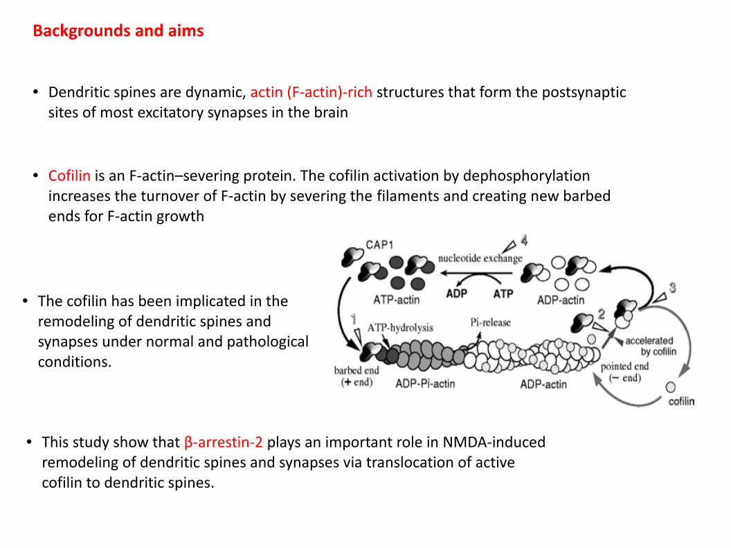
● Dendritic spines are dynamic, actin (F-actin)-rich structures that form the postsynaptic sites of most excitatory synapses in the brain
● Cofilin is an F-actin–severing protein. The cofilin activation by dephosphorylation increases the turnover of F-actin by severing the filaments and creating new barbed ends for F-actin growth
● The cofilin has been implicated in the remodeling of dendritic spines and synapses under normal and pathological conditions.
● This study show that β-arrestin-2 plays an important role in NMDA-induced remodeling of dendritic spines and synapses via translocation of active cofilin to dendritic spines.
Backgrounds and aims
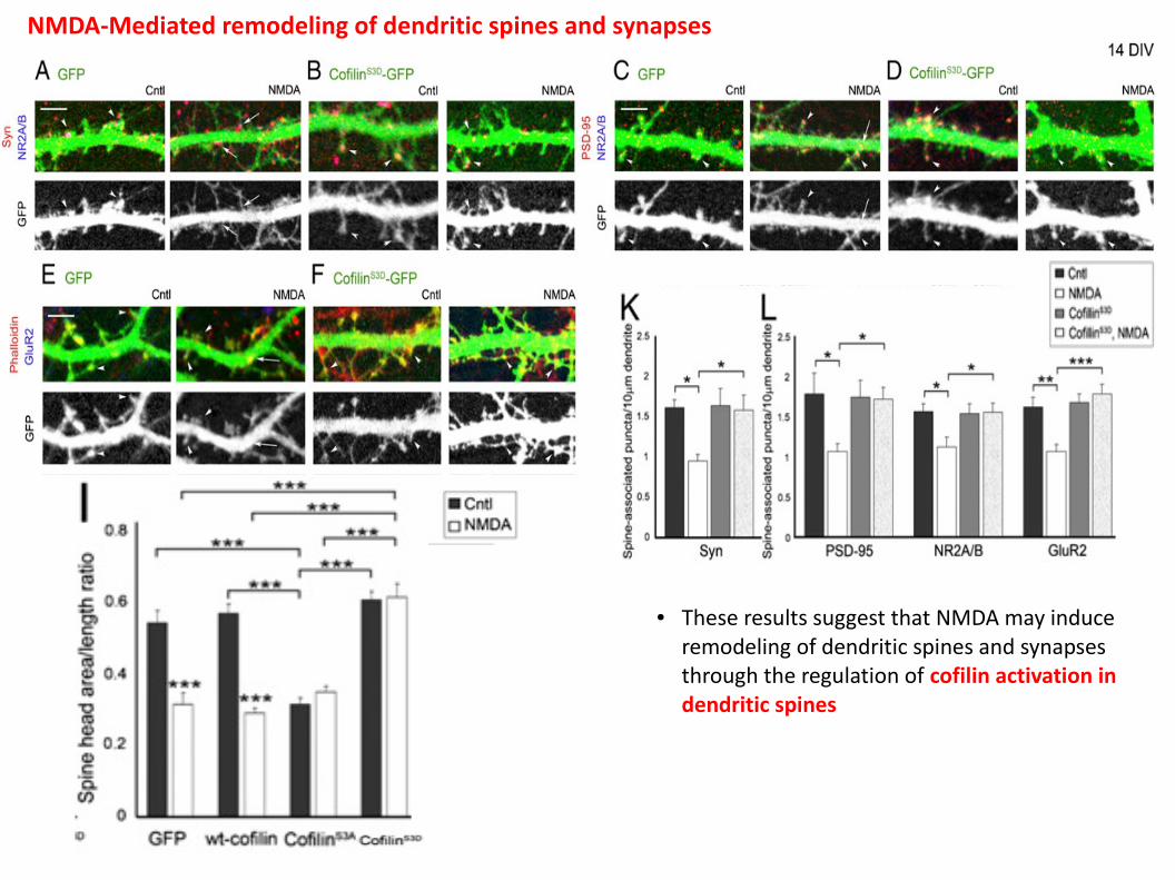
● These results suggest that NMDA may induce remodeling of dendritic spines and synapses through the regulation of cofilin activation in dendritic spines
NMDA-Mediated remodeling of dendritic spines and synapses
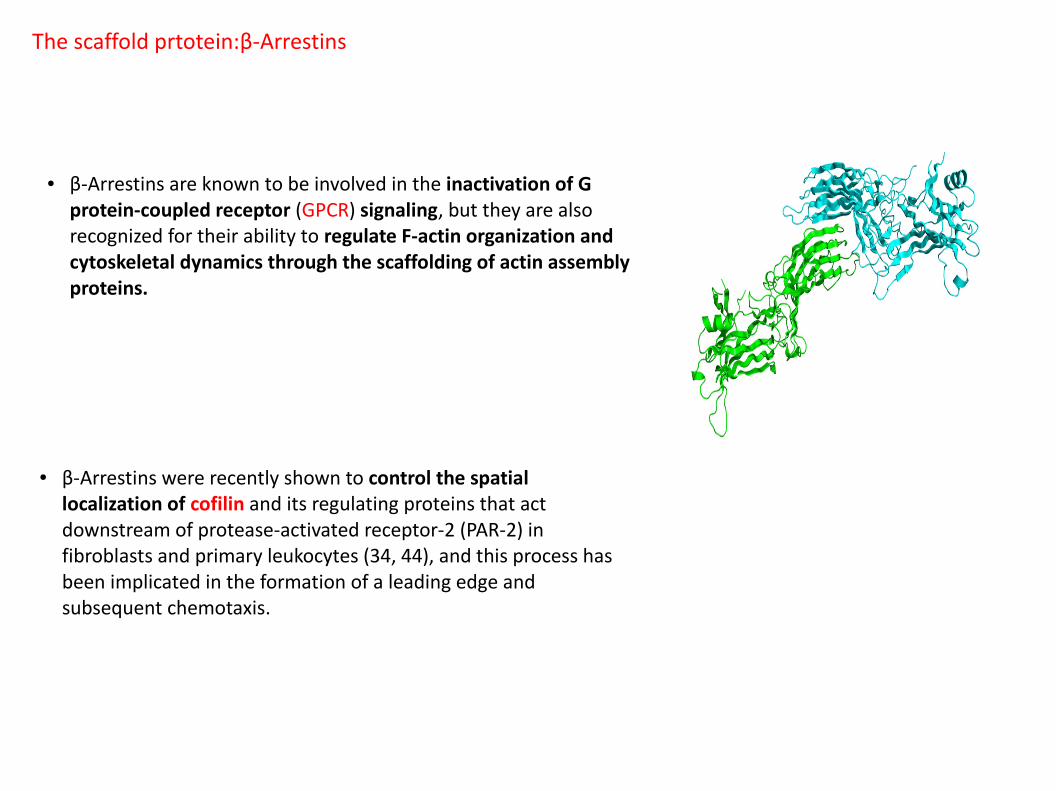
● β-Arrestins are known to be involved in the inactivation of G protein-coupled receptor (GPCR) signaling, but they are also recognized for their ability to regulate F-actin organization and cytoskeletal dynamics through the scaffolding of actin assembly proteins.
● β-Arrestins were recently shown to control the spatial localization of cofilin and its regulating proteins that act downstream of protease-activated receptor-2 (PAR-2) in fibroblasts and primary leukocytes (34, 44), and this process has been implicated in the formation of a leading edge and subsequent chemotaxis.
The scaffold prtotein:β-Arrestins
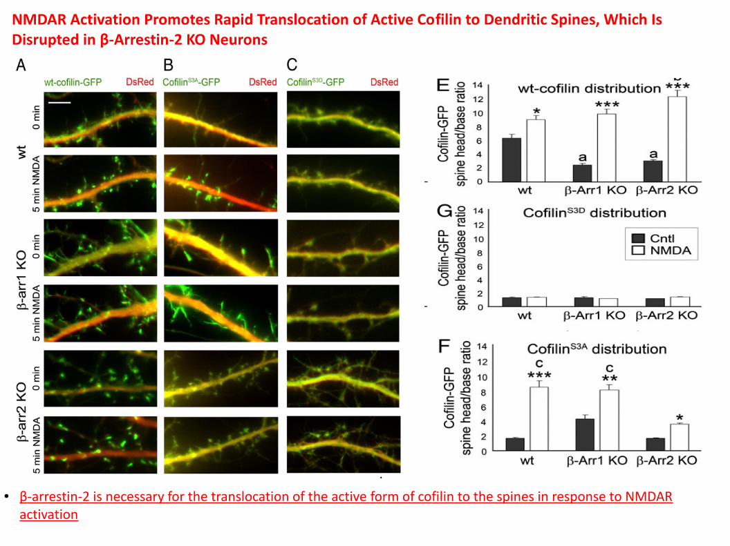
NMDAR Activation Promotes Rapid Translocation of Active Cofilin to Dendritic Spines, Which Is Disrupted in β-Arrestin-2 KO Neurons
● β-arrestin-2 is necessary for the translocation of the active form of cofilin to the spines in response to NMDAR activation
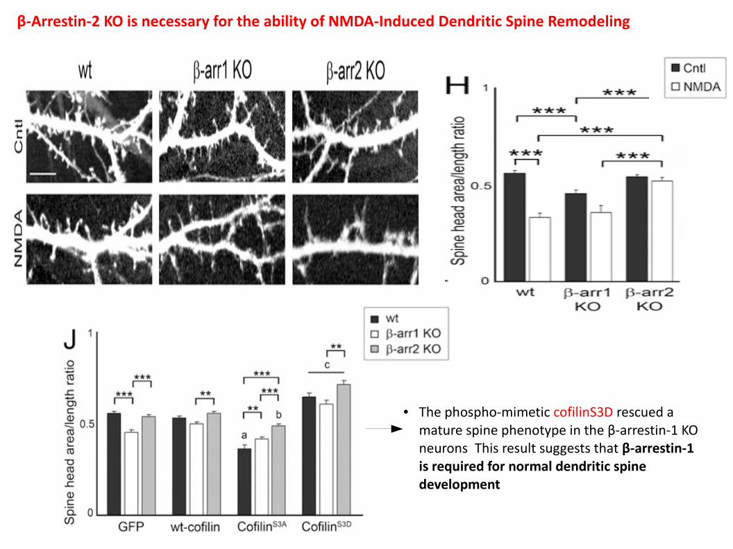
β-Arrestin-2 KO is necessary for the ability of NMDA-Induced Dendritic Spine Remodeling
● The phospho-mimetic cofilinS3D rescued a mature spine phenotype in the β-arrestin-1 KO neurons This result suggests that β-arrestin-1 is required for normal dendritic spine development
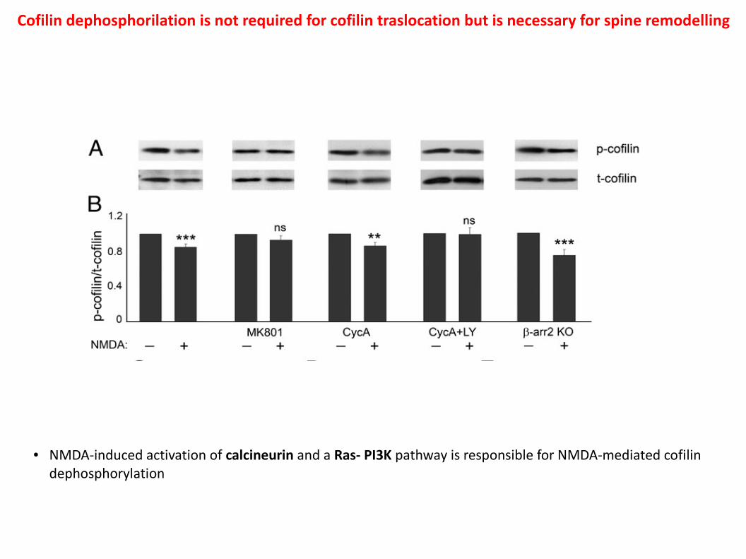
● NMDA-induced activation of calcineurin and a Ras- PI3K pathway is responsible for NMDA-mediated cofilin dephosphorylation
Cofilin dephosphorilation is not required for cofilin traslocation but is necessary for spine remodelling
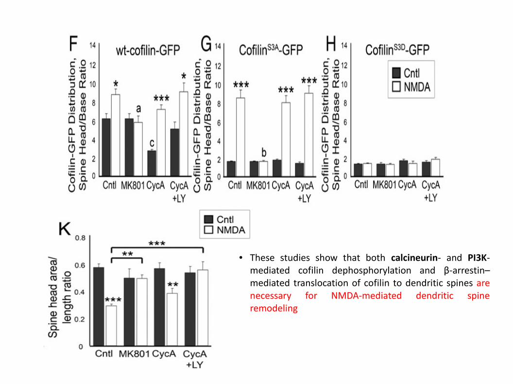
● These studies show that both calcineurin- and PI3K-mediated cofilin dephosphorylation and β-arrestin–mediated translocation of cofilin to dendritic spines are necessary for NMDA-mediated dendritic spine remodeling
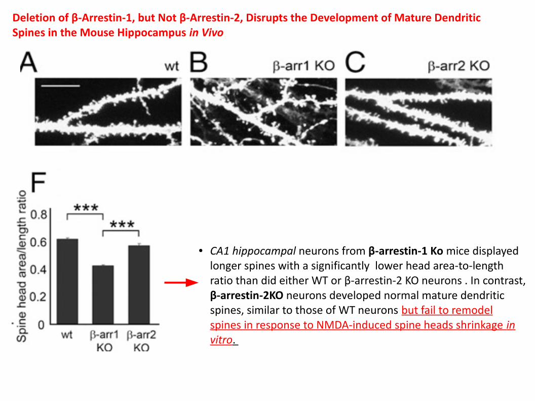
Deletion of β-Arrestin-1, but Not β-Arrestin-2, Disrupts the Development of Mature Dendritic Spines in the Mouse Hippocampus in Vivo
● CA1 hippocampal neurons from β-arrestin-1 Ko mice displayed longer spines with a significantly lower head area-to-length ratio than did either WT or β-arrestin-2 KO neurons . In contrast, β-arrestin-2KO neurons developed normal mature dendritic spines, similar to those of WT neurons but fail to remodel spines in response to NMDA-induced spine heads shrinkage in vitro.
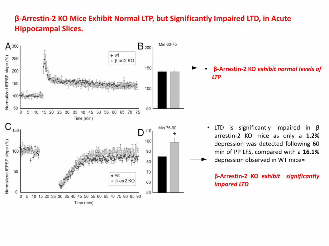
β-Arrestin-2 KO Mice Exhibit Normal LTP, but Significantly Impaired LTD, in Acute Hippocampal Slices.
● β-Arrestin-2 KO exhibit normal levels of LTP
● LTD is significantly impaired in β arrestin-2 KO mice as only a 1.2% depression was detected following 60 min of PP LFS, compared with a 16.1% depression observed in WT mice=
β-Arrestin-2 KO exhibit significantly impared LTD
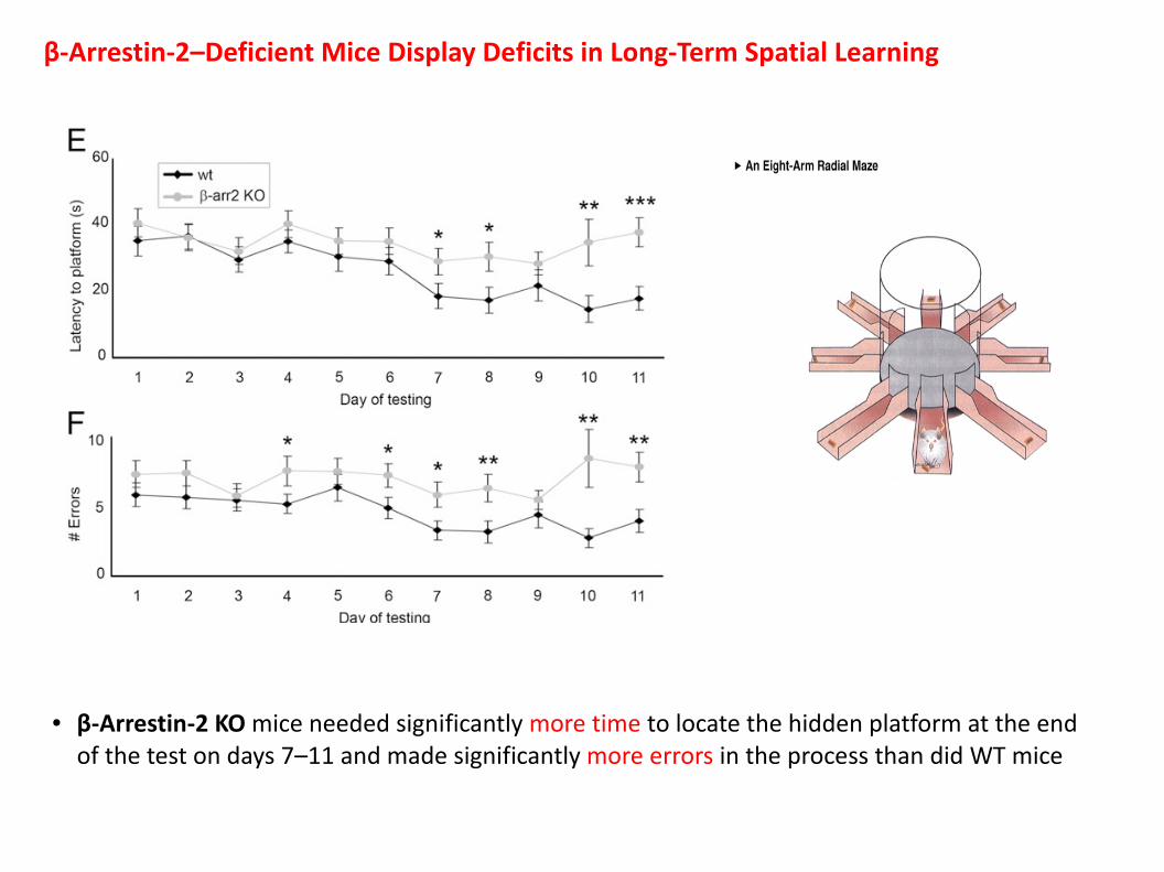
β-Arrestin-2–Deficient Mice Display Deficits in Long-Term Spatial Learning
● β-Arrestin-2 KO mice needed significantly more time to locate the hidden platform at the end of the test on days 7–11 and made significantly more errors in the process than did WT mice
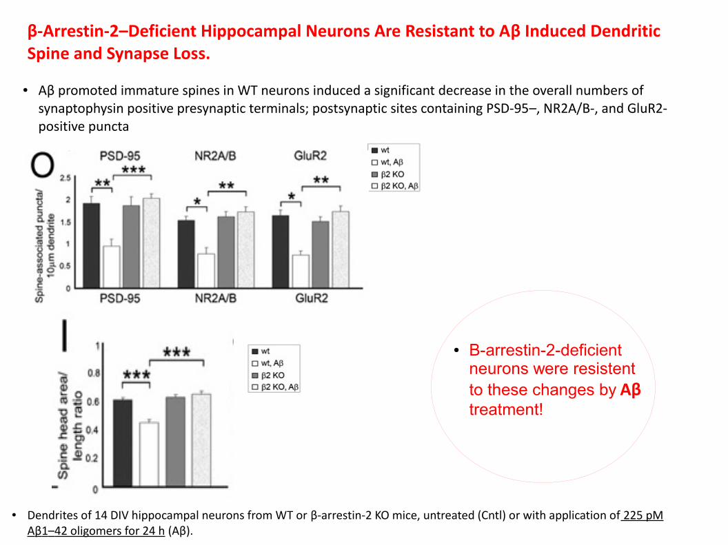
β-Arrestin-2–Deficient Hippocampal Neurons Are Resistant to Aβ Induced Dendritic Spine and Synapse Loss.
● Aβ promoted immature spines in WT neurons induced a significant decrease in the overall numbers of synaptophysin positive presynaptic terminals; postsynaptic sites containing PSD-95–, NR2A/B-, and GluR2-positive puncta
● Dendrites of 14 DIV hippocampal neurons from WT or β-arrestin-2 KO mice, untreated (Cntl) or with application of 225 pM Aβ1–42 oligomers for 24 h (Aβ).
● B-arrestin-2-deficient neurons were resistent to these changes by Aβ treatment!
Conclusion
● This studies implicate β-arrestin-2 in cofilin regulation during dendritic spine plasticity, hippocampal LTD, and spatial learning.
● We have shown here a unique function of β arrestins that does not involve GPCR signaling, but regulates NMDA-mediated dendritic spine/synapse plasticity through the spatial control of cofilin activation.
Raon Cajal- hippocampal neurons