PYR-41 and Thalidomide Impair Dendritic Cell Cross...
15
Research Article PYR-41 and Thalidomide Impair Dendritic Cell Cross-Presentation by Inhibiting Myddosome Formation and Attenuating the Endosomal Recruitments of p97 and Sec61 via NF-κB Inactivation Xiang You, 1 Dan Dan Xu, 1 Di Zhang, 1 Jie Chen , 1 and Feng Guang Gao 1,2 1 Department of Immunology, Basic Medicine Science, Medical College, Xiamen University, Xiamen, Fujian 361102, China 2 State Key Laboratory of Oncogenes and Related Genes, Shanghai Jiao Tong University, Shanghai 200032, China Correspondence should be addressed to Jie Chen; [email protected] and Feng Guang Gao; [email protected] Received 27 December 2017; Revised 1 April 2018; Accepted 29 April 2018; Published 5 July 2018 Academic Editor: Peirong Jiao Copyright © 2018 Xiang You et al. This is an open access article distributed under the Creative Commons Attribution License, which permits unrestricted use, distribution, and reproduction in any medium, provided the original work is properly cited. PYR-41 and thalidomide have therapeutic effects on inflammation-associated diseases with side effects such as tumorigenesis. Cross-presentation allows dendritic cells (DC) to present endogenous antigen and induce protective immunity against microbe infection and tumors. But, up to now, the effects of PYR-41 and thalidomide on cross-presentation are still uncertain. In this study, we investigated the effect and mechanism of PYR-41 and thalidomide on DC cross-presentation by observing Myddosome formation, endosomal recruitment of p97 and Sec61, NF-κB activation, and cross-priming ability. We demonstrated that the inhibition of endosomal recruitment of p97 and Sec61, together with attenuated NF-κB activation and Myddosome formation, contributes to PYR-41- and thalidomide-impaired cross-presentation and thereby reverses cross- activation of T cells. These observations suggest that NF-κB signaling and p97 and Sec61 molecules are candidates for dealing with the side effects of PYR-41 and thalidomide. 1. Introduction Ubiquitination-proteasome is responsible for the degrada- tion of IκB and regulation of NF-κB activity [1]. PYR-41, an inhibitor of ubiquitin-activating enzyme E1, was reported to prevent the reduction of IκB protein from degrading, suggesting the potential issues for E1 inhibitors as therapeutics in cancer [2]. Thalidomide, an inhibitor of E3 ubiquitin ligases, increased the degradation of TNFα mRNA and reduced the production of TNFα, making thalidomide a good candidate for the treatment of multiple myeloma [3]. Therefore, targeting ubiquitination by PYR- 41 or thalidomide to inhibit NF-κB activation may represent a potential strategy for the treatment of Crohn’s disease and rheumatoid arthritis. Despite that PYR-41 and thalidomide have therapeutic properties, they still reveal severe side effects, such as tumorigenesis and angiogenesis [4, 5]. In addition to classical MHC I-restricted endogenous antigen presentation, cross-presentation allows dendritic cells (DC) presenting extracellular antigen and inducing protective immunity against intracellular microbe infection and tumors [6–8]. The side effects of PYR-41 and thalidomide indicate that these drugs might impair DC cross-presentation. Cross-presentation occurs in the vacuolar and endosome- to-cytosol pathways, which degrades antigens within endo- somes by lysosomal proteases or in the cytosol by cytosolic proteinase, respectively [7–10]. In the endosome-to-cytosol pathway, the internalized antigens need to be exported from the endosomes into the cytosol [9, 10]. During this process, p97, an AAA-ATPase that provides the driving force for the transport of misfolded proteins [11], translocated toward endosomes [8, 12, 13]. Sec61, another ER component protein that imports proteins into the ER, was also reported to relocate from the ER toward phagosomes during cross- presentation [14, 15]. Nevertheless, the inhibition of Sec61 Hindawi Journal of Immunology Research Volume 2018, Article ID 5070573, 14 pages https://doi.org/10.1155/2018/5070573
Transcript of PYR-41 and Thalidomide Impair Dendritic Cell Cross...
Research ArticlePYR-41 and Thalidomide Impair Dendritic CellCross-Presentation by Inhibiting Myddosome Formation andAttenuating the Endosomal Recruitments of p97 and Sec61 viaNF-κB Inactivation
Xiang You,1 Dan Dan Xu,1 Di Zhang,1 Jie Chen ,1 and Feng Guang Gao 1,2
1Department of Immunology, Basic Medicine Science, Medical College, Xiamen University, Xiamen, Fujian 361102, China2State Key Laboratory of Oncogenes and Related Genes, Shanghai Jiao Tong University, Shanghai 200032, China
Correspondence should be addressed to Jie Chen; [email protected] and Feng Guang Gao; [email protected]
Received 27 December 2017; Revised 1 April 2018; Accepted 29 April 2018; Published 5 July 2018
Academic Editor: Peirong Jiao
Copyright © 2018 Xiang You et al. This is an open access article distributed under the Creative Commons Attribution License,which permits unrestricted use, distribution, and reproduction in any medium, provided the original work is properly cited.
PYR-41 and thalidomide have therapeutic effects on inflammation-associated diseases with side effects such as tumorigenesis.Cross-presentation allows dendritic cells (DC) to present endogenous antigen and induce protective immunity against microbeinfection and tumors. But, up to now, the effects of PYR-41 and thalidomide on cross-presentation are still uncertain. In thisstudy, we investigated the effect and mechanism of PYR-41 and thalidomide on DC cross-presentation by observingMyddosome formation, endosomal recruitment of p97 and Sec61, NF-κB activation, and cross-priming ability. Wedemonstrated that the inhibition of endosomal recruitment of p97 and Sec61, together with attenuated NF-κB activation andMyddosome formation, contributes to PYR-41- and thalidomide-impaired cross-presentation and thereby reverses cross-activation of T cells. These observations suggest that NF-κB signaling and p97 and Sec61 molecules are candidates for dealingwith the side effects of PYR-41 and thalidomide.
1. Introduction
Ubiquitination-proteasome is responsible for the degrada-tion of IκB and regulation of NF-κB activity [1]. PYR-41,an inhibitor of ubiquitin-activating enzyme E1, wasreported to prevent the reduction of IκB protein fromdegrading, suggesting the potential issues for E1 inhibitorsas therapeutics in cancer [2]. Thalidomide, an inhibitor ofE3 ubiquitin ligases, increased the degradation of TNFαmRNA and reduced the production of TNFα, makingthalidomide a good candidate for the treatment of multiplemyeloma [3]. Therefore, targeting ubiquitination by PYR-41 or thalidomide to inhibit NF-κB activation mayrepresent a potential strategy for the treatment of Crohn’sdisease and rheumatoid arthritis. Despite that PYR-41 andthalidomide have therapeutic properties, they still revealsevere side effects, such as tumorigenesis and angiogenesis[4, 5]. In addition to classical MHC I-restricted endogenous
antigen presentation, cross-presentation allows dendriticcells (DC) presenting extracellular antigen and inducingprotective immunity against intracellular microbe infectionand tumors [6–8]. The side effects of PYR-41 andthalidomide indicate that these drugs might impair DCcross-presentation.
Cross-presentation occurs in the vacuolar and endosome-to-cytosol pathways, which degrades antigens within endo-somes by lysosomal proteases or in the cytosol by cytosolicproteinase, respectively [7–10]. In the endosome-to-cytosolpathway, the internalized antigens need to be exported fromthe endosomes into the cytosol [9, 10]. During this process,p97, an AAA-ATPase that provides the driving force for thetransport of misfolded proteins [11], translocated towardendosomes [8, 12, 13]. Sec61, another ER component proteinthat imports proteins into the ER, was also reported torelocate from the ER toward phagosomes during cross-presentation [14, 15]. Nevertheless, the inhibition of Sec61
HindawiJournal of Immunology ResearchVolume 2018, Article ID 5070573, 14 pageshttps://doi.org/10.1155/2018/5070573
impairing antigen cross-presentation independently ofendosome-to-cytosol export was also documented [16].But, up to now, little is known about the effects of PYR-41 and thalidomide on the endosomal translocation ofp97 and Sec61.
Toll-like receptor 4 (TLR4) signaling, which mediates therecruitment of myeloid differentiation factor 88 (MyD88)and results in I κB kinase degradation, was found to play avital role in cross-presentation [15, 17–19]. For example,TLR4-MyD88 signaling mediated the endosomal relocationof transporter associated with antigen processing (TAP)and Sec61 to allow the entry of antigenic peptides [15, 17,18]. Upon activation, MyD88 and IRAK4 were recruited tothe endosomes to form myddosome (TLR4-MyD88-IRAK4)[20, 21]. Despite that the inhibition of NF-κB by PYR-41 andthalidomide was already documented [2–4], the effects ofPYR-41 and thalidomide on myddosome formation are stillto be elucidated.
In this study, we investigated the mechanism of PYR-41and thalidomide on cross-presentation by observingmyddosome formation, endosomal recruitment of p97 andSec61, inhibition of NF-κB, and cross-priming ability. Wedemonstrated that PYR-41 and thalidomide attenuate LPS-induced NF-κB activation. The inhibition of myddosomeformation and endosomal recruitment of p97 and Sec61lead to PYR-41- and thalidomide-induced impairment ofcross-presentation and thereby reverse cross-priming.These observations suggest that NF-κB signaling and p97and Sec61 molecules are potential candidates for dealingwith the side effects of PYR-41 and thalidomide.
2. Material and Methods
2.1. Mice. Pathogen-free C57BL/6 mice (female, 4–6 weeksold) were bought from the Shanghai Laboratory AnimalCenter of the Chinese Academy of Sciences and kept atthe Animal Center of Xiamen University. The protocolwas approved by the Ethics Committee of Animal Experi-ments of Xiamen University. All surgeries were performedunder sodium pentobarbital anesthesia, and all efforts weremade to minimize suffering.
2.2. Reagents and Antibodies. Reagents were purchased fromthe following companies: albumin from chicken egg white(OVA), LPS from Escherichia coli, and DAPI were obtainedfrom Sigma-Aldrich (St. Louis, MO, USA). Recombinantmouse GM-CSF and IL-4 were obtained from PeproTech(Rocky Hill, NJ, USA). RPMI 1640 medium and fetal bovineserum were purchased from Hyclone (Logan, UT, USA).OVA peptide SIINFEKL of amino acids 257~264 were syn-thesized by Auspep (Tullamarine, VIC, Australia). PYR-41and thalidomide were bought from Selleck Chemicals (Hous-ton, TX, USA). PE-conjugated anti-mouse H2Kb bound toSIINFEKL (25-D1.16) was obtained from BioLegend (SanDiego, CA, USA). Antibodies to TLR4 (D8L5W), MyD88(D80F5), Rab7 (D95F2), phospho-NF-κB p65 (93H1), IκBα(L35A5), and GAPDH were bought from Cell SignalingTechnology (Beverly, MA, USA). Antibodies to EEA1 (E-8),Rab5 (D-11), Sec61α (G-20), Sec61β (E-6), and calnexin
(AF-18) were bought from Santa Cruz Biotechnology(Dallas, TX, USA); antibody to p97 (ABIN681178) wasobtained from ABNOVA (Taipei, Taiwan); antibodies toRabbit IgG (Chromeo 546), Rabbit IgG (Chromeo 488),mouse IgG (Cy3), and mouse IgG (Alexa Fluor) were fromAbcam (Cambridge, UK). BrdU cell proliferation assay kitwas obtained from Roche (Roche Diagnostics GmbH,Germany); IL-12 p70 ELISA kit (BMS6004) was obtainedfrom eBioscience (San Diego, CA, USA); IFN-γ ELISPOTkit was obtained from U-CyTech biosciences (Utrecht,Netherlands). The PrimeScript RT-PCR kit and SYBRPremix Ex Taq™ kit were purchased from Takara Bio(Dalian, Liaoning, China).
2.3. Generation of Murine Bone Marrow-Derived DC. Bonemarrow-derived DC were generated by culturing progenitorsin RPMI 1640 medium supplemented with 30 ng/ml GM-CSF and 1ng/ml IL-4 [22]. Nonadherent cells were gentlywashed out with PBS on day 4 of culture; the remainingloosely adherent clusters were used as semimatured DC. Cellswere synchronized by serum starvation (in RPMI 1640 with0.5% FBS) for 3 h prior to further treatment.
2.4. DC Treatment. To determine the effects of PYR-41 andthalidomide on cross-presentation and the endosomalrecruitment of p97 and Sec61, the DC was treated withthalidomide (30μM) or PYR-41 (5μM) prior to ovalbuminpulse (50μg/ml). To investigate the effects of PYR-41 andthalidomide on LPS-induced NF-κB activation and myddo-some formation, the DC conferred PYR-41 or thalidomidepretreatment prior to LPS stimulation (10 ng/ml).
2.5. Flow Cytometric Measurements. The effect of PYR-41or thalidomide on cross-presentation was determinedvia flow cytometry [23]. Flow cytometry was performedwith FACSCalibur and data were analyzed with Cell-Quest software.
2.6. Confocal Immunofluorescence Microscope. Immunofluo-rescence observation was performed according to previousdescription [24]. Briefly, DC conferred PYR-41 or thalid-omide treatment. Then, the cells were fixed in 2% PFAand permeabilized with 0.2% saponin. The cells wereblocked, washed, and stained with primary antibodiesovernight at 4°C. Fluorescence conjugated secondary anti-bodies were incubated for 1 h at 37°C. DAPI counter-staining was performed to visualize the nuclei. Imageswere acquired on Olympus FluoView FV1000 confocalmicroscope with oil immersion objective at the wave-length of 488 nm.
2.7. Mixed Lymphocyte Reaction Assays. Antigen-specific Tcell proliferation assays were performed as previouslydescribed [25]. Briefly, DC conferred PYR-41 or thalidomidetreatment prior to ovalbumin pulse and referred to stimula-tor cells. Responder cells were prepared by the depletion ofred blood cells from splenocytes of the same H-2 backgroundC57BL/6 mice. Then, the stimulator cells were mixed withresponders at a ratio of 1 : 10 in 200μl volume. After 5 d
2 Journal of Immunology Research
coincubation, DC-dependent T cell proliferation was deter-mined via BrdU cell proliferation assays.
2.8. IL-12 Enzyme-Linked Immunosorbent Assays. To accessTh1 cell differentiation in the mixed lymphocyte reaction,the supernatants were collected and IL-12 concentrationwas determined by enzyme-linked immunosorbent assay(ELISA) according to the manufacturer’s guideline.
2.9. Ag-Specific IFN-γ ELISPOT Assays. To access the effectsof PYR-41 and thalidomide on cross-priming, antigen-specific IFN-γ ELISPOT assays were performed accordingto previous description [26]. Briefly, DC firstly conferredPYR-41 or thalidomide treatment prior to ovalbumin pulse.Then, 1× 10⁴ cells were intraperitoneally transferred toC57BL/6 mouse in 100μl volume. 5 d after that, splenocytesof receipts were isolated and then placed into plates pre-coated with IFN-γ antibody (5× 10⁵ per well ). After16~20 h SIINFEKL peptide stimulation at the final concen-tration of 2μg/ml, the spots were developed and the datawere presented as spot-forming units per million cells.
2.10. Western Blots. The whole protein of PYR-41- orthalidomide-treated DC was obtained and NF-κB activa-tion was determined according to previous description[27]. Briefly, cell lysates were subjected to 10% SDS-PAGE and transferred onto a PVDF membrane (milli-pore). Membranes were blocked with 5% evaporated milkin Tris base SDS 0.05% Tween and were incubated withprimary antibodies and peroxidase-conjugated secondaryantibodies. Bound antibodies were revealed using theECL Western blot reagents (Advansta, CA) according tothe manufacturer’s directions.
2.11. Quantitative Real-Time PCR. The expressions of gran-zyme B in the spleen and lymph node were investigated byRT-qPCR analysis. Briefly, total RNA was isolated fromcells. Reverse transcription was performed using Prime-Script Reverse Transcriptase kit (Takara) and cDNA wasused for subsequent real-time PCR reactions. Quantitativereal-time PCR was conducted on an ABI Prism 7500instrument using the Maxima SYBR green qPCR MasterMix (Takara). The cycling parameters were 95°C for 30 s,followed by 40 cycles of 95°C for 5 s and 60°C for 34 s;each assay was performed in triplicate, and the relativeexpression levels (defined as fold changes) of the targetgenes were normalized. The following primers were used:β-actin (sc-108070-PR) and granzyme B (sc-35508-PR)(Santa Cruz). However, primer sequences are not providedby Santa Cruz, as stated in their datasheets: “semi-quantitative RT-PCR may be performed to monitor geneexpression knockdown using RT-PCR primer: β-actin(m)-PR: sc-108070-PR (600 bp) and granzyme B (m)-PR:sc-35508-PR (536 bp)”.
2.12. Statistical Analysis. All data were expressed as averageof experimental data points, and standard error means weredetermined using the calculated standard deviation. Statisti-cal significance was assessed by one-way ANOVA with the
Newman–Keuls post test, with a value of p < 0 05 consideredstatistically significant.
3. Results
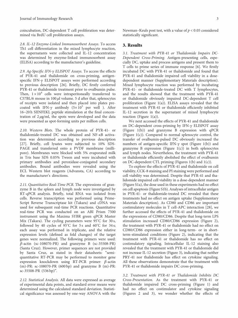
3.1. Treatment with PYR-41 or Thalidomide Impairs DC-Dependent Cross-Priming. Antigen-presenting cells, espe-cially DC, uptake and process antigens and present them toT cells to prime series of immune response [6]. We firstlyincubated DC with PYR-41 or thalidomide and found thatPYR-41 and thalidomide impaired cell viability in a dose-dependent manner (Supplementary Materials description).Mixed lymphocyte reaction was performed by incubatingPYR-41- or thalidomide-treated DC with T lymphocytes,and the results showed that the treatment with PYR-41or thalidomide obviously impaired DC-dependent T cellproliferation (Figure 1(a)). ELISA assays revealed that thetreatment with PYR-41 or thalidomide efficiently inhibitedIL-12 secretion in the supernatant of mixed lymphocytereaction (Figure 1(a)).
We next accessed the effects of PYR-41 and thalidomideon DC-dependent cross-priming by IFN-γ ELISPOT assay(Figure 1(b)) and granzyme B expression with qPCR(Figure 1(c)). Compared to normal splenocyte control, thetransfer of ovalbumin-pulsed DC obviously increased thenumbers of antigen-specific IFN-γ spot (Figure 1(b)) andgranzyme B expression (Figure 1(c)) in both splenocytesand lymph nodes. Nevertheless, the treatment with PYR-41or thalidomide efficiently abolished the effect of ovalbuminon DC-dependent CTL priming (Figures 1(b) and 1(c)).
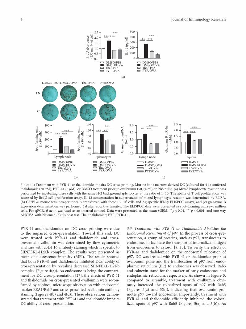
To explore the effects of PYR-41 and thalidomide on cellviability, CCK-8 staining and PI staining were performed andcell viability was determined. Despite that PYR-41 and tha-lidomide impaired cell viability in a dose-dependent manner(Figure S1a), the dose used in these experiments had no effecton cell apoptosis (Figure S1b). Analyses of intracellular antigenin PYR-41- or thalidomide-treated DC revealed that thesetreatments had no effect on antigen uptake (SupplementaryMaterials description). As CD80 and CD86 are importantcostimulatory molecules in T cell-APC interaction [28], wefurther accessed the effects of PYR-41 and thalidomide onthe expressions of CD80/CD86. Despite that long-term LPSstimulation increased CD80/CD86 expression (Figure 2),the treatment with PYR-41 or thalidomide had no effect onCD80/CD86 expression either in long-term- or in short-term-stimulated conditions (Figure 2), indicating that thetreatment with PYR-41 or thalidomide has no effect oncostimulatory signaling. Intracellular IL-12 staining alsorevealed that the treatment with PYR-41 or thalidomide didnot increase IL-12 secretion (Figure 3), indicating that neitherPRY-41 nor thalidomide has effect on cytokine signaling.All these observations demonstrate that the treatment withPYR-41 or thalidomide impairs DC cross-priming.
3.2. Treatment with PYR-41 or Thalidomide Inhibits DCCross-Presentation. As the treatment with PYR-41 orthalidomide impaired DC cross-priming (Figure 1) andhad no effect on costimulator and cytokine signaling(Figures 2 and 3), we wonder whether the effects of
3Journal of Immunology Research
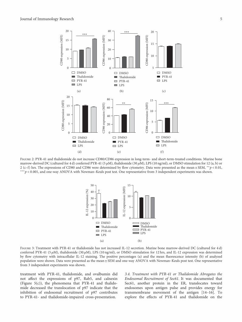
PYR-41 and thalidomide on DC cross-priming were dueto the impaired cross-presentation. Toward this end, DCwere treated with PYR-41 and thalidomide and cross-presented ovalbumin was determined by flow cytometricanalyzes with 25D1.16 antibody staining which is specific toSIINFEKL-H2Kb complex. The results were presented asmean of fluorescence intensity (MFI). The results showedthat both PYR-41 and thalidomide inhibited DCs’ ability ofcross-presentation by revealing decreased SIINFEKL-H2Kbcomplex (Figure 4(a)). As endosome is being the compart-ment for DC cross-presentation [27], the effects of PYR-41and thalidomide on cross-presented ovalbumin were recon-firmed by confocal microscope observation with endosomalmarker EEA1/Rab7 and cross-presented ovalbumin antibodystaining (Figures 4(b) and 4(d)). These observations demon-strated that treatment with PYR-41 and thalidomide impairsDC ability of cross-presentation.
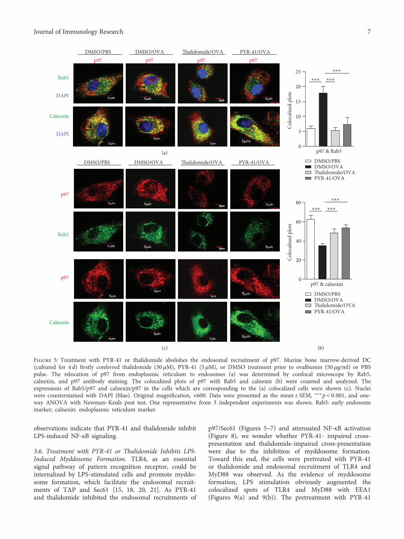
3.3. Treatment with PYR-41 or Thalidomide Abolishes theEndosomal Recruitment of p97. In the process of cross-pre-sentation, a group of proteins, such as p97, translocates toendosomes to facilitate the transport of internalized antigenfrom endosomes to cytosol [8, 13]. To verify the effects ofPYR-41 and thalidomide on the endosomal relocation ofp97, DC was treated with PYR-41 or thalidomide prior toovalbumin pulse and the translocation of p97 from endo-plasmic reticulum (ER) to endosomes was observed. Rab5and calnexin stand for the marker of early endosomes andendoplasmic reticulum, respectively. As shown in Figure 5,compared to scramble, treatment with ovalbumin obvi-ously increased the colocalized spots of p97 with Rab5(Figures 5(a) and 5(b)), indicating that ovalbumin pro-motes p97 toward endosomes. Importantly, treatment withPYR-41 and thalidomide efficiently inhibited the coloca-lized spots of p97 with Rab5 (Figures 5(a) and 5(b)). As
0100200300400500
DMSO/PBSDMSO/OVATha/OVAPYR/OVA
IL-1
2 (p
g/m
l)
0.00.51.01.52.02.5
DMSO/PBSDMSO/OVATha/OVAPYR/OVA
BrdU
abso
rban
ce(4
50 n
m, %
of c
trl)
⁎⁎⁎⁎⁎⁎⁎⁎⁎ ⁎⁎⁎
⁎⁎⁎⁎⁎⁎
(a)
DMSO/PBS DMSO/OVA Tha/OVA PYR/OVA
LN
SP
0
100
200
300
400
Lymph node
Spot
-form
ing
units
(1×
106 ce
lls)
40
60
80
100
120
140
Splenocytes
Spot
-form
ing
units
(1×
106 ce
lls)⁎⁎⁎
⁎⁎⁎⁎⁎⁎
⁎⁎⁎
⁎⁎⁎ ⁎⁎
DMSO/PBSDMSO/OVATha/OVAPYR/OVA
DMSO/PBSDMSO/OVATha/OVAPYR/OVA
(b)
DMSODMSO/OVATha/OVAPYR/OVA
Lymph nodeDMSODMSO/OVATha/OVAPYR/OVA
Spleen
⁎⁎⁎⁎⁎⁎⁎⁎ ⁎⁎⁎
⁎⁎⁎ ⁎⁎⁎
0.0
0.5
1.0
1.5
2.0G
ranz
yme B
mRN
A(fo
lds)
0.0
0.5
1.0
1.5
2.0
2.5
Gra
nzym
e B m
RNA
(fold
s)
(c)
Figure 1: Treatment with PYR-41 or thalidomide impairs DC cross-priming. Murine bone marrow-derived DC (cultured for 4 d) conferredthalidomide (30 μM), PYR-41 (5 μM), or DMSO treatment prior to ovalbumin (50 μg/ml) or PBS pulse. (a) Mixed lymphocyte reaction wasperformed by incubating these cells with the same H-2 background splenocytes at the ratio of 1 : 10. The ability of T cell proliferation wasaccessed by BrdU cell proliferation assay. IL-12 concentration in supernatants of mixed lymphocyte reaction was determined by ELISA.(b) C57BL/6 mouse was intraperitoneally transferred with these 1× 10⁴ cells and Ag-specific IFN-γ ELISPOT assays, and (c) granzyme Bexpression determination was performed 5 d after adoptive transfer. The ELISPOT data were presented as spot-forming units per millioncells. For qPCR, β-actin was used as an internal control. Data were presented as the mean± SEM, ∗∗p < 0 01, ∗∗∗p < 0 001, and one-wayANOVA with Newman–Keuls post test. Tha: thalidomide; PYR: PYR-41.
4 Journal of Immunology Research
treatment with PYR-41, thalidomide, and ovalbumin didnot affect the expressions of p97, Rab5, and calnexin(Figure 5(c)), the phenomena that PYR-41 and thalido-mide decreased the translocation of p97 indicate that theinhibition of endosomal recruitment of p97 contributesto PYR-41- and thalidomide-impaired cross-presentation.
3.4. Treatment with PYR-41 or Thalidomide Abrogates theEndosomal Recruitment of Sec61. It was documented thatSec61, another protein in the ER, translocates towardendosomes upon antigen pulse and provides energy fortransmembrane movement of the antigen [14–16]. Toexplore the effects of PYR-41 and thalidomide on the
0
5
10
15
20
DMSO
PYR-41LPS
Thalidomide
CD80
expr
essio
n (M
FI) ⁎⁎⁎
(a)
0
10
20
30
40
CD86
expr
essio
n (M
FI)
⁎⁎⁎
DMSO
PYR-41LPS
Thalidomide
(b)
5
10
15
20
DMSOPYR-41LPS
CD80
expr
essio
n (M
FI)
(c)
0
5
10
15
20
DMSOThalidomideLPS
CD80
expr
essio
n (M
FI)
(d)
0
20
40
60
80
DMSOPYR-41LPS
CD86
expr
essio
n (M
FI) ⁎⁎
(e)
0
5
10
15
DMSOThalidomideLPS
CD86
expr
essio
n (M
FI) ⁎⁎⁎
(f)
Figure 2: PYR-41 and thalidomide do not increase CD80/CD86 expression in long-term- and short-term-treated conditions. Murine bonemarrow-derived DC (cultured for 4 d) conferred PYR-41 (5 μM), thalidomide (30 μM), LPS (10 ng/ml), or DMSO stimulation for 12 (a, b) or2 (c–f) hrs. The expressions of CD80 and CD86 were determined by flow cytometry. Data were presented as the mean± SEM, ∗∗p < 0 01,∗∗∗p < 0 001, and one-way ANOVA with Newman–Keuls post test. One representative from 3 independent experiments was shown.
0
10
20
30
40
50
DMSOThalidomidePYR-41LPS
IL-1
2 ex
pres
sion
(%)
(a)
0
5
10
15
DMSOThalidomidePYR-41LPS
IL-1
2 ex
pres
sion
(MFI
)
(b)
Figure 3: Treatment with PYR-41 or thalidomide has not increased IL-12 secretion. Murine bone marrow-derived DC (cultured for 4 d)conferred PYR-41 (5 μM), thalidomide (30 μM), LPS (10 ng/ml), or DMSO stimulation for 12 hrs, and IL-12 expression was determinedby flow cytometry with intracellular IL-12 staining. The positive percentages (a) and the mean fluorescence intensity (b) of analyzedpopulation were shown. Data were presented as the mean± SEM and one-way ANOVA with Newman–Keuls post test. One representativefrom 3 independent experiments was shown.
5Journal of Immunology Research
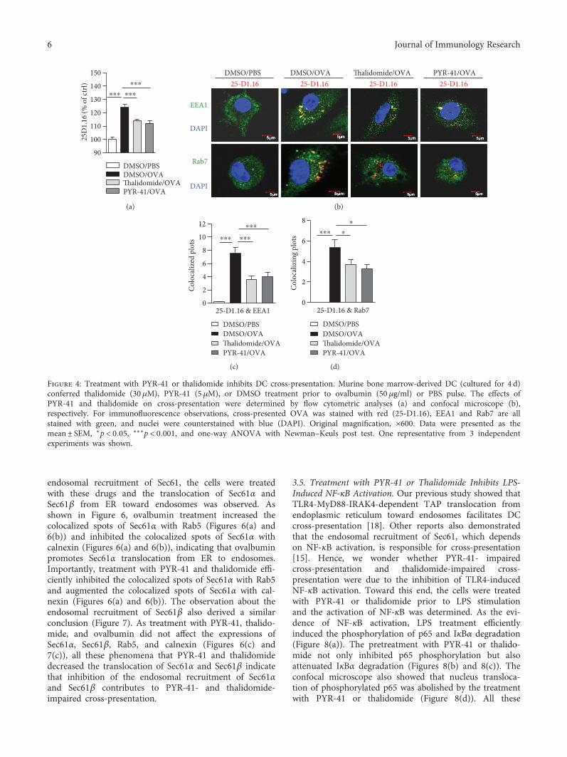
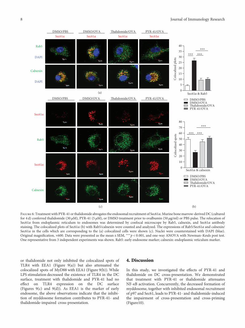
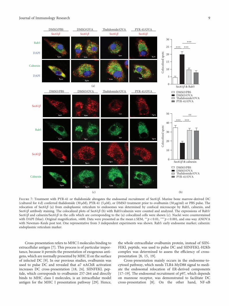
endosomal recruitment of Sec61, the cells were treatedwith these drugs and the translocation of Sec61α andSec61β from ER toward endosomes was observed. Asshown in Figure 6, ovalbumin treatment increased thecolocalized spots of Sec61α with Rab5 (Figures 6(a) and6(b)) and inhibited the colocalized spots of Sec61α withcalnexin (Figures 6(a) and 6(b)), indicating that ovalbuminpromotes Sec61α translocation from ER to endosomes.Importantly, treatment with PYR-41 and thalidomide effi-ciently inhibited the colocalized spots of Sec61α with Rab5and augmented the colocalized spots of Sec61α with cal-nexin (Figures 6(a) and 6(b)). The observation about theendosomal recruitment of Sec61β also derived a similarconclusion (Figure 7). As treatment with PYR-41, thalido-mide, and ovalbumin did not affect the expressions ofSec61α, Sec61β, Rab5, and calnexin (Figures 6(c) and7(c)), all these phenomena that PYR-41 and thalidomidedecreased the translocation of Sec61α and Sec61β indicatethat inhibition of the endosomal recruitment of Sec61αand Sec61β contributes to PYR-41- and thalidomide-impaired cross-presentation.
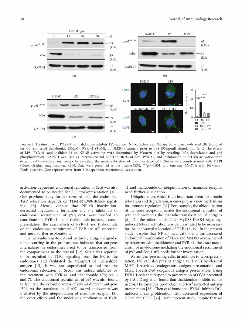
3.5. Treatment with PYR-41 or Thalidomide Inhibits LPS-Induced NF-κB Activation. Our previous study showed thatTLR4-MyD88-IRAK4-dependent TAP translocation fromendoplasmic reticulum toward endosomes facilitates DCcross-presentation [18]. Other reports also demonstratedthat the endosomal recruitment of Sec61, which dependson NF-κB activation, is responsible for cross-presentation[15]. Hence, we wonder whether PYR-41- impairedcross-presentation and thalidomide-impaired cross-presentation were due to the inhibition of TLR4-inducedNF-κB activation. Toward this end, the cells were treatedwith PYR-41 or thalidomide prior to LPS stimulationand the activation of NF-κB was determined. As the evi-dence of NF-κB activation, LPS treatment efficientlyinduced the phosphorylation of p65 and IκBα degradation(Figure 8(a)). The pretreatment with PYR-41 or thalido-mide not only inhibited p65 phosphorylation but alsoattenuated IκBα degradation (Figures 8(b) and 8(c)). Theconfocal microscope also showed that nucleus transloca-tion of phosphorylated p65 was abolished by the treatmentwith PYR-41 or thalidomide (Figure 8(d)). All these
90
100
110
120
130
140
150
DMSO/PBS
Thalidomide/OVAPYR-41/OVA
DMSO/OVA
25D
1.16
(% o
f ctr
l) ⁎⁎⁎
⁎⁎⁎⁎⁎⁎
(a)
EEA1
DAPI
Rab7
DAPI
25-D1.1625-D1.1625-D1.16 25-D1.16DMSO/PBS DMSO/OVA Thalidomide/OVA PYR-41/OVA
(b)
0
2
4
6
8
10
12
DMSO/PBSDMSO/OVAThalidomide/OVAPYR-41/OVA
25-D1.16 & EEA1
Colo
caliz
ed p
lots
⁎⁎⁎
⁎⁎⁎⁎⁎⁎
(c)
0
2
4
6
8
DMSO/PBSDMSO/OVAThalidomide/OVAPYR-41/OVA
25-D1.16 & Rab7
Colo
caliz
ing
plot
s
⁎
⁎⁎⁎⁎
(d)
Figure 4: Treatment with PYR-41 or thalidomide inhibits DC cross-presentation. Murine bone marrow-derived DC (cultured for 4 d)conferred thalidomide (30 μM), PYR-41 (5 μM), or DMSO treatment prior to ovalbumin (50 μg/ml) or PBS pulse. The effects ofPYR-41 and thalidomide on cross-presentation were determined by flow cytometric analyses (a) and confocal microscope (b),respectively. For immunofluorescence observations, cross-presented OVA was stained with red (25-D1.16), EEA1 and Rab7 are allstained with green, and nuclei were counterstained with blue (DAPI). Original magnification, ×600. Data were presented as themean± SEM, ∗p < 0 05, ∗∗∗p < 0 001, and one-way ANOVA with Newman–Keuls post test. One representative from 3 independentexperiments was shown.
6 Journal of Immunology Research
observations indicate that PYR-41 and thalidomide inhibitLPS-induced NF-κB signaling.
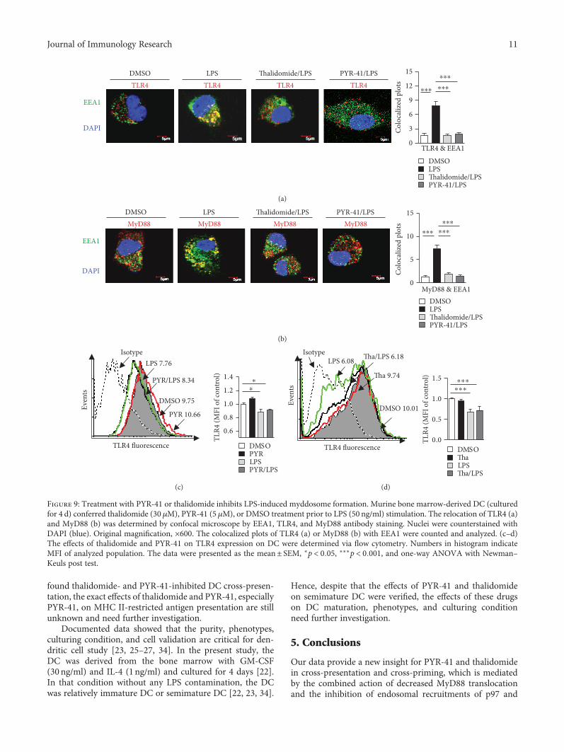
3.6. Treatment with PYR-41 or Thalidomide Inhibits LPS-Induced Myddosome Formation. TLR4, as an essentialsignal pathway of pattern recognition receptor, could beinternalized by LPS-stimulated cells and promote myddo-some formation, which facilitate the endosomal recruit-ments of TAP and Sec61 [15, 18, 20, 21]. As PYR-41and thalidomide inhibited the endosomal recruitments of
p97/Sec61 (Figures 5–7) and attenuated NF-κB activation(Figure 8), we wonder whether PYR-41- impaired cross-presentation and thalidomide-impaired cross-presentationwere due to the inhibition of myddosome formation.Toward this end, the cells were pretreated with PYR-41or thalidomide and endosomal recruitment of TLR4 andMyD88 was observed. As the evidence of myddosomeformation, LPS stimulation obviously augmented thecolocalized spots of TLR4 and MyD88 with EEA1(Figures 9(a) and 9(b)). The pretreatment with PYR-41
Calnexin
p97p97 p97 p97
DAPI
Rab5
DAPI
DMSO/PBS DMSO/OVA Thalidomide/OVA PYR-41/OVA
(a)
Rab5
p97
DMSO/PBS DMSO/OVA Thalidomide/OVA PYR-41/OVA
Calnexin
p97
(c)
0
5
10
15
20
25
DMSO/PBSDMSO/OVAThalidomide/OVAPYR-41/OVA
p97 & Rab5
Colo
caliz
ed p
lots
0
20
40
60
80
DMSO/PBSDMSO/OVAThalidomide/OVAPYR-41/OVA
p97 & calnexinCo
loca
lized
plo
ts
⁎⁎⁎
⁎⁎⁎⁎⁎⁎
⁎⁎⁎
⁎⁎⁎⁎⁎⁎
(b)
Figure 5: Treatment with PYR-41 or thalidomide abolishes the endosomal recruitment of p97. Murine bone marrow-derived DC(cultured for 4 d) firstly conferred thalidomide (30 μM), PYR-41 (5 μM), or DMSO treatment prior to ovalbumin (50 μg/ml) or PBSpulse. The relocation of p97 from endoplasmic reticulum to endosomes (a) was determined by confocal microscope by Rab5,calnexin, and p97 antibody staining. The colocalized plots of p97 with Rab5 and calnexin (b) were counted and analyzed. Theexpressions of Rab5/p97 and calnexin/p97 in the cells which are corresponding to the (a) colocalized cells were shown (c). Nucleiwere counterstained with DAPI (blue). Original magnification, ×600. Data were presented as the mean± SEM, ∗∗∗p < 0 001, and one-way ANOVA with Newman–Keuls post test. One representative from 3 independent experiments was shown. Rab5: early endosomemarker; calnexin: endoplasmic reticulum marker.
7Journal of Immunology Research
or thalidomide not only inhibited the colocalized spots ofTLR4 with EEA1 (Figure 9(a)) but also attenuated thecolocalized spots of MyD88 with EEA1 (Figure 9(b)). WhileLPS stimulation decreased the existence of TLR4 in the DCsurface, treatment with thalidomide and PYR-41 had noeffect on TLR4 expression on the DC surface(Figures 9(c) and 9(d)). As EEA1 is the marker of earlyendosome, the above observations indicate that the inhibi-tion of myddosome formation contributes to PYR-41- andthalidomide-impaired cross-presentation.
4. Discussion
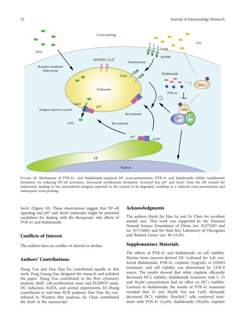
In this study, we investigated the effects of PYR-41 andthalidomide on DC cross-presentation. We demonstratedthat treatment with PYR-41 or thalidomide attenuatesNF-κB activation. Concurrently, the decreased formation ofmyddosome, together with inhibited endosomal recruitmentof p97 and Sec61, leads to PYR-41- and thalidomide-inducedthe impairment of cross-presentation and cross-priming(Figure10).
Calnexin
DAPI
Rab5
DAPI
DMSO/PBS DMSO/OVA Thalidomide/OVA PYR-41/OVASec61�훼Sec61�훼 Sec61�훼 Sec61�훼
(a)
Rab5
DMSO/PBS DMSO/OVA Thalidomide/OVA PYR-41/OVA
Calnexin
Sec61�훼
Sec61�훼
(c)
0
5
10
15
20
25
30
35
40
DMSO/PBSDMSO/OVAThalidomide/OVAPYR-41/OVA
Sec61�훼 & Rab5
Colo
caliz
ed p
lots
0
10
20
30
40
50
60
70
80
DMSO/PBSDMSO/OVAThalidomide/OVAPYR-41/OVA
Sec61�훼 & calnexinCo
loco
lized
spot
s
⁎⁎⁎
⁎⁎⁎⁎⁎⁎
⁎⁎⁎
⁎⁎⁎⁎⁎⁎
(b)
Figure 6: Treatment with PYR-41 or thalidomide abrogates the endosomal recruitment of Sec61α. Murine bonemarrow-derivedDC (culturedfor 4 d) conferred thalidomide (30 μM), PYR-41 (5 μM), or DMSO treatment prior to ovalbumin (50 μg/ml) or PBS pulse. The relocation ofSec61α from endoplasmic reticulum to endosomes was determined by confocal microscope by Rab5, calnexin, and Sec61α antibodystaining. The colocalized plots of Sec61α (b) with Rab5/calnexin were counted and analyzed. The expressions of Rab5/Sec61α and calnexin/Sec61α in the cells which are corresponding to the (a) colocalized cells were shown (c). Nuclei were counterstained with DAPI (blue).Original magnification, ×600. Data were presented as the mean± SEM, ∗∗∗p < 0 001, and one-way ANOVA with Newman–Keuls post test.One representative from 3 independent experiments was shown. Rab5: early endosome marker; calnexin: endoplasmic reticulum marker.
8 Journal of Immunology Research
Cross-presentation refers to MHC I molecules binding toextracellular antigen [7]. This process is of particular impor-tance, because it permits the presentation of exogenous anti-gens, which are normally presented byMHC II on the surfaceof infected DC [9]. In our previous studies, ovalbumin wasused to pulse DC and revealed that α7 nAChR activationincreases DC cross-presentation [18, 24]. SIINFEKL pep-tide, which corresponds to ovalbumin 257-264 and directlybinds to MHC class I molecules, is an intracellular modelantigen for the MHC I presentation pathway [29]. Hence,
the whole extracellular ovalbumin protein, instead of SIIN-FEKL peptide, was used to pulse DC and SIINFEKL-H2Kbcomplex was determined to assess the efficiency of cross-presentation [8, 15, 19].
Cross-presentation mainly occurs in the endosome-to-cytosol pathway, which needs TLR4-MyD88 signal to medi-ate the endosomal relocation of ER-derived components[17–19]. The endosomal recruitment of p97, which dependson mannose receptor, was demonstrated to facilitate DCcross-presentation [8]. On the other hand, NF-κB
Calnexin
DAPI
Rab5
DAPI
DMSO/PBS DMSO/OVA Thalidomide/OVA PYR-41/OVASec61�훽 Sec61�훽 Sec61�훽 Sec61�훽
(a)
Rab5
DMSO/PBS DMSO/OVA Thalidomide/OVA PYR-41/OVA
Calnexin
Sec61�훽
Sec61�훽
(c)
0
5
10
15
20
25
30
DMSO/PBSDMSO/OVAThalidomide/OVAPYR-41/OVA
Sec61�훽 & Rab5
Colo
coliz
ed sp
ots
0
10
20
30
40
50
DMSO/PBSDMSO/OVAThalidomide/OVAPYR-41/OVA
Sec61�훽 & calnexinCo
loco
lized
spot
s
⁎⁎⁎
⁎⁎⁎⁎⁎⁎
⁎⁎⁎ ⁎⁎⁎
⁎⁎
(b)
Figure 7: Treatment with PYR-41 or thalidomide abrogates the endosomal recruitment of Sec61β. Murine bone marrow-derived DC(cultured for 4 d) conferred thalidomide (30 μM), PYR-41 (5 μM), or DMSO treatment prior to ovalbumin (50 μg/ml) or PBS pulse. Therelocation of Sec61β (a) from endoplasmic reticulum to endosomes was determined by confocal microscope by Rab5, calnexin, andSec61β antibody staining. The colocalized plots of Sec61β (b) with Rab5/calnexin were counted and analyzed. The expressions of Rab5/Sec61β and calnexin/Sec61β in the cells which are corresponding to the (a) colocalized cells were shown (c). Nuclei were counterstainedwith DAPI (blue). Original magnification, ×600. Data were presented as the mean± SEM, ∗∗p < 0 01, ∗∗∗p < 0 001, and one-way ANOVAwith Newman–Keuls post test. One representative from 3 independent experiments was shown. Rab5: early endosome marker; calnexin:endoplasmic reticulum marker.
9Journal of Immunology Research
activation-dependent endosomal relocation of Sec6 was alsodocumented to be needed for DC cross-presentation [15].Our previous study further revealed that the endosomalTAP relocation depends on TLR4-MyD88-IRAK4 signal-ing [18]. Hence, despite that NF-κB inactivation-decreased myddosome formation and the inhibition ofendosomal recruitment of p97/Sec61 were verified tocontribute to PYR-41- and thalidomide-impaired cross-presentation, the exact effects of PYR-41 and thalidomideon the endosomal recruitment of TAP are still uncertainand need further explorations.
In the endosome-to-cytosol pathway, antigen degrada-tion occurring in the proteasomes indicates that antigensinternalized in endosomes need to be transported fromthe compartment to the cytosol [12]. Sec61 was reportedto be recruited by TLR4 signaling from the ER to theendosomes and facilitated the transport of internalizedantigen [15]. It was not unpredicted to find that theendosomal relocation of Sec61 was indeed inhibited bythe treatment with PYR-41 and thalidomide (Figures 6and 7). The endosomal recruitment of p97 was also foundto facilitate the cytosolic access of several different antigens[30]. As the translocation of p97 toward endosomes wasmediated by the ubiquitination of mannose receptor [8],the exact effects and the underlying mechanism of PYR-
41 and thalidomide on ubiquitination of mannose receptorneed further elucidation.
Ubiquitination, which is an important event for proteinrelocation and degradation, is emerging as a new mechanismfor immune regulation [31]. For example, the ubiquitinationof mannose receptor mediates the endosomal relocation ofp97 and promotes the cytosolic translocation of antigens[8]. On the other hand, TLR4-MyD88-IRAK4 signaling-induced NF-κB activation was demonstrated to be necessaryfor the endosomal relocation of TAP [18, 19]. In the presentstudy, despite that NF-κB inactivation and the decreasedendosomal translocation of TLR4 and MyD88 were achievedby treatment with thalidomide and PYR-41, the exact mech-anism of myddosome mediating the endosomal recruitmentof p97 and Sec61 still needs further investigation.
As antigen-presenting cells, in addition to cross-presen-tation, DC can also present antigen to T cells by classicalMHC I-restricted endogenous antigen presentation andMHC II-restricted exogenous antigen presentation. UsingDO11.1 cells that respond to presentation of OVA presentedby I-Ad, Deng et al. found that thalidomide inhibits tumornecrosis factor-alpha production and I-Ad restricted antigenpresentation [32]. Chen et al found that PYR41 inhibits DC-induced T cell proliferation with decreased expression ofCD69 and CD25 [33]. In the present study, despite that we
37(kDa)
65(kDa)
39(kDa)
15 30 600 (min)LPS 50 ng/ml
I�휅B�훼
p-p65Ser536
GAPDH
(a)
37(kDa)
65(kDa)
39(kDa)
DMSO LPS/PYRLPS
I�휅B�훼
GAPDH
p-p65Ser536
(b)
p-p65Ser536
37(kDa)
65(kDa)
39(kDa)
DMSO LPS/ThaLPS
I�휅B�훼
GAPDH
(c)
0
10
20
30
DMSOLPSTha/LPSPYR/LPS
⁎⁎⁎⁎⁎⁎⁎⁎⁎
Plot
s in
nucl
eus
p-p65
DAPI
p-p65
DAPI
DMSO LPS
Thalidomide/LPS PYR-41/LPS
(d)
Figure 8: Treatment with PYR-41 or thalidomide inhibits LPS-induced NF-κB activation. Murine bone marrow-derived DC (culturedfor 4 d) conferred thalidomide (30 μM), PYR-41 (5 μM), or DMSO treatment prior to LPS (50 ng/ml) stimulation. (a–c) The effectsof LPS, PYR-41, and thalidomide on NF-κB activation were determined by Western blot by revealing IκBα degradation and p65phosphorylation. GAPDH was used as internal control. (d) The effects of LPS, PYR-41, and thalidomide on NF-κB activation weredetermined by confocal microscope by revealing the nuclei relocation of phosphorylated p65. Nuclei were counterstained with DAPI(blue). Original magnification, ×600. Data were presented as the mean± SEM, ∗∗∗p < 0 001, and one-way ANOVA with Newman–Keuls post test. One representative from 3 independent experiments was shown.
10 Journal of Immunology Research
found thalidomide- and PYR-41-inhibited DC cross-presen-tation, the exact effects of thalidomide and PYR-41, especiallyPYR-41, on MHC II-restricted antigen presentation are stillunknown and need further investigation.
Documented data showed that the purity, phenotypes,culturing condition, and cell validation are critical for den-dritic cell study [23, 25–27, 34]. In the present study, theDC was derived from the bone marrow with GM-CSF(30ng/ml) and IL-4 (1 ng/ml) and cultured for 4 days [22].In that condition without any LPS contamination, the DCwas relatively immature DC or semimature DC [22, 23, 34].
Hence, despite that the effects of PYR-41 and thalidomideon semimature DC were verified, the effects of these drugson DC maturation, phenotypes, and culturing conditionneed further investigation.
5. Conclusions
Our data provide a new insight for PYR-41 and thalidomidein cross-presentation and cross-priming, which is mediatedby the combined action of decreased MyD88 translocationand the inhibition of endosomal recruitments of p97 and
0
3
6
9
12
15
DMSOLPSThalidomide/LPS
⁎⁎⁎ ⁎⁎⁎
⁎⁎⁎
PYR-41/LPS
TLR4 & EEA1
Colo
caliz
ed p
lotsTLR4TLR4
EEA1
DAPI
TLR4 TLR4DMSO LPS Thalidomide/LPS PYR-41/LPS
(a)
DMSOLPSThalidomide/LPSPYR-41/LPS
0
5
10
15
MyD88 & EEA1
Colo
caliz
ed p
lots
EEA1
DAPI
MyD88 MyD88 MyD88MyD88DMSO LPS Thalidomide/LPS PYR-41/LPS
⁎⁎⁎ ⁎⁎⁎
⁎⁎⁎
(b)
0.8
0.6
1.0
1.2
1.4
TLR4
(MFI
of c
ontr
ol)
IsotypeLPS 7.76
DMSO 9.75
PYR/LPS 8.34
TLR4 fluorescence
Even
ts
PYR 10.66
DMSOPYRLPSPYR/LPS
⁎⁎
(c)
0.0
0.5
1.0
1.5
TLR4
(MFI
of c
ontr
ol)
TLR4 fluorescence
IsotypeLPS 6.08
DMSO 10.01
Tha 9.74
Tha/LPS 6.18
Even
ts
DMSOThaLPSTha/LPS
⁎⁎⁎⁎⁎⁎
(d)
Figure 9: Treatment with PYR-41 or thalidomide inhibits LPS-induced myddosome formation. Murine bone marrow-derived DC (culturedfor 4 d) conferred thalidomide (30 μM), PYR-41 (5 μM), or DMSO treatment prior to LPS (50 ng/ml) stimulation. The relocation of TLR4 (a)and MyD88 (b) was determined by confocal microscope by EEA1, TLR4, and MyD88 antibody staining. Nuclei were counterstained withDAPI (blue). Original magnification, ×600. The colocalized plots of TLR4 (a) or MyD88 (b) with EEA1 were counted and analyzed. (c–d)The effects of thalidomide and PYR-41 on TLR4 expression on DC were determined via flow cytometry. Numbers in histogram indicateMFI of analyzed population. The data were presented as the mean± SEM, ∗p < 0 05, ∗∗∗p < 0 001, and one-way ANOVA with Newman–Keuls post test.
11Journal of Immunology Research
Sec61 (Figure 10). These observations suggest that NF-κBsignaling and p97 and Sec61 molecules might be potentialcandidates for dealing with the therapeutic side effects ofPYR-41 and thalidomide.
Conflicts of Interest
The authors have no conflict of interest to declare.
Authors’ Contributions
Xiang You and Dan Dan Xu contributed equally to thiswork. Feng Guang Gao designed the research and polishedthe paper. Xiang You contributed to the flow cytometryanalyses, BrdU cell proliferation assay and ELISPOT assay,DC induction, ELISA, and animal experiments; Di Zhangcontributed to real-time PCR analyses; Dan Dan Xu con-tributed to Western blot analyses; Jie Chen contributedthe draft of the manuscript.
Acknowledgments
The authors thank Jin Hua Su and Fu Chen for excellentanimal care. This work was supported by the NationalNatural Science Foundation of China (no. 81273203 andno. 81771669) and the State Key Laboratory of Oncogenesand Related Genes (no. 90-14-05).
Supplementary Materials
The effects of PYR-41 and thalidomide on cell viability.Murine bone marrow-derived DC (cultured for 4 d) con-ferred thalidomide, PYR-41, cisplatin (4μg/ml), or DMSOtreatment, and cell viability was determined by CCK-8assays. The results showed that while cisplatin efficientlydecreased DC’s viability, thalidomide treatment with 5, 15and 30μM concentration had no effect on DC’s viability.Contract to thalidomide, the results of PYR-41 treatmentrevealed that 15 and 30μM, but not 5μM, obviouslydecreased DC’s viability. Raw264.7 cells conferred treat-ment with PYR-41 (5μM), thalidomide (30μM), cisplatin
OVA
Receptor-medietedendocytosis
SIINFEKL-H2kb
Cross-priming
Translocation
TLR4
LPS
MyD88
Thalidomide
IKK�훾
I�휅B�훼
p65 p50
PYR-41+
Sec61
Nucleus
Sec61
p97
p97
Endosome
TLR4
Recruitment
Recruitment
ER
OVA
Antigen export to cytosol
P
P
Figure 10: Mechanism of PYR-41- and thalidomide-impaired DC cross-presentation. PYR-41 and thalidomide inhibit myddosomeformation via reducing NF-κB activation. Decreased myddosome formation recruited less p97 and Sec61 from the ER toward theendosomes, leading to less internalized antigens exported to the cytosol to be degraded, resulting in a reduced cross-presentation andsubsequent cross-priming.
12 Journal of Immunology Research
(4μg/ml), or DMSO for 12 hrs, and the cell viability wasdetermined by flow cytometric analyses with propidiumiodide (PI) staining. The results revealed that cisplatin,but not PYR-41 and thalidomide, obviously induced cellapoptosis. The concentration of PYR-41 and thalidomideused in this study had no effect on DC endocytosis.Murine bone marrow-derived DC (cultured for 4 d) con-ferred PYR-41 (5μM), thalidomide (30μM), or DMSOtreatment prior to 15min ovalbumin FITC (5μg/ml) pulseat 37°C. The cells incubated with ovalbumin FITC at 4°Cwere used as negative control. The effect of PYR-41 andthalidomide on cell endocytosis was determined by flowcytometry. The results showed that, while DMSO treat-ment reveals 6.83% ovalbumin-positive cells, treatmentwith PYR-41 and thalidomide achieved 5.89% and 5.82%ovalbumin-positive cells, indicating that the concentrationof PYR-41 and thalidomide used in this study had noeffect on DC endocytosis. (Supplementary Materials)
References
[1] K. Schweitzer, A. Pralow, and M. Naumann, “p97/VCP pro-motes Cullin-RING-ubiquitin-ligase /proteasome-dependentdegradation of IκBα and the preceding liberation of RelAfrom ubiquitinated IκBα,” Journal of Cellular and MolecularMedicine, vol. 20, no. 1, pp. 58–70, 2016.
[2] Y. Yang, J. Kitagaki, R. M. Dai et al., “Inhibitors ofubiquitin-activating enzyme (E1), a new class of potentialcancer therapeutics,” Cancer Research, vol. 67, no. 19,pp. 9472–9481, 2007.
[3] T. Moehler, “Clinical experience with thalidomide and lenali-domide in multiple myeloma,” Current Cancer Drug Targets,vol. 12, no. 4, pp. 372–390, 2012.
[4] T. Paravar and D. J. Lee, “Thalidomide: mechanisms ofaction,” International Reviews of Immunology, vol. 27, no. 3,pp. 111–135, 2008.
[5] N. Y. Hakami, G. J. Dusting, and H. M. Peshavariya, “Trichos-tatin A, a histone deacetylase inhibitor suppresses NADPHOxidase 4-derived redox signalling and angiogenesis,” Journalof Cellular and Molecular Medicine, vol. 20, no. 10, pp. 1932–1944, 2016.
[6] J. A. Villadangos, W. R. Heath, and F. R. Carbone, “Outsidelooking in: the inner workings of the crosspresentationpathway within dendritic cells,” Trends in Immunology,vol. 28, no. 2, pp. 45–47, 2007.
[7] O. P. Joffre, E. Segura, A. Savina, and S. Amigorena, “Cross-presentation by dendritic cells,” Nature Reviews. Immunology,vol. 12, no. 8, pp. 557–569, 2012.
[8] M. Zehner, A. I. Chasan, V. Schuette et al., “Mannose receptorpolyubiquitination regulates endosomal recruitment of p97and cytosolic antigen translocation for cross-presentation,”Proceedings of the National Academy of Sciences of the UnitedStates of America, vol. 108, no. 24, pp. 9933–9938, 2011.
[9] V. Schuette and S. Burgdorf, “The ins-and-outs of endosomalantigens for cross-presentation,” Current Opinion in Immu-nology, vol. 26, pp. 63–68, 2014.
[10] M. Kovacsovics-Bankowski and K. L. Rock, “A phagosome-to-cytosol pathway for exogenous antigens presented on MHCclass I molecules,” Science, vol. 267, no. 5195, pp. 243–246,1995.
[11] Y. Ye, H. H. Meyer, and T. A. Rapoport, “The AAA ATPaseCdc48/p97 and Its partners transport proteins from the ERinto the cytosol,”Nature, vol. 414, no. 6864, pp. 652–656, 2001.
[12] M. Zehner and S. Burgdorf, “Regulation of antigen transportinto the cytosol for cross-presentation by ubiquitination ofthe mannose receptor,” Molecular Immunology, vol. 55,no. 2, pp. 146–148, 2013.
[13] J. Ménager, F. Ebstein, R. Oger et al., “Cross-presentation ofsynthetic long peptides by human dendritic cells: a processdependent on ERAD component p97/VCP but not sec61and/or Derlin-1,” PLoS One, vol. 9, no. 2, article e89897, 2014.
[14] M. Zehner and S. Burgdorf, “Sec61 in antigen cross-presenta-tion,” Oncotarget, vol. 6, no. 24, pp. 19954-19955, 2015.
[15] M. Zehner, A. L. Marschall, E. Bos et al., “The translocon pro-tein Sec61 mediates antigen transport from endosomes in thecytosol for cross-presentation to CD8(+) T cells,” Immunity,vol. 42, no. 5, pp. 850–863, 2015.
[16] J. E. Grotzke, P. Kozik, J. D. Morel et al., “Sec61 blockade bymycolactone inhibits antigen cross-presentation indepen-dently of endosome-to-cytosol export,” Proceedings of theNational Academy of Sciences of the United States of America,vol. 114, no. 29, pp. E5910–E5919, 2017.
[17] P. Nair-Gupta, A. Baccarini, N. Tung et al., “TLR signalsinduce phagosomal MHC-I delivery from the endosomalrecycling compartment to allow cross-presentation,” Cell,vol. 158, no. 3, pp. 506–521, 2014.
[18] Y. Y. Wang, C. F. Hu, J. Li, X. You, and F. G. Gao, “Increasedtranslocation of antigens to endosomes and TLR4 mediatedendosomal recruitment of TAP contribute to nicotine aug-mented cross-presentation,” Oncotarget, vol. 7, no. 25,pp. 38451–38466, 2016.
[19] S. Burgdorf, C. Schölz, A. Kautz, R. Tampé, and C. Kurts,“Spatial and mechanistic separation of cross-presentationand endogenous antigen presentation,” Nature Immunology,vol. 9, no. 5, pp. 558–566, 2008.
[20] N. J. Gay, M. Gangloff, and L. A. O'Neill, “What the myddo-some structure tells us about the initiation of innate immu-nity,” Trends in Immunology, vol. 32, no. 3, pp. 104–109, 2011.
[21] R. Ferrao, H. Zhou, Y. Shan et al., “IRAK4 dimerization andtrans-autophosphorylation are induced by myddosomeassembly,” Molecular Cell, vol. 55, no. 6, pp. 891–903, 2014.
[22] M. Zhang, H. Tang, Z. Guo et al., “Splenic stroma drivesmature dendritic cells to differentiate into regulatory den-dritic cells,” Nature Immunology, vol. 5, no. 11, pp. 1124–1133, 2004.
[23] F. G. Gao, D. F. Wan, and J. R. Gu, “Ex vivo nicotinestimulation augments the efficacy of therapeutic bonemarrow–derived dendritic cell vaccination,” Clinical CancerResearch, vol. 13, no. 12, pp. 3706–3712, 2007.
[24] Y. Y. Wang, Y. W. Yang, X. You et al., “Ex vivo nicotinestimulation augments the efficacy of human peripheral bloodmononuclear cell-derived dendritic cell vaccination viaactivating Akt-S6 pathway,” Analytical Cellular Pathology,vol. 2015, Article ID 741487, 13 pages, 2015.
[25] F. G. Gao, H. T. Li, Z. J. Li, and J. R. Gu, “Nicotine stimulateddendritic cells could achieve anti-tumor effects in mouse lungand liver cancer,” Journal of Clinical Immunology, vol. 31,no. 1, pp. 80–88, 2011.
[26] F. Wang, Y. Y. Wang, J. Li et al., “Increased antigen presenta-tion but impaired T cells priming after upregulation ofinterferon-beta induced by lipopolysaccharides is mediated
13Journal of Immunology Research
by upregulation of B7H1 and GITRL,” PLoS One, vol. 9, no. 8,article e105636, 2014.
[27] H. J. Jin, H. T. Li, H. X. Sui et al., “Nicotine stimulated bonemarrow-derived dendritic cells could augment HBV specificCTL priming by activating PI3K-Akt pathway,” ImmunologyLetters, vol. 146, no. 1-2, pp. 40–49, 2012.
[28] A. N. Schweitzer and A. H. Sharpe, “Studies using antigen-presenting cells lacking expression of both B7-1 (CD80) andB7-2 (CD86) show distinct requirements for B7 moleculesduring priming versus restimulation of Th2 but not Th1cytokine production,” The Journal of Immunology, vol. 161,no. 6, pp. 2762–2771, 1998.
[29] S. Burgdorf, A. Kautz, V. Böhnert, P. A. Knolle, and C. Kurts,“Distinct pathways of antigen uptake and intracellular routingin CD4 and CD8 T cell activation,” Science, vol. 316, no. 5824,pp. 612–616, 2007.
[30] J. E. Grotzke and P. Cresswell, “Are ERAD componentsinvolved in cross-presentation?,” Molecular Immunology,vol. 68, no. 2, pp. 112–115, 2015.
[31] V. G. Bhoj and Z. J. Chen, “Ubiquitylation in innate and adap-tive immunity,” Nature, vol. 458, no. 7237, pp. 430–437, 2009.
[32] L. Deng, W. Ding, and R. D. Granstein, “Thalidomide nhibitstumor necrosis factor-alpha production and antigen presenta-tion by Langerhans cells,” The Journal of Investigative Derma-tology, vol. 121, no. 5, pp. 1060–1065, 2003.
[33] C. Chen, Y. Meng, L. Wang et al., “Ubiquitin-activatingenzyme E1 inhibitor PYR41 attenuates angiotensin II-induced activation of dendritic cells via the IκBa/NF-κB andMKP1/ERK/STAT1 pathways,” Immunology, vol. 142, no. 2,pp. 307–319, 2014.
[34] S. X. Hu, H. X. Sui, H. J. Jin et al., “Lipopolysaccharide anddose of nicotine determine the effects of nicotine on murinebone marrow-derived dendritic cells,” Molecular MedicineReports, vol. 5, no. 4, pp. 1005–1010, 2012.
14 Journal of Immunology Research

Stem Cells International
Hindawiwww.hindawi.com Volume 2018
Hindawiwww.hindawi.com Volume 2018
MEDIATORSINFLAMMATION
of
EndocrinologyInternational Journal of
Hindawiwww.hindawi.com Volume 2018
Hindawiwww.hindawi.com Volume 2018
Disease Markers
Hindawiwww.hindawi.com Volume 2018
BioMed Research International
OncologyJournal of
Hindawiwww.hindawi.com Volume 2013
Hindawiwww.hindawi.com Volume 2018
Oxidative Medicine and Cellular Longevity
Hindawiwww.hindawi.com Volume 2018
PPAR Research
Hindawi Publishing Corporation http://www.hindawi.com Volume 2013Hindawiwww.hindawi.com
The Scientific World Journal
Volume 2018
Immunology ResearchHindawiwww.hindawi.com Volume 2018
Journal of
ObesityJournal of
Hindawiwww.hindawi.com Volume 2018
Hindawiwww.hindawi.com Volume 2018
Computational and Mathematical Methods in Medicine
Hindawiwww.hindawi.com Volume 2018
Behavioural Neurology
OphthalmologyJournal of
Hindawiwww.hindawi.com Volume 2018
Diabetes ResearchJournal of
Hindawiwww.hindawi.com Volume 2018
Hindawiwww.hindawi.com Volume 2018
Research and TreatmentAIDS
Hindawiwww.hindawi.com Volume 2018
Gastroenterology Research and Practice
Hindawiwww.hindawi.com Volume 2018
Parkinson’s Disease
Evidence-Based Complementary andAlternative Medicine
Volume 2018Hindawiwww.hindawi.com
Submit your manuscripts atwww.hindawi.com