Chapter 5.9 THE USE OF D-AMINO ACIDS IN PEPTIDE DESIGN
16
In: D-Amino Acids: ISBN 1-60021-075-9 A New Frontier in Amino Acid and Protein Research © 2006 Nova Science Publishers, Inc. Chapter 5.9 THE USE OF D-AMINO ACIDS IN PEPTIDE DESIGN Radhakrishnan Mahalakshmi and Padmanabhan Balaram ∗ Molecular Biophysics Unit, Indian Institute of Science, India. Keywords: peptide hairpins, α-aminoisobutyric acid, Schellman motif, ambidextrous helices, LD peptides 1. INTRODUCTION Proteins and most naturally occurring peptides are composed of amino acids of the L-configuration. D- amino acids are found as constituents of natural peptides produced primarily, by microorganisms, using a non-ribosomal mechanism of synthesis. Research in this field dates back to over 60 years ago when Lipmann et al noted the presence of D-amino acids in tyrocidines and gramicidins [1]. Post-translational epimerization is an infrequently used mechanism in higher organisms for the introduction of D-amino acids into polypeptide chains [2]. Some notable examples are the production of opioid peptides, namely dermorphins and deltorphins form the skin of various frog species belonging to the Phyllomedusinae sub- family [3] and the contryphans produced by cone snails via post-translational epimerization [4]. Recently, total chemical synthesis of proteins containing all-D residues has been carried out and the characteristics and enzymatic activity of such enantiomeric proteins has been analyzed [5-7]. The incorporation of D- amino acids into polypeptide chains imposes local conformational constraints [8]. The purpose of this review is to examine the use of D-amino acids in the design of peptides adopting well-defined folded conformations. 2. CONFORMATIONAL CHARACTERISTICS OF D-AMINO ACIDS The effects of residue configuration on polypeptide chain conformation are readily understood by considering the Ramachandran maps for N-acetyl-L-Ala-NHMe and N-acetyl-D-Ala-NHMe shown in Fig. 1A & 1B [9-12]. The map for the D-residue is derived by inverting the classical L-Ala map through the origin. Most of the stereochemically allowed regions for the L-residues correspond to negative values of φ, while for D-residues, the allowed regions correspond to positive values of φ. The classical 3 10 and α-helical ∗ Correspondence concerning this article should be addressed to Padmanabhan Balaram, E-mail: [email protected], Fax: +91-80-23600535.
Transcript of Chapter 5.9 THE USE OF D-AMINO ACIDS IN PEPTIDE DESIGN
In: D-Amino Acids: ISBN 1-60021-075-9 A New Frontier in Amino Acid and Protein Research © 2006 Nova Science Publishers, Inc.
Chapter 5.9
THE USE OF D-AMINO ACIDS IN PEPTIDE DESIGN
Radhakrishnan Mahalakshmi and Padmanabhan Balaram∗
Molecular Biophysics Unit, Indian Institute of Science, India.
Keywords: peptide hairpins, α-aminoisobutyric acid, Schellman motif, ambidextrous helices, LD peptides
1. INTRODUCTION Proteins and most naturally occurring peptides are composed of amino acids of the L-configuration. D-
amino acids are found as constituents of natural peptides produced primarily, by microorganisms, using a non-ribosomal mechanism of synthesis. Research in this field dates back to over 60 years ago when Lipmann et al noted the presence of D-amino acids in tyrocidines and gramicidins [1]. Post-translational epimerization is an infrequently used mechanism in higher organisms for the introduction of D-amino acids into polypeptide chains [2]. Some notable examples are the production of opioid peptides, namely dermorphins and deltorphins form the skin of various frog species belonging to the Phyllomedusinae sub-family [3] and the contryphans produced by cone snails via post-translational epimerization [4]. Recently, total chemical synthesis of proteins containing all-D residues has been carried out and the characteristics and enzymatic activity of such enantiomeric proteins has been analyzed [5-7]. The incorporation of D-amino acids into polypeptide chains imposes local conformational constraints [8]. The purpose of this review is to examine the use of D-amino acids in the design of peptides adopting well-defined folded conformations.
2. CONFORMATIONAL CHARACTERISTICS OF D-AMINO ACIDS The effects of residue configuration on polypeptide chain conformation are readily understood by
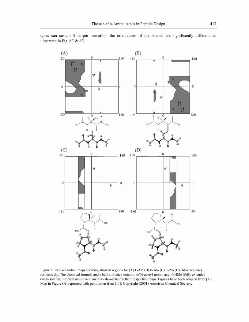
considering the Ramachandran maps for N-acetyl-L-Ala-NHMe and N-acetyl-D-Ala-NHMe shown in Fig. 1A & 1B [9-12]. The map for the D-residue is derived by inverting the classical L-Ala map through the origin. Most of the stereochemically allowed regions for the L-residues correspond to negative values of φ, while for D-residues, the allowed regions correspond to positive values of φ. The classical 310 and α-helical
∗ Correspondence concerning this article should be addressed to Padmanabhan Balaram, E-mail: [email protected], Fax:
+91-80-23600535.
Radhakrishnan Mahalakshmi and Padmanabhan Balaram 416
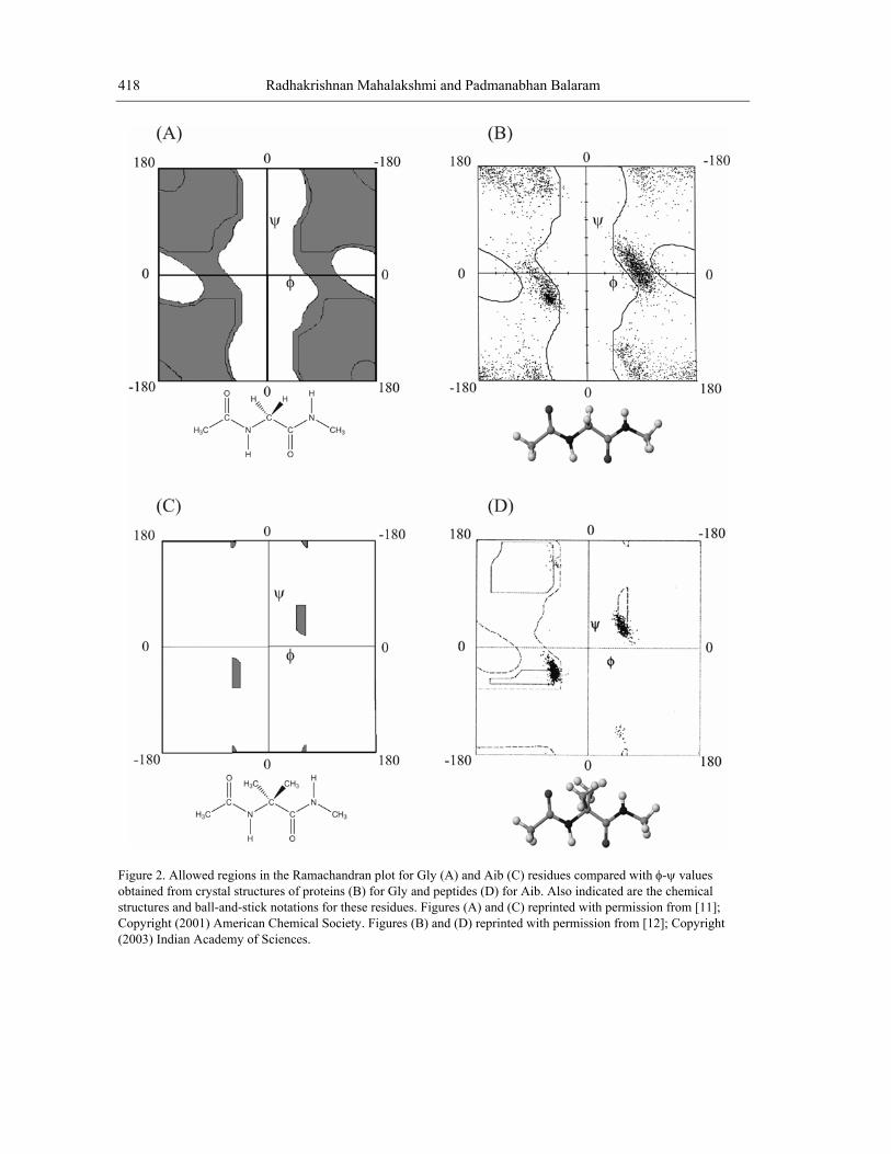
structures formed by all-L polypeptides adopt a right-handed twist (310-R/αR), while the corresponding D-polypeptides form helices with a left-handed twist (310-L/αL). Notably, there are small sterically allowed regions, which correspond to positive φ values for L-residues and negative φ values for D-residues. L-Proline is the most constrained of the amino acids occurring in proteins. The restraints imposed by the formation of the pyrrolidine ring restrict the allowed range of φ values in L-Pro to -60 ± 20° (Fig. 1C) [11,12]. By extension, the allowed values for D-Pro are +60 ± 20° (Fig. 1D). D-Pro can be used to advantage in restricting the local conformation of peptide chains, facilitating the formation of prime β-turns. The only achiral amino acid occurring in proteins is glycine, a residue for which considerably larger regions of φ-ψ space are sterically allowed, as a consequence of the absence of a substituent at the Cα atom (Fig. 2A). The Gly residue can adopt conformations on either side of the φ-ψ map. The observed distribution of Gly residues in proteins (Fig. 2B) establishes an approximately symmetric distribution about the origin [13]. α-Aminoisobutyric acid (Aib) is an achiral residue, which contains a pair of methyl groups at the Cα carbon atom. This residue may be formally viewed as a combination of L-Ala and D-Ala residues. The stereochemically allowed regions of Ramachandran space for Aib are readily derived by considering only the regions of overlap between the L-Ala and D-Ala φ-ψ maps (Fig. 2C). Clearly, the Aib residue is largely limited to the right (αR) and left (αL) handed helical structures. The observed distribution of crystallographically obtained conformations for Aib residues in peptides provides overwhelming support for this expectation (Fig. 2D) [11,12]. Achiral amino acids can, therefore, also be viewed as surrogate D-amino acids in peptide design because they can adopt conformations on both sides of the φ-ψ map. While the achiral Gly and Aib residues can support conformations with both positive and negative values of φ, D-amino acids may be used in design when positive φ values are required to form a desired structure.
3. β-HAIRPIN DESIGN β-Hairpins are widely present in proteins with antiparallel β-sheet formation being facilitated by a
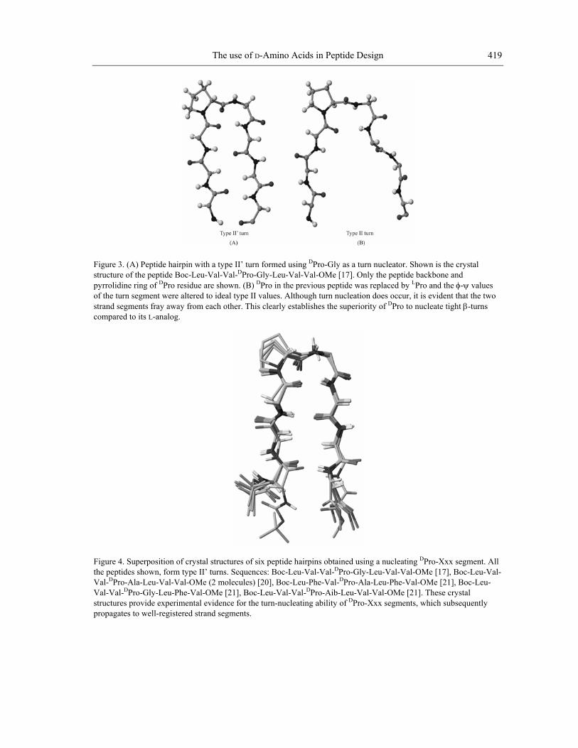
nucleating reverse turn. Two-residue β-turns belonging to the type II’ and I’ category are most often observed as nucleating features for hairpins in proteins [14]. In the prime turns, the residue at position i+1 necessarily adopts the conformation of φ ~ +60°, while ψ values of ~ -120° and ~ +30° are observed for type II’ and I’ turns, respectively [15]. Fig.3A illustrates a view of an ideal hairpin formed by a centrally positioned nucleating type II’ β-turn. When the stereochemistry of the β-turn is type II (φi+1 = -60°, ψi+1 = +120°), registry of the antiparallel strands is not readily achieved. Although chain reversal occurs, the antiparallel strands drift away from one another (Fig. 3B).
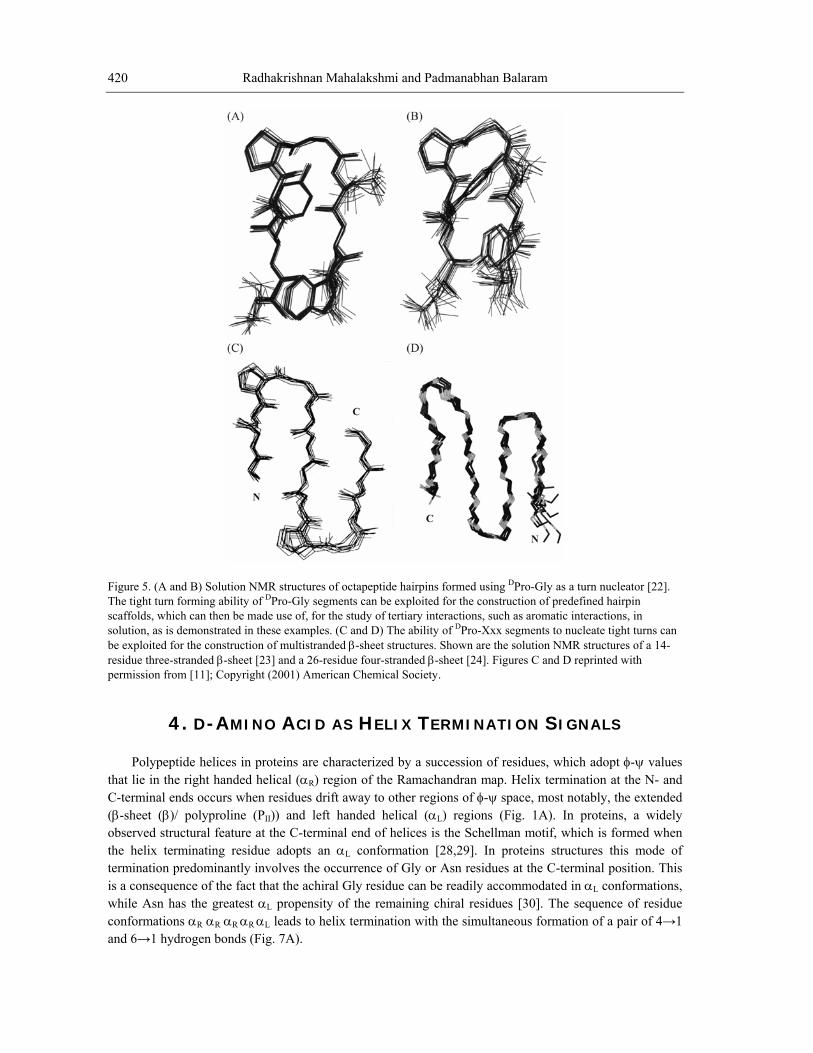
In the design of β-hairpins, DPro-Xxx sequences can provide a site for nucleation of prime turns. Indeed, insertion of DPro residues has proved to be a successful strategy for peptide hairpin design [8,11,12,16-21]. Fig. 4 illustrates the superposition of the crystal structures of six designed peptide hairpins containing a central DPro-Xxx type II’ β-turn. DPro residues provide a strong conformational determinant, when inserted into L-amino acid sequences. In ongoing studies, centrally positioned DPro-Gly sequences have been used to generate well-defined β-hairpin scaffolds which permit spectroscopic probing of cross-strand interactions between aromatic side chains (Fig. 5A & 5B) [22]. Multiple DPro-Xxx segments may be used to nucleate multistranded antiparallel β-sheet structures as exemplified by the design of three, four and five stranded β-sheets (Fig. 5C & 5D) [23-26].
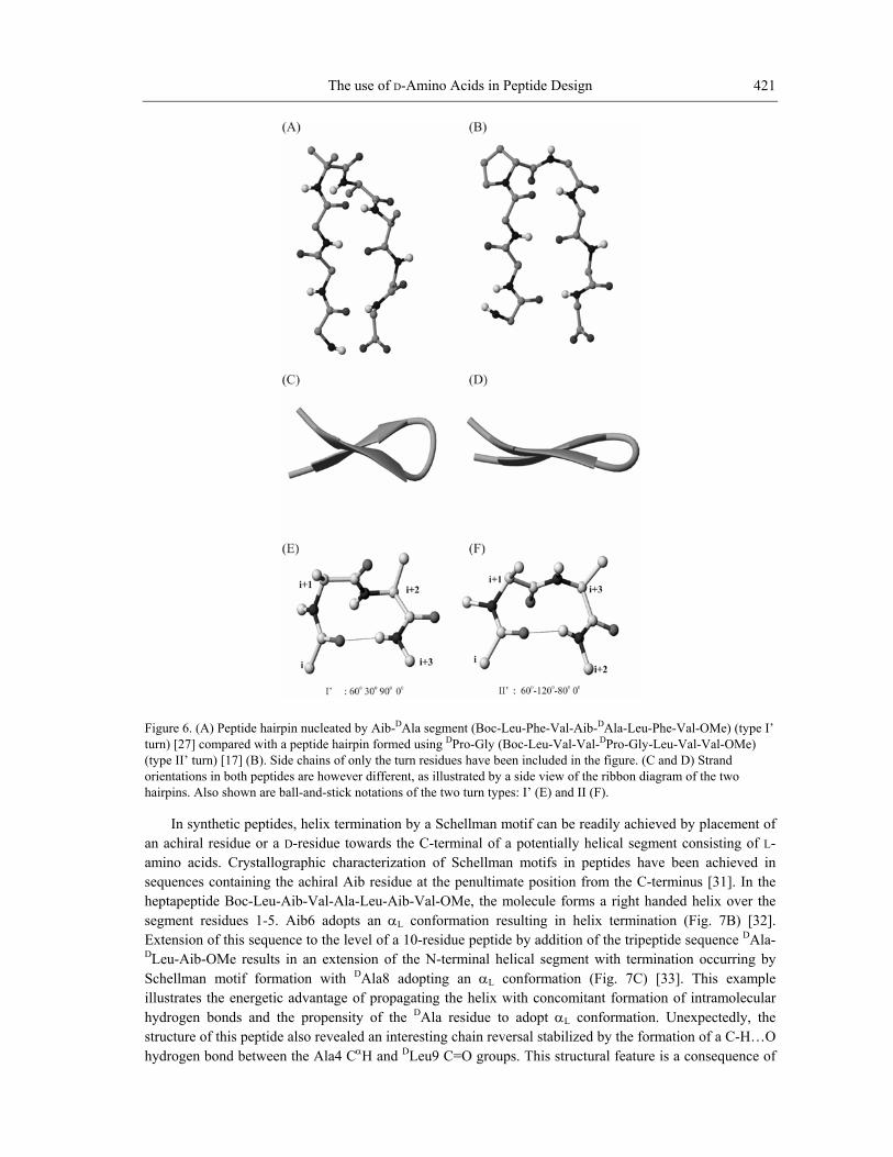
In principle, hairpin nucleation may be achieved by using sequences with a high propensity to form prime β-turns. Specifically, the Aib-DAla segment has been used to nucleate the formation of a β-hairpin in the octapeptide Boc-Leu-Phe-Val-Aib-DAla-Leu-Phe-Val-OMe (Fig. 6A) [27]. In this case, crystallographic studies establish that the Aib-DAla β-turn adopts a type I’ conformation, undoubtedly as a consequence of the tendency of the Aib residue to form αR or αL conformations. Thus, D-amino acid containing dipeptide segments may be used to generate both type II’ and I’ structures. While both turn
The use of D-Amino Acids in Peptide Design 417
types can sustain β-hairpin formation, the orientations of the strands are significantly different, as illustrated in Fig. 6C & 6D.
Figu . (A) L-Ala (B) D-Ala (C) L-Pro (D) D-Pro residues, respectively l-amino acyl-NHMe (fully extended conformation) for each amino acid are also shown below their respective maps. Figures have been adapted from [11]. Map in Figure (A) reprinted with permission from [11]; Copyright (2001) American Chemical Society.
re 1 Ramachandran maps showing allowed regions for. The chemical formula and a ball-and-stick notation of N-acety
Radhakrishnan Mahalakshmi and Padmanabhan Balaram 418
Figure 2. Allowed regions in the Ramachandran plot for Gly (A) and Aib (C) residues compared with φ-ψ values obtained from crystal structures of proteins (B) for Gly (D) for Aib. Also indicated are the chemical structures and ball-and-stick notations for these residues. Figures (A) and (C) reprinted with permission from [11]; Copy ciety. Figures (B) and (D) reprinted with permission from [12]; Copyright (200
and peptides
right (2001) American Chemical So3) Indian Academy of Sciences.
The use of D-Amino Acids in Peptide Design 419
Figu . structure of the peptide Boc-Leu-Val-Val- Pro-Gly-Leu-Val-Val-OMe [17]. Only the peptide backbone and pyrr nof th rstrand segcompared
re 3 (A) Peptide hairpin with a type II’ turn formed using DPro-Gly as a turn nucleator. Shown is the crystal D
Dolidi e ring of Pro residue are shown. (B) DPro in the previous peptide was replaced by LPro and the φ-ψ values e tu n segment were altered to ideal type II values. Although turn nucleation does occur, it is evident that the two
ments fray away from each other. This clearly establishes the superiority of DPro to nucleate tight β-turns to its L-analog.
4. Superposition of crystal structures of six peptide hairpins obtained using a nucleating DPro-Xxx segment. All tides shown, form type II’ turns. Sequences: Boc-Leu-Val-Val-DPro-G
Figurethe pep ly-Leu-Val-Val-OMe [17], Boc-Leu-Val-
DPVal-V Pro-Aib-Leu-Val-Val-OMe [21]. These crystal
ag
Val- ro-Ala-Leu-Val-Val-OMe (2 molecules) [20], Boc-Leu-Phe-Val-DPro-Ala-Leu-Phe-Val-OMe [21], Boc-Leu-al-DPro-Gly-Leu-Phe-Val-OMe [21], Boc-Leu-Val-Val-D
structures provide experimental evidence for the turn-nucleating ability of DPro-Xxx segments, which subsequently prop ates to well-registered strand segments.
Radhakrishnan Mahalakshmi and Padmanabhan Balaram 420
Figure 5. (A and B) Solution NMR structures of octapeptide hairpins formed using DPro-Gly as a turn nucleator [22]. The tight turn forming ability of DPro-Gly segments can be exploited for the construction of predefined hairpin scaffolds, which can then be made use of, for the study of tertiary interactions, such as aromatic interactions, in solution, as is demonstrated in these examples. (C and D) The ability of DPro-Xxx segments to nucleate tight turns can be exploited for the construction of multistranded β-sheet structures. Shown are the solution NMR structures of a 14-residue three-stranded β-sheet [23] and a 26-residue four-stranded β-sheet [24]. Figures C and D reprinted with permission from [11]; Copyright (2001) American Chemical Society.
4. D-AMINO ACID AS HELIX TERMINATION SIGNALS Polypeptide helices in proteins are characterized by a succession of residues, which adopt φ-ψ values
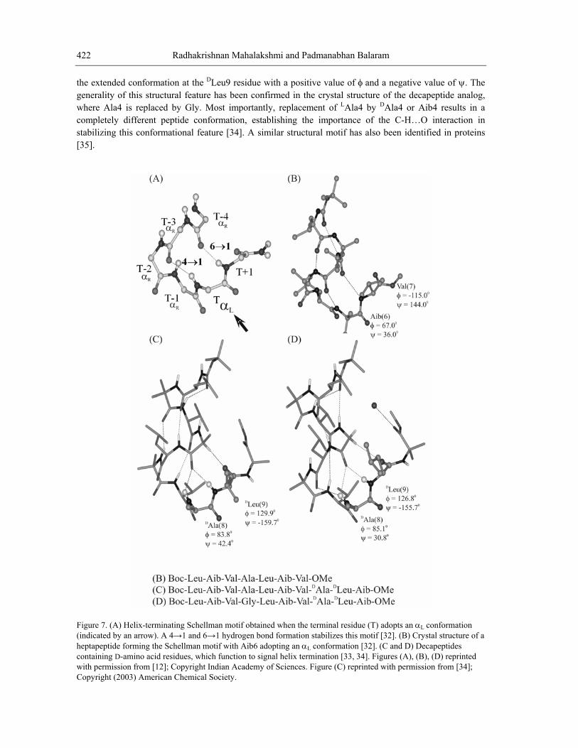
that lie in the right handed helical (αR) region of the Ramachandran map. Helix termination at the N- and C-terminal ends occurs when residues drift away to other regions of φ-ψ space, most notably, the extended (β-sheet (β)/ polyproline (PII)) and left handed helical (αL) regions (Fig. 1A). In proteins, a widely observed structural feature at the C-terminal end of helices is the Schellman motif, which is formed when the helix terminating residue adopts an αL conformation [28,29]. In proteins structures this mode of termination predominantly involves the occurrence of Gly or Asn residues at the C-terminal position. This is a consequence of the fact that the achiral Gly residue can be readily accommodated in αL conformations, while Asn has the greatest αL propensity of the remaining chiral residues [30]. The sequence of residue conformations αR αR αR αR αL leads to helix termination with the simultaneous formation of a pair of 4→1 and 6→1 hydrogen bonds (Fig. 7A).
The use of D-Amino Acids in Peptide Design 421
Figure 6. (A) Peptide hairpin nucleated by Aib-DAla segment (Boc-Leu-Phe-Val-Aib-DAla-Leu-Phe-Val-OMe) (type I’ turn) [27] compared with a peptide hairpin formed using DPro-Gly (Boc-Leu-Val-Val-DPro-Gly-Leu-Val-Val-OMe) (type II’ turn) [17] (B). Side chains of only the turn residues have been included in the figure. (C and D) Strand orientations in both peptides are however different, as illustrated by a side view of the ribbon diagram of the two hairpins. Also shown are ball-and-stick notations of the two turn types: I’ (E) and II (F).
In synthetic peptides, helix termination by a Schellman motif can be readily achieved by placement of an achiral residue or a D-residue towards the C-terminal of a potentially helical segment consisting of L-amino acids. Crystallographic characterization of Schellman motifs in peptides have been achieved in sequences containing the achiral Aib residue at the penultimate position from the C-terminus [31]. In the heptapeptide Boc-Leu-Aib-Val-Ala-Leu-Aib-Val-OMe, the molecule forms a right handed helix over the segm αL conformation resulting in helix termination (Fig. 7B) [32]. Extension of this sequence to the level of ddition of the tripeptide sequence DAla-DLeu-Aib-OMe results in an extension o segment with termination occurring by Schellman motif formation with DAla8 adopting an αL conformation (Fig. 7C) [33]. This example illustrates the energetic advantage of propagating the helix with concomitant formation of intramolecular hydrogen bonds and the propensity of the DAla residue to adopt αL conformation. Unexpectedly, the structure of this peptide also revealed an interesting chain reversal stabilized by the formation of a C-H…O hydrogen bond between the Ala4 CαH and DLeu9 C=O groups. This structural feature is a consequence of
ent residues 1-5. Aib6 adopts an a 10-residue peptide by af the N-terminal helical
Radhakrishnan Mahalakshmi and Padmanabhan Balaram 422
the n lue of ψ. The generality of this structural feature has been confirmed in the crystal structure of the decapeptide analog, where Ala4 is replaced by Gly. Most importantly, replacement of LAla4 by DAla4 or Aib4 results in a completely different peptide conformation, establishing the importance of the C-H…O interaction in stabilizing this conformational feature [34]. A similar structural motif has also been identified in proteins [35].
exte ded conformation at the DLeu9 residue with a positive value of φ and a negative va
Figure 7. (A) Helix-terminating Schellman motif obtained when the terminal residue (T) adopts an α conformation (indi dheptapeptcontaininwith perm oCopyright (2003) American Chemical Society.
Lcate by an arrow). A 4→1 and 6→1 hydrogen bond formation stabilizes this motif [32]. (B) Crystal structure of a
ide forming the Schellman motif with Aib6 adopting an αL conformation [32]. (C and D) Decapeptides g D-amino acid residues, which function to signal helix termination [33, 34]. Figures (A), (B), (D) reprinted issi n from [12]; Copyright Indian Academy of Sciences. Figure (C) reprinted with permission from [34];
The use of D-Amino Acids in Peptide Design 423
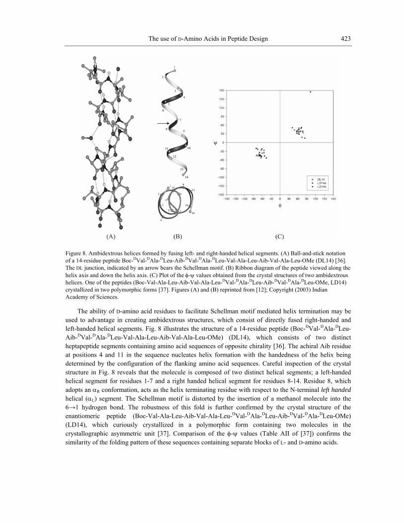
Figure 8. Ambidextrous helices formed by fusing left- and right-handed helical segments. (A) Ball-and-stick notation of a 14-residue peptide Boc-DVal-DAla-DLeu-Aib-DVal-DAla-DLeu-Val-Ala-Leu-Aib-Val-Ala-Leu-OMe (DL14) [36]. The DL junction, indicated by an arrow bears the Schellman motif. (B) Ribbon diagram of the peptide viewed along the helix axis and down the helix axis. (C) Plot of the φ-ψ values obtained from the crystal structures of two ambidextrous helices. One of the peptides (Boc-Val-Ala-Leu-Aib-Val-Ala-Leu-DVal-DAla-DLeu-Aib-DVal-DAla-DLeu-OMe, LD14) crystallized in two polymorphic forms [37]. Figures (A) and (B) reprinted from [12]; Copyright (2003) Indian Academy of Sciences.
The ability of D-amino acid residues to facilitate Schellman motif mediated helix termination may be used to advantage in creating ambidextrous structures, which consist of directly fused right-handed and left-handed helical segments. Fig. 8 illustrates the structure of a 14-residue peptide (Boc-DVal-DAla-DLeu-Aib- Val-DAla-DLeu-Val-Ala-Leu-Aib-Val-Ala-Leu-OMe) (DL14), which consists of two distinct heptapeptide segments containing amino acid sequences of opposite chirality [36]. The achiral Aib residue at positions 4 and 11 in the sequence nucle on with the handedness of the helix being determined by the configuration of the flanking amino acid sequences. Careful inspection of the crystal structure in Fig. 8 reveals that the molecule is composed of two distinct helical segments; a left-handed helical segment for residues 1-7 and a right handed helical segment for residues 8-14. Residue 8, which adopts an αR conformation, acts as the helix terminating residue with respect to the N-terminal left handed helical (αL) segment. The Schellman motif is distorted by the insertion of a methanol molecule into the 6→1 hydrogen bond. The robustness of this fold is further confirmed by the crystal structure of the enantiomeric peptide (Boc-Val-Ala-Leu-Aib-Val-Ala-Leu-DVal-DAla-DLeu-Aib-DVal-DAla-DLeu-OMe) (LD14), which curiously crystallized in a polymorphic form containing two molecules in the crystallographic asymmetric unit [37]. Comparison of the φ-ψ values (Table AII of [37]) confirms the similarity of the folding pattern of these sequences containing separate blocks of L- and D-amino acids.
D
ates helix formati
Radhakrishnan Mahalakshmi and Padmanabhan Balaram 424
FigureOne D
in D D
Aib-DV ]; Copyright (2003) Indian Academy of
In
acids mino acids. The site of D-amino acid insertion becomes resistant
analogthe sit lly chosen in order to minimize the possibility of major
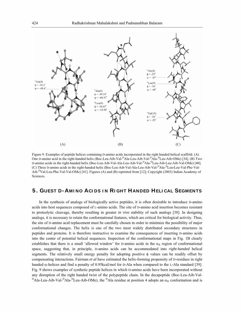
peptidinto the center of potential helical sequences. Inspection of the conformational maps in Fig. 1B clearly establishes that there is a small ‘allowed window’ for D-amino acids in the αR region of conformational space, suggesting that, in principle, D-amino acids can be accommodated into right-handed helical segments. The relatively small energy penalty for adopting positive φ values can be readily offset by compensating interactions. Fairman et al have estimated the helix-forming propensity of D-residues in right handed α-helices and find a penalty of 0.95kcal/mol for D-Ala when compared to the L-Ala standard [39]. Fig. 9 shows examples of synthetic peptide helices in which D-amino acids have been incorporated without any disruption of the right handed twist of the polypeptide chain. In the decapeptide (Boc-Leu-Aib-Val-DAla-Leu-Aib-Val-DAla-DLeu-Aib-OMe), the DAla residue at position 4 adopts an αR conformation and is
9. Examples of peptide helices containing D-amino acids incorporated in the right handed helical scaffold. (A) -amino acid in the right-handed helix (Boc-Leu-Aib-Val-DAla-Leu-Aib-Val-DAla-DLeu-Aib-OMe) [34]. (B) Two o acids in the right-handed helix (Boc-Leu-AD-am ib-Val-Ala-Leu-Aib-Val- Ala- Leu-Aib-Leu-Aib-Val-OMe) [40].
(C) Three D-amino acids in the right-handed helix (Boc-Leu-Aib-Val-Ala-Leu-Aib-Val-DAla-DLeu-Leu-Val-Phe-Val-al-Leu-Phe-Val-Val-OMe) [41]. Figures (A) and (B) reprinted from [12
Sciences.
5. GUEST D-AMINO ACIDS IN RIGHT HANDED HELICAL SEGMENTS
the synthesis of analogs of biologically active peptides, it is often desirable to introduce D-amino into host sequences composed of L-a
to proteolytic cleavage, thereby resulting in greater in vivo stability of such analogs [38]. In designing s, it is necessary to retain the conformational features, which are critical for biological activity. Thus, e of D-amino acid replacement must be carefu
conformational changes. The helix is one of the two most widely distributed secondary structures in es and proteins. It is therefore instructive to examine the consequences of inserting D-amino acids
The use of D-Amino Acids in Peptide Design 425
incorporated into the right-handed helix formed by the segments 1-7. In this case, the observed φ-ψ values for the DAla residue in the two crystallographically independent molecules are -46.6, -52.9 and -54.1, -48.6 respectively [34]. The 13-residue peptide (Boc-Leu-Aib-Val-Ala-Leu-Aib-Val-DAla-DLeu-Aib-Leu-Aib-Val-OMe) provides an interesting example of a double D-segment (DAla8, DLeu9) incorporated into a right-handed helix [40]. The crystal structure of the 19-residue peptide (Boc-Leu-Aib-Val-Ala-Leu-Aib-Val-DAl DLeu-Leu-Val-Phe-Val-Aib-DVal-Leu-Phe-Val-Val-OMe) is a dramatic example of the accommodation of as many as three D-amino acids into a right-handed helical scaffold [41]. These examples clearly emphasize the fact that right-handed helices can readily accommodate D-residues. Selective D-amino acid substitution in strongly helical sequences may provide a convenient means of enhancing proteolytic stability, without significant disruption of overall secondary structure.
a-
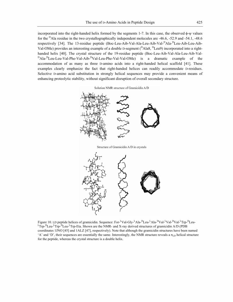
Figure 10. LD peptide helices of gramicidin. Sequence: For-LVal-Gly-LAla-DLeu-LAla-DVal-LVal-DVal-LTrp-DLeu-LTrp-DLeu-LTrp-DLeu-LTrp-Eta. Shown are the NMR- and X-ray derived structures of gramicidin A/D (PDB coordinates 1JNO [45] and 1ALZ [47], respectively). Note that although the gramicidin structures have been named ‘A’ and ‘D’, their sequences are essentially the same. Interestingly, the NMR structure reveals a πLD helical structure for the peptide, whereas the crystal structure is a double helix.
Radhakrishnan Mahalakshmi and Padmanabhan Balaram 426
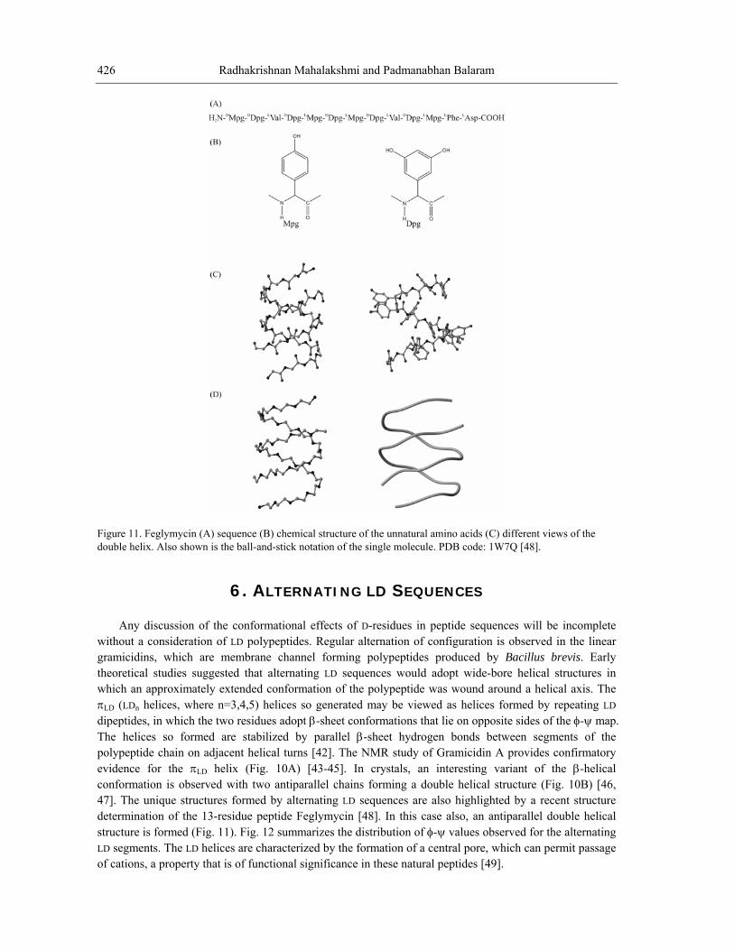
e 11. Feglymycin (A) sequence (B) chemical structure of the unnatural amino acids (C) different views of the le helix. Also shown is the ball-and-stick notation of the single molecule. PDB code: 1W7Q [48].
6. ALTERNATING LD SEQUE
Figurdoub
NCES
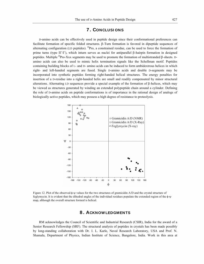
witho s. Regular alternation of configuration is observed in the linear gram channel forming polypeptides produced by Bacillus brevis. Early theoretical studies suggested that alternating LD sequences would adopt wide-bore helical structures in w mation of the polypeptide was wound around a helical axis. The π be viewed as helices formed by repeating LD dipeptides, in which the two residues adopt β-sheet conformations that lie on opposite sides of the φ-ψ map. The helices so formed are stabilized by parallel β-sheet hydrogen bonds between segments of the polypeptide chain on adjacent helical turns [42]. The NMR study of Gramicidin A provides confirmatory evidence for the πLD helix (Fig. 10A) [43-45]. In crystals, an interesting variant of the β-helical conformation is observed with two antiparallel chains forming a double helical structure (Fig. 10B) [46, 47]. The unique structures formed by alternating LD sequences are also highlighted by a recent structure determination of the 13-residue peptide Feglymycin [48]. In this case also, an antiparallel double helical structure is formed (Fig. 11). Fig. 12 summarizes the distribution of φ-ψ values observed for the alternating LD segments. The LD helices are characterized by the formation of a central pore, which can permit passage of cations, a property that is of functional significance in these natural peptides [49].
Any discussion of the conformational effects of D-residues in peptide sequences will be incomplete ut a consideration of LD polypeptideicidins, which are membrane
hich an approximately extended conforLD (LDn helices, where n=3,4,5) helices so generated may
The use of D-Amino Acids in Peptide Design 427
7. CONCLUSIONS D-amino acids can be effectively used in peptide design since their conformational preferences can
facilitate formation of specific folded structures. β-Turn formation is favored in dipeptide sequences of alternating configuration (LD peptides). DPro, a constrained residue, can be used to force the formation of prime turns (ty serves as nuclei for antiparallel β-hairpin formation in designed
ultistranded β-sheets. D-min an motif. Peptides
us helices in which ght ents are fused. Single D-amino acids and double D-segments may be
alterabe vithb
pe II’/I’), which inturneptides. Multiple DPro-Xp xx segments may be used to promote the formation of m
a o acids can also be used to mimic helix termination signals like the Schellmcontaining building blocks of L- and D- amino acids can be induced to form ambidextrori - and left-handed segmincorporated into synthetic peptides forming right-handed helical structures. The energy penalties for insertion of a D-residue into a right-handed helix are small and readily compensated by minor structural
tions. Alternating LD sequences provide a special example of the formation of β-helices, which may ewed as structures generated by winding an extended polypeptide chain around a cylinder. Defining ole of D-amino acids on peptide conformations is of importance in the rational de r esign of analogs of
iologically active peptides, which may possess a high degree of resistance to proteolysis.
Figure 12. Plot of the observed φ-ψ values for the two structures of gramicidin A/D and the crystal structure of feglymycin. It is evident that the dihedral angles of the individual residues populate the extended region of the φ-ψ map, although the overall structure formed is helical.
8. ACKNOWLEDGMENTS RM acknowledges the Council of Scientific and Industrial Research (CSIR), India for the award of a
Senior Research Fellowship (SRF). The structural analysis of peptides in crystals has been made possibly by long-standing collaboration with Dr. I. L. Karle, Naval Research Laboratory, USA and Prof. N. Shamala, Department of Physics, Indian Institute of Science, Bangalore, India. Work in this area at
Radhakrishnan Mahalakshmi and Padmanabhan Balaram 428
Bangalore has been supported by grants from the Department of Science and Technology and the Department of Biotechnology.
REFERENCES
[1] Lipmann, F., Hotchkiss, R. D. & Dubos, J. (1941). The occurrence of D-amino acids in gram
. & Olivera, B. M. (1999). Post-translationally modified neuropeptides from conus venoms. Eur J Biochem, 264, 271-275.
[5] Milton, R. C. L., Milton, S. C. F. & Kent, S. B. H. (1992). Total chemical synthesis of a D-enzyme: The enantiomers of HIV-1 protease show demonstration of reciprocal chiral substrate specificity. Science, 256, 1445-1448.
[6] Zawadzke, L. E. & Berg, J. M. (1992). A racemic protein. J Am Chem Soc, 114, 4002-4003. [7] Fitzgerald, M. C., Chernuschevich, I., Standing, K. G., Kent, S. B. H. & Whitman, C. P. (1995).
Total chemical synthesis and catalytic properties of the enzyme enantiomers L- and D-4-oxalocrotonate tautomerase. J Am Chem Soc, 117, 11075-11080.
[8] Balaram, P. (1999). De novo design: Backbone conformational constraints in nucleating helices and β-hairpins. J Peptide Res, 54, 195-199.
[9] Ramachandran, G. N. & Sasisekharan, V. (1968). Conformation of polypeptides and proteins. Adv Protein Chem, 23, 283-438.
[10] Ramachandran, G. N. & Chandrasekaran, R. Progress in Peptide Research. Lande, S., Ed.; Gordon & Breach: New York, 1972; Vol. II (Proceedings of the Second American Peptide Symposium, Cleveland, 1970), p 195.
[11] Venkatraman, J., Shankaramma, S. C. & Balaram, P. (2001). Design of folded peptides. Chem Rev,
. Non-protein amino acids in peptide design. Proc Indian Acad Sci (Chem Sci), 115, 373-400.
[13] Ramakrishnan, C. & Srinivasan, N. (1990). Gly sis. Curr Sci, 59, 851-861.
[14] Wilmot, C. M. & Thornton, J. M. (1988). Analysis and prediction of the different types of β-turn in
[15] Cr rties (2nd edition), New York, NY:
[16] AwBiophys Res Commun, 216, 375-381.
[17] Karle, I. L., Awasthi, S. K. & Balaram, P. (1996). A designed β-hairpin peptide in crystals. Proc Natl Acad Sci USA, 93, 8189-8193.
[18] Stanger, H. E. & Gellman, S. H. (1998). Rules for antiparallel β-sheet design: D-Pro-Gly is superior to L-Asn-Gly for β-hairpin nucleation. J Am Chem Soc, 120, 4236-4237.
[19] Karle, I. L., Das, C. & Balaram, P. (2000). De novo protein design: Crystallographic characterization of a synthetic peptide containing independent helical and hairpin domains. Proc Natl Acad Sci USA, 97, 3034-3037.
[20] Das, C., Naganagowda, G. A., Karle, I. L. & Balaram, P. (2001). Designed β-hairpin peptides with defined tight turn stereochemistry. Biopolymers, 58, 335-346.
icidin and tyrocidine. J Biol Chem, 141, 163-169.
[2] Kleinkauf, H. & von Dohren, H. (1996). A nonribosomal system of peptide biosynthesis. Eur J Biochem, 236, 335-351.
[3] Kreil, G. (1997). D-amino acids in animal peptides. Annu Rev Biochem, 66, 337-345. [4] Craig, G., Bandyopadhyay, P
101, 3131-3152. [12] Aravinda, S., Shamala, N., Roy, R. S. & Balaram, P. (2003)
cyl residues in proteins and peptides: An analy
proteins. J Mol Biol, 203, 221-232.
eighton, T. E. (1997) Proteins: Structure and molecular propeFreeman Press.
asthi, S. K., Raghothama, S. & Balaram, P. (1995). A designed β-hairpin peptide. Biochem
The use of D-Amino Acids in Peptide Design 429
[21] Aravinda, S., Harini, V. V., Shamala, N., Das, C. & Balaram, P. (2004). Structure and assembly of designed β-hairpin peptides in crystals as models for β-sheet aggregation. Biochemistry, 43, 1832-1846.
[22] Mahalakshmi, R., Raghothama, S. & Balaram, P. (2006). NMR analysis of aromatic interactions in designed peptide β-hairpins. J Am Chem Soc, 128, 1125-1138.
[24] Das, C., Raghothama, S. & Balaram, P. (1999). A four stranded β-sheet structure in a designed synthetic polypeptide. Chem Commun, 967-968.
[25] Venkataraman, J., Naganagowda, G. A., Sudha, R. & Balaram, P. (2001). De novo design of a five-
[27] Aravinda, S., Shamala, N., Rajkishore, R., G , P. (2002). A crystalline β-hairpin peptide nucleated by a type I' Aib-D-Ala β-turn: Evidence for cross-strand aromatic interactions. Angew Chem Int Ed Engl, 41, 3863-3865.
[28] Schellman, C. (1980) The αL conformation at the ends of helices, in Protein folding (Jaenicke, R., ed.), pp. 53-61, Elsevier/North-Holland, New York.
[30] V. S., Ramakrishnan, C., Sowdhamini, R. & Balaram, P. (1994). Conformational characteristics of asparginyl residues in proteins. Int J Peptide Protein Res, 44, 112-122.
[31] Karle, I. L., Flippen-Anderson, J. L., Uma, K. & Balaram, P. (1993). Peptide mimics for structural features in proteins. Crystal structures of three heptapeptide helices with a C-terminal 6→1 hydrogen
[33]
[34] C-H...O hydrogen bond stabilized polypeptide chain reversal at the C-terminus of designed peptide helices. Structural characterization of three decapeptides. J Am Chem Soc, 125, 15065-15075.
[35] Babu, M. M., Singh, S. K. & Balaram, P. (2002). A C-H triple bond O hydrogen bond stabilized polypeptide chain reversal motif at the C terminus of helices in proteins. J Mol Biol, 322, 871-880.
[36] Banerjee, A., Raghothama, S. R., Karle, I. L. & Balaram, P. (1996). Ambidextrous molecules: Cylindrical peptide structures formed by fusing left- and right-handed helices. Biopolymers, 39, 279-285.
[37] Karle, I. L. (2001). Controls exerted by the Aib residue: Helix formation and helix reversal. Biopolymers (Pept Sci), 60, 351-365.
[38] van Regenmortel, M. & Muller, S. (1998). D-peptides as immunogens and diagnostic reagents. Curr Opin Biotechnol, 9, 377-382.
[39] Fairman, R., Anthony-Cahill, S. J. & DeGrado, W. F. (1992). The helix-forming propensity of D-alanine in a right-handed α-helix. J Am Chem Soc, 114, 5458-5459.
[40] Aravinda, S., Shamala, N., Desiraju, S. & Balaram, P. (2002). A right-handed peptide helix containing a central double D-amino acid segment. Chem Commun, 2454-2455.
[23] Das, C., Raghothama, S. & Balaram, P. (1998). A designed three stranded β-sheet peptide. J Am Chem Soc, 120, 5812-5813.
stranded β-sheet anchoring a metal-ion binding site. Chem Commun, 2660-2661. [26] Venkatraman, J., Nagana Gowda, G. A. & Balaram, P. (2002). Design and construction of an open
multistranded β-sheet polypeptide stabilized by a disulfide bridge. J Am Chem Soc, 124, 4987-4994. opi, H. N. & Balaram
[29] Nagarajaram, H. A., Sowdhamini, R., Ramakrishnan, C. & Balaram, P. (1993). Termination of right handed helices in proteins by residues in left-handed helical conformations. FEBS Lett, 321, 79-83. Srinivasan, N., Anuradha,
bond. Int J Pept Protein Res, 42, 401-410. [32] Banerjee, A., Datta, S., Pramanik, A., Shamala, N. & Balaram, P. (1996). Heterogeneity and stability
of helical conformations in peptides: Crystallographic and NMR studies of a model heptapeptide. J Am Chem Soc, 118, 9477-9483. Aravinda, S., Shamala, N., Pramanik, A., Das, C. & Balaram, P. (2000). An unusual C-H...O hydrogen bond mediated reversal of polypeptide chain direction in a synthetic peptide helix. Biochem Biophys Res Commun, 273, 933-36. Aravinda, S., Shamala, N., Bandyopadhyay, A. & Balaram, P. (2003). Probing the role of the
Radhakrishnan Mahalakshmi and Padmanabhan Balaram 430
[41] Karle, I. L., Gopi, H. N. & Balaram, P. (2003). Crystal structure of a hydrophobic 19-residue peptide helix containing three centrally located D amino acids. Proc Natl Acad Sci USA, 100, 13946-13951.
[42] Ramachandran, G. N. & Chandrasekaran, R. (1972). Conformation of peptide chains containing both L- and D-residues: Part I-helical structures with alternating L- and D-residues with special reference to the LD-ribbon and the LD-helices. Indian J Biochem Biophys, 9, 1-11.
[43] Arseniev, A. S., Barsukov, I. L., Lomize, A. L., Orekhov, V. Y. & Bystrov, V. F. (1992). Refinement of the spatial structure of the gramicidin A ion channel. Bioorg Khim, 18, 182-200.
[44] Ketchem, R. R., Lee, K. C., Huo, S. & Cross, T. A. (1996). Macromolecular structural elucidation with solid-state NMR-derived orientational constraints. J Biomol NMR, 8, 1-14.
[45] Townsley, L. E., Tucker, A., Sham, S. & Hinton, J. F. (2001). Structures of gramicidins A, B and C incorporated in sodium dodecyl sulfate micelles. Biochemistry, 40, 11676-86.
[46] Langs, D. A. (1988). Three-dimensional structure at 0.86Å of the uncomplexed form of the transmembrane ion channel peptide gramicidin A. Science, 241, 188-191.
[47] Burkhart, B. M., Gassman, R. M., Langs, D. A., Pangborn, W. A. & Duax, W. L. (1998). Heterodimer formation and crystal nucleation of gramicidin D. Biophys J, 75, 2135-2146.
[48] Bunkoczi, G., Vertesy, L. & Sheldrick, G. M. (2005). The antiviral antibiotic feglymycin: First direct-methods solution of a 1000+ equal-atom structure. Angew Chem Int Ed, 44, 1340-1342.
[49] Hladky, S. B. & Haydon, D. A. (1970). Discreteness of conductance change in bimolecular lipid membranes in the presence of certain antibiotics. Nature, 225, 451-53.