RESEARCH Open Access E-cigarette-induced pulmonary
17
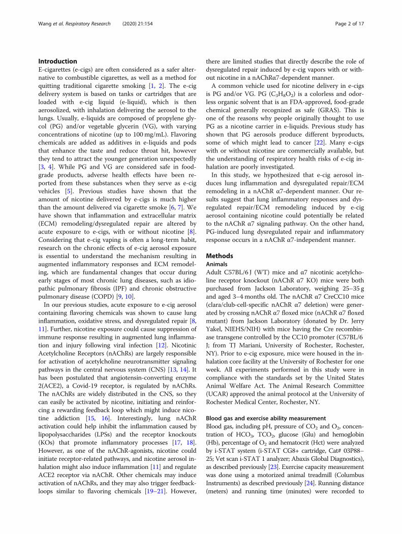
RESEARCH Open Access E-cigarette-induced pulmonary inflammation and dysregulated repair are mediated by nAChR α7 receptor: role of nAChR α7 in SARS-CoV-2 Covid-19 ACE2 receptor regulation Qixin Wang 1 , Isaac K. Sundar 1 , Dongmei Li 2 , Joseph H. Lucas 1 , Thivanka Muthumalage 1 , Samantha R. McDonough 1 and Irfan Rahman 1* Abstract Electronic cigarette (e-cig) vaping is increasing rapidly in the United States, as e-cigs are considered less harmful than combustible cigarettes. However, limited research has been conducted to understand the possible mechanisms that mediate toxicity and pulmonary health effects of e-cigs. We hypothesized that sub-chronic e-cig exposure induces inflammatory response and dysregulated repair/extracellular matrix (ECM) remodeling, which occur through the α7 nicotinic acetylcholine receptor (nAChRα7). Adult wild-type (WT), nAChRα7 knockout (KO), and lung epithelial cell-specific KO (nAChRα7 CreCC10) mice were exposed to e-cig aerosol containing propylene glycol (PG) with or without nicotine. Bronchoalveolar lavage fluids (BALF) and lung tissues were collected to determine e-cig induced inflammatory response and ECM remodeling, respectively. Sub-chronic e-cig exposure with nicotine increased inflammatory cellular influx of macrophages and T-lymphocytes including increased pro- inflammatory cytokines in BALF and increased SARS-Cov-2 Covid-19 ACE2 receptor, whereas nAChRα7 KO mice show reduced inflammatory responses associated with decreased ACE2 receptor. Interestingly, matrix metalloproteinases (MMPs), such as MMP2, MMP8 and MMP9, were altered both at the protein and mRNA transcript levels in female and male KO mice, but WT mice exposed to PG alone showed a sex-dependent phenotype. Moreover, MMP12 was increased significantly in male mice exposed to PG with or without nicotine in a nAChRα7-dependent manner. Additionally, sub-chronic e-cig exposure with or without nicotine altered the abundance of ECM proteins, such as collagen and fibronectin, significantly in a sex-dependent manner, but without the direct role of nAChRα7 gene. Overall, sub-chronic e-cig exposure with or without nicotine affected lung inflammation and repair responses/ECM remodeling, which were mediated by nAChRα7 in a sex-dependent manner. Keywords: E-cig exposure, nAChRα7, Inflammation, Dysregulated repair, Extracellular matrix © The Author(s). 2020 Open Access This article is licensed under a Creative Commons Attribution 4.0 International License, which permits use, sharing, adaptation, distribution and reproduction in any medium or format, as long as you give appropriate credit to the original author(s) and the source, provide a link to the Creative Commons licence, and indicate if changes were made. The images or other third party material in this article are included in the article's Creative Commons licence, unless indicated otherwise in a credit line to the material. If material is not included in the article's Creative Commons licence and your intended use is not permitted by statutory regulation or exceeds the permitted use, you will need to obtain permission directly from the copyright holder. To view a copy of this licence, visit http://creativecommons.org/licenses/by/4.0/. The Creative Commons Public Domain Dedication waiver (http://creativecommons.org/publicdomain/zero/1.0/) applies to the data made available in this article, unless otherwise stated in a credit line to the data. * Correspondence: [email protected] 1 Department of Environmental Medicine, University of Rochester Medical Center, Box 850, 601 Elmwood Avenue, Rochester, NY 14642, USA Full list of author information is available at the end of the article Wang et al. Respiratory Research (2020) 21:154 https://doi.org/10.1186/s12931-020-01396-y
Transcript of RESEARCH Open Access E-cigarette-induced pulmonary
RESEARCH Open Access
E-cigarette-induced pulmonaryinflammation and dysregulated repair aremediated by nAChR α7 receptor: role ofnAChR α7 in SARS-CoV-2 Covid-19 ACE2receptor regulationQixin Wang1, Isaac K. Sundar1, Dongmei Li2, Joseph H. Lucas1, Thivanka Muthumalage1,Samantha R. McDonough1 and Irfan Rahman1*
Abstract
Electronic cigarette (e-cig) vaping is increasing rapidly in the United States, as e-cigs are considered less harmfulthan combustible cigarettes. However, limited research has been conducted to understand the possiblemechanisms that mediate toxicity and pulmonary health effects of e-cigs. We hypothesized that sub-chronic e-cigexposure induces inflammatory response and dysregulated repair/extracellular matrix (ECM) remodeling, whichoccur through the α7 nicotinic acetylcholine receptor (nAChRα7). Adult wild-type (WT), nAChRα7 knockout (KO),and lung epithelial cell-specific KO (nAChRα7 CreCC10) mice were exposed to e-cig aerosol containing propyleneglycol (PG) with or without nicotine. Bronchoalveolar lavage fluids (BALF) and lung tissues were collected todetermine e-cig induced inflammatory response and ECM remodeling, respectively. Sub-chronic e-cig exposurewith nicotine increased inflammatory cellular influx of macrophages and T-lymphocytes including increased pro-inflammatory cytokines in BALF and increased SARS-Cov-2 Covid-19 ACE2 receptor, whereas nAChRα7 KO miceshow reduced inflammatory responses associated with decreased ACE2 receptor. Interestingly, matrixmetalloproteinases (MMPs), such as MMP2, MMP8 and MMP9, were altered both at the protein and mRNAtranscript levels in female and male KO mice, but WT mice exposed to PG alone showed a sex-dependentphenotype. Moreover, MMP12 was increased significantly in male mice exposed to PG with or without nicotine in anAChRα7-dependent manner. Additionally, sub-chronic e-cig exposure with or without nicotine altered theabundance of ECM proteins, such as collagen and fibronectin, significantly in a sex-dependent manner, but withoutthe direct role of nAChRα7 gene. Overall, sub-chronic e-cig exposure with or without nicotine affected lunginflammation and repair responses/ECM remodeling, which were mediated by nAChRα7 in a sex-dependentmanner.
Keywords: E-cig exposure, nAChRα7, Inflammation, Dysregulated repair, Extracellular matrix
© The Author(s). 2020 Open Access This article is licensed under a Creative Commons Attribution 4.0 International License,which permits use, sharing, adaptation, distribution and reproduction in any medium or format, as long as you giveappropriate credit to the original author(s) and the source, provide a link to the Creative Commons licence, and indicate ifchanges were made. The images or other third party material in this article are included in the article's Creative Commonslicence, unless indicated otherwise in a credit line to the material. If material is not included in the article's Creative Commonslicence and your intended use is not permitted by statutory regulation or exceeds the permitted use, you will need to obtainpermission directly from the copyright holder. To view a copy of this licence, visit http://creativecommons.org/licenses/by/4.0/.The Creative Commons Public Domain Dedication waiver (http://creativecommons.org/publicdomain/zero/1.0/) applies to thedata made available in this article, unless otherwise stated in a credit line to the data.
* Correspondence: [email protected] of Environmental Medicine, University of Rochester MedicalCenter, Box 850, 601 Elmwood Avenue, Rochester, NY 14642, USAFull list of author information is available at the end of the article
Wang et al. Respiratory Research (2020) 21:154 https://doi.org/10.1186/s12931-020-01396-y
IntroductionE-cigarettes (e-cigs) are often considered as a safer alter-native to combustible cigarettes, as well as a method forquitting traditional cigarette smoking [1, 2]. The e-cigdelivery system is based on tanks or cartridges that areloaded with e-cig liquid (e-liquid), which is thenaerosolized, with inhalation delivering the aerosol to thelungs. Usually, e-liquids are composed of propylene gly-col (PG) and/or vegetable glycerin (VG), with varyingconcentrations of nicotine (up to 100mg/mL). Flavoringchemicals are added as additives in e-liquids and podsthat enhance the taste and reduce throat hit, howeverthey tend to attract the younger generation unexpectedly[3, 4]. While PG and VG are considered safe in food-grade products, adverse health effects have been re-ported from these substances when they serve as e-cigvehicles [5]. Previous studies have shown that theamount of nicotine delivered by e-cigs is much higherthan the amount delivered via cigarette smoke [6, 7]. Wehave shown that inflammation and extracellular matrix(ECM) remodeling/dysregulated repair are altered byacute exposure to e-cigs, with or without nicotine [8].Considering that e-cig vaping is often a long-term habit,research on the chronic effects of e-cig aerosol exposureis essential to understand the mechanism resulting inaugmented inflammatory responses and ECM remodel-ing, which are fundamental changes that occur duringearly stages of most chronic lung diseases, such as idio-pathic pulmonary fibrosis (IPF) and chronic obstructivepulmonary disease (COPD) [9, 10].In our previous studies, acute exposure to e-cig aerosol
containing flavoring chemicals was shown to cause lunginflammation, oxidative stress, and dysregulated repair [8,11]. Further, nicotine exposure could cause suppression ofimmune response resulting in augmented lung inflamma-tion and injury following viral infection [12]. NicotinicAcetylcholine Receptors (nAChRs) are largely responsiblefor activation of acetylcholine neurotransmitter signalingpathways in the central nervous system (CNS) [13, 14]. Ithas been postulated that angiotensin-converting enzyme2(ACE2), a Covid-19 receptor, is regulated by nAChRs.The nAChRs are widely distributed in the CNS, so theycan easily be activated by nicotine, initiating and reinfor-cing a rewarding feedback loop which might induce nico-tine addiction [15, 16]. Interestingly, lung nAChRactivation could help inhibit the inflammation caused bylipopolysaccharides (LPSs) and the receptor knockouts(KOs) that promote inflammatory processes [17, 18].However, as one of the nAChR-agonists, nicotine couldinitiate receptor-related pathways, and nicotine aerosol in-halation might also induce inflammation [11] and regulateACE2 receptor via nAChR. Other chemicals may induceactivation of nAChRs, and they may also trigger feedback-loops similar to flavoring chemicals [19–21]. However,
there are limited studies that directly describe the role ofdysregulated repair induced by e-cig vapors with or with-out nicotine in a nAChRα7-dependent manner.A common vehicle used for nicotine delivery in e-cigs
is PG and/or VG. PG (C3H8O2) is a colorless and odor-less organic solvent that is an FDA-approved, food-gradechemical generally recognized as safe (GRAS). This isone of the reasons why people originally thought to usePG as a nicotine carrier in e-liquids. Previous study hasshown that PG aerosols produce different byproducts,some of which might lead to cancer [22]. Many e-cigswith or without nicotine are commercially available, butthe understanding of respiratory health risks of e-cig in-halation are poorly investigated.In this study, we hypothesized that e-cig aerosol in-
duces lung inflammation and dysregulated repair/ECMremodeling in a nAChR α7-dependent manner. Our re-sults suggest that lung inflammatory responses and dys-regulated repair/ECM remodeling induced by e-cigaerosol containing nicotine could potentially be relatedto the nAChR α7 signaling pathway. On the other hand,PG-induced lung dysregulated repair and inflammatoryresponse occurs in a nAChR α7-independent manner.
MethodsAnimalsAdult C57BL/6 J (WT) mice and α7 nicotinic acetylcho-line receptor knockout (nAChR α7 KO) mice were bothpurchased from Jackson Laboratory, weighing 25–35 gand aged 3–4months old. The nAChR α7 CreCC10 mice(clara/club-cell-specific nAChR α7 deletion) were gener-ated by crossing nAChR α7 floxed mice (nAChR α7 floxedmutant) from Jackson Laboratory (donated by Dr. JerryYakel, NIEHS/NIH) with mice having the Cre recombin-ase transgene controlled by the CC10 promoter (C57BL/6J; from TJ Mariani, University of Rochester, Rochester,NY). Prior to e-cig exposure, mice were housed in the in-halation core facility at the University of Rochester for oneweek. All experiments performed in this study were incompliance with the standards set by the United StatesAnimal Welfare Act. The Animal Research Committee(UCAR) approved the animal protocol at the University ofRochester Medical Center, Rochester, NY.
Blood gas and exercise ability measurementBlood gas, including pH, pressure of CO2 and O2, concen-tration of HCO3, TCO2, glucose (Glu) and hemoglobin(Hb), percentage of O2 and hematocrit (Hct) were analyzedby i-STAT system (i-STAT CG8+ cartridge, Cat# 03P88–25; Vet scan i-STAT 1 analyzer; Abaxis Global Diagnostics),as described previously [23]. Exercise capacity measurementwas done using a motorized animal treadmill (ColumbusInstruments) as described previously [24]. Running distance(meters) and running time (minutes) were recorded to
Wang et al. Respiratory Research (2020) 21:154 Page 2 of 17
present exercise tolerance. Mice stayed on the treadmill for5min before start of measurement. The treadmill began at8.5m/min with 0° incline for 9min. Then, the speed wasincreased to 10m/min with 5° incline for 3min. After that,the speed was increased to 2.5m/min every 3min, and in-cline was increased to 5° every 9min until the mouse be-came exhausted, judged by observation of failure ofrunning and continuous contact with the electric grid. Fol-lowing this exercise, blood was collected by submandibularvenipuncture for blood gas and cotinine measurement.
E-cig device and e-liquidThe e-cig device used in this study was purchased fromthe Joytech VTC mini (SciReq, Montreal, Canada). Theatomizer/coil (0.15Ω) used for aerosolization of e-liquidwas purchased from Kanger technology (Shenzhen,China). All other components of the e-cig exposurechamber were purchased from SCIREQ, and all thecomponents were cleaned after exposure every day. Theatomizer/coil was replaced on a weekly basis to avoidoverheating and generation of carbon monoxide. The e-liquids used in this study that contain PG alone and PGwith nicotine (25 mg/mL), were purchased from xtreme-vaping.com.
E-cig exposureThe e-cig exposure performed here has been describedin our previous studies [25]. Briefly, the in vivo e-cig ex-posure was setup inside a fume hood and based on theSCIREQ InExpose e-cig extension smoking system. Thee-cig exposure puffing profile used was based on realistictopographical data from e-cig users, with 3.3 s/puff, 2puffs/min and a 70mL puff volume [26]. The aerosoliza-tion of e-liquid was performed by using a 3rd generatione-cig device (Joytech eVIC VTCmini), which was con-trolled by the SCIREQ flexiware software (V8.0). Thewhole-body exposure was done for a total of 2 h/day, 5days/week, for 30 days. During e-cig exposures,temperature, humidity, oxygen and carbon dioxide per-centages were monitored along with carbon monoxidelevels in the chamber. The e-cig aerosol generated waspassed through the condensing chamber and pumpedinto the mixing chamber with a flowrate of 1.0 L/min.The vapor was diluted with air in the mixing chamberand then delivered into the whole-body exposure cham-ber where mice were separated by dividers. Simultan-eously, the e-cig aerosol in the exposure chamber wasexhausted by another pump with a flowrate of 2.0 L/min. Both the pumps were calibrated and adjusted eachtime before exposure. Pumps were cleaned at the end ofeach exposure to minimize the effects of nicotine resi-dues. Mice were divided into air (control), PG, and PGwith nicotine groups, each with an equal number ofmales and females, for both the WT and nAChR α7 KO
conditions. Air group mice stayed in the inhalation facil-ity in a similar environment during the 30 day exposure.Serum cotinine levels measured by ELISA (Calbiotech)were ~ 500 ng/mL for the PG with nicotine group, and ~20 ng/mL for the PG only group, due to nicotine residuein the pumps and exposure chambers which was difficultto fully clean (Additional File 1: Figure S1). As expected,the air group showed no cotinine.
Bronchoalveolar lavage fluid (BALF)Mice were euthanized with Ketamine/Xylazine 24 h afterthe final exposure. Their tracheas were cannulated andtheir lungs were lavaged three times with 0.6 mL salinewith 1% FBS (1.8 mL total). The recovered fluids werecollected and spun down at 1000 x g for 10 min at 4 °Cfor harvesting of BALF cells. The supernatant was storedat − 80 °C for future analysis. The BALF cells were re-suspended in 1.0 mL saline with 1% FBS and stainedwith acridine orange propidium iodide (AO/PI). Totalcell counts per mL were measured from AO/PI stainedcells via cellometer.
Inflammatory cell countThe resuspended BALF cells were used for immune-inflammatory cell counts with cell-type-specific labelledmonoclonal antibodies. Total 1.0 × 10⁵ BALF cells wereused for antibody labeling. Before antibody staining, allcells were blocked with purified anti-mouse CD16/32(Cat# 50–163-432, Fisher Scientific) to prevent non-specific binding, and washed with PBS once. The cellswere stained with F4/80 PE-conjugated antibody formacrophages (Cat# 123109, BioLegend), LY6B.2 Alexafluor488-conjugated antibody for neutrophils (Cat#NBP213077AF488, Novus Biologicals), PE-Cyanine7antibody for CD4a+ T-lymphocytes (Cat# 25–0041-82,Fisher Scientific), and APC conjugated MonoclonalAntibody for CD8a+ T-Lymphocytes (Cat# 17–0081-82,Fisher Scientific). The absolute cell numbers of macro-phages, neutrophils, and CD4a+/CD8a+ T-lymphocyteswere determined by multiplying the percentage of cellsby the total cell counts. Flow cytometry was performedusing the Guava® easyCyte™ flow cytometer (MilliporeSigma) and analyzed using Guava® InCyte™ software.
Measurement of pro-inflammatory cytokines by Luminexin BALFTo measure the pro-inflammatory cytokines present inBALF, a Bio-Plex Pro mouse cytokine 23-plex immuno-assay kit (Cat#: M60009RDPD, BioRad) was used, ac-cording to the manufacturer’s instructions. Briefly, thediluted magnetic beads were placed into the assay plateand rinsed with wash buffer. The BALF samples andstandards were then added into the wells, and shaken at850 rpm at room temperature for 30 min. After sample
Wang et al. Respiratory Research (2020) 21:154 Page 3 of 17
incubation, the plates were washed with wash buffer 3times, then the detection antibody was added and theplates incubated for 30 min at room temperature, shak-ing at 850 rpm. Next, plates were washed with wash buf-fer 3 times, and SA-PE was added for 10 min at roomtemperature, shaking at 850 rpm. After this step, theplates were washed 3 times with wash buffer, and thebeads were resuspended in assay buffer for reading. Re-sults are determined via the Luminex flexmap 3d (Lumi-nex Corp).
Protein isolationLung tissues harvested from the animals during sacrificewere stored at − 80 °C for future analysis. Frozen lungswere homogenized mechanically in RIPA buffer withprotease inhibitor (Cat#: 78440, ThermoFisher Scien-tific). After homogenization, lysates were kept on ice for45 min, followed by centrifugation at 15,000 x g for 30min at 4 °C. The supernatant was collected and proteinconcentration was quantified using the Pierce BCA Pro-tein Assay Kit (Cat#: 23227, ThermoFisher Scientific),based on the manufacturer’s protocol.
Western blotThe protein samples from lung homogenates were run-ning through SDS-polyacrylamide electrophoresis gels(SDS-PAGE: 10%) with 20 μg of protein in each lane. Afterelectrophoresis, the gel was transferred onto a nitrocellu-lose membrane (Cat# 1620112, BioRad). The membranewas washed with tris-buffered saline containing 0.1%Tween 20 (TBS-T) for 15min, then blocked with 5% non-fat dry milk for 1 h at room temperature. Then, the mem-branes were probed with primary antibodies 16 h at 4 °C:anti-MMP2 (1:1000, ab92536, Abcam); anti-MMP9 (1:1000, ab38898, Abcam); anti-MMP8 (1:1000, ab81286,Abcam); anti-MMP12 (1:1000, NBP2–67344, Novus Bio-logicals); anti-Collagen 1α1 (1:1000, NBP1–30054, NovusBiologicals); anti-Collagen 1α2 (1:500, NBP1–57987,Novus Biologicals); anti-fibronectin (1:2000, ab45688,Abcam); and anti-PAI-1 (1:1000, ab182973, Abcam),anti-ACE2 (1:1000, ab15348, Abcam), and the appropriatesecondary antibody (Goat-Anti-Rabbit, 1:10000, Cat#1706515, BioRad) for 1 h at room temperature. After eachantibody incubation, membrane was washed 4 times inTBS-T at room temperature, 15min per wash. The lumi-nescence signals were developed using chemilumines-cence substrate (Perkin Elmer, Waltham, MA). Themembrane exposure and band intensities were detectedvia the Bio-Rad ChemiDoc MP imaging system (Bio-RadLaboratories, Hercules, CA, USA). Protein quantificationwas done by densitometry analysis, and β-actin (1:2500,ab20272, Abcam) was used as housekeeping control.
RNA isolation and Nanostring quantificationFrozen lung tissues from − 80 °C were used for RNA iso-lation. Lung tissue (~ 100 mg per sample was homoge-nized in Trizol for 20s. The RNA was isolated using theDirect-zol™ RNA Miniprep Plus assay kit (Cat# R2073,Zymo Research). Isolated RNA was quantified andchecked for quality by spectrophotometer (ND-1000,NanoDrop Technologies, Wilmington, DE, USA). RNAsamples were aliquoted and sent out to Nanostring facil-ity for analysis of genes using nCounter Mouse MyeloidInnate Immunity v2 Panel (NanoString Technologies,Inc. Cat# XT-CSO-MMII2–12).
Statistical analysisThe statistical analysis was done by either one-wayANOVA or student’s t-test, followed by Tukey’s multiplecomparisons in GraphPad Prism software (Version 8.0,La Jolla, CA). Results were presented as the mean ±SEM. P < 0.05 was considered statistically significant.
ResultsSub-chronic e-cig exposure altered exercise capacity andblood-gas saturationAfter 30 day e-cig exposure, WT (floxed) mice showedno difference in exercise capacity among the groups(Additional File 1: Figure S2). However, nAChR α7 defi-cient (KO) mice showed less capacity to run when ex-posed to PG with nicotine compared to air control andPG alone groups. Similar results were observed innAChR α7 lung epithelial cell-specific conditional KOmice exposed to PG with nicotine, which showed re-duced exercise capacity compared to air control and PGalone groups. E-cig aerosol with nicotine exposure innAChRα7 KO mice show lowered exercise capacity indi-cating the possible role of nAChRα7 in the lung epithe-lium. Blood pH, O2 pressure, and concentrations ofHCO3 and CO2 were shown to be dysregulated whenWT mice were exposed to PG with nicotine comparedto PG alone, while no difference was observed betweenWT and nAChRα7 KO mice exposed to PG with orwithout nicotine (Additional File 2: Table S1).
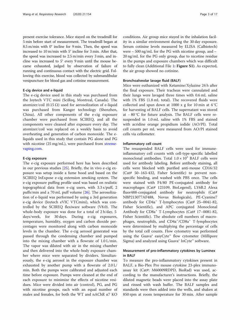
Sub-chronic e-cig exposure induces inflammatory cellularinflux in the mouse lungThe total cell counts were increased in WT mice ex-posed to PG with or without nicotine relative to respect-ive air group, and total cell counts in WT mice exposedto PG with nicotine increased significantly compared toair and PG alone groups (Fig. 1a). The nAChRα7 KOmice showed no change in total cell counts when ex-posed to PG with nicotine (Fig. 1a). However, the totalcell counts were increased but not significantly com-pared to respective control group (Fig. 1a). Macrophagecounts followed a similar trend as observed in total cell
Wang et al. Respiratory Research (2020) 21:154 Page 4 of 17
counts (Fig. 1b). WT and nAChRα7 KO mice exposedto PG with nicotine showed no change in neutrophilcounts in BALF (Fig. 1c). Additionally, CD4a+ andCD8a+ T-lymphocyte counts were significantly increasedwhen WT mice were exposed to PG with nicotine, andloss of nAChR α7 prevents increase in T-lymphocytes(Fig. 1d-e). PG-exposed nAChRα7 KO mice showedhigher levels of neutrophils and CD4a+/CD8a+ T-lymphocytes than PG-exposed WT mice (Fig. 1c-e).
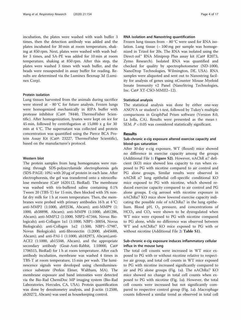
Sub-chronic e-cig exposure augments influx of pro-inflammatory cytokines in BALFWe next determined the level of pro-inflammatory cyto-kines in BALF after sub-chronic e-cig exposure. Most ofthe pro-inflammatory cytokines in BALF were signifi-cantly increased in WT mice exposed to PG with nico-tine compared to PG alone and air control (Fig. 2 andAdditional File 2: Table S2). We have noticed thatmacrophage-mediated pro-inflammatory cytokines, IL-1α, MCP-1, and GM-CSF were significantly increased inWT mice exposed to PG with nicotine, and nAChRα7KO blocks the release of these mediators (Fig. 2). Simi-larly, lymphocytes-related cytokines, IL-2, IL-9, IFNγ,and RANTES showed a nAChRα7-dependent reduction
in cytokine levels (Fig. 2). Additionally, TNFα, MIP-1β,IL-1β and IL-5 were increased in PG with nicotine ex-posed WT mice, but not in nAChRα7 KO mice exposedto PG with nicotine. The remaining cytokines IL-3, IL-4,IL-10, IL-12p40, IL-17A, G-CSF, and MIP-1α were notaffected among any of the exposure groups in WT andnAChRα7 KO mice compared to respective air control(Additional File 2: Table S2).
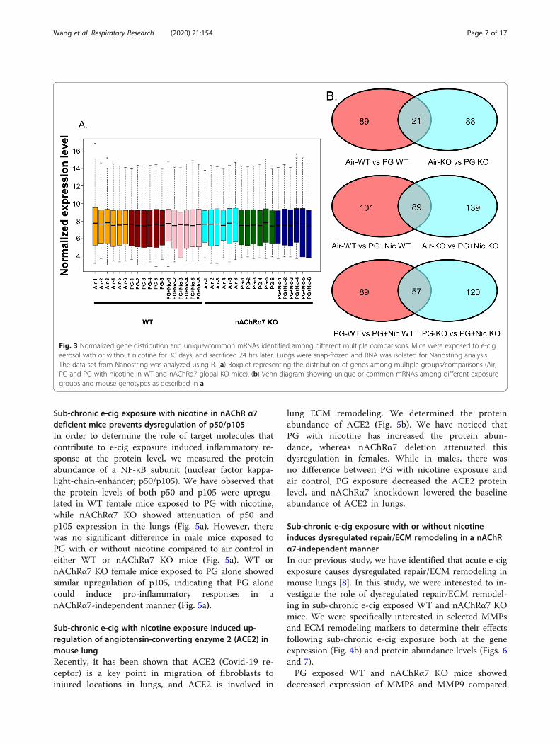
Sub-chronic e-cig exposure alters mRNA transcript levelsof inflammation and dysregulated repair response genesin WT and nAChRα7 KO miceTo further determine whether sub-chronic e-cigexposure-mediated inflammatory responses and dysregu-lated repair/ECM remodeling was caused by altered ex-pression of myeloid innate immune response targetgenes (~ 734 genes), we performed gene expression ana-lysis using nCounter Mouse Myeloid Innate ImmunityPanel (Nanostring Technologies, Inc.). The datasets wereanalyzed by Nanostring nSolver and R programming(Additional file 3: Table S3). The distribution of normal-ized transcript levels among different experimentalgroups was presented as boxplot (Fig. 3a). The pairwisecomparison between PG alone exposed WT and
Fig. 1 Sub-chronic e-cig exposure augments inflammatory cell influx in BALF. Differential inflammatory cell counts were measured in BALF frommice exposed to air, PG or PG with nicotine (PG + Nic) for 30 days (2 h/day). (a) Total inflammatory cell counts were analyzed by Cellometer usingAO/PI staining. Differential inflammatory cell counts were determined as percentages by flow cytometry. Absolute cell counts for (b) F4/80+macrophages, (c) Ly6B.2+ neutrophils, (d) CD4a + T-lymphocytes, and (e) CD8a + T-lymphocytes were normalized to the total cell counts. Data areshown as mean ± SEM (n = 4–6/group; equal number of male and female mice). * P < 0.05, ** P < 0.01 between groups; & P < 0.05, compared toPG exposed WT group; # P < 0.05, compared to PG + Nic exposed WT group
Wang et al. Respiratory Research (2020) 21:154 Page 5 of 17
nAChRα7 KO mice revealed 21 genes that were differen-tially expressed compared to respective air control(Fig. 3b). Additionally, pairwise comparison analysisamong PG with nicotine exposed WT and nAChRα7KO mice revealed 89 genes differentially expressedwhen compared to respective air control (Fig. 3b).Furthermore, pairwise comparison between PG withnicotine and PG alone groups in WT and nAChRα7KO mice revealed 57 differentially expressed genes(Fig. 3b).WT mice showed significantly higher RNA counts in-
dicative of increased mRNA transcript levels for multipletargets in PG with or without nicotine exposed groups,compared to air control (Additional File 1: Figure S3).When we compared WT mice exposed to PG alone, aircontrol or PG with nicotine we found altered gene ex-pression of inflammatory and ECM remodeling targetssuch as matrix metalloproteases (MMP9 and MMP8),S100A8, and collagens (COL17A1 and COL14A1) (Add-itional File 1: Figure S3A). When WT mice exposed toPG with nicotine were compared with air control, wefound a significant difference in transcript levels of in-flammatory targets such as ARG1 and LPL (AdditionalFile 1: Figure S3A). PG with nicotine exposure causedsignificant alterations in the expression of several targetgenes such as SMAD7, KLF4, CDH1, COL4A2, ICAM1,
LDLR, IL1B, TLR5, NFKBIA, and CXCL2 among WTand nAChR α7 KO mice that belong to the broad cat-egories inflammation and dysregulated repaired/ECMremodeling (Additional File 1: Figure S3B). Based onthe above-mentioned gene expression analysis by Nano-string, we chose specific target genes for further analysis(Fig. 4).Based on the Nanostring nCounter analysis, transcript
levels of target genes that showed significant differencesbetween WT and nAChRα7 KO mice following PG withnicotine exposure were selectively represented in Fig. 4a.The gene expression levels of SKIL (Ski-like protein) andLDLR (Low-density lipoprotein receptor) were signifi-cantly decreased in WT mice exposed to PG with nico-tine, but not nAChRα7 KO mice. However, geneexpression levels of CCL9 (Chemokine ligand 9), KLF4(Kruppel-like factor 4), DUSP1 (Dual Specificity Phos-phatase 1), BTLA (B and T Lymphocyte Associated),and SMAD7 (SMAD Family Member 7) were signifi-cantly increased only in nAChRα7 KO mice exposed toPG with nicotine compared to WT mice (Fig. 4a). Fi-nally, NECTIN1 gene expression in WT and nAChRα7KO mice showed a decreased trend, but only WT miceexposed to PG with nicotine showed a significant reduc-tion in the expression of NECTIN1 transcript comparedto air controls (Fig. 4a).
Fig. 2 Sub-chronic e-cig exposure-induced pro-inflammatory mediators in BALF. Bio-Plex Pro mouse cytokine 23-plex assay kit (Bio-Rad) was used todetermined levels of pro-inflammatory cytokines/chemokines in BALF from mice exposed to e-cig with or without nicotine for 30 days (2 h/day).Significant changes were found in cytokines related to macrophages (IL-1α, MCP-1, TNF-α, GM-CSF, and MIP-1β) and T-lymphocytes (IL-2, IL-5, IL-9, RANTES,and IFN-γ). Data are shown as mean ± SEM (n = 6–10/group; equal number of male and female mice). * P < 0.05, ** P < 0.01, *** P < 0.001, compared to aircontrol; # P < 0.05, compared to PG +Nic exposed WT mice
Wang et al. Respiratory Research (2020) 21:154 Page 6 of 17
Sub-chronic e-cig exposure with nicotine in nAChR α7deficient mice prevents dysregulation of p50/p105In order to determine the role of target molecules thatcontribute to e-cig exposure induced inflammatory re-sponse at the protein level, we measured the proteinabundance of a NF-κB subunit (nuclear factor kappa-light-chain-enhancer; p50/p105). We have observed thatthe protein levels of both p50 and p105 were upregu-lated in WT female mice exposed to PG with nicotine,while nAChRα7 KO showed attenuation of p50 andp105 expression in the lungs (Fig. 5a). However, therewas no significant difference in male mice exposed toPG with or without nicotine compared to air control ineither WT or nAChRα7 KO mice (Fig. 5a). WT ornAChRα7 KO female mice exposed to PG alone showedsimilar upregulation of p105, indicating that PG alonecould induce pro-inflammatory responses in anAChRα7-independent manner (Fig. 5a).
Sub-chronic e-cig with nicotine exposure induced up-regulation of angiotensin-converting enzyme 2 (ACE2) inmouse lungRecently, it has been shown that ACE2 (Covid-19 re-ceptor) is a key point in migration of fibroblasts toinjured locations in lungs, and ACE2 is involved in
lung ECM remodeling. We determined the proteinabundance of ACE2 (Fig. 5b). We have noticed thatPG with nicotine has increased the protein abun-dance, whereas nAChRα7 deletion attenuated thisdysregulation in females. While in males, there wasno difference between PG with nicotine exposure andair control, PG exposure decreased the ACE2 proteinlevel, and nAChRα7 knockdown lowered the baselineabundance of ACE2 in lungs.
Sub-chronic e-cig exposure with or without nicotineinduces dysregulated repair/ECM remodeling in a nAChRα7-independent mannerIn our previous study, we have identified that acute e-cigexposure causes dysregulated repair/ECM remodeling inmouse lungs [8]. In this study, we were interested to in-vestigate the role of dysregulated repair/ECM remodel-ing in sub-chronic e-cig exposed WT and nAChRα7 KOmice. We were specifically interested in selected MMPsand ECM remodeling markers to determine their effectsfollowing sub-chronic e-cig exposure both at the geneexpression (Fig. 4b) and protein abundance levels (Figs. 6and 7).PG exposed WT and nAChRα7 KO mice showed
decreased expression of MMP8 and MMP9 compared
Fig. 3 Normalized gene distribution and unique/common mRNAs identified among different multiple comparisons. Mice were exposed to e-cigaerosol with or without nicotine for 30 days, and sacrificed 24 hrs later. Lungs were snap-frozen and RNA was isolated for Nanostring analysis.The data set from Nanostring was analyzed using R. (a) Boxplot representing the distribution of genes among multiple groups/comparisons (Air,PG and PG with nicotine in WT and nAChRα7 global KO mice). (b) Venn diagram showing unique or common mRNAs among different exposuregroups and mouse genotypes as described in a
Wang et al. Respiratory Research (2020) 21:154 Page 7 of 17
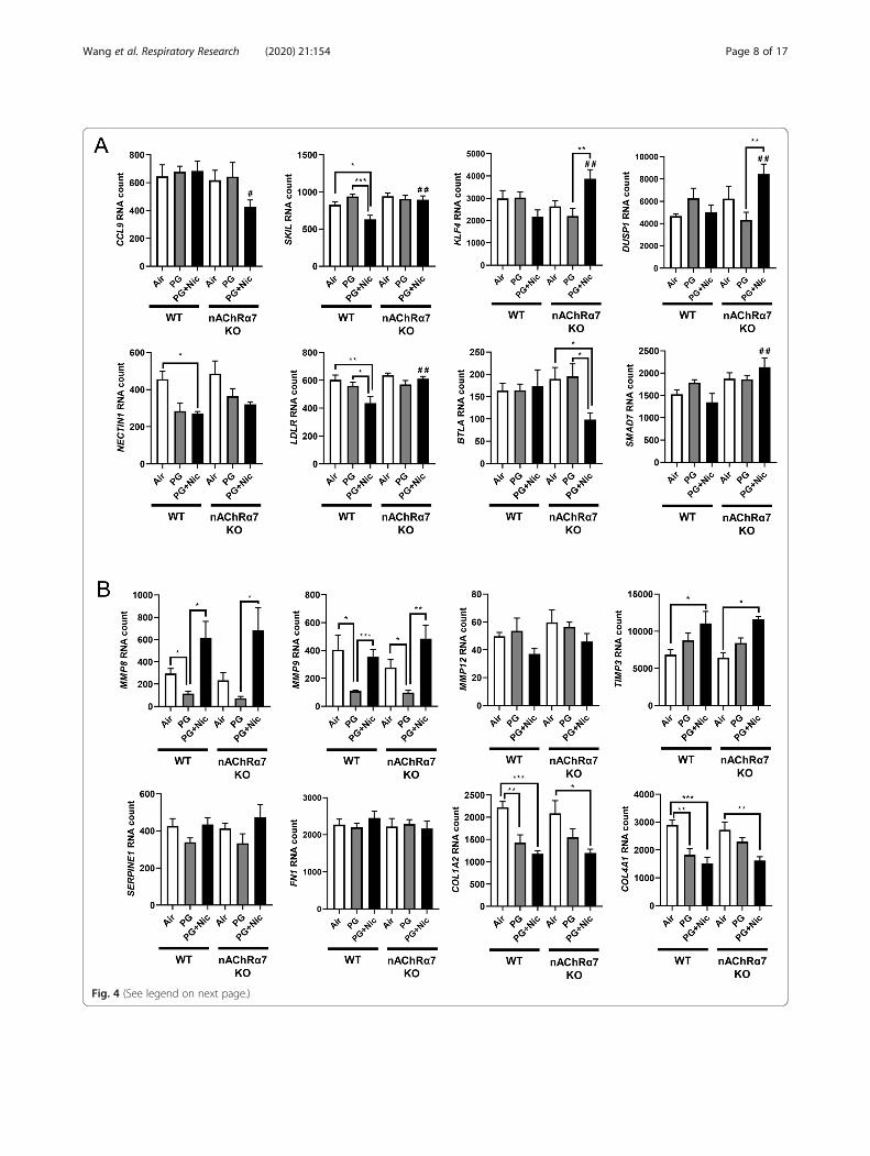
Fig. 4 (See legend on next page.)
Wang et al. Respiratory Research (2020) 21:154 Page 8 of 17
to air control and PG with nicotine groups (Fig. 4b).WT and nAChRα7 KO mice exposed to PG withnicotine showed significant increases in TIMP3, whichis an inhibitor of MMPs (Fig. 4b). ECM target genesfibronectin (FN1) and Plasminogen activatorinhibitor-1 (PAI-1, gene code: SERPINE1) were un-affected, while genes that modulate collagen expres-sion (COL1A2 and COL4A1) significantly decreasedin PG with or without nicotine exposed WT andnAChRα7 KO mice.Based on the gene expression changes in dysregulated
repair markers, we measured the protein abundances ofthe same in e-cig exposed WT and nAChRα7 KO mice.Results from immunoblot analysis correlated with geneexpression of MMP9 and MMP8 in both female andmale mice (Fig. 6a-b). Decreased MMP8 and MMP9were also observed in PG-exposed WT and nAChRα7KO mice compared to PG with nicotine and air control(Fig. 6a-b). MMP2 protein abundance was increased inWT male and female mice exposed to PG alone, com-pared to air control and PG with nicotine-exposed mice.Moreover, nAChRα7 KO female mice show increasedMMP2 protein abundance at baseline, while no changewas observed in male mice (Fig. 6a-b). The protein levelsof MMP9, MMP2, and MMP12 were increased in WTmale mice exposed to PG with nicotine, while nAChRα7KO inhibits protein abundance of MMPs (Fig. 6b).MMP8 protein levels were comparable in female andmale mice and nAChRα7 KO showed decreased proteinabundance of MMP8 at baseline (Fig. 6a-b).ECM proteins/regulators such as collagens, fibronectin,
and PAI-1, were measured in WT and nAChRα7 KOmouse lungs (Fig. 7). The protein abundance of PAI-1showed no difference among air, PG alone and PG withnicotine exposed WT and nAChRα7 KO female mice (Fig.7a). Whereas PAI-1 was increased in WT male mice ex-posed to PG with nicotine, but was reduced in nAChRα7KO male mice (Fig. 7b). Furthermore, two different sub-units of type 1 collagens (COL1A1 and COL1A2) were dif-ferentially expressed at the protein level among theexperimental groups. WT and nAChRα7 KO female andmale mice showed increased expression of COL1A2 whenexposed to PG alone (Fig. 7). However, COL1A1 proteinabundance was reduced in WT male mice exposed to PGwith or without nicotine, and nAChRα7 deficiency furtherdecreased the baseline of COL1A1 levels in male mice.
There is no change in the protein levels of COL1A1 amongPG alone and PG with nicotine exposure in WT andnAChRα7 KO mice. PG with or without nicotine exposurereduced the protein abundance of fibronectin in WT fe-male and nAChRα7 KO mice compared to air control (Fig.7a). However, a decreased fibronectin protein level was ob-served in the PG-exposed WT male mice, and there was noalteration in nAChRα7 KO male mice among all the differ-ent exposure conditions (Fig. 7b).
Sub-chronic e-cig exposure in lung epithelial cell-specificnAChR α7 deletion protects against inflammationConsidering the inflammatory responses seen in WT andnAChRα7 KO mice, we exposed nAChRα7 lung epithelialcell-specific KO mice to e-cig aerosol with or withoutnicotine to determine the role of nAChRα7 in the lungepithelium. Surprisingly, we did not see any significantchanges in pro-inflammatory cytokines following PG withnicotine exposure in nAChRα7 epithelial cell-specific KOmice. However, IL-5, MCP-1, KC, Eotaxin, GM-CSF, andG-CSF levels were significantly increased in PG alonegroup when compared to air control, and these cytokineswere inhibited in nAChRα7 epithelial cell-specific KOmice (Additional File 1: Figure S4A). The rest of the cyto-kines from BALF showed no changes among all exposuregroups in nAChRα7 epithelial cell-specific KO mice (Add-itional File 1: Figure S4B-C).
DiscussionThe popularity of e-cig vaping has been rising recently inthe United States, and hence health concerns about vapinghave recently attracted public attention [27]. Our previousstudies have shown that acute exposure to e-cigarettescould cause inflammatory responses, oxidative stress, andECM remodeling [8, 11, 28]. Although in the past we haveshown the health risks caused by acute e-cig exposures inmice [8, 11], sub-chronic or chronic effects of e-cigexposure-induced toxicity and respiratory health effectsremain elusive. Based on our knowledge, no study has yetshown that long-term exposure of e-cig aerosol can resultin persistent dysregulated repair/ECM remodeling and in-flammatory responses implicating the role of thenAChRα7 receptor in the lung.Similar to our acute exposure results, e-cig aerosol
with nicotine induced inflammatory cell counts in BALFcompared to air control, especially in macrophages and
(See figure on previous page.)Fig. 4 Sub-chronic e-cig exposure affects mRNA expression of myeloid and innate immune response target genes analyzed by Nanostringanalysis. Mice were exposed to e-cig aerosol with or without nicotine for 30 days (2 hrs/day), and sacrificed 24 hrs after final exposure. RNA wasisolated from lungs and screened via nCounter Mouse Myeloid Innate Immunity Panel using Nanostring analysis. Targets were selected based onsignificant differences, especially (a). between) PG+Nic exposed WT and nAChRα7 KO mice, or (b) ECM remodeling focused. RNA counts werenormalized to multiple housekeeping genes and quantified using nSolver. Data are shown as mean ± SEM (n=6/group; equal number of maleand female mice). * P < 0.05, ** P < 0.01, *** P < 0.001 between groups; # P < 0.05, # # P < 0.01 compared with PG+Nic exposed WT group
Wang et al. Respiratory Research (2020) 21:154 Page 9 of 17
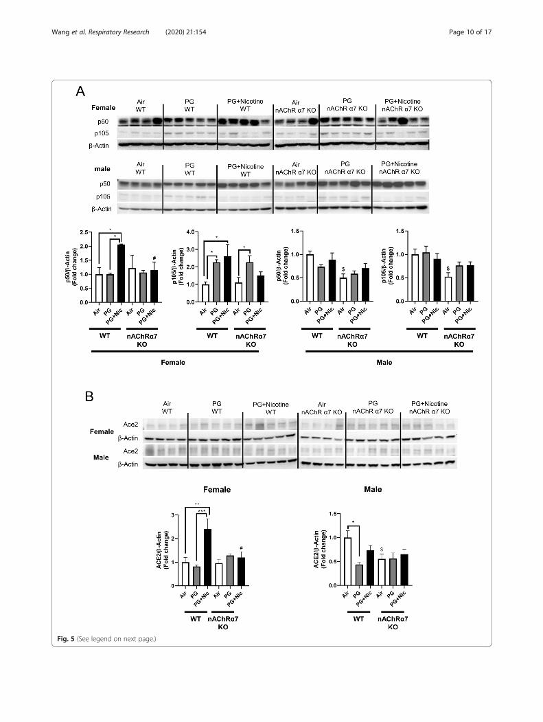
Fig. 5 (See legend on next page.)
Wang et al. Respiratory Research (2020) 21:154 Page 10 of 17
CD4a+/CD8a+ T-lymphocytes. Previous report has dem-onstrated that two weeks of e-cig (with nicotine) expos-ure increased the number of macrophages but notneutrophils, which supports our findings [29]. Anotherstudy reported that acute exposure of e-cig aerosol in-creased total cell and macrophage counts; and even thevehicle exposure alone showed a trend towards increasein total cells and macrophages [30]. Based on our results,e-cig exposure with or without nicotine causes increasedinflammatory cellular influx into the lungs (BALF). PGalone (vehicle) exposure was considered safe by e-cigusers, but our data reveals that sub-chronic e-cig expos-ure with PG (without nicotine) induced lung inflamma-tion. The genetic ablation of nAChRα7 protects againste-cig exposure induced increase in macrophages andCD4a+/CD8a+ T-lymphocytes. Probably, e-cig exposurecontaining PG with nicotine causes lung inflammation/remodeling response via the nAChRα7 receptor medi-ated signaling pathway, and blocking the receptor couldattenuate nicotine-dependent effects in the lungs. Priorreports show that activation of nAChR can also inhibitinflammation [31], and we have observed increased neu-trophils and CD4a+/CD8a+ T-lymphocytes when ex-posed to PG alone in nAChRα7 KO mice. The nAChRdeletion may promote inflammation independent ofnicotine exposure in the lungs. The chemicals derivedfrom e-cig aerosols containing PG, such as formaldehyde,acetaldehyde, and methylglyoxal, in nicotine-free aerosol[32] may result in increased inflammation. As reportedpreviously, both e-cig with or without nicotine caused in-filtration of inflammatory cells and pro-inflammatory me-diators that are key players for causing lung inflammatoryresponse [8, 11, 30]. As an agonist of nAChRα7, nicotinemight be both pro-inflammatory and anti-inflammatoryroles, and further research is needed to study the relation-ship between nicotine and nicotinic receptors in the con-text of sub-chronic e-cig exposure.Apart from the inflammatory cells, some pro-
inflammatory mediators in BALF were significantly in-creased following e-cig exposure with nicotine. Specificmacrophage-mediated pro-inflammatory cytokines such asIL-1α, MCP-1, and GM-CSF, were increased in BALF afterPG with nicotine exposure in a nAChRα7-dependent man-ner. However, a few other macrophage-driven cytokines,IL-1β, TNF-α, and MIP-1β, did not show nAChRα7 de-pendency. Our results paralleled reports that e-cig aerosol
containing nicotine induced cytotoxicity in alveolar macro-phage and significant release of inflammatory mediators(TNF-α, CXCL-8, MCP-1, and IL-6) [33]. Additional stud-ies have shown specific T-lymphocyte-related cytokines/chemokines (IL-2, IL-9, IFN-γ, and RANTES) and their rolein inflammatory signaling [34–37], and our results indi-cated that these altered cytokine responses were nAChR-dependent in e-cig exposed mouse lungs. Mice exposed toPG with nicotine showed increased IL-5 release in anAChR independent manner. Another report shows in-creased IL-4, IL-5, and IL-13 levels following e-cig contain-ing nicotine when challenged to ovalbumin treated miceresulting in exacerbation of allergic airway inflammation[38]. The increased IFN-γ production has been related tocytotoxicity and infiltration of CD8a+ T-cells [39, 40]. Thisaltered pro-inflammatory cytokine response and cellular in-flux in the lung are inter-connected to each other partlydue to nAChRα7 mediated signaling. It is possible thatnAChRα7 deletion could attenuate the inflammatory re-sponses induced by e-cig aerosol contained nicotine in thelungs. However, PG-induced increase in CD4a+/CD8a+ T-lymphocytes does not corroborate with the cytokine levels.Future studies will address immune cell-type specific rolein pulmonary toxicity of e-cig with and without nicotineduring long-term exposure in mice.We measured the mRNA expression using the mouse
myeloid innate immunity panel from Nanostring technol-ogy to screen the potentially affected target genes. Our re-sults showed altered expression of SKIL and LDLR in PGwith nicotine-exposed WT mice, but the same genes re-main unaffected in nAChRα7 KO exposed groups. Thesetarget genes have been shown to play vital roles in TGFβ/SMAD canonical signaling and TGFβ-induced epithelial–mesenchymal transition (EMT) may be inhibited by SKILbinding to SMAD [41, 42] In this study, we found in-creased protein abundance of PAI-1 in WT mice exposedto PG with nicotine, but PAI-1 was further reduced innAChRα7 KO mice. Our results suggest that e-cig expos-ure of PG with nicotine may affect SKIL-TGFβ-PAI-1 axisin the lung possibly through nAChRα7 activation. Theother important target significantly altered by PG withnicotine is LDLR gene. Overexpression of LDLR is capableof promoting macrophage differentiation and exaggeratedinflammatory response, and silencing LDLR results in re-duced inflammatory responses in THP-1 cells [43]. In ourstudy, LDLR was reduced in PG with nicotine exposed
(See figure on previous page.)Fig. 5 Sub-chronic e-cig exposure lead to dysregulateddifferentially affects the protein abundance of NF-κB subunits (p50/p105) and Angiotensin-converting enzyme 2 (ACE2) in mouse lungs with sex differences. The protein abundance of (a) p50/p105 and (b) Angiotensin-converting enzyme 2(ACE2) were measured in whole lung homogenates via Western blot. Representative blot images for female and male mice are shown. Densitometryanalysis of individual blots was performed for p50/p105 and ACE2 for both female and male, and β-actin was used as an endogenous control. Data areshown as mean ± SEM. (n=4-5/group). * P < 0.05 significant compared between groups; P < 0.05, compared with air exposed WT group; # P < 0.05,compared with PG+Nic exposed WT group
Wang et al. Respiratory Research (2020) 21:154 Page 11 of 17
Fig. 6 Sub-chronic e-cig exposure affects protein abundance of matrix metalloproteinases (MMPs) in mouse lungs. Protein levels of several MMPs(MMP9, MMP2, MMP12, and MMP8) were measured in whole mouse lung homogenates via Western blot. Representative blot images anddensitometry analyses for female (a) and male (b) mice are shown. β-actin was used as an endogenous control. Data are shown as mean ± SEM.(n = 4–5/group). * P < 0.05 between groups; $ P < 0.05, compared with air exposed WT group; & P < 0.05, compared with PG exposed WT group.# P < 0.05, compared with PG + Nic exposed WT group
Wang et al. Respiratory Research (2020) 21:154 Page 12 of 17
Fig. 7 Sub-chronic e-cig exposure affects protein abundance of ECM-related markers in mouse lungs. The abundance of ECM proteins (PAI-1,COL1A1, COL1A2, and fibronectin) were measured in whole mouse lung homogenates via Western blot. Representative blot images anddensitometry analyses for female (a) and male (b) mice are shown. β-actin was used as an endogenous control. Data are shown as mean ± SEM.(n = 4–5/group). (* P < 0.05 between groups; $ P < 0.05, compared with air exposed WT group; & P < 0.05, compared with PG exposed WT group.# P < 0.05, compared with PG + Nic exposed WT group)
Wang et al. Respiratory Research (2020) 21:154 Page 13 of 17
WT mice, while LDLR transcript levels remain unchangedin both PG with and without nicotine exposed nAChRα7KO groups. These findings suggest that nAChRα7 may bean important gateway that affects specific inflammatoryresponse/ECM remodeling target genes during sub-chronic e-cig exposure with nicotine.To further study the inflammatory responses, we mea-
sured the protein abundance of NF-κB (p50/p105) in bothfemale and male mice. The activation of NF-κB is welldocumented in different lung injury models [44] includingcigarette smoke-induced inflammation, wheras researchon e-cig aerosol induced NF-κB activation remains lessexplored [45–48]. Although the anti-inflammatory effects(i.e. inhibition of NF-κB signaling) via activation ofnAChRs is well-known and has been studied previously,nicotine has been shown induce inflammation through ac-tivation of the NF-κB signaling pathway [31, 49]. Our find-ings suggest that e-cig aerosol containing nicotineincreased p50 and p105 protein abundance in female micein a nAChRα7-dependent manner. We believe nicotine isa potent inducer of altered lung inflammation via acti-vated nAChRα7 compared to the anti-inflammatory ef-fects regulated by nAChR-related signaling. We foundlack of nAChRα7 showing attenuation of inflammatory re-sponse following e-cig exposure with nicotine. However,nicotine-free e-cig exposure increased p105 protein levels,which suggests this subunit may act through an alternativepathway that may be independent of nAChRα7 signaling.We for the first time show that lung inflammation causedby e-cig aerosol containing nicotine is nAChRα7-dependent, probably in a sex-specific manner. However,more detailed studies are needed to further illustrate theexact mechanism by which sex-differences in lung inflam-matory responses occur following e-cig exposure throughcell-type specific nAChRα7-mediated signaling.As we recently reported, acute exposure to e-cig aero-
sol with or without nicotine resulted in dysregulated re-pair and ECM remodeling [8]. We noticed similar effectsin our sub-chronic exposure model as well. E-cig aerosolwith or without nicotine alters MMPs and ECM remod-eling proteins in a sex-dependent manner, which arenAChRα7 receptor independent. It is known that nico-tine exposure causes harmful respiratory effects, such asairway remodeling and airspace enlargement [50, 51],which are key features of emphysema and COPD [51–53]. However, dysregulation of MMPs serves not only asa factor for ECM remodeling, but also as a marker of in-flammation mediated by dysregulated macrophages.Based on our results, nicotine-free e-cig aerosol inducedaugmented MMPs dysregulation more so than e-cigaerosol with nicotine. Increased MMP2/MMP12, alongwith decreased MMP9/MMP8, might suggest a compen-satory feedback loop [54]. We observed that ACE2 is in-creased by e-cig with nicotine exposure, whereas the
level is decreased in nAChRα7-deficient mice, suggestingthat ACE2 upregulation by nicotine is mediated bynAChRα7. It is thought that ACE2 is upregulated bysmoking, and that its expression level is increased insmokers and COPD patients [55, 56], as well as servingas the entry gate for the novel coronavirus (SARS-CoV-2) [57]. Hence, e-cig with nicotine inhalation would pro-mote SARS-CoV-2 infection, and nAChRα7 deletion anddownregulation of ACE2 may have some ramificationson viral infection.One of the major substrates of MMPs is ECM related
proteins. From our previous study, mice exposed to e-cig aerosols for 3 days showed dysregulated repair/ECMremodeling both at the mRNA and protein levels in asex-dependent manner [8]. The current study shows thatsub-chronic e-cig exposure with or without nicotine af-fects ECM remodeling to a certain extent dependent onnAChRα7 with some sex-specific phenotypes. We haveobserved increased PAI-1 only in WT male mice ex-posed to PG with nicotine in a nAChRα7-dependentmanner, and PAI-1 is a primary ECM regulator and pro-fibrotic marker [58]. Therefore, increased PAI-1 follow-ing inhalation of e-cig aerosol with nicotine indicatesthat e-cig vapor containing nicotine could increase riskof chronic fibrotic disease, implicating the crucial role ofnAChRα7-related signaling. Some other ECM proteins,such as type 1 collagen and fibronectin, were alteredafter exposure of e-cig aerosol with or without nicotine.ECM proteins affected by e-cig exposure are associatedwith dysregulated wound healing and fibrotic disease[59]. The observed increase in COL1A2 following e-cigwith PG alone exposure in both male and female micedemonstrates the significant health risk imposed by PGalone, in addition to the risks associated with PG withnicotine. As mentioned above, nicotine might not be theonly target that can activate nAChRα7, and PG alone viaother indirect mechanisms could activate nicotinic re-ceptors, which may be in agreement with our previousstudies [8, 19]. It is well known that collagen and fibro-nectin are required during the wound healing processand delayed wound healing is observed when epithelialcells were unable to synthesize fibronectin [60]. Wefound down-regulation of collagen and fibronectin fol-lowing e-cig exposure, which will make lungs more vul-nerable later in life when exposed to other noxious gasesand environmental toxicants. Further studies are re-quired to understand the mechanisms underlying how e-cig exposures may induce damaging responses, leadingto lung disease and injury.Based on our findings, long-term studies are needed
to provide further insights on how nAChRα7 depend-ency plays an important cell-type specific role in thelung (epithelial and fibroblast) during e-cig exposure,and its involvement in ACE2 regulation. We know
Wang et al. Respiratory Research (2020) 21:154 Page 14 of 17
that several factors, such as duration of exposure,route of exposure, dose, e-liquid composition, settingused for delivering the e-cig aerosols, concentrationof PG mixture and nicotine, as well as mouse strain,background, and sex of mice may equally contributeto the difference and phenotype observed in themeasure outcomes. Chronic e-cig exposures are un-derway that will help us to understand how these sig-naling mechanisms are altered by e-cig exposure withand without nicotine, and how these changes maycontribute to the respiratory toxicity observed inmice, which could translate to clinical relevance inhuman e-cig users. Ongoing human studies are beingcarried out to identify potential targets and toxicitybiomarkers that may relate to human respiratoryhealth effects of e-cigarettes.
ConclusionIn conclusion, nAChRα7 ablation attenuates the in-flammation induced by sub-chronic e-cig exposure,which may be partly due to e-cig induced ECM re-modeling/dysregulated repair response in the lung. Ina sub-chronic e-cig exposure, nAChRα7 plays a novelrole in an anti-inflammatory response induced bynicotine. We show a significant difference in terms ofaltered genes that belong to inflammation, dysregu-lated repair and ECM remodeling in WT mice com-pared to nAChRα7 KO mice when exposed to PGwith nicotine. The differential inflammatory cellcounts and pro-inflammatory cytokines in BALF werecomparable to our previous findings. Hence,nAChRα7 may play a vital role in the inflammatoryresponses induced by nicotine-containing e-cig aero-sols. Our data suggest that ECM remodeling/dysregu-lated repair caused by e-cig exposure is mediated bynAChRα7, and occurs in a sex-dependent manner.Overall, e-cig aerosol with nicotine causes lung in-flammation due to altered dysregulated repair re-sponse and ECM remodeling mediated by nAChRα7.Deletion of nAChRα7 may possibly protect againstlung inflammation and injury by attenuating associ-ated signaling targets for ECM remodeling. Hence,nAChRα7 is considered a valid target for inflamma-tion induced by e-cig aerosol with nicotine in thelung.
Supplementary informationSupplementary information accompanies this paper at https://doi.org/10.1186/s12931-020-01396-y.
Additional file 1 Figure S1. Sub-chronic e-cig with PG + Nic exposureincreased cotinine levels in mouse serum. Mice were exposed to e-cigwith or without nicotine for 30 days, and blood was collected throughsubmandibular venipuncture following the final exposure. Cotinine levelswere measured in serum samples from WT and nAChR α7 KO mice
exposed to PG or PG with nicotine by ELISA (Cat# CO096D, Calbiotech),(n = 3–10/group). Figure S2. Sub-chronic e-cig exposure decreased exer-cise capacity in nAChR α7 KO mice. Mice were exposed to e-cig with orwithout nicotine for 30 days, and treadmill for exercise ability measure-ments were performed before the day of sacrifice, after the last exposure.The running distance and running time for nAChR α7 global KO and WTmice (A and B, respectively) and nAChR α7 lung epithelial cell-specificfloxed/Cre-CC10 KO mice (C and D, respectively) were recorded separ-ately. (n = 3–6/group), (*P < 0.05 ***P < 0.001 between groups). FigureS3. Volcano plots represent the differentially expressed myeloid genesamong air, PG with and without nicotine exposed WT and nAChR α7 KOmice. Nanostring mRNA data set obtained from Nanostring Technologiesand analyzed using nSolver. (A) Comparisons between Air vs PG, Air vsPG + Nic, and PG vs PG + Nic in WT mice, with P < 0.05 considered a sig-nificant difference. (B) Comparison between WT and nAChR α7 KO miceexposed to air, PG or PG + Nic that will specifically determine thegenotype-dependent change in expression of myeloid genes followinge-cig exposure. Figure S4. Sub-chronic e-cig exposure induced pro-inflammatory mediators in BALF of nAChRα7 lung epithelial cell-specificKO mice. Bio-Plex Pro mouse cytokine 23-plex assay kit (Bio-Rad) wasused to determine levels of pro-inflammatory cytokines/chemokines inBALF from nAChRα7 lung epithelial cell-specific KO mice exposed to e-cig for 30 days (2 h/day). Significant changes were found in pro-inflammatory cytokines IL-5, MCP-1, KC, Eotaxin, GM-CSF, and G-CSF (A).All the other pro-inflammatory cytokines such as IL-1α, TNFα, MIP-1β, IL-2,IL-9, IFNγ, RANTES, IL-6, IL-12p70, IL-13, IL-1β, Eotaxin, IL-3, IL-4, IL-10, IL-12p40, IL-17A, and MIP-1α (B and C) did not show any significantchanges between the groups. Data are shown as mean ± SEM (n = 3–5/group; equal number of male and female mice), (* P < 0.05, comparedwith air control).
Additional file 2 Table S1. Sub-chronic e-cig exposure using PG withor without nicotine altered blood gas but not hematology. Table S2.Sub-chronic e-cig exposure using PG with nicotine increased pro-inflammatory cytokines in BALF.
Additional file 3 Table S3.
AbbreviationsE-cig: Electronic cigarette; ECM: Extracellular matrix; nAChRs: NicotinicAcetylcholine Receptors; WT: Wild-type; nAChRα7 KO: nAChRα7 knockout;nAChRα7 CreCC10: Lung epithelial-cell-specific nAChRα7 KO; PG: Propyleneglycol; VG: Vegetable glycerin; BALF: Bronchoalveolar lavage fluids;MMPs: Matrix metalloproteinases; IPF: Idiopathic pulmonary fibrosis;COPD: Chronic obstructive pulmonary disease; CNS: Central nervous system;LPSs: Lipopolysaccharides; Glu: Glucose; Hb: Hemoglobin; Hct: Hematocrit;AO/PI: Acridine orange propidium iodide; CCL9: Chemokine ligand 9;KLF4: Kruppel-like factor 4; DUSP1: Dual Specificity Phosphatase 1; BTLA: Band T Lymphocyte Associated; SMAD7: SMAD Family Member 7; NF-κB: Nuclear factor kappa-light-chain-enhancer of activated B cells; PAI-1: Plasminogen activator inhibitor-1; COL1A1: Type 1 collagens chain 1;COL1A2: Type 1 collagens chain 2; ACE2: Angiotensin-converting enzyme 2;SARS-CoV2: Severe acute respiratory syndrome coronavirus 2
AcknowledgmentsThe authors thank Dr. Ye, Dongxia, for technical help of running ELISA/Luminex assays.
Human participants, human data and human tissueNot applicable.
Human ethical approvalNot applicable.
Authors’ contributionsQW, IKS, IR conceived and designed the experiments. QW, DL, JHL, TM, SRMconducted the experiments. QW, IKS, DL, TM, SRM analyzed the data. QW,IKS, SRM, IR wrote and revised the manuscript. The author(s) read andapproved the final manuscript.
Wang et al. Respiratory Research (2020) 21:154 Page 15 of 17
FundingThis study was supported by the National Institutes of Health (NIH)1R01HL135613 and U54 CA228110. The funding body has no role in designof the study, data collection, analysis, and interpretation of data and inwriting the manuscript.
Availability of data and materialsAll data and materials are described in the manuscript.
Ethics approval and consent to participateThis study was performed according to the standards from the United StatesAnimal Welfare Act, National Institutes of Health (NIH). All the animalexperiments were followed the protocol approved by The Animal ResearchCommittee of the University of Rochester (UCAR).
Consent for publicationNot applicable.
Competing interestsThe authors have declared that no competing interest.
Author details1Department of Environmental Medicine, University of Rochester MedicalCenter, Box 850, 601 Elmwood Avenue, Rochester, NY 14642, USA.2Department of Clinical and Translational Research, University of RochesterMedical Center, Rochester, NY, USA.
Received: 14 February 2020 Accepted: 14 May 2020
References1. Farsalinos KE, Polosa R. Safety evaluation and risk assessment of electronic
cigarettes as tobacco cigarette substitutes: a systematic review. Ther AdvDrug Saf. 2014;5:67–86.
2. Javed F, Kellesarian SV, Sundar IK, Romanos GE, Rahman I. Recent updateson electronic cigarette aerosol and inhaled nicotine effects on periodontaland pulmonary tissues. Oral Dis. 2017;23:1052–7.
3. Kaur G, Muthumalage T, Rahman I. Mechanisms of toxicity and biomarkersof flavoring and flavor enhancing chemicals in emerging tobacco and non-tobacco products. Toxicol Lett. 2018;288:143–55.
4. Lawyer GR, Jackson M, Prinz M, Lamb T, Wang Q, Muthumalage T, RahmanI. Classification of flavors in cigarillos and little cigars and their variablecellular and acellular oxidative and cytotoxic responses. PLoS One. 2019;14:e0226066.
5. Madison MC, Landers CT, Gu BH, Chang CY, Tung HY, You R, Hong MJ,Baghaei N, Song LZ, Porter P, Putluri N, Salas R, Gilbert BE, Levental I,Campen MJ, Corry DB, and Kheradmand F. Electronic cigarettes disrupt lunglipid homeostasis and innate immunity independent of nicotine. J ClinInvest. 2019;129:4290–304.
6. Goniewicz ML, Kuma T, Gawron M, Knysak J, Kosmider L. Nicotine levels inelectronic cigarettes. Nicotine Tobacco Res. 2012;15:158–66.
7. Grana RA, Popova L, Ling PM. A longitudinal analysis of electronic cigaretteuse and smoking CessationElectronic cigarette use and smokingCessationLetters. JAMA Intern Med. 2014;174:812–3.
8. Wang Q, Ahmad Khan N, Muthumalage T, Lawyer GR, McDonough SR,Chuang T-D, Gong M, Sundar IK, Rehan VK, Rahman I. Dysregulated repairand inflammatory responses by e-cigarette-derived inhaled nicotine andhumectant propylene glycol in a sex-dependent manner in mouse lung.FASEB BioAdvances. 2019;1:609–23.
9. Burgstaller G, Oehrle B, Gerckens M, White ES, Schiller HB, Eickelberg O. Theinstructive extracellular matrix of the lung: basic composition andalterations in chronic lung disease. Eur Respir J. 2017;50.
10. Wight TN, Frevert CW, Debley JS, Reeves SR, Parks WC, Ziegler SF. Interplayof extracellular matrix and leukocytes in lung inflammation. Cell Immunol.2017;312:1–14.
11. Lerner CA, Sundar IK, Yao H, Gerloff J, Ossip DJ, McIntosh S, Robinson R,Rahman I. Vapors produced by electronic cigarettes and e-juices withflavorings induce toxicity, oxidative stress, and inflammatory response inlung epithelial cells and in mouse lung. PLoS One. 2015;10:e0116732.
12. Wu Q, Jiang D, Minor M, Chu HW. Electronic cigarette liquid increasesinflammation and virus infection in primary human airway epithelial cells.PLoS One. 2014;9:e108342.
13. Zia S, Ndoye A, Nguyen VT, Grando SA. Nicotine enhances expressionof the alpha 3, alpha 4, alpha 5, and alpha 7 nicotinic receptorsmodulating calcium metabolism and regulating adhesion and motilityof respiratory epithelial cells. Res Commun Mol Pathol Pharmacol. 1997;97:243–62.
14. Jensen K, Nizamutdinov D, Guerrier M, Afroze S, Dostal D, Glaser S. Generalmechanisms of nicotine-induced fibrogenesis. FASEB J. 2012;26:4778–87.
15. Dani JA, MJPB D, B. Behavior: Cellular mechanisms of nicotine addiction.Pharmacol Biochem Behav. 2001;70:439–46.
16. Benowitz NL. Nicotine addiction. N Engl J Med. 2010;362:2295–303.17. Mabley J, Gordon S, PJI P. Nicotine exerts an anti-inflammatory effect in a
murine model of acute lung injury. Inflammation. 2011;34:231–7.18. Su X, Matthay MA, ABJTjoi M. Requisite role of the cholinergic α7 nicotinic
acetylcholine receptor pathway in suppressing gram-negative sepsis-induced acute lung inflammatory injury. J Immunol. 2010;184:401–10.
19. Avelar AJ, Akers AT, Baumgard ZJ, Cooper SY, Casinelli GP, Henderson BJ.Why flavored vape products may be attractive: Green apple tobacco flavorelicits reward-related behavior, upregulates nAChRs on VTA dopamineneurons, and alters midbrain dopamine and GABA neuron function.Neuropharmacology. 2019;158:107729.
20. Henderson BJ, Wall TR, Henley BM, Kim CH, Nichols WA, Moaddel R, Xiao C,Lester HA. Menthol alone upregulates midbrain nAChRs, alters nAChRsubtype stoichiometry, alters dopamine neuron firing frequency, andprevents nicotine reward. J Neurosci. 2016;36:2957–74.
21. Henderson BJ, Wall TR, Henley BM, Kim CH, McKinney S, Lester HA. Mentholenhances nicotine reward-related behavior by potentiating nicotine-induced changes in nAChR function, nAChR upregulation, and DA neuronexcitability. Neuropsychopharmacology. 2017;42:2285.
22. Jensen RP, Strongin RM, DH. Solvent chemistry in the electronic cigarettereaction vessel. Sci Rep. 2017;7:42549.
23. Khan NA, Sundar IK, Rahman I. Strain- and sex-dependent pulmonarytoxicity of waterpipe smoke in mouse. Physiol Rep. 2018;6:e13579.
24. Yao H, Arunachalam G, Hwang JW, Chung S, Sundar IK, Kinnula VL, CrapoJD, Rahman I. Extracellular superoxide dismutase protects againstpulmonary emphysema by attenuating oxidative fragmentation of ECM.Proc Natl Acad Sci U S A. 2010;107:15571–6.
25. Khan NA, Yogeswaran S, Wang Q, Muthumalage T, Sundar IK, Rahman I.Waterpipe smoke and e-cigarette vapor differentially affect circadian molecularclock gene expression in mouse lungs. PLoS One. 2019;14:e0211645.
26. Behar RZ, Hua M, Talbot P. Puffing topography and nicotine intake ofelectronic cigarette users. PLoS One. 2015;10:e0117222.
27. Tegin G, Mekala HM, Sarai SK, Lippmann S. E-cigarette toxicity? South Med J.2018;111:35–8.
28. Gerloff J, Sundar IK, Freter R, Sekera ER, Friedman AE, Robinson R, Pagano T,Rahman I. Inflammatory response and barrier dysfunction by different e-cigarette flavoring chemicals identified by gas chromatography-massspectrometry in e-liquids and e-vapors on human lung epithelial cells andfibroblasts. Appl In Vitro Toxicol. 2017;3:28–40.
29. Sussan TE, Gajghate S, Thimmulappa RK, Ma J, Kim JH, Sudini K, Consolini N,Cormier SA, Lomnicki S, Hasan F, et al. Exposure to electronic cigarettesimpairs pulmonary anti-bacterial and anti-viral defenses in a mouse model.PLoS One. 2015;10:e0116861.
30. Glynos C, Bibli S-I, Katsaounou P, Pavlidou A, Magkou C, Karavana V,Topouzis S, Kalomenidis I, Zakynthinos S, Papapetropoulos A.Comparison of the effects of e-cigarette vapor with cigarette smoke onlung function and inflammation in mice. Am J Physiol Lung Cell MolPhysiol. 2018;315:L662–72.
31. Patel H, McIntire J, Ryan S, Dunah A, Loring R. Anti-inflammatory effects ofastroglial alpha7 nicotinic acetylcholine receptors are mediated byinhibition of the NF-kappaB pathway and activation of the Nrf2 pathway. JNeuroinflammation. 2017;14:192.
32. Uchiyama S, Noguchi M, Sato A, Ishitsuka M, Inaba Y, Kunugita N.Determination of thermal decomposition products generated from E-cigarettes. Chem Res Toxicol. 2020.
33. Scott A, Lugg ST, Aldridge K, Lewis KE, Bowden A, Mahida RY, GrudzinskaFS, Dosanjh D, Parekh D, Foronjy R, Sapey E, Naidu B, and Thickett DR. Pro-inflammatory effects of e-cigarette vapour condensate on human alveolarmacrophages. Thorax. 2018;73:1161–9.
Wang et al. Respiratory Research (2020) 21:154 Page 16 of 17
34. Crawford A, Angelosanto JM, Nadwodny KL, Blackburn SD, Wherry EJ. A rolefor the chemokine RANTES in regulating CD8 T cell responses duringchronic viral infection. PLoS Pathog. 2011;7:e1002098.
35. Ross SH, Cantrell DA. Signaling and function of Interleukin-2 in Tlymphocytes. Annu Rev Immunol. 2018;36:411–33.
36. Li H, Nourbakhsh B, Cullimore M, Zhang GX, Rostami A. IL-9 is important forT-cell activation and differentiation in autoimmune inflammation of thecentral nervous system. Eur J Immunol. 2011;41:2197–206.
37. De Cunto G, Lunghi B, Bartalesi B, Cavarra E, Fineschi S, Ulivieri C, LungarellaG, Lucattelli M. Severe reduction in number and function of peripheral Tcells does not afford protection toward emphysema and bronchialremodeling induced in mice by cigarette smoke. Am J Pathol. 2016;186:1814–24.
38. Lim HB, Kim SH. Inhallation of e-cigarette cartridge solution aggravatesallergen-induced airway inflammation and hyper-responsiveness in mice.Toxicol Res. 2014;30:13–8.
39. Bhat P, Leggatt G, Waterhouse N, Frazer IH. Interferon-gamma derived fromcytotoxic lymphocytes directly enhances their motility and cytotoxicity. CellDeath Dis. 2017;8:e2836.
40. Whitmire JK, Tan JT, Whitton JL. Interferon-gamma acts directly on CD8+ Tcells to increase their abundance during virus infection. J Exp Med. 2005;201:1053–9.
41. Tecalco-Cruz AC, Rios-Lopez DG, Vazquez-Victorio G, Rosales-Alvarez RE,Macias-Silva M. Transcriptional cofactors ski and SnoN are major regulatorsof the TGF-beta/Smad signaling pathway in health and disease. SignalTransduct Target Ther. 2018;3:15.
42. Yang H, Zhan L, Yang T, Wang L, Li C, Zhao J, Lei Z, Li X, Zhang HT.Ski prevents TGF-beta-induced EMT and cell invasion by repressingSMAD-dependent signaling in non-small cell lung cancer. Oncol Rep.2015;34:87–94.
43. Ricci C, Ruscica M, Camera M, Rossetti L, Macchi C, Colciago A, Zanotti I,Lupo MG, Adorni MP, Cicero AFG, et al. PCSK9 induces a pro-inflammatoryresponse in macrophages. Sci Rep. 2018;8:2267.
44. Wang J, Liu Y-T, Xiao L, Zhu L, Wang Q, Yan T. Anti-inflammatory effects ofapigenin in lipopolysaccharide-induced inflammatory in acute lung injuryby suppressing COX-2 and NF-kB pathway. Inflammation. 2014;37:2085–90.
45. Zhang C, Qin S, Qin L, Liu L, Sun W, Li X, Li N, Wu R, Wang X. Cigarettesmoke extract-induced p120-mediated NF-κB activation in human epithelialcells is dependent on the RhoA/ROCK pathway. Sci Rep. 2016;6:23131.
46. Tartell HE. Are E-Cigarettes a safe alternative to smoking? Using airway cellcultures to assess early indicators of COPD progression. Inquiries J StudentPulse. 2015;7.
47. Hasnis E, Bar-Shai M, Burbea Z, Reznick AZ. Mechanisms underlyingcigarette smoke-induced NF-kappaB activation in human lymphocytes:the role of reactive nitrogen species. J Physiol Pharmacol. 2007;58(Suppl5):275–87.
48. Liu X, Togo S, Al-Mugotir M, Kim H, Fang Q, Kobayashi T, Wang X, Mao L,Bitterman P, Rennard S. NF-kappaB mediates the survival of humanbronchial epithelial cells exposed to cigarette smoke extract. Respir Res.2008;9:66.
49. Iho S, Tanaka Y, Takauji R, Kobayashi C, Muramatsu I, Iwasaki H, Nakamura K,Sasaki Y, Nakao K, Takahashi T. Nicotine induces human neutrophils toproduce IL-8 through the generation of peroxynitrite and subsequentactivation of NF-kappaB. J Leukoc Biol. 2003;74:942–51.
50. Hahn HL, Lang M, Bleicher S, Zwerenz S, Rausch C. Nicotine-induced airwaysmooth muscle contraction: neural mechanisms involving the airwayepithelium. Functional and histologic studies in vitro. Clin Investig. 1992;70:252–62.
51. Garcia-Arcos I, Geraghty P, Baumlin N, Campos M, Dabo AJ, Jundi B,Cummins N, Eden E, Grosche A, Salathe M, Foronjy R. Chronic electroniccigarette exposure in mice induces features of COPD in a nicotine-dependent manner. Thorax. 2016;71:1119–29.
52. McDonough JE, Yuan R, Suzuki M, Seyednejad N, Elliott WM, Sanchez PG,Wright AC, Gefter WB, Litzky L, Coxson HO, et al. Small-airway obstructionand emphysema in chronic obstructive pulmonary disease. N Engl J Med.2011;365:1567–75.
53. Shapiro SD. Animal models for COPD. Chest. 2000;117:223S–7S.54. Kato H, Duarte S, Liu D, Busuttil RW, Coito AJ. Matrix Metalloproteinase-2
(MMP-2) gene deletion enhances MMP-9 activity, impairs PARP-1degradation, and exacerbates hepatic ischemia and reperfusion injury inmice. PLoS One. 2015;10:e0137642.
55. Leung JM, Yang CX, Tam A, Shaipanich T, Hackett TL, Singhera GK, DorscheidDR, Sin DD. ACE-2 expression in the small airway epithelia of smokers andCOPD patients: implications for COVID-19. Eur Respir J. 2020;55(5).
56. Brake SJ, Barnsley K, Lu W, McAlinden KD, Eapen MS, Sohal SS. SmokingUpregulates angiotensin-converting Enzyme-2 receptor: a potentialadhesion site for novel coronavirus SARS-CoV-2 (Covid-19). J Clin Med.20209(3).
57. Hoffmann M, Kleine-Weber H, Schroeder S, Kruger N, Herrler T, Erichsen S,Schiergens TS, Herrler G, Wu NH, Nitsche A, et al. SARS-CoV-2 Cell EntryDepends on ACE2 and TMPRSS2 and Is Blocked by a Clinically ProvenProtease Inhibitor. Cell. 2020;181:271–80 e278.
58. Rabieian R, Boshtam M, Zareei M, Kouhpayeh S, Masoudifar A, Mirzaei H.Plasminogen activator inhibitor Type-1 as a regulator of fibrosis. J CellBiochem. 2018;119:17–27.
59. Xue M, Jackson CJ. Extracellular matrix reorganization during wound healingand its impact on abnormal scarring. Adv Wound Care (New Rochelle).2015;4:119–36.
60. Kicic A, Hallstrand TS, Sutanto EN, Stevens PT, Kobor MS, Taplin C, Pare PD,Beyer RP, Stick SM, Knight DA. Decreased fibronectin productionsignificantly contributes to dysregulated repair of asthmatic epithelium. AmJ Respir Crit Care Med. 2010;181:889–98.
Publisher’s NoteSpringer Nature remains neutral with regard to jurisdictional claims inpublished maps and institutional affiliations.
Wang et al. Respiratory Research (2020) 21:154 Page 17 of 17