TNF-α-Induced cPLA2 Expression via NADPH … · NADPH Oxidase/Reactive Oxygen Species-Dependent...
15
ORIGINAL RESEARCH published: 25 November 2016 doi: 10.3389/fphar.2016.00447 Edited by: Wenliang Song, Vanderbilt University Medical Center, USA Reviewed by: Aida Habib, American University of Beirut, Lebanon Hui Huang, Vanderbilt University, USA *Correspondence: Chuen-Mao Yang [email protected] † These authors have contributed equally to this work. Specialty section: This article was submitted to Inflammation Pharmacology, a section of the journal Frontiers in Pharmacology Received: 28 July 2016 Accepted: 08 November 2016 Published: 25 November 2016 Citation: Lin C-C, Lin W-N, Cho R-L, Wang C-y, HsiaoL-D and Yang C-M (2016) TNF-α-Induced cPLA 2 Expression via NADPH Oxidase/Reactive Oxygen Species-Dependent NF-κ B Cascade on Human Pulmonary Alveolar Epithelial Cells. Front. Pharmacol. 7:447. doi: 10.3389/fphar.2016.00447 TNF-α-Induced cPLA 2 Expression via NADPH Oxidase/Reactive Oxygen Species-Dependent NF-κB Cascade on Human Pulmonary Alveolar Epithelial Cells Chih-Chung Lin 1† , Wei-Ning Lin 2† , Rou-Ling Cho 3 , Chen-yu Wang 3 , Li-Der Hsiao 1 and Chuen-Mao Yang 1,3,4 * 1 Department of Anesthetics, Chang Gung Memorial Hospital at Linkou and College of Medicine, Chang Gung University, Tao-Yuan, Taiwan, 2 Graduate Institute of Basic Medicine, Fu Jen Catholic University, New Taipei City, Taiwan, 3 Department of Physiology and Pharmacology and Health Aging Research Center, College of Medicine, Chang Gung University, Tao-Yuan, Taiwan, 4 Research Center for Industry of Human Ecology, Research Center for Chinese Herbal Medicine, and Graduate Institute of Health Industry Technology, Chang Gung University of Science and Technology, Tao-Yuan, Taiwan Tumor necrosis factor-α (TNF-α) triggers activation of cytosolic phospholipase A 2 (cPLA 2 ) and then enhancing the synthesis of prostaglandin (PG) in inflammatory diseases. However, the detailed mechanisms of TNF-α induced cPLA 2 expression were not fully defined in human pulmonary alveolar epithelial cells (HPAEpiCs). We found that TNF-α-stimulated increases in cPLA 2 mRNA (5.2 folds) and protein (3.9 folds) expression, promoter activity (4.3 folds), and PGE 2 secretion (4.7 folds) in HPAEpiCs, determined by Western blot, real-time PCR, promoter activity assay and PGE 2 ELISA kit. These TNF-α-mediated responses were abrogated by the inhibitors of NADPH oxidase [apocynin (APO) and diphenyleneiodonium chloride (DPI)], ROS [N- acetyl cysteine, (NAC)], NF-κB (Bay11-7082) and transfection with siRNA of ASK1, p47 phox , TRAF2, NIK, IKKα, IKKβ, or p65. TNF-α markedly stimulated NADPH oxidase activation and ROS including superoxide and hydrogen peroxide production which were inhibited by pretreatment with a TNFR1 neutralizing antibody, APO, DPI or transfection with siRNA of TRAF2, ASK1, or p47 phox . In addition, TNF-α also stimulated p47 phox phosphorylation and translocation in a time-dependent manner. On the other hand, TNF-α induced TNFR1, TRAF2, ASK1, and p47 phox complex formation in HPAEpiCs, which were attenuated by a TNF-α neutralizing antibody. We found that pretreatment with NAC, DPI, or APO also attenuated the TNF-α-stimulated IKKα/β and NF-κB p65 phosphorylation, NF-κB (p65) translocation, and NF-κB promoter activity in HPAEpiCs. Finally, we observed that TNF-α-stimulated NADPH oxidase activation and ROS generation activates NF-κB through the NIK/IKKα/β pathway. Taken together, our results demonstrated that in HPAEpiCs, up-regulation of cPLA 2 by TNF-α is, at least in part, mediated through the cooperation of TNFR1, TRAF2, ASK1, and NADPH oxidase leading to ROS generation and ultimately activates NF-κB pathway. Keywords: ASK1, cytokines, cytosolic phospholipase A 2 , lung inflammation, signaling transduction Frontiers in Pharmacology | www.frontiersin.org 1 November 2016 | Volume 7 | Article 447
Transcript of TNF-α-Induced cPLA2 Expression via NADPH … · NADPH Oxidase/Reactive Oxygen Species-Dependent...
fphar-07-00447 November 23, 2016 Time: 17:3 # 1
ORIGINAL RESEARCHpublished: 25 November 2016
doi: 10.3389/fphar.2016.00447
Edited by:Wenliang Song,
Vanderbilt University Medical Center,USA
Reviewed by:Aida Habib,
American University of Beirut,Lebanon
Hui Huang,Vanderbilt University, USA
*Correspondence:Chuen-Mao Yang
†These authors have contributedequally to this work.
Specialty section:This article was submitted toInflammation Pharmacology,
a section of the journalFrontiers in Pharmacology
Received: 28 July 2016Accepted: 08 November 2016Published: 25 November 2016
Citation:Lin C-C, Lin W-N, Cho R-L,
Wang C-y, Hsiao L-D and Yang C-M(2016) TNF-α-Induced cPLA2
Expression via NADPHOxidase/Reactive Oxygen
Species-Dependent NF-κB Cascadeon Human Pulmonary Alveolar
Epithelial Cells.Front. Pharmacol. 7:447.
doi: 10.3389/fphar.2016.00447
TNF-α-Induced cPLA2 Expression viaNADPH Oxidase/Reactive OxygenSpecies-Dependent NF-κB Cascadeon Human Pulmonary AlveolarEpithelial CellsChih-Chung Lin1†, Wei-Ning Lin2†, Rou-Ling Cho3, Chen-yu Wang3, Li-Der Hsiao1 andChuen-Mao Yang1,3,4*
1 Department of Anesthetics, Chang Gung Memorial Hospital at Linkou and College of Medicine, Chang Gung University,Tao-Yuan, Taiwan, 2 Graduate Institute of Basic Medicine, Fu Jen Catholic University, New Taipei City, Taiwan, 3 Departmentof Physiology and Pharmacology and Health Aging Research Center, College of Medicine, Chang Gung University,Tao-Yuan, Taiwan, 4 Research Center for Industry of Human Ecology, Research Center for Chinese Herbal Medicine, andGraduate Institute of Health Industry Technology, Chang Gung University of Science and Technology, Tao-Yuan, Taiwan
Tumor necrosis factor-α (TNF-α) triggers activation of cytosolic phospholipase A2
(cPLA2) and then enhancing the synthesis of prostaglandin (PG) in inflammatorydiseases. However, the detailed mechanisms of TNF-α induced cPLA2 expressionwere not fully defined in human pulmonary alveolar epithelial cells (HPAEpiCs). Wefound that TNF-α-stimulated increases in cPLA2 mRNA (5.2 folds) and protein (3.9folds) expression, promoter activity (4.3 folds), and PGE2 secretion (4.7 folds) inHPAEpiCs, determined by Western blot, real-time PCR, promoter activity assay andPGE2 ELISA kit. These TNF-α-mediated responses were abrogated by the inhibitors ofNADPH oxidase [apocynin (APO) and diphenyleneiodonium chloride (DPI)], ROS [N-acetyl cysteine, (NAC)], NF-κB (Bay11-7082) and transfection with siRNA of ASK1,p47phox, TRAF2, NIK, IKKα, IKKβ, or p65. TNF-α markedly stimulated NADPH oxidaseactivation and ROS including superoxide and hydrogen peroxide production whichwere inhibited by pretreatment with a TNFR1 neutralizing antibody, APO, DPI ortransfection with siRNA of TRAF2, ASK1, or p47phox. In addition, TNF-α also stimulatedp47phox phosphorylation and translocation in a time-dependent manner. On the otherhand, TNF-α induced TNFR1, TRAF2, ASK1, and p47phox complex formation inHPAEpiCs, which were attenuated by a TNF-α neutralizing antibody. We found thatpretreatment with NAC, DPI, or APO also attenuated the TNF-α-stimulated IKKα/β andNF-κB p65 phosphorylation, NF-κB (p65) translocation, and NF-κB promoter activity inHPAEpiCs. Finally, we observed that TNF-α-stimulated NADPH oxidase activation andROS generation activates NF-κB through the NIK/IKKα/β pathway. Taken together, ourresults demonstrated that in HPAEpiCs, up-regulation of cPLA2 by TNF-α is, at least inpart, mediated through the cooperation of TNFR1, TRAF2, ASK1, and NADPH oxidaseleading to ROS generation and ultimately activates NF-κB pathway.
Keywords: ASK1, cytokines, cytosolic phospholipase A2, lung inflammation, signaling transduction
Frontiers in Pharmacology | www.frontiersin.org 1 November 2016 | Volume 7 | Article 447
fphar-07-00447 November 23, 2016 Time: 17:3 # 2
Lin et al. TNF-α-Induced cPLA2 Expression
INTRODUCTION
The occurrence and exacerbation of lung diseases, includingchronic obstructive pulmonary disease (COPD) and asthma,is dependent on the severity of lung inflammation (Lee andYang, 2012). Eicosanoids, one of lipid mediators generatingfrom conversion of arachidonic acid (AA), have been found insitu in airway secretion of asthmatics (Barnes, 1989; Hendersonet al., 2002). Phospholipase A2 (PLA2) enzymes catalyze thehydrolysis of membrane phospholipids resulting in the releaseof AA (Borsch-Haubold et al., 1998). The constitutive enzymecyclooxygenase (COX)-1 or the inducible COX-2 then convertsAA to prostaglandins (PGs), such as PGE2 (Yang et al.,2002; Hsieh et al., 2006). Three PLA2 have been identifiedincluding secretory PLA2, the 85 kDa cytosolic group IVPLA2 (cPLA2), and a calcium-independent group VI PLA2in mammalian cells (Six and Dennis, 2000). cPLA2 playsa major role in agonist-induced AA release and eicosanoidproduction (Leslie, 1997). Involvement of cPLA2 in sepsis-related acute lung injury (Nagase et al., 2000) and anaphylaxis-associated bronchial reactivity has been proved (Uozumi et al.,1997). Furthermore, PGE2 synthesis increases are dependent onupregulation of cPLA2 activity in various cell types (Dieter et al.,2002; Gilroy et al., 2004). Elevated levels of TNF-α have beendetected in the bronchoalveolar lavage fluid of asthmatic patients.TNF-α could exaggerate inflammatory responses through up-regulation of inflammatory genes, such as cPLA2 (Hulkoweret al., 1994; Van Putten et al., 2001). Up-regulation of cPLA2further catalyzes the hydrolysis of membrane phospholipidsand releases AA served as a substrate for PGs synthesis(i.e., PGE2) that augments lung inflammation. Moreover, ourprevious findings also provided insights into the correlationbetween COX-2 and cPLA2 expression in ATPγS-stimulatedvascular smooth muscle cells (VSMCs) with similar molecularmechanisms and functional coupling to amplify the occurrenceof vascular inflammation (Lin et al., 2009). Therefore, thesynthesis of PGE2 could be a good index of AA release thatis more sensitive than [3H]AA mobilization (Berenbaum et al.,2003). In this study, although the effect of TNF-α on COX-2 expression was not investigated, we tested the effect ofTNF-α on PGE2 synthesis as a parameter of cPLA2 activityin human pulmonary alveolar epithelial cells (HPAEpiCs).Therefore, up-regulation of cPLA2 may play a key role inlocal and systemic inflammation in airway diseases. However,the molecular mechanisms by which TNF-α induces cPLA2expression and PGE2 synthesis in HPAEpiCs are not completelyunderstood.
Previous report indicates that TNF-α binds to distinctreceptors, TNFR1 and TNFR2, and triggers various inflammatoryresponses (Lee et al., 2009). The association of TNF-α andTNFR1 modulates the severity of tissue injury via activationof proinflammatory or programmed cell death pathway (vanVliet et al., 2005; Lee et al., 2009). TNF receptor associatedfactor 2 (TRAF2) plays an important role in innate immune andinflammatory responses. However, the interaction among TNF-α,TNFR1, TRAF2 and downstream components leading to cPLA2expression is still unknown in HPAEpiCs.
Reactive oxygen species (ROS) are products of normalcellular metabolism acting as second messengers (Lee andYang, 2012). However, either reduced nicotinamide adeninedinucleotide phosphate (NADPH) by pro-inflammatorycytokines such as TNF-α or the mitochondrial electron transportchain and xanthine oxidase leads to increased productionof ROS and unbalance of cellular oxidative stress, which arecauses of airway/lung damages and subsequently respiratoryinflammatory diseases/injuries (Lee and Yang, 2012). Apoptosissignal-regulating kinase 1 (ASK1), a mitogen-activated proteinkinase kinase kinase, participates in regulating stress andimmune responses. ASK1 is activated by cytokines and variousenvironmental and cellular stresses. Hsu et al. indicated thatpeptidoglycan (PGN) induced COX-2 expression via an ASK1signaling in A549 cells (Hsu et al., 2010). Therefore, we exploredwhether TNFR1, TRAF2, ASK1, and NADPH oxidase/ROS areinvolved in TNF-α-induced cPLA2 expression and PGE2 release.
NF-κB plays major roles not only in the evolution butalso in the resolution of inflammatory responses. A widespectrum of biological effects including immune and stress-induced responses, proliferation, differentiation, tumorigenesis,apoptosis, and tissue remodeling are all controlled by activatedNF-κB (Lee and Yang, 2012). The activation of NF-κB can beregulated by various extracellular stimuli, including cytokinesand oxidative stress (Lee and Yang, 2012). We noticed that ROSgeneration can impact NF-κB signaling pathways (Morgan andLiu, 2011). In addition, NF-κB modulates cPLA2 gene activity invarious cell types (Luo et al., 2006; Huwiler et al., 2012; Chi et al.,2014). Therefore, we examined whether TNF-α regulates cPLA2expression via ROS-dependent NF-κB activation in HPAEpiCs.
In addressing these questions, the experiments wereperformed to investigate the mechanisms underlyingTNF-α-induced cPLA2 expression and PGE2 synthesis inHPAEpiCs. These findings suggested that TNF-α-inducedcPLA2-expression associated PGE2 release is, at least in part,mediated through a TNFR1/TRAF2/ASK1/p47phox/NADPHoxidase/ROS/NIK/IKKα/β/NF-κB pathway in these cells.
MATERIALS AND METHODS
Materials
Recombinant human TNF-α was from R&D System(Minneapolis, MN, USA). Anti-cPLA2 (sc-454), anti-p47phox
(sc-14015), anti-Gαs (sc-823), anti-TRAF2 (sc-7346), anti-ASK1 (sc-5294), anti-TNFR1 (sc-52739), anti-NIK (sc7211),anti-IKKα (sc7218), anti-IKKβ (sc8014), anti-p65 (sc-7151)and anti-phospho-serine (sc-81515) antibodies were fromSanta Cruz (Santa Cruz, CA). An anti-GAPDH antibody(#MCA-1D4) was from Encor (Gainesville, FL, USA). HumanTNF-α neutralizing antibody (#7321), anti-phospho-tyrosine(#9411), anti-phospho-ASK1 (#3765), anti-phospho-p65(#3031), and anti-phospho-IKKα/β (#2697) antibodies werefrom Cell Signaling (Danvers, MA, USA). N-Acetyl cysteine(NAC), diphenyleneiodonium chloride (DPI), apocynin(APO), and Bay11-7082 were from Biomol (Plymouth
Frontiers in Pharmacology | www.frontiersin.org 2 November 2016 | Volume 7 | Article 447
fphar-07-00447 November 23, 2016 Time: 17:3 # 3
Lin et al. TNF-α-Induced cPLA2 Expression
Meeting, PA, USA). Dihydroethidium (DHE) and 5-(and-6)-chloromethyl-2′,7′-dichlorodihydrofluorescein diacetate, acetylester (CM-H2DCFDA) were from Molecular Probes (Eugene,OR, USA). SDS-PAGE supplies were from MDBio Inc (Taipei,Taiwan). All other reagents were from Sigma (St. Louis, MO,USA).
Cell Culture and TreatmentHuman pulmonary alveolar epithelial cells (HPAEpiCs) wereordered from ScienCell Research Lab (San Diego, CA, USA).The passages 4–7 were used throughout this study. HPAEpiCswere cultured in DMEM/F12 medium containing 10% FBS,as previously described Lee et al. (2008). The growth mediumwas changed after 48 h and then every 3 days. The viability ofHPAEpiCs after treatment with DMSO or the pharmacologicalinhibitors alone was determined by an XTT [2,3-bis-(2-methoxy-4-nitro-5-sulfophenyl)-2H-tetrazolium-5-carboxanilide] assay,which showed no significant differences (data not shown).
Western Blot AnalysisSerum-starved HPAEpiCs were incubated with TNF-α at 37◦Cfor the various time points. At the end of treatment, the cellswere harvested and centrifuged at 45000 × g at 4◦C for 1 hto prepare the whole cell lysate, as previously described (Chiet al., 2012). The denatured samples were analyzed by 10%SDS-PAGE gels and transferred to nitrocellulose membrane.The Western blot was performed by incubation membrane withan anti-cPLA2, anti-p47phox, anti-Gαs, anti-TRAF2, anti-ASK1,anti-TNFR1, anti-NIK, anti-IKKα, anti-IKKβ, anti-p65, anti-phospho-serine or anti-GAPDH (1:1000) antibody for 24 h,and then incubated with an anti-mouse horseradish peroxidaseantibody (1:2000) for 1 h. ECL reagents and UVP BioSpectrum500 Imaging System (Upland, CA, USA) were used to detect andcapture the immunoreactive bands. The image densitometry ofeach immunoreactive band was analyzed and quantified by theUN-SCAN-IT gel software (Orem, UT, USA).
Real-Time PCR AnalysisTotal RNA of HPAEpiCs was extracted using TRIzol reagentand reverse-transcribed into cDNA. Real-time PCR usingSYBR Green PCR reagents (Applied Biosystems, Branchburg,NJ, USA) and primers specific for cPLA2 and GAPDH geneswere used, as previously described (Chi et al., 2012). Thereal-time primers were as follows: cPLA2α, forward primer:5′-ATGATAGCTCGGACAGTGATGATGA-3′; reverse primer:5′-CATACGATGAATCCAACTTGCTTGA-3′ and GAPDH,forward primer: 5′-CTCTGCTCCTCCTGTTCGAC-3′; reverseprimer: 5′-TTAAAAGCAGCCCTGGTGAC-3′. The expressionof cPLA2 was quantified by normalization to the GAPDHexpression.
Measurement of Intracellular ROSAccumulationThe intracellular H2O2 levels were determined by measuringfluorescence of 2′,7′-dichloro fluorescein diacetate (DCF-DA)and the O•−2 levels were determined by measuring the level
of dihydroethidium (DHE), as previously described Hsu et al.(2014). The fluorescence for DCF and DHE staining was detectedat 495/529 and 518/605 nm, respectively, using a fluorescencemicroscope (Zeiss, Axiovert 200M). For the purpose of theseexperiments, HPAEpiCs were washed with warm HBSS andincubated in HBSS or cell medium containing 10 µM DCFH-DA or DHE at 37◦C for 45 min. Subsequently, HBSS ormedium containing DCFH-DA or DHE was removed andreplaced with fresh medium. HPAEpiCs were then incubatedwith TNF-α. Cells were washed twice with PBS and detachedwith trypsin/EDTA, and the fluorescence intensity of the cells wasanalyzed using a multi-technology reader (Thermo, Appliskan) atEx/Em: 485/530 nm.
Determination of NADPH OxidaseActivity by Chemiluminescence AssayHPAEpiCs (2 × 10⁶ cells/ml) were cultured in 6-well platesand then incubated with TNF-α after growth to confluence andserum-starved. At the end of incubation, cells were harvestedand centrifuged at 400 × g for 10 min at 4◦C. The cell pelletwas kept on ice after re-suspended by 35 µl/per well of ice-coldRPMI-1640 medium. The complex including NADPH (1 µM)or lucigenin (20 µM), 5 µl of cell suspension (0.2 × 10⁵
cells) and a final 200 µl volume of pre-warmed (37◦C) RPMI-1640 medium were prepared and the chemiluminescence wasimmediate recorded with an Appliskan luminometer (Thermo R©)in out-of-coincidence mode. Appropriate blanks and controlswere used. There was no background chemiluminescence oflucigenin in NADPH or NADH alone group (30–40 counts permin), as previously described Hsu et al. (2014). The signals ofchemiluminescence were continuously measured for 12 min, andthe activity of NADPH oxidase was expressed as counts permillion cells.
Measurement of cPLA2 LuciferaseActivityHuman cPLA2 promoter spanning − 2375 to +75 bp as clonedinto pGL3-basic vector (Promega, Madison, WI, USA) as cPLA2-luc plasmid. The activity of cPLA2-luc was detected usinga luciferase assay system (Promega, Madison, WI, USA), aspreviously described Chi et al. (2012). The detected luciferaseactivities were standardized with the activity of β-gal.
Measurement of PGE2 GenerationThe serum-starved cells were treated with TNF-α for the differenttime points. At the end of treatment, culture media were collectedand stored at −80◦C. The concentrations of PGE2 were detectedby a PGE2 enzyme immunoassay kit (Cayman) according to themanufacturer’s instructions, as previously described Lee et al.(2008).
Transient Transfection with siRNAsAll human ASK1, p47phox, TRAF2, NIK, IKKα, IKKβ, and p65siRNA together with scramble siRNA were purchased from Sigma(St. Louis, MO, USA). LipofectamineTM RNAiMAX reagentswere used to prepare siRNA liposome complexes (100 nM
Frontiers in Pharmacology | www.frontiersin.org 3 November 2016 | Volume 7 | Article 447
fphar-07-00447 November 23, 2016 Time: 17:3 # 4
Lin et al. TNF-α-Induced cPLA2 Expression
of siRNAs) according to the manufacturer’s instructions, aspreviously described Lee et al. (2008).
Co-immunoprecipitation AssayCell lysates containing 1 mg of protein were incubated with2 µg of an anti-p47phox, anti-TNFR1, anti-TRAF2, or anti-ASK1 antibody at 4◦C for 24 h, and then 10 µl of 50%protein A-agarose beads was added and mixed at 4◦C for 24 h.The immunoprecipitates were collected and washed thrice witha lysis buffer without Triton X-100. 5x Laemmli buffer wasadded and subjected to electrophoresis on SDS-PAGE, and thenblotted using an anti-phospho-tyrosine, anti-phospho-serine,anti-p47phox, anti-TRAF2, anti-TNFR1, or anti-ASK1 antibody,as previously described (Yang et al., 2014).
Immunofluorescence StainingGrowth-arrested HPAEpiCs were incubated with TNF-α forthe indicated time intervals. After washing twice with ice-cold PBS, cells were fixed, permeabilized and stained using ananti-p65 antibody, as previously described (Chi et al., 2011).A fluorescence microscope (Zeiss, Axiovert 200M) were used toobserve images.
Cell Fractions IsolationThe cell lysates were sonicated for 5 s at output 1.5 using asonicator (Misonix, Farmingdale, NY, USA) and then centrifugedat 6800 × g for 15 min at 4◦C, as previously described Leeet al. (2008). The pellet was collected as the nuclear fraction.The supernatant was further centrifuged at 20,000 × g at 4◦Cfor 60 min to yield the pellet (membrane fraction) and thesupernatant (cytosolic fraction).
Statistical Analysis of DataData were showed as the mean or mean± SEM of five individualexperiments and estimated using GraphPad Prism Program(GraphPad, San Diego, CA, USA). All the data were analyzedby paired two-tailed Student’s t-test or by one-way analysis ofvariance (ANOVA) followed with Tukey’s post hoc test at p< 0.05level of significance.
RESULTS
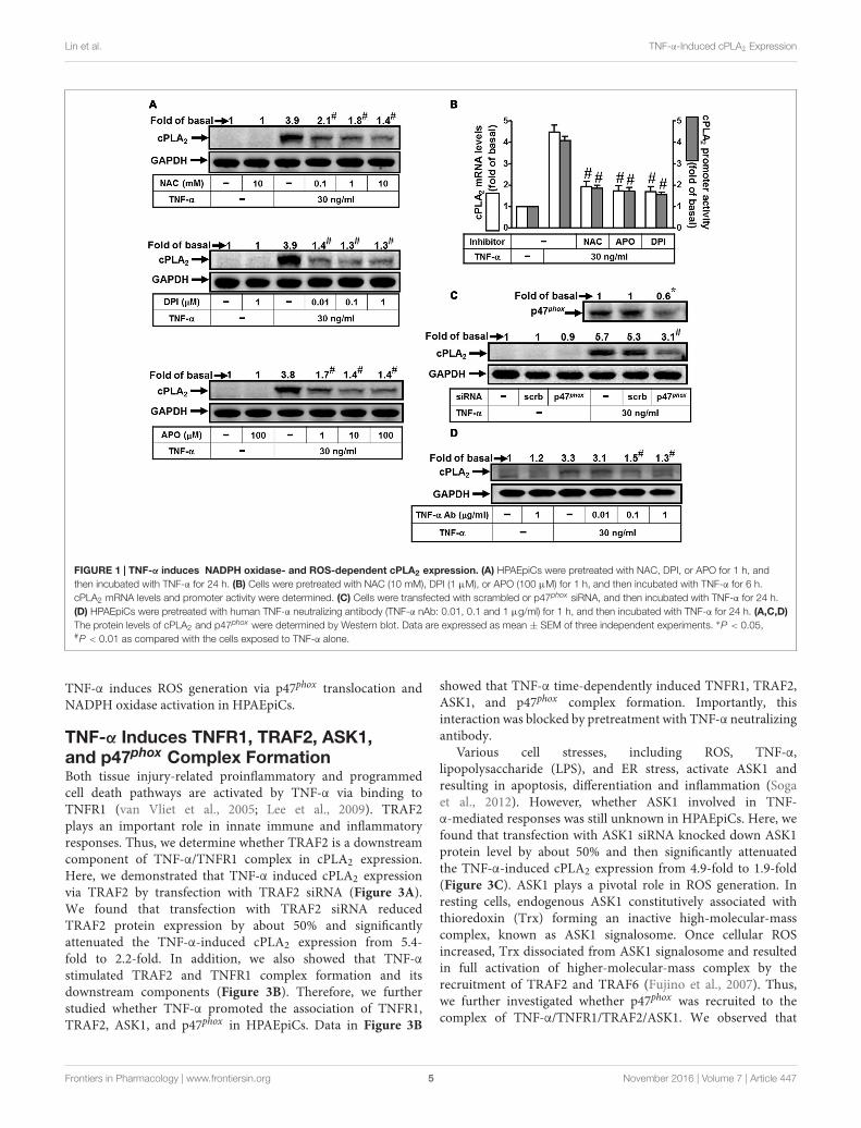
TNF-α Induces cPLA2 Expression viaNADPH Oxidase and ROSReactive oxygen species play both deleterious and beneficialroles. Accumulation of ROS and cellular oxidative stresstrigger expression of inflammatory genes and result in tissuedamages and various diseases (Lee and Yang, 2012). Thus,we attempted to investigate the roles of NADPH Oxidaseand ROS in cPLA2 expression. Here we reported that TNF-α-induced cPLA2 protein levels were significantly reduced bypretreatment with a ROS scavenger (NAC) or the inhibitorsof NADPH oxidase (APO and DPI) (Figure 1A). In addition,pretreatment with NAC, DPI, or APO also attenuated theTNF-α-stimulated cPLA2 mRNA expression and promoter
activity (Figure 1B). The p47phox protein, one of cytosolicsubunits of NADPH oxidase, contributed to acute NADPHoxidase activation via being phosphorylated and binding top22phox (Lee et al., 2009). Thus, we confirmed the role ofp47phox in cPLA2 expression by transfection with p47phox siRNAwhich knocked down protein expression of p47phox and thenmarkedly inhibited TNF-α-induced cPLA2 protein expressionin these cells (Figure 1C). To confirm this cPLA2 expressionis mediated through TNF-α-dependent induction, HPAEpiCswere pretreated with a human TNF-α neutralizing antibodyfollowed by TNF-α treatment. We found that TNF-α neutralizingantibody significantly blocked the cPLA2 induction in a dose-dependent manner (Figure 1D). These results indicated thatTNF-α induces cPLA2 expression via NADPH oxidase and ROSin HPAEpiCs.
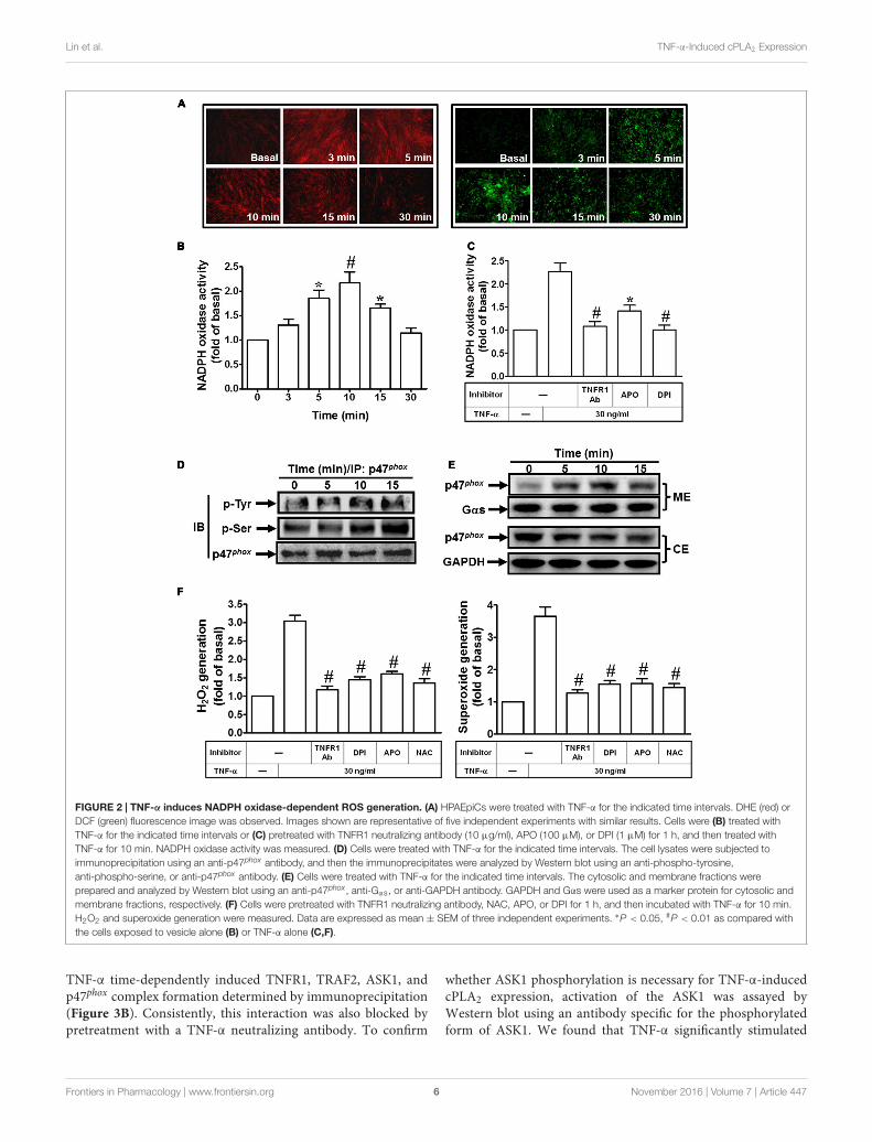
TNF-α Induces NADPHOxidase-Dependent Superoxide andHydrogen Peroxide ProductionTNF-α may stimulate ROS production by several sources, suchas mitochondria, but recent studies have strongly suggested thata major source of ROS is a phagocyte-type NADPH oxidase.Several reports also demonstrate that TNF-α triggers severalsignal transduction pathways to activate the NOX activity andenhances intracellular ROS generation leading to expression ofinflammatory genes (Rahman et al., 1998; Hashimoto et al., 2001;Li et al., 2005; Lee et al., 2013; Yang et al., 2014). Therefore, weinvestigated whether TNF-α-induced cPLA2 expression is dueto activation of NADPH oxidase and ROS generation. Here, wefound that TNF-α markedly induced superoxide and hydrogenperoxide production, determined by using DHE or DCF undera fluorescence microscope (Figure 2A). On the other hand, wealso observed that TNF-α time-dependently induces NADPHoxidase activation (Figure 2B), which was reduced by TNFR1neutralizing antibody, APO, or DPI (Figure 2C). These resultssuggested that TNF-α induced ROS generation via NADPHoxidase activation. The p47phox is phosphorylated on severalserine residues within the polybasic region of the protein, andthese multiple phosphorylation events induce conformationalchanges that permit p47phox to interact with the cytoplasmictail of p22phox and to initiate the formation of the activeNADPH oxidase complex (Dang et al., 2006). Previous reportalso indicates that Src modulates tyrosine phosphorylation ofp47phox in hyperoxia-induced activation of NADPH oxidaseand generation of ROS in lung endothelial cells (Chowdhuryet al., 2005). Thus, we investigated whether TNF-α stimulates thephosphorylation of tyrosine or serine on p47phox. As shown inFigure 2D, TNF-α increased tyrosine and serine phosphorylationof p47phox in a time-dependent manner. Moreover, p47phox wastranslocated from the cytosol to the membrane fractions in TNF-α-stimulated cells (Figure 2E). Finally, we investigated whetherTNF-α-induced superoxide and hydrogen peroxide productionare mediated through NADPH oxidase activation. Here, weobserved that TNF-α-induced superoxide and hydrogen peroxideproduction were inhibited by TNFR1 neutralizing antibody,APO, DPI and NAC (Figure 2F). These results suggested that
Frontiers in Pharmacology | www.frontiersin.org 4 November 2016 | Volume 7 | Article 447
fphar-07-00447 November 23, 2016 Time: 17:3 # 5
Lin et al. TNF-α-Induced cPLA2 Expression
FIGURE 1 | TNF-α induces NADPH oxidase- and ROS-dependent cPLA2 expression. (A) HPAEpiCs were pretreated with NAC, DPI, or APO for 1 h, andthen incubated with TNF-α for 24 h. (B) Cells were pretreated with NAC (10 mM), DPI (1 µM), or APO (100 µM) for 1 h, and then incubated with TNF-α for 6 h.cPLA2 mRNA levels and promoter activity were determined. (C) Cells were transfected with scrambled or p47phox siRNA, and then incubated with TNF-α for 24 h.(D) HPAEpiCs were pretreated with human TNF-α neutralizing antibody (TNF-α nAb: 0.01, 0.1 and 1 µg/ml) for 1 h, and then incubated with TNF-α for 24 h. (A,C,D)The protein levels of cPLA2 and p47phox were determined by Western blot. Data are expressed as mean ± SEM of three independent experiments. ∗P < 0.05,#P < 0.01 as compared with the cells exposed to TNF-α alone.
TNF-α induces ROS generation via p47phox translocation andNADPH oxidase activation in HPAEpiCs.
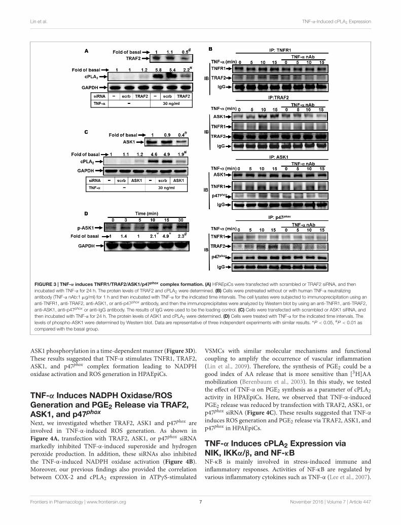
TNF-α Induces TNFR1, TRAF2, ASK1,and p47phox Complex FormationBoth tissue injury-related proinflammatory and programmedcell death pathways are activated by TNF-α via binding toTNFR1 (van Vliet et al., 2005; Lee et al., 2009). TRAF2plays an important role in innate immune and inflammatoryresponses. Thus, we determine whether TRAF2 is a downstreamcomponent of TNF-α/TNFR1 complex in cPLA2 expression.Here, we demonstrated that TNF-α induced cPLA2 expressionvia TRAF2 by transfection with TRAF2 siRNA (Figure 3A).We found that transfection with TRAF2 siRNA reducedTRAF2 protein expression by about 50% and significantlyattenuated the TNF-α-induced cPLA2 expression from 5.4-fold to 2.2-fold. In addition, we also showed that TNF-αstimulated TRAF2 and TNFR1 complex formation and itsdownstream components (Figure 3B). Therefore, we furtherstudied whether TNF-α promoted the association of TNFR1,TRAF2, ASK1, and p47phox in HPAEpiCs. Data in Figure 3B
showed that TNF-α time-dependently induced TNFR1, TRAF2,ASK1, and p47phox complex formation. Importantly, thisinteraction was blocked by pretreatment with TNF-α neutralizingantibody.
Various cell stresses, including ROS, TNF-α,lipopolysaccharide (LPS), and ER stress, activate ASK1 andresulting in apoptosis, differentiation and inflammation (Sogaet al., 2012). However, whether ASK1 involved in TNF-α-mediated responses was still unknown in HPAEpiCs. Here, wefound that transfection with ASK1 siRNA knocked down ASK1protein level by about 50% and then significantly attenuatedthe TNF-α-induced cPLA2 expression from 4.9-fold to 1.9-fold(Figure 3C). ASK1 plays a pivotal role in ROS generation. Inresting cells, endogenous ASK1 constitutively associated withthioredoxin (Trx) forming an inactive high-molecular-masscomplex, known as ASK1 signalosome. Once cellular ROSincreased, Trx dissociated from ASK1 signalosome and resultedin full activation of higher-molecular-mass complex by therecruitment of TRAF2 and TRAF6 (Fujino et al., 2007). Thus,we further investigated whether p47phox was recruited to thecomplex of TNF-α/TNFR1/TRAF2/ASK1. We observed that
Frontiers in Pharmacology | www.frontiersin.org 5 November 2016 | Volume 7 | Article 447
fphar-07-00447 November 23, 2016 Time: 17:3 # 6
Lin et al. TNF-α-Induced cPLA2 Expression
FIGURE 2 | TNF-α induces NADPH oxidase-dependent ROS generation. (A) HPAEpiCs were treated with TNF-α for the indicated time intervals. DHE (red) orDCF (green) fluorescence image was observed. Images shown are representative of five independent experiments with similar results. Cells were (B) treated withTNF-α for the indicated time intervals or (C) pretreated with TNFR1 neutralizing antibody (10 µg/ml), APO (100 µM), or DPI (1 µM) for 1 h, and then treated withTNF-α for 10 min. NADPH oxidase activity was measured. (D) Cells were treated with TNF-α for the indicated time intervals. The cell lysates were subjected toimmunoprecipitation using an anti-p47phox antibody, and then the immunoprecipitates were analyzed by Western blot using an anti-phospho-tyrosine,anti-phospho-serine, or anti-p47phox antibody. (E) Cells were treated with TNF-α for the indicated time intervals. The cytosolic and membrane fractions wereprepared and analyzed by Western blot using an anti-p47phox , anti-Gαs, or anti-GAPDH antibody. GAPDH and Gαs were used as a marker protein for cytosolic andmembrane fractions, respectively. (F) Cells were pretreated with TNFR1 neutralizing antibody, NAC, APO, or DPI for 1 h, and then incubated with TNF-α for 10 min.H2O2 and superoxide generation were measured. Data are expressed as mean ± SEM of three independent experiments. ∗P < 0.05, #P < 0.01 as compared withthe cells exposed to vesicle alone (B) or TNF-α alone (C,F).
TNF-α time-dependently induced TNFR1, TRAF2, ASK1, andp47phox complex formation determined by immunoprecipitation(Figure 3B). Consistently, this interaction was also blocked bypretreatment with a TNF-α neutralizing antibody. To confirm
whether ASK1 phosphorylation is necessary for TNF-α-inducedcPLA2 expression, activation of the ASK1 was assayed byWestern blot using an antibody specific for the phosphorylatedform of ASK1. We found that TNF-α significantly stimulated
Frontiers in Pharmacology | www.frontiersin.org 6 November 2016 | Volume 7 | Article 447
fphar-07-00447 November 23, 2016 Time: 17:3 # 7
Lin et al. TNF-α-Induced cPLA2 Expression
FIGURE 3 | TNF-α induces TNFR1/TRAF2/ASK1/p47phox complex formation. (A) HPAEpiCs were transfected with scrambled or TRAF2 siRNA, and thenincubated with TNF-α for 24 h. The protein levels of TRAF2 and cPLA2 were determined. (B) Cells were pretreated without or with human TNF-α neutralizingantibody (TNF-α nAb:1 µg/ml) for 1 h and then incubated with TNF-α for the indicated time intervals. The cell lysates were subjected to immunoprecipitation using ananti-TNFR1, anti-TRAF2, anti-ASK1, or anti-p47phox antibody, and then the immunoprecipitates were analyzed by Western blot by using an anti-TNFR1, anti-TRAF2,anti-ASK1, anti-p47phox or anti-IgG antibody. The results of IgG were used to be the loading control. (C) Cells were transfected with scrambled or ASK1 siRNA, andthen incubated with TNF-α for 24 h. The protein levels of ASK1 and cPLA2 were determined. (D) Cells were treated with TNF-α for the indicated time intervals. Thelevels of phospho-ASK1 were determined by Western blot. Data are representative of three independent experiments with similar results. ∗P < 0.05, #P < 0.01 ascompared with the basal group.
ASK1 phosphorylation in a time-dependent manner (Figure 3D).These results suggested that TNF-α stimulates TNFR1, TRAF2,ASK1, and p47phox complex formation leading to NADPHoxidase activation and ROS generation in HPAEpiCs.
TNF-α Induces NADPH Oxidase/ROSGeneration and PGE2 Release via TRAF2,ASK1, and p47phox
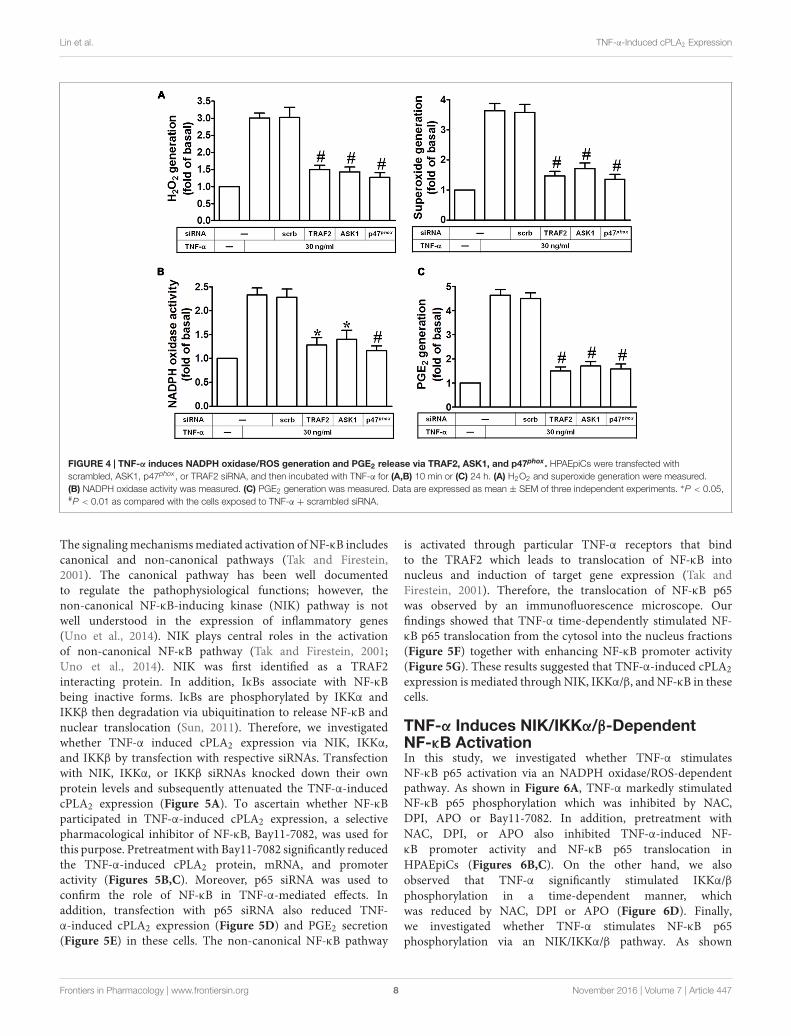
Next, we investigated whether TRAF2, ASK1 and p47phox areinvolved in TNF-α-induced ROS generation. As shown inFigure 4A, transfection with TRAF2, ASK1, or p47phox siRNAmarkedly inhibited TNF-α-induced superoxide and hydrogenperoxide production. In addition, these siRNAs also inhibitedthe TNF-α-induced NADPH oxidase activation (Figure 4B).Moreover, our previous findings also provided the correlationbetween COX-2 and cPLA2 expression in ATPγS-stimulated
VSMCs with similar molecular mechanisms and functionalcoupling to amplify the occurrence of vascular inflammation(Lin et al., 2009). Therefore, the synthesis of PGE2 could be agood index of AA release that is more sensitive than [3H]AAmobilization (Berenbaum et al., 2003). In this study, we testedthe effect of TNF-α on PGE2 synthesis as a parameter of cPLA2activity in HPAEpiCs. Here, we observed that TNF-α-inducedPGE2 release was reduced by transfection with TRAF2, ASK1, orp47phox siRNA (Figure 4C). These results suggested that TNF-αinduces ROS generation and PGE2 release via TRAF2, ASK1, andp47phox in HPAEpiCs.
TNF-α Induces cPLA2 Expression viaNIK, IKKα/β, and NF-κBNF-κB is mainly involved in stress-induced immune andinflammatory responses. Activities of NF-κB are regulated byvarious inflammatory cytokines such as TNF-α (Lee et al., 2007).
Frontiers in Pharmacology | www.frontiersin.org 7 November 2016 | Volume 7 | Article 447
fphar-07-00447 November 23, 2016 Time: 17:3 # 8
Lin et al. TNF-α-Induced cPLA2 Expression
FIGURE 4 | TNF-α induces NADPH oxidase/ROS generation and PGE2 release via TRAF2, ASK1, and p47phox. HPAEpiCs were transfected withscrambled, ASK1, p47phox , or TRAF2 siRNA, and then incubated with TNF-α for (A,B) 10 min or (C) 24 h. (A) H2O2 and superoxide generation were measured.(B) NADPH oxidase activity was measured. (C) PGE2 generation was measured. Data are expressed as mean ± SEM of three independent experiments. ∗P < 0.05,#P < 0.01 as compared with the cells exposed to TNF-α + scrambled siRNA.
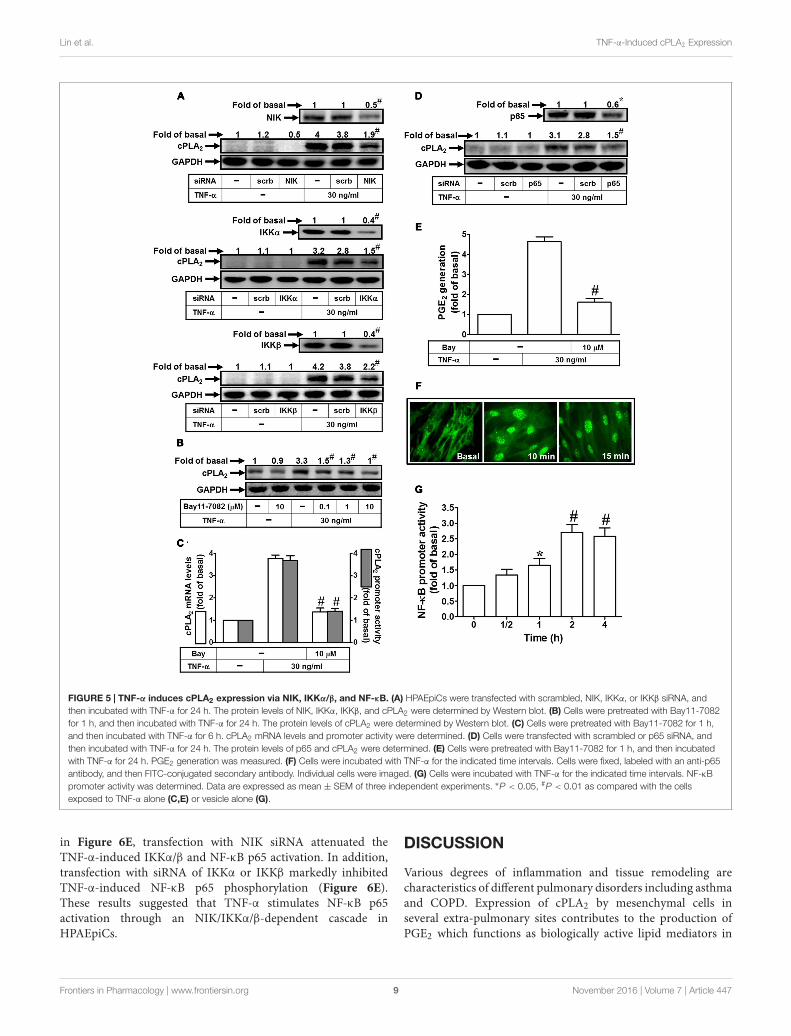
The signaling mechanisms mediated activation of NF-κB includescanonical and non-canonical pathways (Tak and Firestein,2001). The canonical pathway has been well documentedto regulate the pathophysiological functions; however, thenon-canonical NF-κB-inducing kinase (NIK) pathway is notwell understood in the expression of inflammatory genes(Uno et al., 2014). NIK plays central roles in the activationof non-canonical NF-κB pathway (Tak and Firestein, 2001;Uno et al., 2014). NIK was first identified as a TRAF2interacting protein. In addition, IκBs associate with NF-κBbeing inactive forms. IκBs are phosphorylated by IKKα andIKKβ then degradation via ubiquitination to release NF-κB andnuclear translocation (Sun, 2011). Therefore, we investigatedwhether TNF-α induced cPLA2 expression via NIK, IKKα,and IKKβ by transfection with respective siRNAs. Transfectionwith NIK, IKKα, or IKKβ siRNAs knocked down their ownprotein levels and subsequently attenuated the TNF-α-inducedcPLA2 expression (Figure 5A). To ascertain whether NF-κBparticipated in TNF-α-induced cPLA2 expression, a selectivepharmacological inhibitor of NF-κB, Bay11-7082, was used forthis purpose. Pretreatment with Bay11-7082 significantly reducedthe TNF-α-induced cPLA2 protein, mRNA, and promoteractivity (Figures 5B,C). Moreover, p65 siRNA was used toconfirm the role of NF-κB in TNF-α-mediated effects. Inaddition, transfection with p65 siRNA also reduced TNF-α-induced cPLA2 expression (Figure 5D) and PGE2 secretion(Figure 5E) in these cells. The non-canonical NF-κB pathway
is activated through particular TNF-α receptors that bindto the TRAF2 which leads to translocation of NF-κB intonucleus and induction of target gene expression (Tak andFirestein, 2001). Therefore, the translocation of NF-κB p65was observed by an immunofluorescence microscope. Ourfindings showed that TNF-α time-dependently stimulated NF-κB p65 translocation from the cytosol into the nucleus fractions(Figure 5F) together with enhancing NF-κB promoter activity(Figure 5G). These results suggested that TNF-α-induced cPLA2expression is mediated through NIK, IKKα/β, and NF-κB in thesecells.
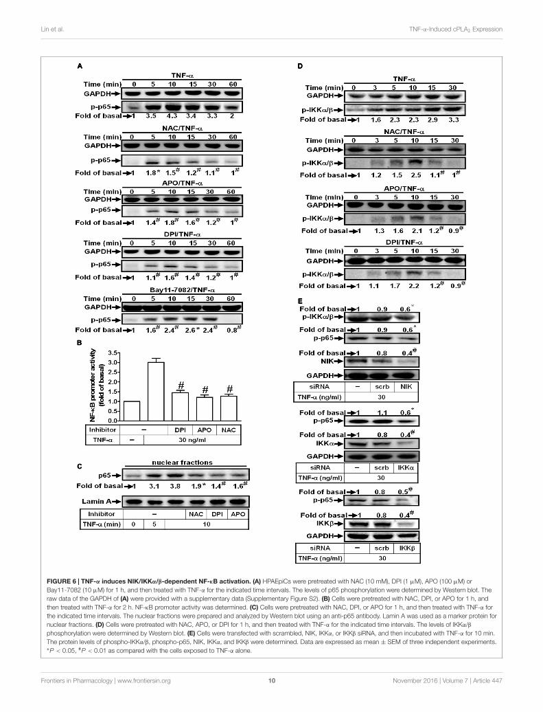
TNF-α Induces NIK/IKKα/β-DependentNF-κB ActivationIn this study, we investigated whether TNF-α stimulatesNF-κB p65 activation via an NADPH oxidase/ROS-dependentpathway. As shown in Figure 6A, TNF-α markedly stimulatedNF-κB p65 phosphorylation which was inhibited by NAC,DPI, APO or Bay11-7082. In addition, pretreatment withNAC, DPI, or APO also inhibited TNF-α-induced NF-κB promoter activity and NF-κB p65 translocation inHPAEpiCs (Figures 6B,C). On the other hand, we alsoobserved that TNF-α significantly stimulated IKKα/βphosphorylation in a time-dependent manner, whichwas reduced by NAC, DPI or APO (Figure 6D). Finally,we investigated whether TNF-α stimulates NF-κB p65phosphorylation via an NIK/IKKα/β pathway. As shown
Frontiers in Pharmacology | www.frontiersin.org 8 November 2016 | Volume 7 | Article 447
fphar-07-00447 November 23, 2016 Time: 17:3 # 9
Lin et al. TNF-α-Induced cPLA2 Expression
FIGURE 5 | TNF-α induces cPLA2 expression via NIK, IKKα/β, and NF-κB. (A) HPAEpiCs were transfected with scrambled, NIK, IKKα, or IKKβ siRNA, andthen incubated with TNF-α for 24 h. The protein levels of NIK, IKKα, IKKβ, and cPLA2 were determined by Western blot. (B) Cells were pretreated with Bay11-7082for 1 h, and then incubated with TNF-α for 24 h. The protein levels of cPLA2 were determined by Western blot. (C) Cells were pretreated with Bay11-7082 for 1 h,and then incubated with TNF-α for 6 h. cPLA2 mRNA levels and promoter activity were determined. (D) Cells were transfected with scrambled or p65 siRNA, andthen incubated with TNF-α for 24 h. The protein levels of p65 and cPLA2 were determined. (E) Cells were pretreated with Bay11-7082 for 1 h, and then incubatedwith TNF-α for 24 h. PGE2 generation was measured. (F) Cells were incubated with TNF-α for the indicated time intervals. Cells were fixed, labeled with an anti-p65antibody, and then FITC-conjugated secondary antibody. Individual cells were imaged. (G) Cells were incubated with TNF-α for the indicated time intervals. NF-κBpromoter activity was determined. Data are expressed as mean ± SEM of three independent experiments. ∗P < 0.05, #P < 0.01 as compared with the cellsexposed to TNF-α alone (C,E) or vesicle alone (G).
in Figure 6E, transfection with NIK siRNA attenuated theTNF-α-induced IKKα/β and NF-κB p65 activation. In addition,transfection with siRNA of IKKα or IKKβ markedly inhibitedTNF-α-induced NF-κB p65 phosphorylation (Figure 6E).These results suggested that TNF-α stimulates NF-κB p65activation through an NIK/IKKα/β-dependent cascade inHPAEpiCs.
DISCUSSION
Various degrees of inflammation and tissue remodeling arecharacteristics of different pulmonary disorders including asthmaand COPD. Expression of cPLA2 by mesenchymal cells inseveral extra-pulmonary sites contributes to the production ofPGE2 which functions as biologically active lipid mediators in
Frontiers in Pharmacology | www.frontiersin.org 9 November 2016 | Volume 7 | Article 447
fphar-07-00447 November 23, 2016 Time: 17:3 # 10
Lin et al. TNF-α-Induced cPLA2 Expression
FIGURE 6 | TNF-α induces NIK/IKKα/β-dependent NF-κB activation. (A) HPAEpiCs were pretreated with NAC (10 mM), DPI (1 µM), APO (100 µM) orBay11-7082 (10 µM) for 1 h, and then treated with TNF-α for the indicated time intervals. The levels of p65 phosphorylation were determined by Western blot. Theraw data of the GAPDH of (A) were provided with a supplementary data (Supplementary Figure S2). (B) Cells were pretreated with NAC, DPI, or APO for 1 h, andthen treated with TNF-α for 2 h. NF-κB promoter activity was determined. (C) Cells were pretreated with NAC, DPI, or APO for 1 h, and then treated with TNF-α forthe indicated time intervals. The nuclear fractions were prepared and analyzed by Western blot using an anti-p65 antibody. Lamin A was used as a marker protein fornuclear fractions. (D) Cells were pretreated with NAC, APO, or DPI for 1 h, and then treated with TNF-α for the indicated time intervals. The levels of IKKα/βphosphorylation were determined by Western blot. (E) Cells were transfected with scrambled, NIK, IKKα, or IKKβ siRNA, and then incubated with TNF-α for 10 min.The protein levels of phospho-IKKα/β, phospho-p65, NIK, IKKα, and IKKβ were determined. Data are expressed as mean ± SEM of three independent experiments.∗P < 0.05, #P < 0.01 as compared with the cells exposed to TNF-α alone.
Frontiers in Pharmacology | www.frontiersin.org 10 November 2016 | Volume 7 | Article 447
fphar-07-00447 November 23, 2016 Time: 17:3 # 11
Lin et al. TNF-α-Induced cPLA2 Expression
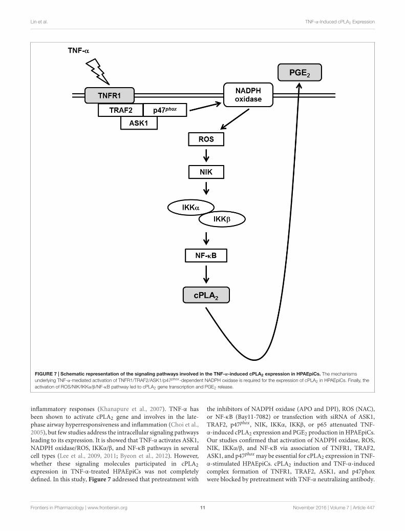
FIGURE 7 | Schematic representation of the signaling pathways involved in the TNF-α-induced cPLA2 expression in HPAEpiCs. The mechanismsunderlying TNF-α-mediated activation of TNFR1/TRAF2/ASK1/p47phox-dependent NADPH oxidase is required for the expression of cPLA2 in HPAEpiCs. Finally, theactivation of ROS/NIK/IKKα/β/NF-κB pathway led to cPLA2 gene transcription and PGE2 release.
inflammatory responses (Khanapure et al., 2007). TNF-α hasbeen shown to activate cPLA2 gene and involves in the late-phase airway hyperresponsiveness and inflammation (Choi et al.,2005), but few studies address the intracellular signaling pathwaysleading to its expression. It is showed that TNF-α activates ASK1,NADPH oxidase/ROS, IKKα/β, and NF-κB pathways in severalcell types (Lee et al., 2009, 2011; Byeon et al., 2012). However,whether these signaling molecules participated in cPLA2expression in TNF-α-treated HPAEpiCs was not completelydefined. In this study, Figure 7 addressed that pretreatment with
the inhibitors of NADPH oxidase (APO and DPI), ROS (NAC),or NF-κB (Bay11-7082) or transfection with siRNA of ASK1,TRAF2, p47phox, NIK, IKKα, IKKβ, or p65 attenuated TNF-α-induced cPLA2 expression and PGE2 production in HPAEpiCs.Our studies confirmed that activation of NADPH oxidase, ROS,NIK, IKKα/β, and NF-κB via association of TNFR1, TRAF2,ASK1, and p47phox may be essential for cPLA2 expression in TNF-α-stimulated HPAEpiCs. cPLA2 induction and TNF-α-inducedcomplex formation of TNFR1, TRAF2, ASK1, and p47phoxwere blocked by pretreatment with TNF-α neutralizing antibody.
Frontiers in Pharmacology | www.frontiersin.org 11 November 2016 | Volume 7 | Article 447
fphar-07-00447 November 23, 2016 Time: 17:3 # 12
Lin et al. TNF-α-Induced cPLA2 Expression
These results suggested that up-regulation of cPLA2 by TNF-αis, at least in part, mediated through the cooperation of TNFR1,TRAF2, ASK1, and NADPH oxidase leading to ROS generationand ultimately activates NF-κB pathway.
Several reports indicate that TNF-α may regulateinflammatory protein expression via activating variousdownstream protein kinases (Lin et al., 2004; Lee et al.,2009). Effects of TNF-α are achieved by binding to one oftwo distinct receptors, known as TNFR1 and TNFR2 (Leeet al., 2009). However, it is reported that TNF-α activates theproinflammatory or the programmed cell death pathwaysleading to tissue injury via binding to TNFR1 (van Vlietet al., 2005; Lee et al., 2009). In contrast, TNFR2 is involvedin promoting tissue repair and angiogenesis (Bradley, 2008).Indeed, transfection with TNFR1 siRNA attenuated NADPHoxidase and ROS generation, revealing that TNFR1 plays a keyrole in modulating inflammatory responses in TNF-α-stimulatedHPAEpiCs.
Reactive oxygen species are generated by various enzymaticreactions and chemical processes or can directly be inhaled fromenvironment (Lee and Yang, 2012). Formation of ROS takesplace constantly in every cell during normal metabolic processes.Activated phagocytic cells could produce large amounts ofROS induced by inhaled particles, microorganisms, or othermediators leading to the activation of the membrane-boundNADPH oxidase complex and the generation of superoxide anion(Lee and Yang, 2012). NADPH oxidase is recognized to be akey player in the generation of ROS when the cells or tissuesexposed to various insults. Upon activation of NADPH oxidaseby various stimuli, the complex of catalytic subunit (gp91phox)and p22phox is anchored in plasma membrane which assembleswith the regulatory subunits (p47phox, p40phox, p67phox, andsmall GTPase Rac) distributed in cytoplasm and leading to ROSgeneration (El-Benna et al., 2008). The initiation of the assemblyNADPH oxidase is first dependent on the phosphorylation ofp47phox triggered by various activated protein kinases. Thus,p47phox plays a role in the membrane translocation and ROSgeneration. Indeed, our results confirmed that blockage ROSaccumulation either by a ROS scavenger (NAC), the inhibitorsof NADPH oxidase (DPI and APO) or transfection with p47phox
siRNA significantly attenuated TNF-α-induced cPLA2 expressionand PGE2 synthesis. DPI or APO pretreatment reduced TNF-α-stimulated ROS generation. These results revealed that TNF-α increased cPLA2/PGE2 expression via NADPH oxidase-dependent ROS generation. p47phox is phosphorylated on severalserine residues within the polybasic region of the protein, andthese multiple phosphorylation events induce conformationalchanges that permit p47phox to interact with the cytoplasmic tailof p22phox and to initiate the formation of the active oxidase(Dang et al., 2006). In lung endothelial cells, Src regulates tyrosinephosphorylation of p47phox in hyperoxia-induced activation ofNADPH oxidase and generation of ROS (Chowdhury et al.,2005). Moreover, in HPAEpiCs, we found that TNF-α couldstimulate both serine and tyrosine phosphorylation of p47phox,and then promotes p47phox translocation from the cytosol to themembrane.
TRAF2 plays a central role in the cellular responses to stressand cytokines via their regulation of stress kinases, resultingin the activation of key transcription factors, including NF-κB,c-Jun, and ATF2. Upon exposure to TNF-α, TRAF2 is recruiteddirectly to TNFR2 or via TRADD to TNFR1, which results inthe activation of JNK1/2, p38 MAPK and NF-κB (Cabal-Hierroand Lazo, 2012). However, our previous report indicated thatJak2 also mediates the TNF-α-induced cPLA2 expression (Yanget al., 2014). To evaluate the relationship between Jak2 andMAPKs pathways, we have performed some more experimentsto determine whether there is any connection between MAPKsand Jak2 using pharmacological inhibitors (AG490, SB202190,SP600125 and U0126). We found that pretreatment with AG490had no effect on TNF-α-stimulated MAPKs phosphorylation, orwith MAPK inhibitors (SB202190, SP600125 and U0126) failed toinhibit Jak2 phosphorylation (Supplementary Figure S1). Theseresults suggested that Jak2 and MAPKs are independent pathwaysto mediate the TNF-α-induced cPLA2 expression. Here, we alsofound that TNF-α-induced cPLA2 expression and PGE2 releasewere markedly inhibited by blockage TRAF2. In addition, wealso observed that TNF-α induced TNFR1 and TRAF2 complexformation. Previous study indicated that TNF-α stimulated theformation of a TNFR1/c-Src/p47phox complex in human airwaysmooth muscle cells (Lee et al., 2009). Here, we demonstrated thatTNF-α induced TNFR1, TRAF2, and p47phox complex formationwhich was blocked by pretreatment with TNF-α neutralizingantibody, and leading to cPLA2 expression in HPAEpiCs.
ASK1, a member of the MAP3K family, regulates theactivation of MAPK kinase 4 (MKK4)/MKK7-JNK and MKK3/6-p38 pathways. ASK1 can be activated by various types of stresses,such as ROS, TNF-α, and ER stress, and exerts pivotal rolesin regulating cell apoptosis, differentiation, and inflammation(Fujino et al., 2007). Therefore, unregulated ASK1 activationis tightly related to various diseases. Moreover, we foundthat inhibition of ASK1 markedly reduced TNF-α-inducedcPLA2 expression and PGE2 release. TNF-α also stimulatedASK1 phosphorylation in these cells. Upon exposure to ROS,Trx dissociated from the N-terminal of ASK1, which thenbecame fully activated by recruitment of TRAF2 and TRAF6(Fujino et al., 2007). Indeed, we found consistent resultsthat TNF-α induces TRAF2 and ASK1 complex formationwhich is also blocked by pretreatment with TNF-α neutralizingantibody. In this study, we are the first to show a novel roleof TNFR1/TRAF2/ASK1/p47phox complex formation in TNF-α-induced NADPH oxidase activation and ROS production inHPAEpiCs. In the future, we will further determine whichdomains of TNFR1, TRAF2, ASK1, and p47phox are involved inprotein-protein interactions caused by TNF-α. Although ROShave been shown to regulate ASK1 activation (Fujino et al.,2007), in this study, we emphasized the critical role of ASK1in TNF-α-induced ROS generation. Thus, in addition to therole of ROS in ASK1 activation, our results supported that theopposite hierarchical relationship exists between ROS and ASK1.Therefore, NADPH oxidase/ROS may act both as upstreamregulators and downstream effectors of ASK1 in various celltypes.
Frontiers in Pharmacology | www.frontiersin.org 12 November 2016 | Volume 7 | Article 447
fphar-07-00447 November 23, 2016 Time: 17:3 # 13
Lin et al. TNF-α-Induced cPLA2 Expression
The NF-κB/Rel family complex, a redox-sensitivetranscription factor, participates in controlling expressionof many inflammatory genes (Sun, 2011). NF-κB usually formsherterodimer by p50 and p65/RelA subunits. In resting cells,nuclear translocation signal of NF-κB is masked by binding withan inhibitor protein called inhibitory κB (IκB) as an inactivenon-DNA-binding form. Upon the stimulation by various NF-κBinducers, two serine residues of IκBα is phosphorylated, whichthen being ubiquinated by E3 ubiquitin-ligases (E3RSIκB) andsubsequently degraded by the 26S proteasome (Sun, 2011). Thereleased NF-κB dimers can then translocate into the nucleusand bind to the κB elements on the promoter of target genes.NIK was first identified as a TRAF2 interacting protein (Takand Firestein, 2001). In inactive form, NF-κB transcriptionfactors binds with the inhibitory proteins IκBs. IKKα and IKKβ
regulates the phosphorylation of IκB proteins, which then beingubiquitination and degradation leading to nuclear localizationof NF-κB transcription factors (Sun, 2011). In this study,inhibition of NIK, IKKα, IKKβ, and NF-κB markedly inhibitedTNF-α-induced cPLA2 expression. Various extracellular stimulisuch as TNF-α and IL-1β, viruses and environmental particulates(PM10s), and oxidative stress regulate the activation of NF-κB(Sun, 2011). Here, we reported that TNF-α time-dependentlyinduced phosphorylation and translocation of NF-κB p65 andNF-κB promoter activity via an NADPH oxidase/ROS pathway.Otherwise, we also proved that TNF-α significantly inducedIKKα/β phosphorylation via an NIK-dependent signaling, andthen promoted NF-κB activation. Thus, we recognized thatTNF-α-induced ROS generation may promote activation of theNIK/IKKα/β/NF-κB pathway in HPAEpiCs.
COX-2 and cPLA2 are tightly regulated by various mediatorsin several species (Beasley, 1999; Ali et al., 2008; Pavicevic et al.,2008). cPLA2 hydrolyzes the membrane phospholipids, resultingin the release of AA, which is further converted by the constitutiveenzyme COX-1 or by the inducible COX-2, and PG synthases tobiologically active PGs (DeWitt, 1999). On the other hand, severalreports indicated that the levels of PGE2 are also degraded byan important enzyme, 15-hydroxyprostaglandin dehydrogenase(15-PGDH) to regulate the levels of PGEs (Pomini et al., 1999;Otani et al., 2006; Thiel et al., 2009). In our previous study, up-regulation of COX-2 in TSMCs has been shown to enhance PGE2synthesis induced by LPS (Luo et al., 2003). Moreover, we alsoprovided insights into the correlation between COX-2 and cPLA2expression in ATPγS-stimulated VSMCs with similar molecularmechanisms and functional coupling to amplify the occurrenceof vascular inflammation (Lin et al., 2009). PGE2 treatment alsoinduced cPLA2 expression in VSMCs, which exerted as a positivefeedback regulator in this response (Lin et al., 2009). Therefore,the synthesis of PGE2 could be a good index of AA releasethat is more sensitive than [3H]AA mobilization (Berenbaumet al., 2003) and PGE2 may further induce cPLA2 expressionto amplify the occurrence of pulmonary inflammation. In thisstudy, although the effect of PGE2 on COX-2 expression was notinvestigated, we tested the effect of TNF-α on PGE2 synthesis as aparameter of cPLA2 activity which may be an important issue forfurther study in HPAEpiCs.
CONCLUSION
According to the literature reports and our results, Figure 7addresses a model for the molecular mechanisms of TNF-α-stimulated cPLA2 expression and PGE2 release inHPAEpiCs. To our knowledge, this is the first study toindicate that in HPAEpiCs, TNF-α-regulated activationof TNFR1/TRAF2/ASK1/p47phox-dependent NADPHoxidase is required for the expression of cPLA2. Finally,activation of the ROS/NIK/IKKα/β/NF-κB pathwayleads to cPLA2 gene activation and expression. It is animportant link for TNF-α-regulated cPLA2 expressionin the pathogenesis of lung inflammatory diseases.Therefore, uncovering the signaling components in TNF-α-mediated cPLA2 expression in HPAEpiCs is importantto develop new therapeutic strategies in pulmonarydiseases.
AUTHOR CONTRIBUTIONS
C-CL, W-NL, R-LC, C-yW, L-DH, and C-MY substantiallycontributed to the conception or design of the work, theacquisition, analysis, and interpretation of data for the work.C-CL, W-NL, R-LC, C-yW, L-DH, and C-MY drafted the workand revised it critically for important intellectual content. C-CL,W-NL, R-LC, CyW, L-DH, and C-MY finally approved theversion to be published. C-CL, W-NL, R-LC, C-yW, L-DH, andC-MY agreed to be accountable for all aspects of the work inensuring that questions related to the accuracy or integrity of anypart of the work are appropriately investigated and resolved.
FUNDING
This work was supported by the Ministry of Education,Taiwan, Grant number: EMRPD1E1641; the Ministry ofScience and Technology, Taiwan, Grant number: MOST104-2320-B-182A-003-MY3, MOST104-2320-B-182-010 andMOST105-2320-B-182-005-MY3; Chang Gung MedicalResearch Foundation, Grant number: CMRPD1C0103, CMRPD1C0563, CMRPD1B0332, CMRPD1F0021, CMRPG3B1093,CMRPG3C1303, CMRPG3A0051, and CMRPG3E2231; and FuJen Catholic University Research Foundation, Grant number:9991A01.
ACKNOWLEDGMENTS
We thank Ms. Yu-Wen Chen for her technical assistance.
SUPPLEMENTARY MATERIAL
The Supplementary Material for this article can be found onlineat: http://journal.frontiersin.org/article/10.3389/fphar.2016.00447/full#supplementary-material
Frontiers in Pharmacology | www.frontiersin.org 13 November 2016 | Volume 7 | Article 447
fphar-07-00447 November 23, 2016 Time: 17:3 # 14
Lin et al. TNF-α-Induced cPLA2 Expression
REFERENCESAli, K., Lund-Katz, S., Lawson, J., Phillips, M. C., and Rader, D. J. (2008). Structure
function properties of the apoE-dependent COX-2 pathway in vascular smoothmuscle cells. Atherosclerosis 196, 201–209. doi: 10.1016/j.atherosclerosis.2007.03.038
Barnes, P. J. (1989). New concepts in the pathogenesis of bronchialhyperresponsiveness and asthma. J. Allergy Clin. Immunol. 83, 1013–1026.doi: 10.1016/0091-6749(89)90441-7
Beasley, D. (1999). COX-2 and cytosolic PLA2 mediate IL-1β-induced cAMPproduction in human vascular smooth muscle cells. Am. J. Physiol. 276, H1369–H1378.
Berenbaum, F., Humber, L., Bereziat, G., and Thirion, S. (2003). Concomitantrecruitment of ERK1/2 and p38 MAPK signalling pathway is required foractivation of cytoplasmic phospholipase A2 via ATP in articular chondrocytes.J. Biol. Chem. 278, 13680–13687. doi: 10.1074/jbc.M211570200
Borsch-Haubold, A. G., Bartoli, F., Asselin, J., Dudler, T., Kramer, R. M., Apitz-Castro, R., et al. (1998). Identification of the phosphorylation sites of cytosolicphospholipase A2 in agonist-stimulated human platelets and HeLa cells. J. Biol.Chem. 273, 4449–4458. doi: 10.1074/jbc.273.8.4449
Bradley, J. R. (2008). TNF-mediated inflammatory disease. J. Pathol. 214, 149–160.doi: 10.1002/path.2287
Byeon, H. E., Um, S. H., Yim, J. H., Lee, H. K., and Pyo, S. (2012). Ohioensin Fsuppresses TNF-α-induced adhesion molecule expression by inactivation of theMAPK, Akt and NF-κB pathways in vascular smooth muscle cells. Life Sci. 90,396–406. doi: 10.1016/j.lfs.2011.12.017
Cabal-Hierro, L., and Lazo, P. S. (2012). Signal transduction by tumor necrosisfactor receptors. Cell. Signal. 24, 1297–1305. doi: 10.1016/j.cellsig.2012.02.006
Chi, P. L., Chen, Y. W., Hsiao, L. D., Chen, Y. L., and Yang, C. M. (2012). Hemeoxygenase-1 attenuates IL-1β-induced cPLA2 expression via a decrease inNADPH oxidase/ROS/AP-1 activation in human rheumatoid arthritis synovialfibroblasts. Arthritis Rheum. 64, 2114–2125. doi: 10.1002/art.34371
Chi, P. L., Liu, C. J., Lee, I. T., Chen, Y. W., Hsiao, L. D., and Yang, C. M.(2014). HO-1 induction by CO-RM2 attenuates TNF-α-induced cytosolicphospholipase A2 expression via inhibition of PKCα-dependent NADPHoxidase/ROS and NF-κB. Mediators Inflamm. 2014:279171. doi: 10.1155/2014/279171
Chi, P. L., Luo, S. F., Hsieh, H. L., Lee, I. T., Hsiao, L. D., Chen, Y. L., et al. (2011).cPLA2 induction and PGE2 release by IL-1β via the MyD88-dependent andcooperation of p300, Akt, and NF-κB pathway in human rheumatoid arthritissynovial fibroblasts. Arthritis Rheum. 63, 2905–2917. doi: 10.1002/art.30504
Choi, I. W., Sun, K., Kim, Y. S., Ko, H. M., Im, S. Y., Kim, J. H., et al. (2005). TNF-α induces the late-phase airway hyperresponsiveness and airway inflammationthrough cytosolic phospholipase A2 activation. J. Allergy Clin. Immunol. 116,537–543. doi: 10.1016/j.jaci.2005.05.034
Chowdhury, A. K., Watkins, T., Parinandi, N. L., Saatian, B., Kleinberg, M. E.,Usatyuk, V., et al. (2005). Src-mediated tyrosine phosphorylation of p47phoxin hyperoxia-induced activation of NADPH oxidase and generation of reactiveoxygen species in lung endothelial cells. J. Biol. Chem. 280, 20700–20711. doi:10.1074/jbc.M411722200
Dang, P. M., Elbim, C., Marie, J. C., Chiandotto, M., Gougerot-Pocidalo,M. A., and El-Benna, J. (2006). Anti-inflammatory effect of interleukin-10 onhuman neutrophil respiratory burst involves inhibition of GM-CSF-inducedp47phox phosphorylation through a decrease in ERK1/2 activity. FASEB J. 20,1504–1506. doi: 10.1096/fj.05-5395fje
DeWitt, D. L. (1999). Cox-2-selective inhibitors: the new super aspirins. Mol.Pharmacol. 55, 625–631.
Dieter, P., Kolada, A., Kamionka, S., Schadow, A., and Kaszkin, M. (2002).Lipopolysaccharide-induced release of arachidonic acid and prostaglandins inliver macrophages: regulation by Group IV cytosolic phospholipase A2, butnot by Group V and Group IIA secretory phospholipase A2. Cell. Signal. 14,199–204. doi: 10.1016/S0898-6568(01)00243-1
El-Benna, J., Dang, P. M. C., and Gougerot-Pocidalo, M. A. (2008). Priming ofthe neutrophil NADPH oxidase activation: role of p47phox phosphorylationand NOX2 mobilization to the plasma membrane. Semin. Immunopathol. 30,279–289. doi: 10.1007/s00281-008-0118-3
Fujino, G., Noguchi, T., Matsuzawa, A., Yamauchi, S., Saitoh, M., Takeda, K.,et al. (2007). Thioredoxin and TRAF family proteins regulate reactive oxygen
species-dependent activation of ASK1 through reciprocal modulation of theN-terminal homophilic interaction of ASK1. Mol. Cell. Biol. 27, 8152–8163.doi: 10.1128/MCB.00227-07
Gilroy, D. W., Newson, J., Sawmynaden, P., Willoughby, D. A., and Croxtall, J. D.(2004). A novel role for phospholipase A2 isoforms in the checkpoint control ofacute inflammation. FASEB J. 18, 489–498. doi: 10.1096/fj.03-0837com
Hashimoto, S., Gon, Y., Matsumoto, K., Takeshita, I., and Horie, T. (2001).N-acetylcysteine attenuates TNF-α-induced p38 MAP kinase activation andp38 MAP kinase-mediated IL-8 production by human pulmonary vascularendothelial cells. Br. J. Pharmacol. 132, 270–276. doi: 10.1038/sj.bjp.0703787
Henderson, W. R. Jr., Tang, L. O., Chu, S. J., Tsao, S. M., Chiang, G. K., Jones, F.,et al. (2002). A role for cysteinyl leukotrienes in airway remodeling in a mouseasthma model. Am. J. Respir. Crit. Care Med. 165, 108–116. doi: 10.1164/ajrccm.165.1.2105051
Hsieh, H. L., Wu, C. Y., Hwang, T. L., Yen, M. H., Parker, P., and Yang, C. M.(2006). BK-induced cytosolic phospholipase A2 expression via sequential PKC-α, p42/p44 MAPK, and NF-κB activation in rat brain astrocytes. J. Cell. Physiol.206, 246–254. doi: 10.1002/jcp.20457
Hsu, C. K., Lee, I. T., Lin, C. C., Hsiao, L. D., and Yang, C. M. (2014). Nox2/ROS-dependent human antigen R translocation contributes to TNF-α-inducedSOCS-3 expression in human tracheal smooth muscle cells. Am. J. Physiol. LungMol. Cell. Biol. 306, L521–L533. doi: 10.1152/ajplung.00274.2013
Hsu, M. J., Chang, C. K., Chen, M. C., Chen, B. C., Ma, H. P., Hong, C. Y., et al.(2010). Apoptosis signal-regulating kinase 1 in peptidoglycan-induced COX-2 expression in macrophages. J. Leukoc. Biol. 87, 1069–1082. doi: 10.1189/jlb.1009668
Hulkower, K. I., Wertheimer, S. J., Levin, W., Coffey, J. W., Anderson, C. M.,Chen, T., et al. (1994). Interleukin-1β induces cytosolic phospholipase A2 andprostaglandin H synthase in rheumatoid synovial fibroblasts. Evidence for theirroles in the production of prostaglandin E2. Arthritis Rheum. 37, 653–661.doi: 10.1002/art.1780370508
Huwiler, A., Feuerherm, A. J., Sakem, B., Pastukhov, O., Filipenko, I., Nguyen, T.,et al. (2012). The omega3-polyunsaturated fatty acid derivatives AVX001and AVX002 directly inhibit cytosolic phospholipase A2 and suppress PGE2formation in mesangial cells. Br. J. Pharmacol. 167, 1691–1701. doi: 10.1111/j.1476-5381.2012.02114.x
Khanapure, S. P., Garvey, D. S., Janero, D. R., and Letts, L. G. (2007). Eicosanoidsin inflammation: biosynthesis, pharmacology, and therapeutic frontiers. Curr.Top. Med. Chem. 7, 311–340. doi: 10.2174/156802607779941314
Lee, C. W., Lin, C. C., Lee, I. T., Lee, H. C., and Yang, C. M. (2011). Activation andinduction of cytosolic phospholipase A2 by TNF-α mediated through Nox2,MAPKs, NF-κB, and p300 in human tracheal smooth muscle cells. J. Cell.Physiol. 226, 2103–2114. doi: 10.1002/jcp.22537
Lee, C. W., Lin, C. C., Lin, W. N., Liang, K. C., Luo, S. F., Wu, C. B., et al. (2007).TNF-α induces MMP-9 expression via activation of Src/EGFR, PDGFR/PI3K/Akt cascade and promotion of NF-κB/p300 binding in human tracheal smoothmuscle cells. Am. J. Physiol. Lung Cell. Mol. Physiol. 292, L799–L812. doi:10.1152/ajplung.00311.2006
Lee, I. T., Lin, C. C., Lee, C. Y., Hsieh, P. W., and Yang, C. M. (2013).Protective effects of (-)-epigallocatechin-3-gallate against TNF-α-induced lunginflammation via ROS-dependent ICAM-1 inhibition. J. Nutr. Biochem. 24,124–136. doi: 10.1016/j.jnutbio.2012.03.009
Lee, I. T., Luo, S. F., Lee, C. W., Wang, S. W., Lin, C. C., Chang,C. C., et al. (2009). Overexpression of HO-1 protects against TNF-α-mediated airway inflammation by down-regulation of TNFR1-dependentoxidative stress. Am. J. Pathol. 175, 519–532. doi: 10.2353/ajpath.2009.090016
Lee, I. T., Wang, S. W., Lee, C. W., Chang, C. C., Lin, C. C., Luo, S. F., et al.(2008). Lipoteichoic acid induces HO-1 expression via the TLR2/MyD88/c-Src/NADPH oxidase pathway and Nrf2 in human tracheal smooth muscle cells.J. Immunol. 181, 5098–5110. doi: 10.4049/jimmunol.181.7.5098
Lee, I. T., and Yang, C. M. (2012). Role of NADPH oxidase/ROS in pro-inflammatory mediators-induced airway and pulmonary diseases. Biochem.Pharmacol. 84, 581–590. doi: 10.1016/j.bcp.2012.05.005
Leslie, C. C. (1997). Properties and regulation of cytosolic phospholipase A2. J. Biol.Chem. 272, 16709–16712. doi: 10.1074/jbc.272.27.16709
Li, J. M., Fan, L. M., Christie, M. R., and Shah, A. M. (2005). Acute tumor necrosisfactor α a signaling via NADPH oxidase in microvascular endothelial cells:
Frontiers in Pharmacology | www.frontiersin.org 14 November 2016 | Volume 7 | Article 447
fphar-07-00447 November 23, 2016 Time: 17:3 # 15
Lin et al. TNF-α-Induced cPLA2 Expression
role of p47phox phosphorylation and binding to TRAF4. Mol. Cell. Biol. 25,2320–2330. doi: 10.1128/MCB.25.6.2320-2330.2005
Lin, C. C., Hsiao, L. D., Chien, C. S., Lee, C. W., Hsieh, J. T., and Yang,C. M. (2004). Tumor necrosis factor-α-induced cyclooxygenase-2 expression inhuman tracheal smooth muscle cells: involvement of p42/p44 and p38 mitogen-activated protein kinases and nuclear factor-κB. Cell. Signal. 16, 597–607. doi:10.1016/j.cellsig.2003.10.002
Lin, C. C., Lin, W. N., Wang, W. J., Sun, C. C., Tung, W. H., Wang, H. H., et al.(2009). Functional coupling expression of COX-2 and cPLA2 induced by ATPin rat vascular smooth muscle cells: role of ERK1/2, p38 MAPK, and NF-κB.Cardiovasc. Res. 82, 522–531. doi: 10.1093/cvr/cvp069
Luo, S. F., Lin, W. N., Yang, C. M., Lee, C. W., Liao, C. H., Leu, Y. L., et al. (2006).Induction of cytosolic phospholipase A2 by lipopolysaccharide in caninetracheal smooth muscle cells: involvement of MAPKs and NF-κB pathways.Cell. Signal. 18, 1201–1211. doi: 10.1016/j.cellsig.2005.09.011
Luo, S. F., Wang, C. C., Chien, C. S., Hsiao, L. D., and Yang, C. M. (2003).Induction of cyclooxygenase-2 by lipopolysaccharide in canine tracheal smoothmuscle cells: involvement of p42/p44 and p38 mitogen-activated protein kinasesand nuclear factor-κB pathways. Cell. Signal. 15, 497–509. doi: 10.1016/S0898-6568(02)00135-3
Morgan, M. J., and Liu, Z. G. (2011). Crosstalk of reactive oxygen species andNF-κB signaling. Cell Res. 21, 103–115. doi: 10.1038/cr.2010.178
Nagase, T., Uozumi, N., Ishii, S., Kume, K., Izumi, T., Ouchi, Y., et al. (2000). Acutelung injury by sepsis and acid aspiration: a key role for cytosolic phospholipaseA2. Nat. Immunol. 1, 42–46. doi: 10.1038/76897
Otani, T., Yamaguchi, K., Scherl, E., Du, B., Tai, H. H., Greifer, M., et al.(2006). Levels of NAD+-dependent 15-hydroxyprostaglandin dehydrogenaseare reduced in inflammatory bowel disease: evidence for involvement of TNF-α. Am. J. Physiol Gastrointest. Liver Physiol. 290, G361–G368. doi: 10.1152/ajpgi.00348.2005
Pavicevic, Z., Leslie, C. C., and Malik, K. U. (2008). cPLA2 phosphorylation at S515and S505 is required for arachidonic acid release in vascular smooth muscle cell.J. Lipid Res. 49, 724–737. doi: 10.1194/jlr.M700419-JLR200
Pomini, F., Caruso, A., and Challis, J. R. G. (1999). Interleukin-10 modifiesthe effects of interleukin-1β and tumor necrosis factor-α on the activityand expression of prostaglandin H synthase-2 and the NAD+-dependent15-hydroxyprostaglandin dehydrogenase in cultured term human villoustrophoblast and chorion trophoblast cells. J. Clin. Endocrinol. Metab. 84,4645–4651.
Rahman, A., Kefer, J., Bando, M., Niles, W. D., and Malik, A. B. (1998). E-selectinexpression in human endothelial cells by TNF-α-induced oxidant generationand NF-κB activation. Am. J. Physiol. 275, L533–L544.
Six, D. A., and Dennis, E. A. (2000). The expanding superfamily of phospholipaseA2 enzymes: classification and characterization. Biochim. Biophys. Acta 1488,1–19. doi: 10.1016/S1388-1981(00)00105-0
Soga, M., Matsuzawa, A., and Ichijo, H. (2012). Oxidative stress-induced diseasesvia the ASK1 signaling pathway. Int. J. Cell Biol. 2012:439587. doi: 10.1155/2012/439587
Sun, S. C. (2011). Non-canonical NF-kappaB signaling pathway. Cell Res. 21,71–85. doi: 10.1038/cr.2010.177
Tak, P. P., and Firestein, G. S. (2001). NF-κB: a key role in inflammatory diseases.J. Clin. Invest. 107, 7–11. doi: 10.1172/JCI11830
Thiel, A., Ganesan, A., Mrena, J., Junnila, S., Nykänen, A., Hemmes, A., et al.(2009). 15-hydroxyprostaglandin dehydrogenase is down-regulated in gastriccancer. Clin. Cancer Res. 15, 4572–4580. doi: 10.1158/1078-0432.CCR-08-2518
Uno, M., Saitoh, Y., Mochida, K., Tsuruyama, E., Kiyono, T., Imoto, I.,et al. (2014). NF-κB inducing kinase, a central signaling componentof the non-canonical pathway of NF-κB, contributes to ovariancancer progression. PLoS ONE 9:e88347. doi: 10.1371/journal.pone.0088347
Uozumi, N., Kume, K., Nagase, T., Nakatani, N., Ishii, S., Tashiro, F., et al. (1997).Role of cytosolic phospholipase A2 in allergic response and parturition. Nature390, 618–622. doi: 10.1038/37622
Van Putten, V., Refaat, Z., Dessev, C., Blaine, S., Wick, M., Butterfield, L., et al.(2001). Induction of cytosolic phospholipase A2 by oncogenic Ras is mediatedthrough the JNK and ERK pathways in rat epithelial cells. J. Biol. Chem. 276,1226–1232. doi: 10.1074/jbc.M003581200
van Vliet, C., Bukczynska, P. E., Puryer, M. A., Sadek, C. M., Shields, B. J., Tremblay,M. L., et al. (2005). Selective regulation of tumor necrosis factor-induced Erksignaling by Src family kinases and the T cell protein tyrosine phosphatase. Nat.Immunol. 6, 253–260. doi: 10.1038/ni1169
Yang, C. M., Chien, C. S., Hsiao, L. D., Luo, S. F., and Wang, C. C. (2002).Interleukin-1β-induced cyclooxygenase-2 expression is mediated throughactivation of p42/44 and p38 MAPKS, and NF-κB pathways in canine trachealsmooth muscle cells. Cell. Signal. 14, 899–911. doi: 10.1016/S0898-6568(02)00037-2
Yang, C. M., Lee, I. T., Chi, P. L., Cheng, S. E., Hsiao, L. D., and Hsu, C. K.(2014). TNF-α induces cytosolic phospholipase A2 expression via Jak2/PDGFR-dependent Elk-1/p300 activation in human lung epithelial cells. Am. J.Physiol. Lung Cell. Mol. Biol. 306, L543–L551. doi: 10.1152/ajplung.00320.2013
Conflict of Interest Statement: The authors declare that the research wasconducted in the absence of any commercial or financial relationships that couldbe construed as a potential conflict of interest.
The reviewer HH and handling Editor declared their shared affiliation, and thehandling Editor states that the process nevertheless met the standards of a fair andobjective review.
Copyright © 2016 Lin, Lin, Cho, Wang, Hsiao and Yang. This is an open-access articledistributed under the terms of the Creative Commons Attribution License (CC BY).The use, distribution or reproduction in other forums is permitted, provided theoriginal author(s) or licensor are credited and that the original publication in thisjournal is cited, in accordance with accepted academic practice. No use, distributionor reproduction is permitted which does not comply with these terms.
Frontiers in Pharmacology | www.frontiersin.org 15 November 2016 | Volume 7 | Article 447