cPLA2 translocation to the fagocitic cup in human … · Abstract . Group IVA cytosolic...
40
The Cationic Cluster of Group IVA Phospholipase A 2 (Lys 488 /Lys 541 /Lys 543 /Lys 544 ) Is Involved in Translocation of the Enzyme to Phagosomes in Human Macrophages Javier Casas,* Martín Valdearcos,* § José Pindado,* Jesús Balsinde, 1 * § and María A. Balboa 2 * § *Instituto de Biología y Genética Molecular, Consejo Superior de Investigaciones Científicas (CSIC),47003 Valladolid, Spain, and § Centro de Investigación Biomédica en Red de Diabetes y Enfermedades Metabólicas Asociadas (CIBERDEM), 08036 Barcelona, Spain 1 Corresponding author: Instituto de Biología y Genética Molecular, Calle Sanz y Forés s/n, 47003 Valladolid, Spain. Phone: 34-983-423-062; FAX: 34-983-184-800; E-mail: [email protected] 2 Corresponding author: Instituto de Biología y Genética Molecular, Calle Sanz y Forés s/n, 47003 Valladolid, Spain. Phone: 34-983-184-833; FAX: 34-983-184-800; E-mail: [email protected] RUNNING TITLE: cPLA 2 α Translocation to Phagosomes KEY WORDS: Lipid Mediators; Monocytes/Macrophages; Phagocytosis; Phospholipase A 2 ; Inflammation 1 by guest, on September 22, 2018 www.jlr.org Downloaded from
Transcript of cPLA2 translocation to the fagocitic cup in human … · Abstract . Group IVA cytosolic...
The Cationic Cluster of Group IVA Phospholipase A2
(Lys488/Lys541/Lys543/Lys544) Is Involved in Translocation of the
Enzyme to Phagosomes in Human Macrophages
Javier Casas,* Martín Valdearcos,*§ José Pindado,* Jesús Balsinde,1*§
and María A. Balboa2*§
*Instituto de Biología y Genética Molecular, Consejo Superior de Investigaciones
Científicas (CSIC),47003 Valladolid, Spain, and
§Centro de Investigación Biomédica en Red de Diabetes y Enfermedades Metabólicas
Asociadas (CIBERDEM), 08036 Barcelona, Spain
1Corresponding author: Instituto de Biología y Genética Molecular, Calle Sanz y Forés
s/n, 47003 Valladolid, Spain. Phone: 34-983-423-062; FAX: 34-983-184-800; E-mail:
2Corresponding author: Instituto de Biología y Genética Molecular, Calle Sanz y Forés
s/n, 47003 Valladolid, Spain. Phone: 34-983-184-833; FAX: 34-983-184-800; E-mail:
RUNNING TITLE: cPLA2α Translocation to Phagosomes
KEY WORDS: Lipid Mediators; Monocytes/Macrophages; Phagocytosis;
Phospholipase A2; Inflammation
1
by guest, on Septem
ber 22, 2018w
ww
.jlr.orgD
ownloaded from
Abstract
Group IVA cytosolic phospholipase A2α (cPLA2α) plays a role in the microbicidal
machinery of immune cells by translocating to phagosomes to initiate the production of
antimicrobial eicosanoids. In the present work we have studied the involvement of the
cationic cluster of cPLA2α (Lys488/Lys541/Lys543/Lys544) in the translocation of cPLA2α
to the phagosomal cup in human macrophages responding to opsonized zymosan.
Phagocytosis was accompanied by an increased mobilization of free arachidonic acid,
which was almost completely abrogated by pyrrophenone. In transfected cells a
catalytically active EGFP-cPLA2α translocated to the phagocytic cup, which was
corroborated by frustrated phagocytosis experiments utilizing immunoglobulin G-coated
plates. However, a cPLA2α mutant in the polybasic cluster Lys488/Lys541/Lys543/Lys544,
that cannot bind the anionic phospholipid phosphatidylinositol 4, 5-bisphosphate (PIP2),
did not translocate to the phagocytic cup. Moreover, an EYFP-cPLA2α and an ECFP-
pleckstrin homology (PH) domain of the PLCδ1 construct that specifically recognizes
endogenous PIP2 in the cells, both localized at the same sites on the phagosome. High
cellular expression of the PH domain inhibited EYFP-cPLA2α translocation. On the
other hand, two other members of the phospholipase A2 superfamily of enzymes were
studied as well, namely group V secreted phospholipase A2 and group VIA calcium-
independent phospholipase A2, but the results indicated that neither of these are
involved in zymosan phagocytosis by human macrophages. Collectively, these data
indicate that the polybasic cluster Lys488/Lys541/Lys543/Lys544 of cPLA2α regulates the
subcellular localization of cPLA2α in intact immune cells under physiologically relevant
conditions.
2
by guest, on Septem
ber 22, 2018w
ww
.jlr.orgD
ownloaded from
ABBREVIATIONS: AA, arachidonic acid; PLA2, phospholipase A2; cPLA2α, group
IVA cytosolic phospholipase A2; iPLA2, calcium-independent phospholipase A2; sPLA2,
secreted phospholipase A2; sPLA2-V, group V secreted phospholipase A2; BEL,
bromoenol lactone; bis-BODIPY FL C11-PC, 1,2-bis-(4,4-difluoro-5,7-dimethyl-4-
bora-3a, 4a-diaza-sindecane-3-undecanoyl)-sn-glycero-3-phosphocholine; PLCδ-PH,
pleckstrin homology domain of PLCδ-PH; PIP2, phosphatidylinositol 4,5-bisphosphate.
3
by guest, on Septem
ber 22, 2018w
ww
.jlr.orgD
ownloaded from
Introduction
Phospholipase A2 (PLA2) enzymes cleave membrane phospholipids at the sn-2 position
of the glycerol backbone, releasing a free fatty acid and a lysophospholipid [1]. One of
the better studied roles of PLA2 enzymes is their involvement in inflammatory
processes, due to their ability to liberate free arachidonic acid (AA) from membrane
phospholipids [2-5]. Free AA will in turn be oxygenated by specific enzymes to
generate a wide variety of compounds with potent pro- and anti-inflammatory actions,
collectively called the eicosanoids [6, 7]. Some of these compounds have also been
described as important bactericidal agents [8].
Of the many existing PLA2 enzymes, only one, the group IVA PLA2 (also
known as cytosolic phospholipase A2α, (cPLA2α) manifests specificity for AA-
containing phospholipids [9]. Today it is widely accepted that cPLA2α is the key
enzyme in AA release leading to physiological and/or pathophysiological eicosanoid
production [9-11]. Structurally, cPLA2α possesses a C2 domain containing the binding
site for Ca2+ --and a binding site for ceramide-1 phosphate as well [12]--, and a catalytic
domain, where the residues involved in enzymatic activity are located [9]. The catalytic
domain also contains a cluster of cationic amino acids (Lys488, Lys541, Lys543, and
Lys544) that may mediate enzyme binding to anionic phospholipids in membranes [13].
In fact, in vitro, anionic phospholipids such as PIP2, PI(3,4,5)P3 and PI(3,4)P2 strongly
increase the cPLA2α specific activity when incorporated into the vesicle substrate [14,
15]. Mutation of the aforementioned polybasic 4-Lys cluster eliminates the activating
effect of PIP2 on cPLA2α activity [13]. Also, by plasmon resonance experiments it has
been demonstrated that PIP2 activates cPLA2α by increasing the catalytic efficiency of
the enzyme as a result of increasing membrane penetration [16]. In live cells, exogenous
PIP2 and PI(3,4)P2 induce the translocation of cPLA2α to the intracellular membranes
4
by guest, on Septem
ber 22, 2018w
ww
.jlr.orgD
ownloaded from
[17]. More recently, it has been described that mutation of the Lys488, Lys543, and Lys544,
decreases AA release in cells activated by serum, pointing to an important role for that
cluster in cPLA2 activation under physiological stimulation [18].
Phagocytosis is a specialized process of cells of the innate immune system to
engulf invading microorganisms, foreign particles and apoptotic cells and debris.
Phagocytosis is initiated at the site of particle attachment, producing a polarized region
within the membrane that enlarges as a pseudopod to engulf the particle and drive it into
the cytoplasm, where it will be degraded [19]. It has been previously shown that
cPLA2α translocates to the phagosomal cup during the ingestion of particles, and such a
translocation appears to be important for in situ eicosanoid generation and efficient
elimination of microbes [20-23]. However, the mechanism regulating this translocation
remains largely unknown. Utilizing human macrophages responding to opsonized
zymosan, we demonstrate in this work that translocation of cPLA2α to the phagocytic
cup depends on an intact cationic cluster Lys488/Lys541/Lys543/Lys544) in the catalytic
domain of the enzyme. These results reveal a novel role for the cluster and provide new
insights into the complex cellular regulation of the cPLA2α.
5
by guest, on Septem
ber 22, 2018w
ww
.jlr.orgD
ownloaded from
Materials and Methods
Materials - [5,6,8,9,11,12,14,15-3H]AA 200 Ci/mmol was purchased from Amersham
Ibérica (Madrid, Spain). Zymosan labeled with Alexa Fluor 594 and bis-BODIPY FL
C11-PC were from Molecular Probes (Carlsbad, CA). Ficoll-PaqueTM Plus was from GE
Healthcare (Uppsala, Sweden). Gentamicin was purchased from Biowhittaker
(Walkersville, MD). Human macrophage Nucleofection solution was from Amaxa
(Gaithersburg, MD). Macrophage serum free medium and RPMI 1640 were purchased
from GIBCO (Carlsbad, CA). The pEGFP-PLCδ1-PH domain construct with the
pleckstrin domain from the PLCδ1 was kindly provided by Dr. Tobias Meyer (Stanford
University Medical Center, Stanford, CA) [24]. The EGFP-cPLA2α plasmid, the mutant
for the PIP2 binding site EGFP-4KE/A-cPLA2α, the iPLA2-VIA-EYFP plasmid, and the
sPLA2-V-EGFP plasmid have been described elsewhere [17, 25, 26]. pDsRed-
Monomer-C1 was obtained from Clontech (Mountain View, CA). All other reagents
were from Sigma.
Cells - Human macrophages were obtained from Buffy coats of healthy volunteer
donors obtained from the Centro de Hemoterapia y Hemodonación de Castilla y León
(Valladolid, Spain). Briefly, blood cells were diluted 1:1 with PBS, layered over a
cushion of Ficoll-Paque and centrifuged at 750 x g during 30 min. The mononuclear
cellular layer was then recovered and washed three times with PBS, resuspended in
RPMI supplemented with 2 mM L-glutamine, 40 μg/ml of gentamicin and allowed to
adhere to plastic in sterile dishes for 2 h. Non-adherent cells were then removed by
extensively washing with PBS. Macrophage differentiation was achieved by incubating
the adhered monocytes in RPMI supplemented with 2 mM L-glutamine, 40 μg/ml of
6
by guest, on Septem
ber 22, 2018w
ww
.jlr.orgD
ownloaded from
gentamicin and 5% human serum for two weeks, in the absence of exogenous cytokine
mixtures.
Plasmid transfection - Human macrophages were transfected by the Nucleofection
technique (Amaxa), following the kit specifications for human macrophages. Briefly,
cells were harvested by treatment with trypsin for 90 min and then by gentle scrapping.
After washing, the cells were resuspended in 100 μl Human Macrophage Nucleofector
solution plus 5 μg of plasmid. Nucleofection was carried out using the program Y-010,
and the cells were resuspended in 400 μl of macrophage serum free medium (GIBCO)
plus 5% heat inactivated human serum.
Synchronized phagocytosis - These experiments were carried out as described elsewhere
with minor modifications [27]. Macrophages were seeded over glass coverslips, allowed
to adhere and then washed with RPMI supplemented with 40 μg/ml gentamicin. Cells
were then kept at 4ºC for 5 min, and opsonized zymosan was then added. After 15 min
incubation, coverslips were transferred to plates with RPMI at 37ºC, and the
phagocytosis was allowed to proceed for different periods of time. Reactions were
stopped by fixation with 3% paraformaldehyde and 3% sacarose for 15 min if they were
to be analyzed by microscopy. After 3 washes with PBS, the coverslips were mounted in
glass slides with antifade medium. Samples were than analyzed by epifluorescent or
confocal microscopy.
Confocal microscopy. Transfected cells were seeded in MatTek dishes and allowed to
adhere for 24 h in RPMI supplemented with 2 mM L-glutamine, 40 μg/ml of gentamicin
and 5% human serum. Medium was then changed by HBSS with 10 mM HEPES and
7
by guest, on Septem
ber 22, 2018w
ww
.jlr.orgD
ownloaded from
1.3 mM CaCl2 and fluorescence was monitored by confocal a Bio-Rad Radiance 2100
laser-scanning system coupled to a Nikon TE-2000U with a thermostatized chamber
(Warner Instruments). The objective was CFI Plan Apo 60X, 1.4 numerical aperture, oil
immersion. The fluorescence of ECFP was monitored at 457 nm argon excitation using
the combination of a long pass barrier filter HQ470LP and a short pass filter HQ520SP.
The fluorescence of EGFP was monitored at 488 nm Argon excitation using the
combination of a long pass filter HQ500LP and a short pass filter HQ560SP. The EYFP
was monitored at 514 nm Argon excitation and the filters HQ520LP and HQ560 SP.
The Alexa Fluor 594 fluorescence was monitored at 543 nm HeNe excitation using a
long band pass filter HQ570LP.
AA release. Cells were labeled with 0.5 μCi/ml of [3H]AA overnight. Afterward, the
cells were washed three times with PBS supplemented with 0.5 mg/ml fatty acid free-
bovine serum albumin and incubated with RPMI supplemented with 0.5 mg/ml fatty
acid-free bovine serum albumin and 1% ITS. Cells were then stimulated and
supernatants were removed at different time periods. Cells monolayers were overlaid
with ice-cold phosphate buffer containing 0.05% Triton X-100 and scraped.
Radioactivity was quantified by liquid scintillation counting and AA release was
referred to total radioactivity for each condition.
Frustrated phagocytosis – This was carried out as described by Marshall et al. [28].
Briefly, human macrophages were dislodged from the culture plates by trypsin
treatment, and were resuspended in RPMI containing 10 mM HEPES and 2 mM EDTA.
Cells were gently stirred for 2-3 hours to allow for the re-expression of membrane
receptors. Cells were then washed, resuspended in RPMI containing 10 mM HEPES and
2 mM MgCl2, and plated over MatTek dishes treated or not with 10 mg/ml pure IgG.
8
by guest, on Septem
ber 22, 2018w
ww
.jlr.orgD
ownloaded from
After 30 min at 37°C, the cells were monitored by confocal microscopy. Some pictures
were obtained in the XZ axis to have a better view of the cell membranes attached to the
glass. In some experiments, the cells were labeled with 5 μM bis-BODIPY FL C11-PC
for 30 min, washed twice and processed for frustrated phagocytosis. Fluorescence was
monitored by confocal microscopy using 488 argon excitation and the combination of a
HQ500 long band pass filter and HQ560 short band pass filter.
9
by guest, on Septem
ber 22, 2018w
ww
.jlr.orgD
ownloaded from
Results
Translocation of cPLA2α to the phagosome in zymosan-stimulated human macrophages.
Human macrophages are capable of recognizing yeast-derived zymosan particles and
engulf them. By using transfected cells, we detected the translocation of a chimeric
construct EGFP-cPLA2α from the cytosol to the phagocytic cups (Fig. 1). Translocation
of the enzyme was particularly prominent in non-sealed phagocytic cups (Fig 1, 10-15
min). Once the phagosome was sealed and internalized, the EGFP-cPLA2α separated
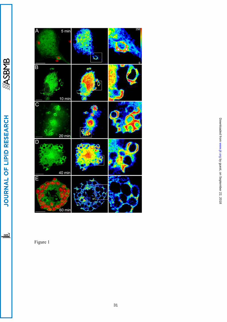
from it (Fig. 1, 60 min). Experiments were performed next to rule out the possibility that
the increased fluorescence arising from EGFP-cPLA2α in the forming phagosome was
due to an increased volume of cytoplasm imaged in the plane. This was addressed by
imaging in the same cell the construct EGFP-cPLA2α and a monomeric form of DsRed
—to correct for local variations in cytoplasmic volume— . Fig. 2 shows a clear increase
in EGFP-cPLA2α fluorescence in the phagosomes that does not correspond with an
increase in cytoplasmic volume (ratio EGFP-cPLA2α /DsRed) at any time, thus
indicating that enzyme translocation actually occurs.
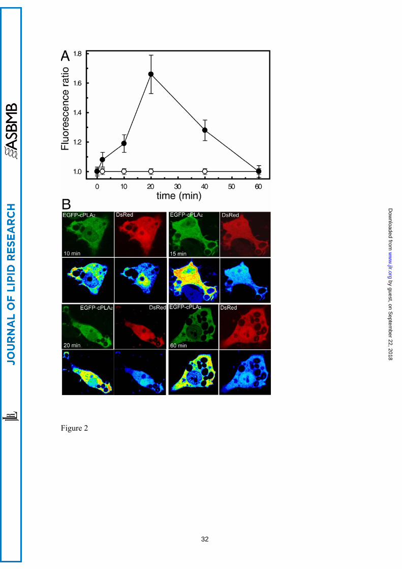
Phagocytosis of opsonized zymosan activates cPLA2α in human macrophages -
Zymosan induced a significant release of AA (and metabolites) to the extracellular
medium, suggesting the activation of a PLA2 (Fig. 3A). This PLA2 was identified as
cPLA2α on the basis of complete inhibition of the response by a low concentration of
pyrrophenone, a cPLA2α inhibitor [29] (Fig. 3B).
To confirm that the cPLA2α that translocates to the phagocytic cup is
functionally active, an experiment of frustrated phagocytosis was performed, utilizing
glass plates coated with IgG [28]. Macrophages exposed to IgG-coated glass surfaces
10
by guest, on Septem
ber 22, 2018w
ww
.jlr.orgD
ownloaded from
responded by translocating the EGFP-cPLA2α to the membranes more proximal to the
glass surface, but not to other cellular membranes (Fig. 4A, B and E). In this
experiment, the IgG-coated glass would represent the phagocytosable particle [28].
Next, we loaded the cells with the fluorogenic phospholipase substrate 1,2-bis-(4,4-
difluoro-5,7-dimethyl-4-bora-3a, 4a-diaza-sindecane-3-undecanoyl)-sn-glycero-3-
phosphocholine (bis-BODIPY FL C11-PC) [30], and subjected them to the frustrated
phagocytosis assay. A dramatic increase in fluorescence was observed, especially in the
proximity of the IgG-coated glass (Fig. 4C, D and F), indicating that an A-type
phospholipase is acting at that place (where the “phagosome” is being initiated). To
confirm that such a phospholipase is actually cPLA2α, we conducted experiments in the
presence of pyrrophenone (Fig. 4D and F). As expected, pyrrophenone, at doses as low
as 1 µM, strongly blocked the fluorescence increase in the cells exposed to IgG-coated
glass, indicating that such a fluorescence increase is mainly due to cPLA2α activation.
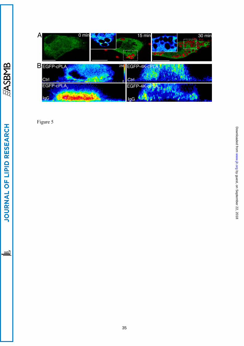
Mutation of a four-lysine cluster in cPLA2α that is involved in binding of anionic
phospholipids suppresses EGFP-cPLA2α translocation to the phagocytic cup - We
have previously described that mutations on Lys488, Lys541, Lys543 and Lys544 of
cPLA2α result in a defective translocation of cPLA2α to intracellular membranes in
response to exogenous PIP2 [17]. Because it is well described that PIP2 increases in the
phagosome [31], we studied next the behavior of the 4-Lys mutant (EGFP-4KE/A-
cPLA2α) in macrophages exposed to opsonized zymosan. The results, as shown in Fig.
5A, clearly indicated that this mutant does not translocate to the phagocytic cup in
activated cells at any time tested, suggesting that a functional binding site in cPLA2α for
anionic phospholipids is necessary for such a translocation to be observed. To further
substantiate this observation, we performed experiments of frustrated phagocytosis,
similar to those shown in Fig. 4, utilizing cells transfected with the EGFP-4KE/A-
11
by guest, on Septem
ber 22, 2018w
ww
.jlr.orgD
ownloaded from
cPLA2α mutant. Fig. 5B shows that, unlike the wild type enzyme, the mutant did not
translocate to the membranes that are closer to the IgG-coated plate. These data
highlight the importance of the 4-Lys cluster for proper binding of cPLA2α to the
phagosomal membranes.
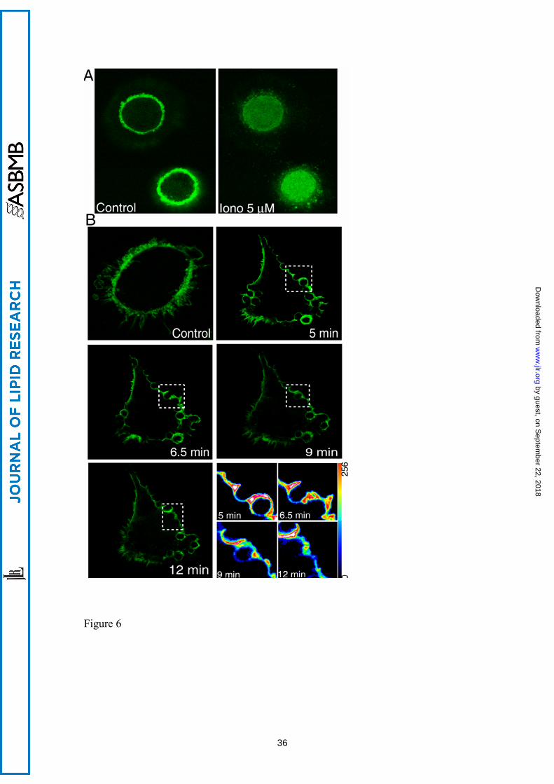
Experiments were conducted next to study a possible role for anionic
phospholipids in cPLA2α translocation to the phagocytic cup. To this end, we took
advantage of the PIP2-binding properties of a fluorescent chimeric protein of EGFP with
the pleckstrin homology domain of the PLCδ1 (EGF-PLCδ1-PH) [28]. Figure 6A shows
that, when transfected into human macrophages, the chimera labels the main cellular
reservoir of PIP2, i.e. the plasma membrane [24]. Immediately after promoting PIP2
hydrolysis by activating the cells with a calcium ionophore, the fluorescence disappears
from the plasma membrane, and accumulates in the cytoplasm, as would be expected
from a functional PH domain (Fig. 6A). By using this chimera, we confirmed in human
macrophages previous observations by Botelho et al. [31], indicating that PIP2 levels
increase at the phagocytic cup while the phagosome is being formed, and decrease when
it seals (Fig. 6B).
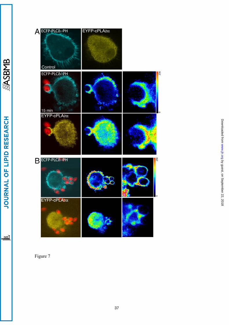
Subsequently, the cells were co-transfected with both constructs, namely ECFP-
PLCδ1-PH and EYFP-cPLA2α. In resting cells, ECFP-PLCδ1-PH was present only in
the plasma membrane, whereas EYFP-cPLA2α was found primarily in the cytoplasm
(Fig. 7). However, after exposure of the cells to opsonized zymosan both chimeric
proteins localized at the forming phagocytic cups (Fig. 7B). There was no co-
localization in the cytoplasm of resting or stimulated cells. We noticed also that in those
cells where ECFP-PLCδ1-PH construct was expressed at higher levels the translocation
of the cPLA2α to the phagosomes was inhibited (Fig. 7 C).
The behavior of EGFP-PLCδ1-PH and EGFP-cPLA2α were analyzed in more
12
by guest, on Septem
ber 22, 2018w
ww
.jlr.orgD
ownloaded from

detail by confocal analysis of z-stack series from cells that were engulfing particles at
similar stages of phagocytosis (Figs. 8 and 9). We observed that both constructs were
enriched in the same sites of the phagosomes, especially along the particles and in the
base. It is worth noting that translocation of the EGFP-cPLA2α to the base of some of
the phagosome was more prominent than that of EGFP-PLCδ1-PH. It is possible that in
the base of the phagosome other factors, in addition to or independently of PIP2,
contribute to the translocation of cPLA2α.
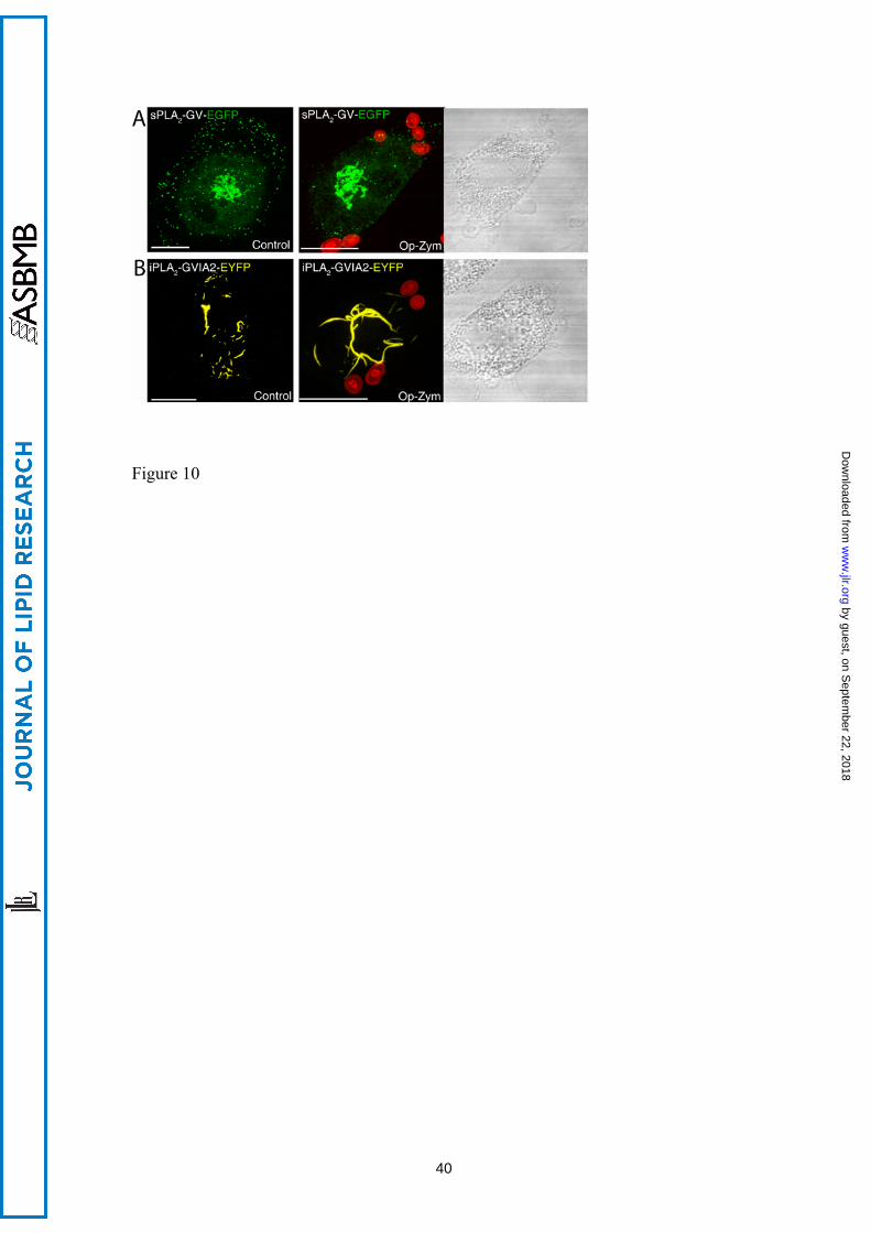
Secreted group V and cytosolic calcium-independent group VIA PLA2s do not
translocate to the phagocytic cup - To address whether other PLA2s in addition to
cPLA2α could also translocate to the phagocytic cup during phagocytosis of opsonized
zymosan, we utilized human macrophages transfected with either sPLA2-V-EGFP or
iPLA2-VIA-EYFP. In agreement with our previous studies in murine macrophages [25,
32], sPLA2-V-EGFP was found in resting cells associated with secretory granules and
Golgi-like structures (Fig 10A). iPLA2-VIA-EYFP had a mitochondrial localization in
unstimulated cells (Fig. 10B), which is in accordance with previous estimates [33]. After
the cells where challenged with opsonized zymosan, localization was studied between
the chimeric proteins and the fluorescent particles. We failed to detect association of the
fluorescence arising from either sPLA2-V-EGFP or iPLA2-VIA-EYFP to the phagocytic
cups under any condition (Fig. 10). Thus, of the three major PLA2 classes potentially
capable of generating lipid mediators during inflammation [2-5, 34], only the cPLA2α
translocates to the phagocytic cup in human macrophages.
13
by guest, on Septem
ber 22, 2018w
ww
.jlr.orgD
ownloaded from
Discussion
A major regulatory mechanism of cPLA2α activity in cells is the Ca2+-dependent control
of the physical state of the enzyme. In resting cells, the enzyme resides in the cytosol
and hence, having no access to its substrate in the membrane, has no activity. In
stimulated cells cPLA2α translocates to the membrane in a Ca2+-dependent process, this
resulting in phospholipid hydrolysis and free AA release [9]. Most of the research
carried out to date has thoroughly documented the translocation of cPLA2α to
intracellular membranes such as those of the nuclear envelope, Golgi complex or
endoplasmic reticulum [9]. A few instances have been reported of translocation of
cPLA2α to cellular membranes different to those indicated above [20, 35, 36]. This
study adds to these studies by showing that the phagosomal membrane of human
macrophages is also a site for cPLA2α translocation during activation conditions.
Particularly relevant to our report is the work by Girotti et. al. showing translocation of
cPLA2α to forming phagosomes in murine macrophages [20]. Although the molecular
mechanism was not investigated, the authors noted that chelation of extracellular
calcium decreased the total number of phagocytic events, but cPLA2α still remained
associated to the lasting phagosomes at intracellular calcium levels equaling those of
resting cells [20]. In previous work from our laboratory, we showed that introducing
exogenous short-chain PIP2 into cells promotes the translocation of cPLA2α from
cytosol to perinuclear membranes at basal levels of intracellular Ca2+ [17]. Moreover,
other studies have demonstrated that PIP2 transiently increases in the forming
phagosome and disappears after phagosome sealing, being undetectable in the sealed
phagosome [31]. Intrigued by these observations, we speculated that translocation of
cPLA2α to the forming phagosome could require the cationic cluster of four Lys
14
by guest, on Septem
ber 22, 2018w
ww
.jlr.orgD
ownloaded from
(Lys488/Lys541/Lys543/Lys544) that is present in cPLA2α and has been shown to bind
anionic phospholipids such as PIP2 [13]. Using confocal microscopy techniques, we
document the localization of cPLA2α and a PIP2-binding construct, ECFP-PLCδ1-PH, in
forming phagosomes. Also, high level expression of the ECFP-PLCδ1-PH inhibits the
translocation of the cPLA2α to phagosomes. Moreover, mutation of the cationic cluster
of Lys488, Lys541, Lys543 and Lys544 that serves as a PIP2-binding site, eliminates the
ability of the enzyme to translocate to the phagosomal membrane. Collectively, these
results provide the first example of a physiologically-relevant condition where the
cationic cluster of cPLA2α participates in regulating enzyme association to membranes
and hence, its activity in intact cells.
The presence of cPLA2α at the membrane of the nascent phagosome may serve
important pathophysiological roles, since during phagocytosis large quantities of
eicosanoids are produced which could be involved in the killing of the ingested
microorganism at the phagosome [8, 21-23]. In the mouse model, other PLA2s in
addition of cPLA2α have been suggested to be related to phagocytic events. Particularly
relevant to this work, it has been shown that group V secreted PLA2 also translocates to
the phagosome in zymosan-stimulated murine peritoneal macrophages [23]. Intrigued
by this report, we also studied the possible movement of sPLA2-V to phagosomes in our
human macrophage cell system. We failed to detect translocation of a chimeric sPLA2-
V-EGFP protein to phagosomes in our systems. We had previously demonstrated that
this chimeric sPLA2-V behaves the same as the native sPLA2-V protein in terms of
biochemical properties, enzymatic activity and subcellular localization [25, 32]. There
are many differences between the murine and human macrophage models of zymosan
phagocytosis that may explain these different results. For instance, in murine
macrophages, zymosan induces abundant AA release [37-43] and may be internalized
15
by guest, on Septem
ber 22, 2018w
ww
.jlr.orgD
ownloaded from
primarily via dectin-1 receptors [44]. However, human macrophages do not respond
readily to zymosan by releasing AA, and opsonization of the particle appears to be
required for full responses, which may occur primarily via Fc receptors. Thus, it appears
likely that the different mechanisms of internalization of phagocytosable particles in
mouse versus human may account, at least in part, for the remarkable differences in the
translocation ability of sPLA2–V to the phagosomes.
The one other PLA2 that we investigated is iPLA2-VIA, a calcium-independent
enzyme. Unlike cPLA2α and sPLA2 enzymes, the involvement of iPLA2-VIA in
receptor-mediated AA mobilization appears not to be a general one, but to depend on
cell type and stimulation conditions [2, 45-48]. Using a chimeric construct, iPLA2-VIA-
EYFP, we have detected no appreciable change in the subcellular localization of this
enzyme during opsonized zymosan challenge; the enzyme always remained associated
to mitochondria, which is consistent with previous data [33]. Thus, from the three major
PLA2 families potentially capable of effecting AA release for eicosanoid production,
only one, cPLA2α, translocates to the phagosome in human cells.
Acknowledgments
We thank Montse Duque and Yolanda Sáez for expert technical assistance, and the
personnel from Centro de Hemoterapia y Hemodonación de Castilla y León for
supplying Buffy coats from healthy volunteers. This work was supported by the Spanish
Ministry of Science and Innovation (Grants SAF2007-60055 and BFU2007-67154) and
the Regional Government of Castile and León (Grant CSI09-A08). CIBERDEM is an
initiative of Instituto de Salud Carlos III.
16
by guest, on Septem
ber 22, 2018w
ww
.jlr.orgD
ownloaded from
References
1. Schaloske R.H., and E. A. Dennis. 2006. The phospholipase A2 superfamily and its
group numbering system. Biochim. Biophys. Acta, 1761, 1246-1259.
2. Balsinde J., M.A. Balboa, P.A. Insel, and E.A. Dennis. 1999. Regulation and
inhibition of phospholipase A2. Annu. Rev. Pharmacol. Toxicol. 39, 175-189.
3. Balsinde J., M.V. Winstead, and E.A. Dennis. 2002. Phospholipase A2 regulation of
arachidonic acid mobilization. FEBS Lett. 531, 2-6
4. Balsinde J., and M.A. Balboa. 2005 Cellular regulation and proposed biological
functions of group VIA calcium-independent phospholipase A2 in activated cells. Cell
Signal. 17, 1052-1062.
5. Balboa M.A., and J. Balsinde. 2006 Oxidative estress and arachidonic acid
mobilization. Biochim. Biophys. Acta 1761, 385-391.
6. Funk C.D. 2001. Prostaglandins and leukotrienes: advances in eicosanoid biology.
Science 294, 1871-1875.
7. Serhan C.N., N. Chiang, and T.E. Van Dyke. 2008 Resolving inflammation: dual
anti-inflammatory and pro-resolution lipid mediators. Nat. Rev. Immunol. 8: 349-361.
8. Bailie M. B., T. J. Standiford, L. L. Laichalk, M. J. Coffery, R. Strieter, and M. Peters-
Golden. 1996 Leukotriene-deficient mice manifest enhanced lethality from Klebsiella
17
by guest, on Septem
ber 22, 2018w
ww
.jlr.orgD
ownloaded from
pneumonia in association with decreased alveolar macrophage phagocytic and bactericidal
activities. J. Immunol. 157: 5221-5224.
9. Ghosh, M., D.E. Tucker, S.A. Burchett, and C.C. Leslie. 2006. Properties of the
Group IV phospholipase A2 family. Prog. Lipid Res. 45, 487–510.
10. Bonventre J.V., Z. Huang, M.R. Taheri, E. O'Leary, E. Li, M.A. Moskowitz, and A.
Sapirstein. 1997. Reduced fertility and postischaemic brain injury in mice deficient in
cytosolic phospholipase A2. Nature 390: 622-625.
11. Uozumi N, K. Kume , T. Nagase , N. Nakatani , S. Ishii , F. Tashiro , Y. Komagata ,
K. Maki , K. Ikuta , Y. Ouchi , J. Miyazaki, and T. Shimizu. 1997. Role of cytosolic
phospholipase A2 in allergic response and parturition. Nature 390, 618-622.
12. Subramanian P., R.V. Stahelin, Z. Szulc, A. Bielawska, W. Cho, and C.E. Chalfant.
2005. Ceramide 1-phosphate acts as a positive allosteric activator of group IVA
cytosolic phospholipase A2α and enhances the interaction of the enzyme with
phosphatidylcholine. J. Biol. Chem. 280, 17601-17607.
13. Das S., and W. Cho. 2002. Roles of catalytic domain residues in interfacial binding
and activation of group IV cytosolic phospholipase A2. J. Biol. Chem. 277, 23838–
23846.
14. Mosior M., D.A. Six, and E.A. Dennis. 1998. Group IV cytosolic phospholipase A2
binds with high affinity and specificity to phosphatidylinositol 4,5-bisphosphate
18
by guest, on Septem
ber 22, 2018w
ww
.jlr.orgD
ownloaded from
resulting in dramatic increases in activity. J. Biol. Chem. 273, 2184-2191.
15. Six D.A., and E.A. Dennis. 2003. Essential Ca2+-independent role of the group IVA
cytosolic phospholipase A2 C2 domain for interfacial activity. J. Biol. Chem. 278,
23842–23850.
16. Subramanian P, M. Vora, L.B. Gentile, R.V. Stahelin, and C.E. Chalfant. 2007
Anionic lipids activate group IVA cytosolic phospholipase A2 via distinct and separate
mechanisms. J Lipid Res. 48, 2701-8.
17. Casas J., M. A. Gijón, A. G. Vigo, M. S. Crespo, J. Balsinde, and M.A. Balboa.
2006. Phosphatidylinositol 4,5-bisphosphate anchors cytosolic group IVA
phospholipase A2 to perinuclear membranes and decreases its calcium requirement for
translocation in live cells. Mol. Biol. Cell 17, 155-162.
18. Tucker D.E., M. Ghosh, F. Ghomashchi, R. Loper, S. Suram, B.S. John, M. Girotti,
J.G. Bollinger, M.H. Gelb, and C.C. Leslie. 2009 Role of phosphorylation and basic
residues in the catalytic domain of cytosolic phospholipase A2α in regulating interfacial
kinetics and binding and cellular function. J Biol Chem. 284, 9596-611.
19. Stuart L.M., and A.B. Ezekowitz. 2005. Phagocytosis: elegant complexity.
Immunity 22, 539-550.
19
by guest, on Septem
ber 22, 2018w
ww
.jlr.orgD
ownloaded from
20. Girotti, M., J. H. Evans, D. Burke, and C. C. Leslie. 2004. Cytosolic phospholipase
A2 translocates to forming phagosomes during phagocytosis of zymosan in
macrophages. J. Biol. Chem. 279:19113-19121.
21. Tomioka, H., C. Sano, K. Sato, K. Ogasawara, T. Akaki, K. Sano, S. S. Cai, and T.
Shimizu. 2005. Combined effects of ATP on the therapeutic efficacy of antimicrobial
drug regimens against Mycobacterium avium complex infection in mice and roles of
cytosolic phospholipase A2-dependent mechanisms in the ATP-mediated potentiation of
antimycobacterial host resistance. J. Immunol. 175: 6741-6749.
22. Rubin, B. B., G. P. Downey, A. Koh, N. Degousee, F. Ghomashchi, L. Nallan, E.
Stefanski, D. W. Harkin, C. Sun, B. P. Smart, T. F. Lindsay, V. Cherepanov, E. Vachon,
D. Kelvin, M. Sadilek, G. E. Brown, M. B. Yaffe, J. Plumb, S. Grinstein, M. Glogauer,
and M. H. Gelb. 2005. Cytosolic phospholipase A2α is necessary for platelet-activating
factor biosynthesis, efficient neutrophil mediated bacterial killing, and the innate
immune response to pulmonary infection: cPLA2α does not regulate neutrophil NADPH
oxidase activity. J. Biol. Chem. 280: 7519-7529.
23. Balestrieri, B., V. W. Hsu, H. Gilbert, C. C. Leslie, W. K. Han, J. V. Bonventre, and
J. P. Arm. 2006. Group V secretory phospholipase A2 translocates to the phagosome
after zymosan stimulation of mouse peritoneal macrophages and regulates phagocytosis.
J. Biol. Chem. 281: 6691-6698.
20
by guest, on Septem
ber 22, 2018w
ww
.jlr.orgD
ownloaded from
24. Stauffer, T.P., S. Ahn, and T. Meyer. 1998 Receptor-induced transient reduction in
plasma membrane PtdIns(4,5)P2 concentration monitored in living cells. Curr. Biol. 8,
343-346.
25. Shirai Y., J. Balsinde, and E.A. Dennis. 2005. Localization and functional
interrelationships among cytosolic group IV, secreted group V, and Ca2+-independent
group VI phospholipase A2s in P388D1 macrophages using GFP/RFP constructs.
Biochim. Biophys. Acta 1735, 119-129.
26. Casas J., M.A. Gijón, A.G. Vigo, M.S. Crespo, J. Balsinde, and M.A. Balboa. 2006.
Overexpression of cytosolic group IVA phospholipase A2 protects cells from calcium-
dependent death. J. Biol. Chem. 281, 6106-6116.
27. Larsen, E.C., J. A. DiGennaro, N. Saito, S. Mehta, D.J. Loegering, J.E.
Mazurkiewicz, and M.R. Lennartz. 2000. Differential requirement for classic and novel
PKC isoforms in respiratory burst and phagocytosis in RAW 264.7 cells. J. Immunol.
165, 2809-2817.
28. Marshall J.G., J.W. Booth, V. Stambolic, T. Mak, T. Balla, A.D. Schreiber, T.
Meyer, and S. Grinstein. 2001. Restricted accumulation of phosphatidylinositol 3-kinase
products in a plasmalemmal subdomain during Fc gamma receptor-mediated
phagocytosis. J. Cell. Biol. 153, 1369-1380.
21
by guest, on Septem
ber 22, 2018w
ww
.jlr.orgD
ownloaded from
29. Ono T., K. Yamada, Y. Chikazawa, M. Ueno, S. Nakamoto, T. Okuno, and K. Seno.
2002. Characterization of a novel inhibitor of cytosolic phospholipase A2α,
pyrrophenone. Biochem. J. 363, 727-735.
30. Manna D., and W. Cho. 2007. Real-time cell assays of phospholipase A2s using
fluorogenic phospholipids. Methods Enzymol. 434, 15-27.
31. Botelho, R.J., M. Teruel, R. Dierckman, R. Anderson, A. Wells, J.D. York, Y.
Meyer, and S. Grinstein. 2000. Localized biphasic changes in phosphatidylinositol-4,5-
bisphosphate at sites of phagocytosis. J. Cell Biol. 151, 1353-1367.
32. Balboa M.A., Y. Shirai, G. Gaietta, M.E. Ellisman, J. Balsinde, and E.A. Dennis.
2003. Localization of group V phospholipase A2 in caveolin-enriched granules in
activated P388D1 macrophage-like cells. J. Biol. Chem. 278, 48059-48065.
33. Seleznev K., C. Zhao, X.H. Zhang, K. Song, and Z.A. Ma. 2006. Calcium-
independent phospholipase A2 localizes in and protects mitochondria during apoptotic
induction by staurosporine. J. Biol. Chem. 281, 22275-22288.
34. Fitzpatrik F.A., and R. Soberman. 2001. Regulated formation of eicosanoids. J. Clin.
Invest. 107, 1347-1351.
35. Shmelzer Z., N. Haddad, E. Admon, I. Pessach, T.L. Leto, Z. Eitan-Hazan, M.
Hershfinkel, and R. Levy. 2003. Unique targeting of cytosolic phospholipase A2 to
22
by guest, on Septem
ber 22, 2018w
ww
.jlr.orgD
ownloaded from
plasma membranes mediated by the NADPH oxidase in phagocytes. J. Cell Biol. 162,
683-692.
36. Wooten, R.E., M.C. Willingham, L.W. Daniel, C.C. Leslie, L.C. Rogers, S.
Sergeant, and J.T. O'Flaherty. 2008. Novel translocation responses of cytosolic
phospholipase A2alpha fluorescent proteins. Biochim Biophys Acta. 1783:1544-50.
37. Balsinde, B. Fernandez, and E. Diez, E. 1990. Regulation of arachidonic acid release
in mouse peritoneal macrophages. The role of extracellular calcium and protein kinase
C. J. Immunol. 144, 4298-4304.
38. Balsinde J., B. Fernández, J.A. Solís-Herruzo, and E. Diez. 1992. Pathways for
arachidonic acid mobilization in zymosan-stimulated mouse peritoneal macrophages.
Biochim. Biophys. Acta. 1136, 75-82.
39. Qiu Z.H., M.S. de Carvalho, and C.C. Leslie. 1993. Regulation of phospholipase A2
activation by phosphorylation in mouse peritoneal macrophages. 268, 24506-24513.
40. Balsinde, J., M. A. Balboa, P. A. Insel, and E. A. Dennis. 1997. Differential
regulation of phospholipase D and phospholipase A2 by protein kinase C in P388D1
macrophages. Biochem. J. 321, 805-809.
41. Balsinde, J., M. A. Balboa, S. Yedgar, and E. A. Dennis. 2000. Group V
phospholipase A2-mediated oleic acid mobilization in lipopolysaccharide-stimulated
P388D1 macrophages. J. Biol. Chem. 275, 4783-4786.
23
by guest, on Septem
ber 22, 2018w
ww
.jlr.orgD
ownloaded from
42. Gijón, M.A., D.M. Spencer, A.R. Siddiqui, J.V. Bonventre, and C.C. Leslie. 2000.
Cytosolic phospholipase A2 is required for macrophage arachidonic acid release by
agonists that do and do not mobilize calcium. Novel role of mitogen-activated protein
kinase pathways in cytosolic phospholipase A2 regulation. J. Biol. Chem. 275, 20146-
20156.
43. Balsinde J., M.A. Balboa, and E.A. Dennis. 2000. Identification of a third pathway
for arachidonic acid mobilization and prostaglandin production in activated P388D1
macrophage-like cells. J. Biol. Chem. 275, 22544-22549.
44. Suram S., G.D. Brown, M. Ghosh, S. Gordon, R. Loper, P.R. Taylor, S. Akira, S.
Uematsu, D.L. Williams, and C.C. Leslie. 2006 Regulation of cytosolic phospholipase
A2 activation and cyclooxygenase 2 expression in macrophages by the beta-glucan
receptor. J. Biol. Chem. 281, 5506-5514.
45. Balsinde, J. 2002. Roles of various phospholipases A2 in providing lysophospholipid
acceptors for fatty acid phospholipid incorporation and remodelling. Biochem. J. 364:
695-702.
46. Balboa, M. A., Y. Sáez, and J. Balsinde. 2003. Calcium-independent phospholipase
A2 is required for lysozyme secretion in U937 promonocytes. J. Immunol. 170: 5276-
5280
24
by guest, on Septem
ber 22, 2018w
ww
.jlr.orgD
ownloaded from
47. Balboa, M. A., and J. Balsinde. 2002. Involvement of calcium-independent
phospholipase A2 in hydrogen peroxide-induced accumulation of free fatty acids in
human U937 cells. J. Biol. Chem. 277: 40384-40389.
48. Pérez, R., R. Melero, M. A. Balboa, and J. Balsinde. 2004. Role of group VIA
calcium-independent phospholipase A2 in arachidonic acid release, phospholipid fatty
acid incorporation, and apoptosis in U937 cells responding to hydrogen peroxide. J.
Biol. Chem. 279: 40385-40391.
25
by guest, on Septem
ber 22, 2018w
ww
.jlr.orgD
ownloaded from

Figure Legends
Figure 1. Translocation of cPLA2α to the phagosome in zymosan-treated human
macrophages. Human macrophages were transfected with the construct EGFP-cPLA2α,
treated with 1 mg/ml opsonized-zymosan for the indicated times, fixed and analyzed by
confocal microscopy. In panels A and E Alexa Fluor 594-labeled zymosan was used to
better visualize the particles. Fluorescence arising from the EGFP-cPLA2α (green) or
zymosan (red) is shown on the left column. Middle-column panels show a
pseudocolored fluorescence intensity from the EGFP-cPLA2α. Panels on the right
column show a detailed amplification of the phagosomes framed in the middle panels.
Scale bar = 10 μM.
Figure 2. Analysis of the translocation of cPLA2α and the monomeric DsRed to
phagosomal membranes. Human macrophages were co-transfected with the construct
EGFP-cPLA2α or the soluble fluorescent protein DsRed (monomeric form). Cells were
stimulated with opsonized zymosan, fixed, and fluorescence was analyzed by confocal
microscopy at different times. A) The figure represents the fluorescence ratio EGFP-
cPLA2α / DsRed in phagosomes versus cytosol at each time point. At least 4
phagosomes per cell were analyzed from many cells. B) Pictures of the cellular
fluorescence of the EGFP-cPLA2α and the DsRed at different times of phagocytosis are
shown.
Figure 3. AA release in human macrophages challenged with opsonized zymosan. (A)
Human macrophages labeled with [3H]AA were treated with 1 mg/ml opsonized
26
by guest, on Septem
ber 22, 2018w
ww
.jlr.orgD
ownloaded from
zymosan (black triangles) or vehicle (black squares). AA release was assessed at
different times as described under Materials and Methods. (B) Cells labeled with
[3H]AA were pretreated with 1 μM pyrrophenone (pyrr) or vehicle (control) for 30 min
and then treated with 1 mg/ml opsonized zymosan (black bars) or vehicle (grey bars) for
1 h.
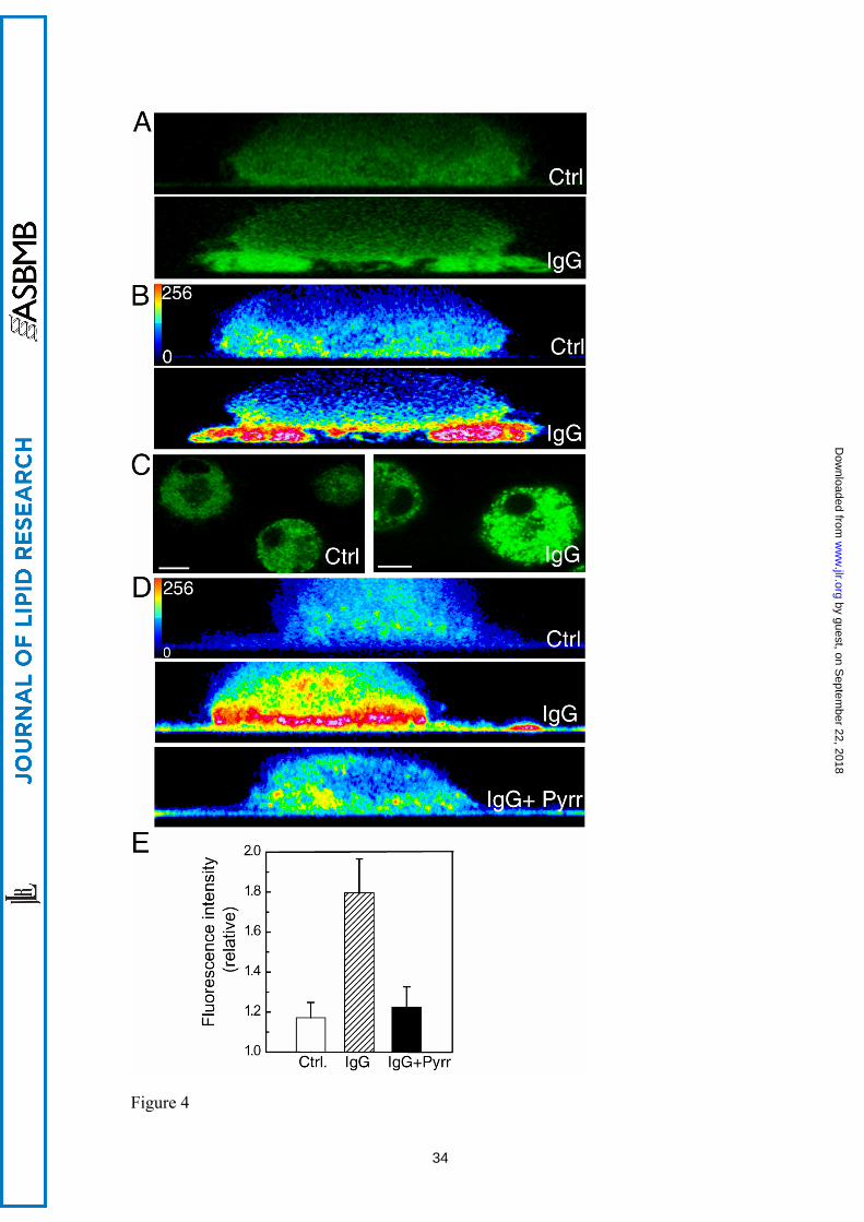
Figure 4. cPLA2α is functionally active in the phagosomal cup in human macrophages.
(A) Cells transfected with EGFP-cPLA2 were subjected to a frustrated phagocytosis
assay on non-coated glass (Ctrl) or IgG-coated glass (IgG), as indicated, and analyzed
by confocal microscopy. Pictures of the XZ axis of the cells were taken. (B) The
intensity of the fluorescence obtained in A was analyzed in pseudocolor. (C) Cells,
labeled with bis-BODIPY FL C11-PC, were plated on non-coated glass (Ctrl) or on IgG-
coated glass (IgG) for 30 min, and fluorescence was analyzed by confocal microscopy.
The mean of fluorescence intensity in Ctrl was 66 and in IgG was 128. (D) The cells
were labeled with bis-BODIPY FL C11-PC and subjected to a frustrated phagocytosis
assay. Pictures of the XZ axis of the cells were taken, and the intensity of the
fluorescence was analyzed in pseudocolor. In the picture on the bottom, the cells were
treated with 1 μM pyrrophenone (Pyrr). White bar = 10 μm. E) Statistical analysis of the
fluorescence intensity in cells assayed for frustrated phagocytosis as in D. Data are
represented as relative fluorescence intensities (mean fluorescence intensity in
membranes closer to the glass/mean fluorescence intensity in the cytosol). At least 15
different cells were analyzed.
Figure 5. The mutant EGFP-4KE/A-cPLA2α does not translocate to the phagosome in
human macrophages. A) Human macrophages transfected with the mutant EGFP-
27
by guest, on Septem
ber 22, 2018w
ww
.jlr.orgD
ownloaded from
4KE/A-cPLA2α were subjected to synchronized phagocytosis with Alexa Fluor 594-
labeled opsonized zymosan for the indicted times, fixed and analyzed by confocal
microscopy. Framed phagosomes have been enlarged and the intensity of the
fluorescence analyzed by pseudocolor (left upper inserts at 15 and 30 min). B) Cells
transfected with the construct EGFP-cPLA2α or EGFP-4KE/A-cPLA2α were subjected
to a frustrated phagocytosis assay, and plated over non-coated glasses (Ctrl) or IgG-
coated glasses (IgG) as indicated, and analyzed by confocal microscopy. Pictures of the
XZ axis of the cells were taken and the intensity of the fluorescence obtained was
analyzed in pseudocolor. The figure is representative of more than 40 cells that were
analyzed per experiment, and the experiment was repeated four times. White bar = 10
μm.
Figure 6. PIP2 accumulation in the forming phagosomes during zymosan phagocytosis
in human macrophages. (A) Cells were transfected with the construct EGFP-PLCδ-PH
and analyzed in vivo by confocal microscopy. Pictures were taken before (control) and
after 5 min of treatment with 5 μM ionomycin (Iono 5 μM). (B) Cells were transfected
with the construct EGFP-PLCδ-PH, treated with opsonized zymosan, an analyzed in
vivo by confocal microscopy. Pictures were taken at different time points after exposure
to zymosan, as indicated. The pictures on the bottom right correspond to the
phagosomes selected with a dotted white box at each time point, and the fluorescence
intensity has been analyzed in pseudocolor.
Figure 7. Localization of ECFP-PLCδ1-PH and EYFP-cPLA2α during phagocytosis in
human macrophages. Human macrophages, co-transfected with the ECFP-PLCδ1-PH
and the EYFP-cPLA2α constructs, were subjected to synchronized phagocytosis using
28
by guest, on Septem
ber 22, 2018w
ww
.jlr.orgD
ownloaded from
Alexa Fluor 594-labeled opsonized zymosan, fixed at 0 (Control) and 15 min and
analyzed by confocal microscopy (A). B) A cell with high expression of the construct
ECFP-PLCδ1-PH is shown. In A and B, middle panels, fluorescence intensities are
shown in pseudocolor, and detailed fluorescence in forming phagosomes is shown in
panels to the right.
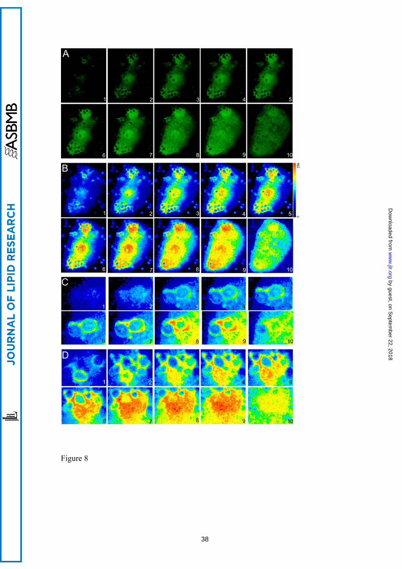
Figure 8. Analysis of human macrophages transfected with EGFP-cPLA2α during
phagocytosis of zymosan. Human macrophages transfected with the construction EGFP-
cPLA2α were treated with opsonized zymosan for 20 min, fixed, and fluorescence
analyzed by confocal microscopy. A) Ten different z-stacks of the same cell are shown
(from top to bottom). B) Pseudocolor analysis of the intensity of fluorescence of the cell
in A is shown. C and D) Detailed phagosomes from B.
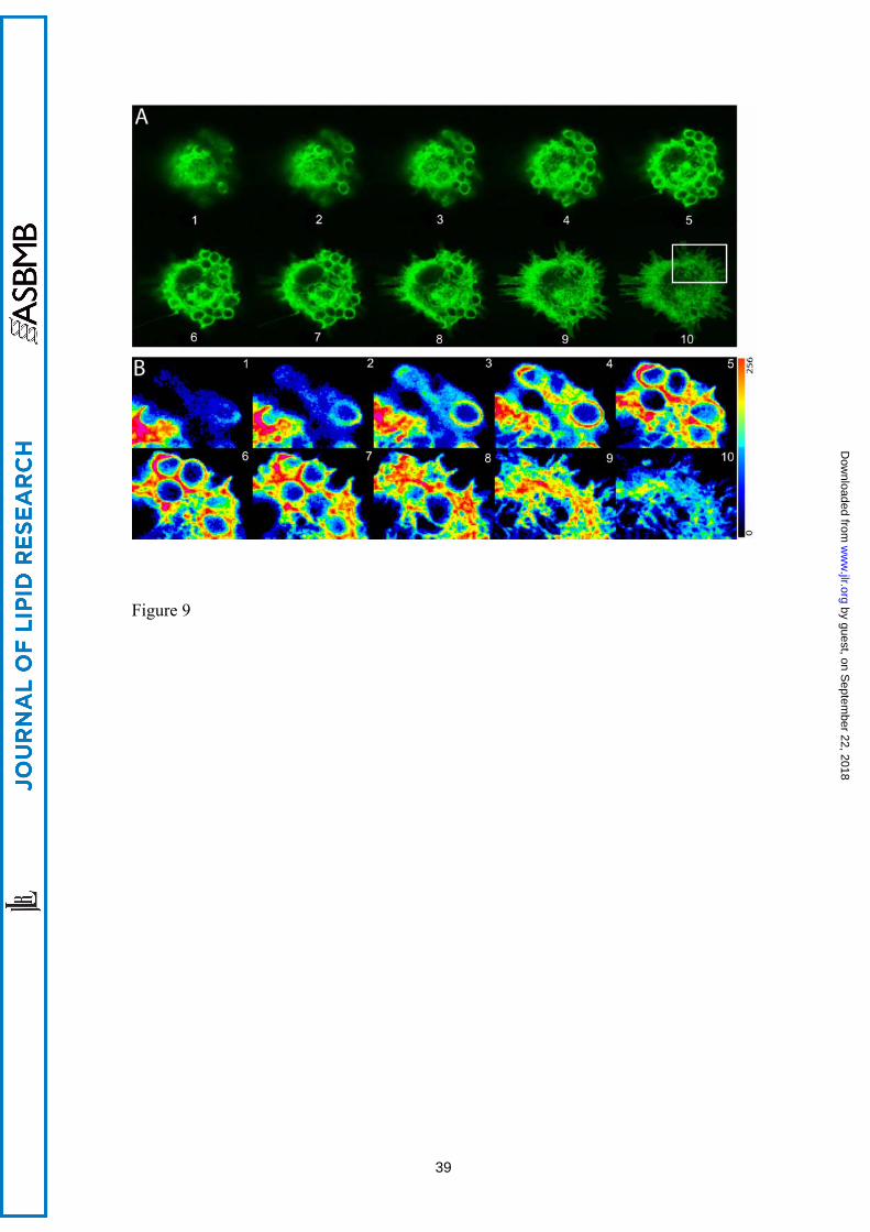
Figure 9. Analysis of a human macrophage transfected with EGFP-PLCδ1-PH during
phagocytosis of zymosan. Human macrophages transfected with the construction EGFP-
PLCδ1-PH were treated with zymosan for 20 min, fixed, and fluorescence analyzed by
confocal microscopy. A) Ten different z-stacks of the same cell are shown (from top to
bottom). B) Pseudocolor analysis of the intensity of fluorescence from some
phagosomes is shown (white square in A).
Figure 10. sPLA2-V and iPLA2-VIA do not translocate to the phagocytic cup in human
macrophages. Cells transfected with the fluorescent constructs sPLA2-V-EGFP (A) or
iPLA2-VIA-EYFP (B) were subjected to synchronized phagocytosis using Alexa Fluor
594-labeled opsonized zymosan (op-zym) or vehicle (control) for 15 min, fixed and
analyzed by confocal microscopy. Images are the projection to the Z axis of more than
29
by guest, on Septem
ber 22, 2018w
ww
.jlr.orgD
ownloaded from
13 stacks (0.25 μm each). Transmission images are shown in the right panel. White bar
= 10 μm.
30
by guest, on Septem
ber 22, 2018w
ww
.jlr.orgD
ownloaded from