Suppressive Effects of GSS on Lipopolysaccharide-Induced ...
14
Research Article Suppressive Effects of GSS on Lipopolysaccharide-Induced Endothelial Cell Injury and ALI via TNF-α and IL-6 Lei Yi, 1 Zengding Zhou, 1 Yijuan Zheng , 2 Mengling Chang, 1 Xiaoqin Huang, 1 Feng Guo , 3 Quanming Zhao , 4 and Jingning Huan 1 1 Department of Burn and Plastic Surgery, Ruijin Hospital, School of Medicine, Shanghai Jiao Tong University, Shanghai, China 2 Department of Critical Care Medicine, Zhongshan Hospital, Fudan University, Shanghai, China 3 Department of Plastic Surgery, Shanghai Jiaotong University Affiliated Sixth People’s Hospital, Shanghai, China 4 Department of Orthopedic Surgery, The Second Affiliated Hospital of Nantong University, Nantong, Jiangsu Province, China Correspondence should be addressed to Feng Guo; [email protected], Quanming Zhao; [email protected], and Jingning Huan; [email protected] Received 25 February 2019; Revised 1 September 2019; Accepted 23 October 2019; Published 30 December 2019 Academic Editor: Stefanie B. Flohé Copyright © 2019 Lei Yi et al. This is an open access article distributed under the Creative Commons Attribution License, which permits unrestricted use, distribution, and reproduction in any medium, provided the original work is properly cited. Background. Under septic conditions, LPS induced lung vascular endothelial cell (EC) injury, and the release of inflammatory mediator launches and aggravates acute lung injury (ALI). There are no effective therapeutic options for ALI. Genistein-3 ′ -sodium sulfonate (GSS) is a derivative of native soy isoflavone, which exhibits neuroprotective effects via its antiapoptosis property. However, whether GSS protect against sepsis-induced EC injury and release of inflammatory mediators has not been determined. In this study, we found that GSS not only downregulated the levels of TNF-α and IL-6 in the lung and serum of mice in vivo but also inhibited the expression and secretion of TNF-α and IL-6 in ECs. Importantly, we also found that GSS blocked LPS-induced TNF-α and IL-6 expression in ECs via the Myd88/NF-κB signaling pathway. Taken together, our results demonstrated that GSS might be a promising candidate for sepsis-induced ALI via its regulating effects on inflammatory response in lung ECs. 1. Background Genistein is the major isoflavone in soybeans and a natural tyrosine kinase inhibitor [1]. Recently, scientific interest in genistein has grown enormously owing to genistein putative multiple pharmacological properties such as antitumor, anti-inflammatory, and antioxidant effects [2, 3]. Though genistein has many beneficial effects, its poor water solubility and fat solubility limits its widespread application [4]. To improve the bioavailability and activity of genistein, Li et al. synthesized genistein-3 ′ -sodium sulfonate (GSS), a new compound derived from genistein that protected cortical neurons from ischemia-induced neuronal apoptosis [5]. However, there is no report about the effects of GSS on bac- terial infection-induced acute lung injury (ALI) under sepsis condition until now. Sepsis is characterized by an imbalance of proinflam- matory responses that induce the dysfunction of multiple organs, such as ALI and acute respiratory distress syn- drome (ARDS) [6]. ALI is a severe clinical syndrome characterized by diffuse pulmonary edema. Much of the pathogenesis of ALI is due to lung vascular endothelial cell injury, which leads to increased lung vascular perme- ability and extensive inflammatory pulmonary infiltrates [7]. Endothelial cell injury-induced disruption of lung endothelial barrier integrity and excessive inflammatory response are central to the pathogenesis of sepsis- induced ALI [8–10]. Some studies have demonstrated that the inflammation response of lung vascular ECs in WT mice was upregulated in LPS-induced ALI [11], and blocking pulmonary inflammation effectively attenuated histopathologic lung injury [12, 13]. Hindawi Mediators of Inflammation Volume 2019, Article ID 4251394, 13 pages https://doi.org/10.1155/2019/4251394
Transcript of Suppressive Effects of GSS on Lipopolysaccharide-Induced ...

Research ArticleSuppressive Effects of GSS on Lipopolysaccharide-InducedEndothelial Cell Injury and ALI via TNF-α and IL-6
Lei Yi,1 Zengding Zhou,1 Yijuan Zheng ,2Mengling Chang,1 Xiaoqin Huang,1 Feng Guo ,3
Quanming Zhao ,4 and Jingning Huan 1
1Department of Burn and Plastic Surgery, Ruijin Hospital, School of Medicine, Shanghai Jiao Tong University, Shanghai, China2Department of Critical Care Medicine, Zhongshan Hospital, Fudan University, Shanghai, China3Department of Plastic Surgery, Shanghai Jiaotong University Affiliated Sixth People’s Hospital, Shanghai, China4Department of Orthopedic Surgery, The Second Affiliated Hospital of Nantong University, Nantong, Jiangsu Province, China
Correspondence should be addressed to Feng Guo; [email protected], Quanming Zhao; [email protected],and Jingning Huan; [email protected]
Received 25 February 2019; Revised 1 September 2019; Accepted 23 October 2019; Published 30 December 2019
Academic Editor: Stefanie B. Flohé
Copyright © 2019 Lei Yi et al. This is an open access article distributed under the Creative Commons Attribution License, whichpermits unrestricted use, distribution, and reproduction in any medium, provided the original work is properly cited.
Background. Under septic conditions, LPS induced lung vascular endothelial cell (EC) injury, and the release ofinflammatory mediator launches and aggravates acute lung injury (ALI). There are no effective therapeutic options forALI. Genistein-3′-sodium sulfonate (GSS) is a derivative of native soy isoflavone, which exhibits neuroprotective effects via itsantiapoptosis property. However, whether GSS protect against sepsis-induced EC injury and release of inflammatory mediatorshas not been determined. In this study, we found that GSS not only downregulated the levels of TNF-α and IL-6 in the lung andserum of mice in vivo but also inhibited the expression and secretion of TNF-α and IL-6 in ECs. Importantly, we also found thatGSS blocked LPS-induced TNF-α and IL-6 expression in ECs via the Myd88/NF-κB signaling pathway. Taken together, ourresults demonstrated that GSS might be a promising candidate for sepsis-induced ALI via its regulating effects on inflammatoryresponse in lung ECs.
1. Background
Genistein is the major isoflavone in soybeans and a naturaltyrosine kinase inhibitor [1]. Recently, scientific interest ingenistein has grown enormously owing to genistein putativemultiple pharmacological properties such as antitumor,anti-inflammatory, and antioxidant effects [2, 3]. Thoughgenistein has many beneficial effects, its poor water solubilityand fat solubility limits its widespread application [4]. Toimprove the bioavailability and activity of genistein, Li et al.synthesized genistein-3′-sodium sulfonate (GSS), a newcompound derived from genistein that protected corticalneurons from ischemia-induced neuronal apoptosis [5].However, there is no report about the effects of GSS on bac-terial infection-induced acute lung injury (ALI) under sepsiscondition until now.
Sepsis is characterized by an imbalance of proinflam-matory responses that induce the dysfunction of multipleorgans, such as ALI and acute respiratory distress syn-drome (ARDS) [6]. ALI is a severe clinical syndromecharacterized by diffuse pulmonary edema. Much of thepathogenesis of ALI is due to lung vascular endothelialcell injury, which leads to increased lung vascular perme-ability and extensive inflammatory pulmonary infiltrates[7]. Endothelial cell injury-induced disruption of lungendothelial barrier integrity and excessive inflammatoryresponse are central to the pathogenesis of sepsis-induced ALI [8–10]. Some studies have demonstrated thatthe inflammation response of lung vascular ECs in WTmice was upregulated in LPS-induced ALI [11], andblocking pulmonary inflammation effectively attenuatedhistopathologic lung injury [12, 13].
HindawiMediators of InflammationVolume 2019, Article ID 4251394, 13 pageshttps://doi.org/10.1155/2019/4251394

Sepsis can be initiated by extensive vascular endothelialcell (EC) dysfunction in response to gram-negative bacterialinfection [14]. Lipopolysaccharide (LPS) is the major compo-nent of the outer surface of gram-negative bacteria and isrecognized as the vital factor involved in the pathogenesisof EC injury [14]. LPS binds and activates Toll-like receptor4 (TLR4) of ECs, which results in massive release of variousproinflammatory mediators, such as interleukin 6 (IL-6)and tumor necrosis factor α (TNF-α). Overexpression ofTNF-α and IL-6 is associated with ALI, multiple organ dys-function syndrome (MODS), and mortality [15, 16].
Nuclear factor κB (NF-κB) is a ubiquitous nucleartranscription factor which plays an important role in theinflammatory response to infection and is involved in theregulation of a variety of genes activated upon inflammationincluding the promoter region of the TNF-α and IL-6 gene[17]. Our previous studies suggested that after the interactionof LPS and TLR4 in the membrane of ECs, LPS induced acti-vation of downstream NF-κB in ECs via both the Myd88-dependent manner (Myd88 pathway) and the Myd88-independent manner (GEF-H1 pathway) [18]. Recently,Jeong et al. showed that genistein had anti-inflammatoryeffects in LPS-stimulated microglial cells via suppression ofthe TLR4 signaling [19]. However, the underlying effects ofGSS on LPS-induced activation of TLR4 pathway and subse-quent TNF-α and IL-6 overexpression in lung microvascularECs are largely unknown. These experiments were designedto investigate whether GSS confers protective effects againstLPS-induced overexpression of TNF-α and IL-6 in lungmicrovascular ECs and to determine the related signalingpathway.
In the present study, we not only found that GSS pro-tected against sepsis-induced ALI but also revealed that theabove protective effects of GSS are achieved through inhibi-tion of LPS-induced Myd88/NF-κB/TNF-α/IL-6 signalingactivation instead of GEF-H1/NF-κB pathway in lung micro-vascular ECs. Our findings provided new perspectives on theprotective role of GSS in LPS-induced lung microvascular ECinjury, and the findings of this study may contribute to theprevention and treatment of sepsis-induced ALI.
2. Methods (We Followed the Methods of Yiet al. [20])
2.1. Animal Studies (ALI Model, Lung Histopathology, Ratiosof Wet/Dry, Ratios of PaO2/FiO2, and Evans BlueExtravasation). Male C57BL/6 mice (6-8 week old, 18-20 g)were obtained from the Experimental Animal Center ofRuijin Hospital, Shanghai, China. All of the mice were pro-vided with standard laboratory food and water and housedin accordance with institutional animal care policies. All pro-cedures and animal care were performed in accordance withthe National Institutes of Health Guide for the Care and Useof Laboratory Animals with the approval (SYXK-2011-0113)of the Scientific Investigation Board of Shanghai Jiao TongUniversity School of Medicine, Shanghai, China. The ani-mals were acclimatized to the laboratory conditions (25°C,12 h/12 h light/dark, 50% humidity, and ad libitum accessto food and water) for one week prior to experimentation.
The mice were firstly pretreated with or without BAY11-7082 by intraperitoneal injection for 2 hours, and then, thesepsis-associated ALI was induced by a cecal ligation andpuncture (CLP) model; the mice were anesthetized with 1%sodium pentobarbital (40mg/kg). For histological assess-ment of lung injury, the mouse lungs were harvested 24hafter CLP application and were quickly removed and fixedin 10% paraformaldehyde. The paraformaldehyde-fixed lobeof the lungs was embedded in paraffin and cut into 5μm sec-tions. H&E staining was performed using standard protocols.The slides of each group were assessed under high-powerfields (sections were evaluated at ×100 magnification). Forassessment of sepsis-induced lung edema, the lungs wereimmediately weighed to obtain the wet weight and thenplaced in an oven at 80°C for 48 hours to obtain the dryweight. The ratio of the wet lung to the dry lung was calcu-lated to assess lung edema. For analysis of sepsis-inducedlung vascular leak, Evans blue dye (30ml/kg) was injectedinto the caudal vein 2 hours before termination of the exper-iment. Measurement of Evans blue accumulation in the lungtissue was performed by spectrofluorimetric analysis of lungtissue lysates as before [21]. For measurement of oxygenationindex changes, the carotid arteries were cannulated, and thearterial blood samples were collected for analysis [22].
2.2. Cell Culture and Chemicals. For cell culture andreagents, primary mouse pulmonary microvascular endo-thelial cells were obtained from Angio-Proteomie (Boston,MA, US) and maintained in Angio-Proteomie EndothelialCell Medium in a humidified 37°C, 5% CO2 incubator.The medium was changed at 48-hour intervals. Genistein-3′-sodium sulfonate (GSS) was obtained from ShanghaiTianxi Chemical Co., Ltd. (Shanghai, China), and BAY11-7082 was purchased from Beyotime of China. The LPS (fromEscherichia coli 055:B5) was obtained from Sigma-Aldrich(St. Louis, MO). The anti-GEF-H1 rabbit monoclonal anti-body (mAb), anti-Myd88 rabbit mAb, anti-NF-κB p65 rabbitmAb, and anti-rabbit phospho-NF-κB-p65 rabbit antibodywere obtained from Cell Signaling Technologies (Danvers,MA). Alexa Fluor 594-conjugated goat anti-rabbit secondaryantibody and the ProLong Gold Antifade Mountant withDAPI were obtained from Invitrogen Life Science.
2.3. Cell Viability Assay. The Cell Counting Kit-8 (CCK-8;CK04, DOJINDO) was used to assess cell viability accordingto the manufacturer’s protocol. ECs were seeded in 96-wellplates and cultured for 24–48 h. When the monolayer of cellswas 90% confluent, the ECs were exposed to various concen-trations of GSS for different times. Next, 10μl of CCK-8 solu-tion was added to each well, and the cells were incubated foran additional 3 h. The absorbance at 450nm was measuredusing a microplate reader. The cell survival rate was calcu-lated using the average of pooled data from three separateexperiments of six wells.
2.4. ELISA. The supernatants were assayed for TNF-α andIL-6 protein content using ELISA kits for TNF-α and IL-6(BD Biosciences, San Diego, CA) according to the manufac-turer’s instructions. Briefly, each well of the 96-well plate
2 Mediators of Inflammation
was coated overnight with capture antibody before beingwashed with PBS containing 0.05% Tween; then, thesupernatant was added to the appropriate wells. After havingbeen incubated for 1 hour at room temperature, the detectionantibody was added and incubated for another 1 hour. Thewells were then washed with PBS/Tween, and horseradishperoxidase-conjugated streptavidin was added for further 1hour at room temperature. Finally, the color was developedby adding peroxidase substrate to each well before readingthe absorbance at 450nm using the Dynatec plate reader(Denkendorf, Germany).
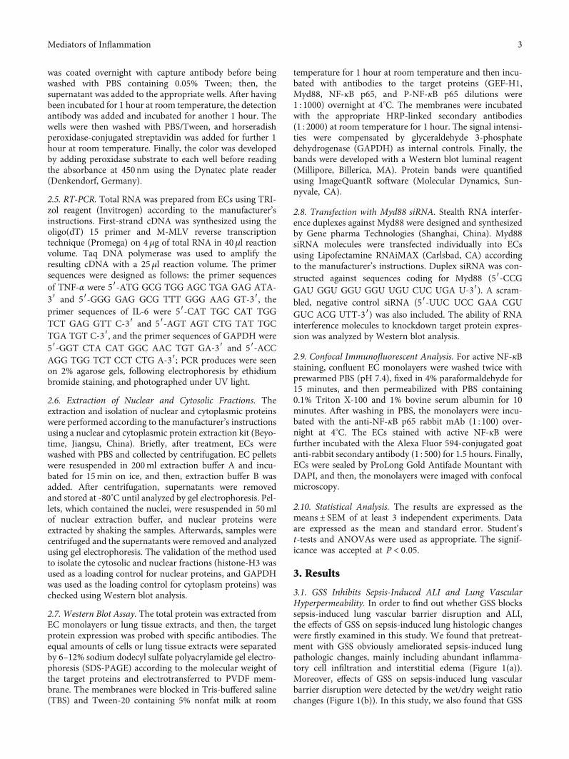
2.5. RT-PCR. Total RNA was prepared from ECs using TRI-zol reagent (Invitrogen) according to the manufacturer’sinstructions. First-strand cDNA was synthesized using theoligo(dT) 15 primer and M-MLV reverse transcriptiontechnique (Promega) on 4μg of total RNA in 40μl reactionvolume. Taq DNA polymerase was used to amplify theresulting cDNA with a 25μl reaction volume. The primersequences were designed as follows: the primer sequencesof TNF-α were 5′-ATG GCG TGG AGC TGA GAG ATA-3′ and 5′-GGG GAG GCG TTT GGG AAG GT-3′, theprimer sequences of IL-6 were 5′-CAT TGC CAT TGGTCT GAG GTT C-3′ and 5′-AGT AGT CTG TAT TGCTGA TGT C-3′, and the primer sequences of GAPDH were5′-GGT CTA CAT GGC AAC TGT GA-3′ and 5′-ACCAGG TGG TCT CCT CTG A-3′; PCR produces were seenon 2% agarose gels, following electrophoresis by ethidiumbromide staining, and photographed under UV light.
2.6. Extraction of Nuclear and Cytosolic Fractions. Theextraction and isolation of nuclear and cytoplasmic proteinswere performed according to the manufacturer’s instructionsusing a nuclear and cytoplasmic protein extraction kit (Beyo-time, Jiangsu, China). Briefly, after treatment, ECs werewashed with PBS and collected by centrifugation. EC pelletswere resuspended in 200ml extraction buffer A and incu-bated for 15min on ice, and then, extraction buffer B wasadded. After centrifugation, supernatants were removedand stored at -80°C until analyzed by gel electrophoresis. Pel-lets, which contained the nuclei, were resuspended in 50mlof nuclear extraction buffer, and nuclear proteins wereextracted by shaking the samples. Afterwards, samples werecentrifuged and the supernatants were removed and analyzedusing gel electrophoresis. The validation of the method usedto isolate the cytosolic and nuclear fractions (histone-H3 wasused as a loading control for nuclear proteins, and GAPDHwas used as the loading control for cytoplasm proteins) waschecked using Western blot analysis.
2.7. Western Blot Assay. The total protein was extracted fromEC monolayers or lung tissue extracts, and then, the targetprotein expression was probed with specific antibodies. Theequal amounts of cells or lung tissue extracts were separatedby 6–12% sodium dodecyl sulfate polyacrylamide gel electro-phoresis (SDS-PAGE) according to the molecular weight ofthe target proteins and electrotransferred to PVDF mem-brane. The membranes were blocked in Tris-buffered saline(TBS) and Tween-20 containing 5% nonfat milk at room
temperature for 1 hour at room temperature and then incu-bated with antibodies to the target proteins (GEF-H1,Myd88, NF-κB p65, and P-NF-κB p65 dilutions were1 : 1000) overnight at 4°C. The membranes were incubatedwith the appropriate HRP-linked secondary antibodies(1 : 2000) at room temperature for 1 hour. The signal intensi-ties were compensated by glyceraldehyde 3-phosphatedehydrogenase (GAPDH) as internal controls. Finally, thebands were developed with a Western blot luminal reagent(Millipore, Billerica, MA). Protein bands were quantifiedusing ImageQuantR software (Molecular Dynamics, Sun-nyvale, CA).
2.8. Transfection with Myd88 siRNA. Stealth RNA interfer-ence duplexes against Myd88 were designed and synthesizedby Gene pharma Technologies (Shanghai, China). Myd88siRNA molecules were transfected individually into ECsusing Lipofectamine RNAiMAX (Carlsbad, CA) accordingto the manufacturer’s instructions. Duplex siRNA was con-structed against sequences coding for Myd88 (5′-CCGGAU GGU GGU GGU UGU CUC UGA U-3′). A scram-bled, negative control siRNA (5′-UUC UCC GAA CGUGUC ACG UTT-3′) was also included. The ability of RNAinterference molecules to knockdown target protein expres-sion was analyzed by Western blot analysis.
2.9. Confocal Immunofluorescent Analysis. For active NF-κBstaining, confluent EC monolayers were washed twice withprewarmed PBS (pH 7.4), fixed in 4% paraformaldehyde for15 minutes, and then permeabilized with PBS containing0.1% Triton X-100 and 1% bovine serum albumin for 10minutes. After washing in PBS, the monolayers were incu-bated with the anti-NF-κB p65 rabbit mAb (1 : 100) over-night at 4°C. The ECs stained with active NF-κB werefurther incubated with the Alexa Fluor 594-conjugated goatanti-rabbit secondary antibody (1 : 500) for 1.5 hours. Finally,ECs were sealed by ProLong Gold Antifade Mountant withDAPI, and then, the monolayers were imaged with confocalmicroscopy.
2.10. Statistical Analysis. The results are expressed as themeans ± SEM of at least 3 independent experiments. Dataare expressed as the mean and standard error. Student’st-tests and ANOVAs were used as appropriate. The signif-icance was accepted at P < 0:05.
3. Results
3.1. GSS Inhibits Sepsis-Induced ALI and Lung VascularHyperpermeability. In order to find out whether GSS blockssepsis-induced lung vascular barrier disruption and ALI,the effects of GSS on sepsis-induced lung histologic changeswere firstly examined in this study. We found that pretreat-ment with GSS obviously ameliorated sepsis-induced lungpathologic changes, mainly including abundant inflamma-tory cell infiltration and interstitial edema (Figure 1(a)).Moreover, effects of GSS on sepsis-induced lung vascularbarrier disruption were detected by the wet/dry weight ratiochanges (Figure 1(b)). In this study, we also found that GSS
3Mediators of Inflammation

reverses sepsis-induced PaO2/FiO2 decrease (Figure 1(c))and Evans blue dye accumulation (Figures 1(d) and 1(e)) inthe mouse lung.
3.2. GSS Attenuates Sepsis-Induced Inflammatory Response inMice. TNF-α and IL-6 are the important proinflammatory
mediators, which are associated with systemic inflammatoryresponse syndrome, acute lung injury, and mortality [15]. Tofurther determine whether GSS inhibits sepsis-induced lunginflammation in vivo, we use ELISA analysis to check thechanges of TNF-α and IL-6 in the lung tissue and serum,respectively. Compared with the control group, the CLP
Control CLP
CLP+GSS (1.5 mg/kg) CLP+GSS (3 mg/kg)
(a)
W/D
ratio
in lu
ng
Cont
rol
CLP
CLP+
GSS
(1.5
mg/
kg)
CLP+
GSS
(3 m
g/kg
)
#
10
8
6
4
2
0
⁎
(b)
PaO 2/F
iO2
600#
400
200
0
Cont
rol
CLP
CLP+
GSS
(1.5
mg/
kg)
CLP+
GSS
(3 m
g/kg
)
⁎
(c)
#60
40
20
0
Extr
avas
atio
ns o
fEv
ans b
lue (𝜇
g/g)
Cont
rol
CLP
CLP+
GSS
(1.5
mg/
kg)
CLP+
GSS
(3 m
g/kg
)
⁎
(d)
Evans blue extravasation
Control CLP CLP+GSS (1.5) CLP+GSS (3.0)
(e)
Figure 1: GSS inhibits sepsis-induced acute lung injury and vascular barrier dysfunction in mice. The mouse lungs harvested 24 h after CLPapplication. (a) Sepsis in C57BL/6J mice was induced by the CLP procedure after pretreatment of GSS, and the histological analysis of lungtissue was analyzed by hematoxylin and eosin staining. (b) Wet/dry ratio of lungs was represented as a histogram according to data. (c) ThePaO2/FiO2 ratio was detected after CLP inmice with and without GSS treatment. (d) The quantitative analysis of Evans blue extravasation wasperformed by spectrophotometric analysis of Evans blue extracted from the lung tissue samples. (e) Evans blue dye was injected 2 hours beforetermination of the experiment. ∗P < 0:05 versus negative control. #P < 0:05 versus the corresponding CLP treatment group.
4 Mediators of Inflammation
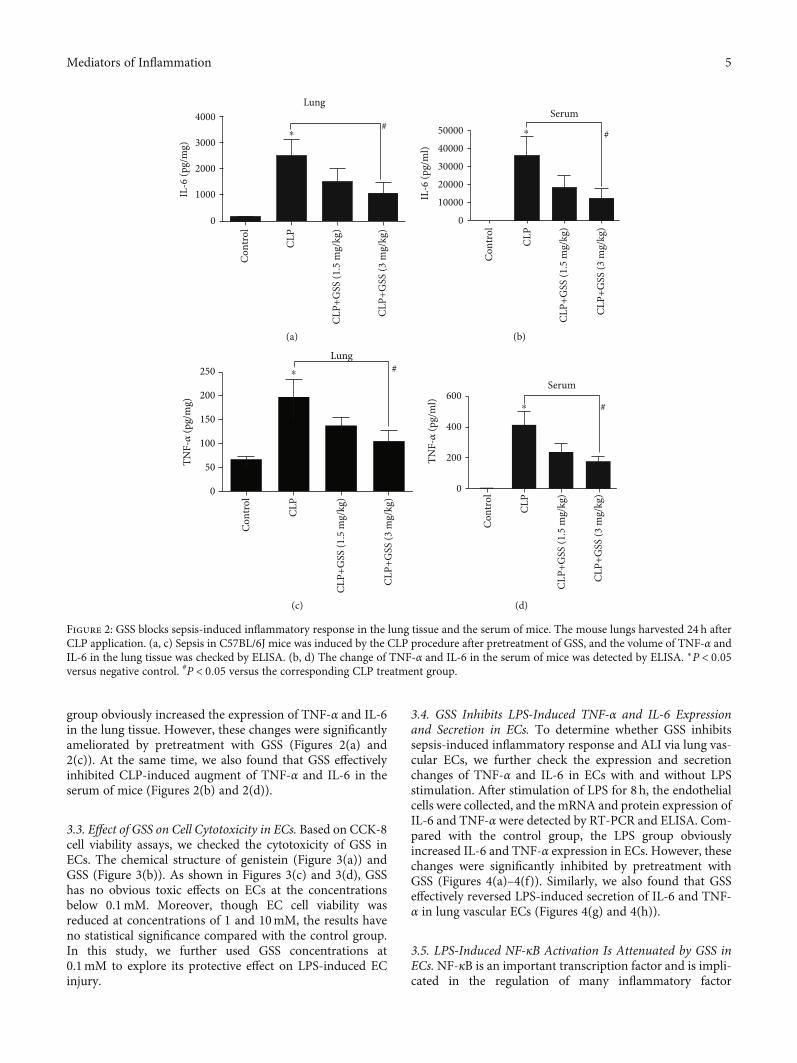
group obviously increased the expression of TNF-α and IL-6in the lung tissue. However, these changes were significantlyameliorated by pretreatment with GSS (Figures 2(a) and2(c)). At the same time, we also found that GSS effectivelyinhibited CLP-induced augment of TNF-α and IL-6 in theserum of mice (Figures 2(b) and 2(d)).
3.3. Effect of GSS on Cell Cytotoxicity in ECs. Based on CCK-8cell viability assays, we checked the cytotoxicity of GSS inECs. The chemical structure of genistein (Figure 3(a)) andGSS (Figure 3(b)). As shown in Figures 3(c) and 3(d), GSShas no obvious toxic effects on ECs at the concentrationsbelow 0.1mM. Moreover, though EC cell viability wasreduced at concentrations of 1 and 10mM, the results haveno statistical significance compared with the control group.In this study, we further used GSS concentrations at0.1mM to explore its protective effect on LPS-induced ECinjury.
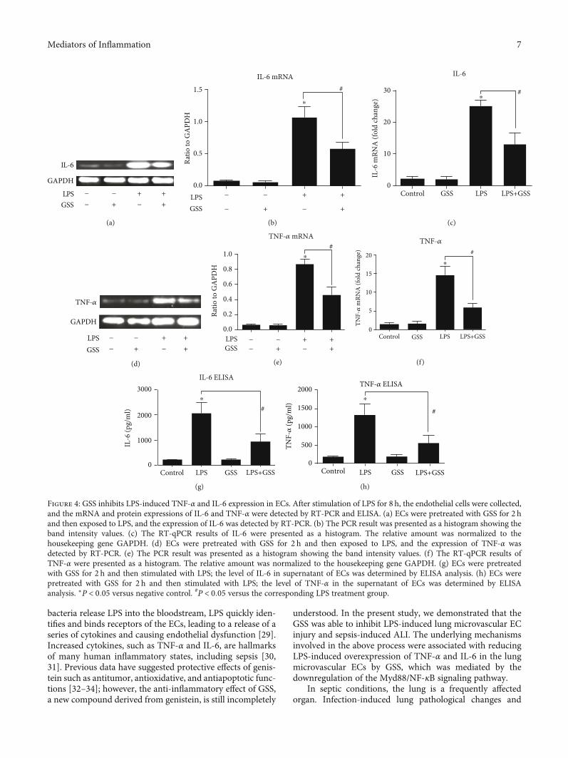
3.4. GSS Inhibits LPS-Induced TNF-α and IL-6 Expressionand Secretion in ECs. To determine whether GSS inhibitssepsis-induced inflammatory response and ALI via lung vas-cular ECs, we further check the expression and secretionchanges of TNF-α and IL-6 in ECs with and without LPSstimulation. After stimulation of LPS for 8 h, the endothelialcells were collected, and the mRNA and protein expression ofIL-6 and TNF-α were detected by RT-PCR and ELISA. Com-pared with the control group, the LPS group obviouslyincreased IL-6 and TNF-α expression in ECs. However, thesechanges were significantly inhibited by pretreatment withGSS (Figures 4(a)–4(f)). Similarly, we also found that GSSeffectively reversed LPS-induced secretion of IL-6 and TNF-α in lung vascular ECs (Figures 4(g) and 4(h)).
3.5. LPS-Induced NF-κB Activation Is Attenuated by GSS inECs. NF-κB is an important transcription factor and is impli-cated in the regulation of many inflammatory factor
Lung
#4000
3000
2000
1000
0
IL-6
(pg/
mg)
Cont
rol
CLP
CLP+
GSS
(1.5
mg/
kg)
CLP+
GSS
(3 m
g/kg
)
⁎
(a)
Serum
#50000
40000
30000
20000
10000
0
IL-6
(pg/
ml)
⁎
Cont
rol
CLP
CLP+
GSS
(1.5
mg/
kg)
CLP+
GSS
(3 m
g/kg
)
(b)
TNF-𝛼
(pg/
mg)
Lung#250
200
150
100
50
0
⁎
Cont
rol
CLP
CLP+
GSS
(1.5
mg/
kg)
CLP+
GSS
(3 m
g/kg
)
(c)
Serum
#600
400TN
F-𝛼
(pg/
ml)
200
0
⁎
Cont
rol
CLP
CLP+
GSS
(1.5
mg/
kg)
CLP+
GSS
(3 m
g/kg
)(d)
Figure 2: GSS blocks sepsis-induced inflammatory response in the lung tissue and the serum of mice. The mouse lungs harvested 24 h afterCLP application. (a, c) Sepsis in C57BL/6J mice was induced by the CLP procedure after pretreatment of GSS, and the volume of TNF-α andIL-6 in the lung tissue was checked by ELISA. (b, d) The change of TNF-α and IL-6 in the serum of mice was detected by ELISA. ∗P < 0:05versus negative control. #P < 0:05 versus the corresponding CLP treatment group.
5Mediators of Inflammation

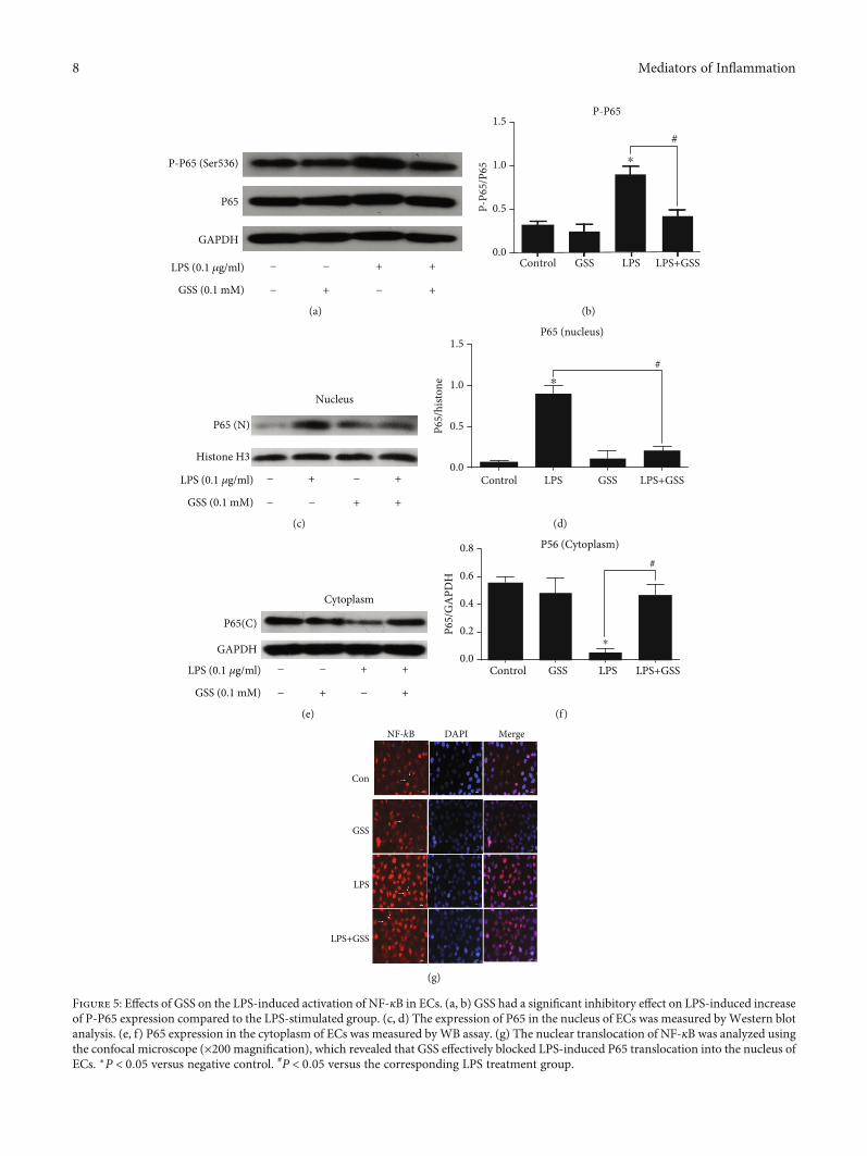
liberation, such as TNF-α and IL-6 [23]. In order to examinewhether GSS inhibited LPS-induced TNF-α and IL-6increase in ECs through NF-κB signaling, we checked theexpression and activation of NF-κB in ECs. In this study,ECs were pretreated with GSS for 2 hours and then stimu-lated with LPS. Western blot analysis was conducted toexamine the effects of GSS on the LPS-induced upregulationof NF-κB in ECs. The results showed that GSS exhibited sig-nificant inhibitory effect on LPS-induced increase in NF-κBexpression (Figures 5(a) and 5(b)). NF-κB is an importanttranscription factor, and the activation of NF-κB will mediateits translocation from cytoplasm to nucleus. In order toexamine the effect of GSS on LPS-induced NF-κB activation,the nuclear and cytosolic extracts were isolated from ECs.The expression of NF-κB in the cytosolic and nuclear frac-tions of ECs after treatment of LPS with/without GSS waschecked using Western blot analysis. We found that LPSincreased nuclear translocation of NF-κB in ECs, but pre-treatment of GSS prior to LPS stimulation significantlydecrease the levels of NF-κB in the nucleus of ECs(Figures 5(c)–5(f)). We further used fluorescence confocalmicroscopy to examine the subcellular localization and fluo-rescence intensity of NF-κB. We found that NF-κB wasmainly localized in the nucleus of ECs in the LPS-stimulated group. However, preincubation with GSS effec-tively decreased LPS-induced NF-κB translocation into thenucleus (Figure 5(g)).
3.6. GSS Regulates LPS-Induced Myd88 Expression in ECs.NF-κB activation is mediated by a wide variety of signals.Our previous studies found that both GEF-H1 and Myd88are involved in the regulation of NF-κB during LPS-induced EC injury [18]. In order to investigate the underlyingmechanism of GSS on LPS-induced NF-κB activation, we
pretreated ECs with GSS and then added LPS. We performeda Western blot assay to verify whether GSS has the blockeffects on GEF-H1 andMyd88.We found that GSS effectivelyinhibited LPS-induced Myd88 expression (Figures 6(a) and6(b)). However, GSS could not effectively block LPS-induced overexpression of GEF-H1 protein (Figures 6(c)and 6(d)). The above data indicated that GSS might protectagainst LPS-induced EC apoptosis via Myd88/NF-κBsignaling.
3.7. GSS Inhibits LPS-Induced TNF-α and IL-6 Expression inECs via Myd88/NF-κB Signaling Pathway. To further clarifythe protective role of GSS on LPS-induced EC inflammatoryresponse, we pretreated ECs with GSS, Myd88 siRNA, andBAY11-7082 (the specific inhibitor of NF-κB) (10μmol/l)before the stimulation of LPS. As shown in Figures 7(a) and7(b) and Figures 8(a) and 8(b), not only GSS but also inhibi-tion of Myd88 or NF-κB significantly reversed LPS-inducedincrease of TNF-α and IL-6. The above data indicates thatGSS protects ECs against LPS-induced TNF-α and IL-6 aug-ment via inactivation of Myd88/NF-κB signaling pathway.
4. Discussion
ALI is a common complication of sepsis in which pulmonaryendothelial cell dysfunction is regarded to be the primary ini-tiator [24]. A large number of studies reported that the lungvascular endothelial cells are key modulators and orchestra-tors of ALI [25], and LPS cause lung endothelial cell dysfunc-tion and ALI [26]. Lung endothelial cell injury andendothelial barrier failure are the key event in edema forma-tion and inflammation in ALI [27].
ECs play a vital role in the progress of sepsis as first-lineresponders against invading pathogens [28]. When infectious
OHOH O
OHO
(a)
OH
SO3Na
OH O
OHO
(b)
OD
val
ue
0
1.0
0.8
0.6
0.4
0.2
0.001 0.01 0.1 1 100.0
Concentration of GSS (mM)
(c)Ce
ll vi
abili
ty (%
)
1.5
1.0
0.5
0.00 0.001 0.01 0.1 1 10
Concentration of GSS (mM)
(d)
Figure 3: Chemical structure of GSS and its effect on EC viability. (a) The chemical structure of genistein. (b) The chemical structure ofgenistein-3′-sodium sulfonate. To observe the effect of GSS on the viability of ECs, cells were stimulated with 0, 0.001, 0.01, 0.1, 1, and10mM GSS for 24 h, respectively. The OD value (c) and EC viability (d) were measured by CCK-8 assay.
6 Mediators of Inflammation
bacteria release LPS into the bloodstream, LPS quickly iden-tifies and binds receptors of the ECs, leading to a release of aseries of cytokines and causing endothelial dysfunction [29].Increased cytokines, such as TNF-α and IL-6, are hallmarksof many human inflammatory states, including sepsis [30,31]. Previous data have suggested protective effects of genis-tein such as antitumor, antioxidative, and antiapoptotic func-tions [32–34]; however, the anti-inflammatory effect of GSS,a new compound derived from genistein, is still incompletely
understood. In the present study, we demonstrated that theGSS was able to inhibit LPS-induced lung microvascular ECinjury and sepsis-induced ALI. The underlying mechanismsinvolved in the above process were associated with reducingLPS-induced overexpression of TNF-α and IL-6 in the lungmicrovascular ECs by GSS, which was mediated by thedownregulation of the Myd88/NF-κB signaling pathway.
In septic conditions, the lung is a frequently affectedorgan. Infection-induced lung pathological changes and
IL-6
GAPDH
LPSGSS
−
−
−
+
+
−
+
+
(a)
LPS0.0
0.5
1.0
1.5
GSS
Ratio
to G
APD
H
#
−
−
−
+
+
−
+
+
⁎
IL-6 mRNA
(b)
Control GSS LPS LPS+GSS
#30
20
IL-6
mRN
A (f
old
chan
ge)
10
0
⁎
IL-6
(c)
TNF-𝛼
GAPDH
LPSGSS
−
−
−
+
+
−
+
+
(d)
Ratio
to G
APD
H
#1.0
0.8
0.6
0.4
0.2
0.0
TNF-𝛼 mRNA
LPSGSS
−
−
−
+
+
−
+
+
⁎
(e)
Control GSS LPS LPS+GSS
#20
15
10
5
0
TNF-𝛼
mRN
A (f
old
chan
ge)
⁎
TNF-𝛼
(f)
Control LPS GSS LPS+GSS
#
3000
2000
IL-6
(pg/
ml)
1000
0
IL-6 ELISA
⁎
(g)
Control LPS GSS LPS+GSS
#
2000
1500
1000
500
0
TNF-𝛼
(pg/
ml)
⁎
TNF-𝛼 ELISA
(h)
Figure 4: GSS inhibits LPS-induced TNF-α and IL-6 expression in ECs. After stimulation of LPS for 8 h, the endothelial cells were collected,and the mRNA and protein expressions of IL-6 and TNF-α were detected by RT-PCR and ELISA. (a) ECs were pretreated with GSS for 2 hand then exposed to LPS, and the expression of IL-6 was detected by RT-PCR. (b) The PCR result was presented as a histogram showing theband intensity values. (c) The RT-qPCR results of IL-6 were presented as a histogram. The relative amount was normalized to thehousekeeping gene GAPDH. (d) ECs were pretreated with GSS for 2 h and then exposed to LPS, and the expression of TNF-α wasdetected by RT-PCR. (e) The PCR result was presented as a histogram showing the band intensity values. (f) The RT-qPCR results ofTNF-α were presented as a histogram. The relative amount was normalized to the housekeeping gene GAPDH. (g) ECs were pretreatedwith GSS for 2 h and then stimulated with LPS; the level of IL-6 in supernatant of ECs was determined by ELISA analysis. (h) ECs werepretreated with GSS for 2 h and then stimulated with LPS; the level of TNF-α in the supernatant of ECs was determined by ELISAanalysis. ∗P < 0:05 versus negative control. #P < 0:05 versus the corresponding LPS treatment group.
7Mediators of Inflammation
P65
GAPDH
P-P65 (Ser536)
LPS (0.1 𝜇g/ml)
GSS (0.1 mM)
−
−
−
+
+
−
+
+
(a)
Control GSS LPS LPS+GSS
#1.5
1.0
0.5P-P6
5/P6
5
0.0
⁎
P-P65
(b)
P65 (N)
Histone H3
Nucleus
LPS (0.1 𝜇g/ml)
GSS (0.1 mM)
−
−
−
+
+
−
+
+
(c)
Control LPS GSS LPS+GSS
P65/
histo
ne
P65 (nucleus)
#
1.5
1.0
0.5
0.0
⁎
(d)
GAPDH
P65(C)
Cytoplasm
LPS (0.1 𝜇g/ml)
GSS (0.1 mM)
−
−
−
+
+
−
+
+
(e)
P65/
GA
PDH
Control GSS LPS LPS+GSS
#0.8
0.6
0.4
0.2
0.0
P56 (Cytoplasm)
⁎
(f)
LPS+GSS
LPS
GSS
Con
DAPI MergeNF-kB
(g)
Figure 5: Effects of GSS on the LPS-induced activation of NF-κB in ECs. (a, b) GSS had a significant inhibitory effect on LPS-induced increaseof P-P65 expression compared to the LPS-stimulated group. (c, d) The expression of P65 in the nucleus of ECs was measured byWestern blotanalysis. (e, f) P65 expression in the cytoplasm of ECs was measured byWB assay. (g) The nuclear translocation of NF-κB was analyzed usingthe confocal microscope (×200 magnification), which revealed that GSS effectively blocked LPS-induced P65 translocation into the nucleus ofECs. ∗P < 0:05 versus negative control. #P < 0:05 versus the corresponding LPS treatment group.
8 Mediators of Inflammation

Myd88
GAPDH
LPSGSS
−
−
−
+
+
−
+
+
(a)
Control GSS LPS LPS+GSS
Ratio
to G
APD
H
#
Myd881.5
1.0
0.5
0.0
⁎
(b)
GEF-H1
GAPDH
LPSGSS
−
−
−
+
+
−
+
+
(c)
Control GSS LPS LPS+GSS
Ratio
to G
APD
H
NS1.5
1.0
0.5
0.0
⁎
GEF-H1
(d)
Figure 6: GSS block LPS-induced Myd88 expression in ECs. (a, b) ECs were incubated with GSS for 2 hours before LPS stimulation, and theexpression of Myd88 was determined by Western blot. (c, d) ECs were pretreated with GSS for 2 hours followed by LPS stimulation, and theinhibitory effect on GEF-H1 was determined by Western blot. ∗P < 0:05 versus negative control. #P < 0:05 versus the corresponding LPStreatment group.
GAPDHTNF-𝛼
LPS
GSS
BAY11-7082
Con siRNA
Myd88 siRNA
Ratio
to G
APD
H
#
##
$
1.5
1.0
0.5
0.0
⁎
⁎
− − − − − + + + + + + +
− − − − + − − + − − + +
− − − + − − − − + − + −
− − + − − + − − − − − −
− + − − − − − − − + − +
mRNA
# #
(a)
IL-6
mRN
A (f
old
chan
ge)
####
#
$30
20
10
0
IL-6 mRNA⁎
⁎
LPSGSS
BAY11-7082Con siRNA
Myd88 siRNA
− − − − − + + + + + + +
− − − − + − − + − − + +
− − − + − − − − + − + −
− − + − − + − − − − − −
− + − − − − − − − + − +
(b)
Figure 7: GSS inhibit LPS-induced IL-6 expression in ECs via Myd88/NF-κB signaling pathway. (a) After transfection withMyd88 siRNA for48 hours, ECs were treated with GSS for another 2 hours prior to the stimulation of LPS; the expression of IL-6 was determined by RT-PCR.Similarly, ECs were pretreated with BAY11-7082 (10 μmol/l) and GSS for 2 hours and then incubated with LPS; the IL-6 expression waschecked by RT-PCR. (b) The RT-qPCR results of IL-6 was presented as a histogram. The relative amount was normalized to thehousekeeping gene GAPDH. ∗P < 0:05 versus negative control. #P < 0:05 versus the corresponding LPS treatment group. $P < 0:05 versusthe corresponding GSS treatment group.
9Mediators of Inflammation

subsequent ALI are the major cause of death in septicpatients [7, 35, 36]. Multiple previous studies have revealedthe mechanisms involved in LPS-induced ALI [20, 37, 38];however, synthetic chemical inhibitors of pathogenesis hadmore side effects, limiting their widespread clinical use. Thenatural flavones that occurred in high concentrations infruits gradually attracted people’s attention. Some previousstudies have focused on the effects of genistein on braininjury and suggested that genistein protected the brainagainst inflammation. For example, Mirahmadi et al.reported that genistein alleviated neural inflammation andcognitive dysfunctions [39]. Jeong et al. demonstrated thatgenistein inhibited activation of microglial cells and releaseof inflammatory cytokines [19]. Other scholars recentlyrevealed that GSS could protect cortical neurons from focalcerebral ischemia-induced apoptosis of neurocytes [5].Therefore, it was of interest to understand the effects ofGSS on LPS-induced ALI. The lesions in LPS-induced ALIprimarily include pulmonary edema and infiltration of mul-tiple inflammatory cells [37]. In the present study, first, weevaluated the role of GSS in change of pulmonary histopa-thology, oxygen index, and ratios of wet/dry. Our resultsshow that GSS can effectively reverse the LPS-induceddecrease of the oxygen index, increase of wet/dry ratios,and lung pathological changes. These results indicated thatGSS plays a substantial protective role in LPS-induced lunginjury and mouse mortality. To further characterize the effectof GSS on LPS-induced lung overinflammation, we analyzedthe expression of TNF-α and IL-6 in pulmonary tissue and
serum of mice by ELISA. We found that GSS significantlyinhibited LPS-induced overexpression of TNF-α and IL-6.Together with previous reports [5], these results suggest thatGSS not only plays a protective role in the brain injury butalso effectively inhibits LPS-induced lung injury via suppres-sion of inflammation.
To elucidate the mechanism by which GSS exerts protec-tive effect on lung microvascular ECs to provide cell-basedevidence for the therapeutic effect of GSS in LPS-inducedALI, we further tested the role of GSS on the expressionand release of TNF-α and IL-6 in ECs in vitro. Consistentwith our above findings in vivo, we also observed that GSSsignificantly inhibited the expression of TNF-α and IL-6 inECs. Analogous to previous reports [23], these data providefurther evidence that the LPS-induced EC injury and releaseof TNF-α and IL-6 are the leading reason of LPS-inducedALI.
NF-κB is a pleiotropic transcription factor that playsan important role in activation of proinflammatory genesand is believed to be a promising target for the treatmentof inflammation [40]. In general, the inactive form of NF-κB is present in the cytoplasm. When it is activated byphosphorylation, NF-κB translocates into the nucleus,where active NF-κB binds to specific DNA sequences,thereby increasing the expression of many downstreamgenes, including TNF-α and IL-6 [41]. In this study, GSSexhibited statistically significant inhibition of the activationof NF-κB and attenuated LPS-induced NF-κB transloca-tion from the cytoplasm to the nucleus in ECs. Our data
Ratio
to G
APD
H
#
$
1.5
1.0
0.5
0.0
TNF-𝛼 mRNA
#
##
#
⁎⁎
GAPDH
TNF-𝛼
LPS
GSS
BAY11-7082
Con siRNA
Myd88 siRNA
− − − − − + + + + + + +
− − − − + − − + − − + +
− − − + − − − − + − + −
− − + − − + − − − − − −
− + − − − − − − − + − +
(a)
####
#
$
20
10
15
0
5
IL-𝛼
mRN
A (f
old
chan
ge)
TNF-𝛼 mRNA
⁎⁎
LPSGSS
BAY11-7082Con siRNA
Myd88 siRNA
− − − − − + + + + + + +
− − − − + − − + − − + +
− − − + − − − − + − + −
− − + − − + − − − − − −
− + − − − − − − − + − +
(b)
Figure 8: GSS inhibit LPS-induced TNF-α expression in ECs via Myd88/NF-κB signaling pathway. (a) After transfection with Myd88 siRNAfor 48 hours, ECs were treated with GSS for another 2 hours prior to the stimulation of LPS; the expression of TNF-α was determined by RT-PCR. Similarly, ECs were pretreated with BAY11-7082 (10 μmol/l) and GSS for 2 hours and then incubated with LPS; the TNF-α expressionwas checked by RT-PCR. (b) The RT-qPCR results of TNF-α were presented as a histogram. The relative amount was normalized to thehousekeeping gene GAPDH. ∗P < 0:05 versus negative control. #P < 0:05 versus the corresponding LPS treatment group. $P < 0:05 versusthe corresponding GSS treatment group.
10 Mediators of Inflammation

suggested that NF-κB signaling might be implicated inGSS-induced inhibition of TNF-α and IL-6 in LPS-stimulated ECs.
It is well known that there are many upstream proteinsregulating the activity of NF-κB [42]. Among these signalingpathways, Myd88-dependent and GEF-H1-dependentmanner-mediated regulation of NF-κB is the main pathwaysimplicated in LPS-induced endothelium injury [18]. To gainfurther insight into the protective role of the GSS on LPS-induced overexpression of TNF-α and IL-6 and to under-stand the intrinsic molecular mechanism, we assessed theexpression and activity changes of Myd88 and GEF-H1 afterpretreatment with GSS in LPS-incubated ECs. In the presentstudy, we found that GSS protected against LPS-inducedoverexpression of proinflammatory mediators in endothelialcells via Myd88 signaling pathway. Furthermore, combinedapplication of GSS and Myd88 siRNA and also combinedapplication of GSS and NF-κB inhibitor produce anincreased inhibitory effect on LPS-induced expression ofTNF-α and IL-6 compared with GSS alone. These resultsindicate that GSS partly block proinflammation cytokine-associated EC injury through the Myd88/NF-κB/TNF-α/IL-6 signaling pathway in vitro.
As shown in Figure 9, we explored for the first time therole of GSS on the anti-inflammatory properties of lungmicrovascular endothelial cells in this study. We also providenew evidence that the molecular mechanism of the protectiveeffect of GSS on the production of LPS-induced TNF-α andIL-6 is mediated in a Myd88/NF-κB-dependent manner.However, accompanied by the injury of lung epithelial cellsand vascular endothelial cells, sepsis-induced acute lunginjury began to develop [43]. In addition to LPS-inducedoverexpression of inflammatory mediators in endothelialcell, LPS-induced inflammation activation in epithelial cellsalso plays an important role in the process of sepsis-induced lung injury [44]. Hence, GSS might also block LPS-induced release of inflammatory mediators in lung epithelialcells, which need to be studied further.
5. Conclusion
In summary, the present study indicated that GSS has theinhibitory effect on LPS-induced acute lung injury and pul-monary vascular EC inflammation response. GSS signifi-cantly attenuated lung histopathological changes byreducing lung microvascular permeability and inflammatoryfactor release. We further revealed that the Myd88/NF-κB/TNF-α/IL-6 signaling pathway was the important targetof GSS. All above results suggest that GSS may be a noveltherapeutic agent for the prevention of EC inflammation-associated lung injury during septic condition.
Data Availability
The data used to support the findings of this study areincluded within the article at the last part of this manuscript.
Conflicts of Interest
The authors declare that they have no conflicts of interest.
Authors’ Contributions
Lei Yi, Zengding Zhou, and Yijuan Zheng contributedequally to this work and therefore are all first authors.
Acknowledgments
This study was supported by the National Natural ScienceFoundation of China (nos. 81772078, 81772077, and81971832) and by a grant from Shanghai Jiao Tong Univer-sity (No. YG2015MS58) and by the Natural Science Founda-tion of Shanghai (19ZR1432100).
References
[1] T. Akiyama, J. Ishida, S. Nakagawa et al., “Genistein, a spe-cific inhibitor of tyrosine-specific protein kinases,” The
Acute lung injury
Vascular endothelial cell
LPS
Myd88
TLR4
NF-kB
GSS
IL-6, TNF-𝛼
NucleusCytoplasm
Micro-vascular
TNF-𝛼IL-6
Figure 9: Schematic figure of possible signaling mechanisms of GSS in the inhibition of LPS-induced lung vascular endothelial cellinflammatory response and subsequent ALI in mice.
11Mediators of Inflammation

Journal of Biological Chemistry, vol. 262, no. 12, pp. 5592–5595, 1987.
[2] O. Benavente-Garcia and J. Castillo, “Update on uses andproperties of citrus flavonoids: new findings in anticancer, car-diovascular, and anti-inflammatory activity,” Journal of Agri-cultural and Food Chemistry, vol. 56, no. 15, pp. 6185–6205,2008.
[3] E. M. Varoni, G. Lodi, A. Sardella, A. Carrassi, and M. Iriti,“Plant polyphenols and oral health: old phytochemicals fornew fields,” Current Medicinal Chemistry, vol. 19, no. 11,pp. 1706–1720, 2012.
[4] P. Del Gaudio, F. Sansone, T. Mencherini, F. De Cicco,P. Russo, and R. P. Aquino, “Nanospray drying as a novel toolto improve technological properties of soy isoflavone extracts,”Planta Medica, vol. 83, no. 5, pp. 426–433, 2017.
[5] L. Li, J. Xue, R. Liu et al., “Neuroprotective effects of genistein-3′-sodium sulfonate on focal cerebral ischemia in rats,”Neuro-science Letters, vol. 646, pp. 43–48, 2017.
[6] K. S. Houschyar, M. P. Chelliah, S. Rein et al., “Role of Wntsignaling during inflammation and sepsis: a review of the liter-ature,” The International Journal of Artificial Organs, vol. 41,no. 5, pp. 247–253, 2018.
[7] L. B. Ware and M. A. Matthay, “The acute respiratory distresssyndrome,” The New England Journal of Medicine, vol. 342,no. 18, pp. 1334–1349, 2000.
[8] W. Xu, X. Wang, J. Jin et al., “Inhibition of GGPPS1 attenuatedLPS-induced acute lung injury and was associated with NLRP3inflammasome suppression,” American Journal of Physiology-Lung Cellular and Molecular Physiology, vol. 316, no. 3,pp. L567–L577, 2019.
[9] S. M. Opal and T. van der Poll, “Endothelial barrier dysfunc-tion in septic shock,” Journal of Internal Medicine, vol. 277,no. 3, pp. 277–293, 2015.
[10] S. Mu, Y. Liu, J. Jiang et al., “Unfractionated heparin amelio-rates pulmonary microvascular endothelial barrier dysfunc-tion via microtubule stabilization in acute lung injury,”Respiratory Research, vol. 19, no. 1, p. 220, 2018.
[11] J. Lou, Y. Hu, M. D. Wu et al., “Endothelial cell-specific antic-oagulation reduces inflammation in a mouse model of acutelung injury,” Acta Pharmacologica Sinica, vol. 40, no. 6, article175, pp. 769–780, 2018.
[12] G. Choi, J. J. Hofstra, J. J. Roelofs et al., “Antithrombin inhibitsbronchoalveolar activation of coagulation and limits lunginjury during Streptococcus pneumoniae pneumonia in rats,”Critical Care Medicine, vol. 36, no. 1, pp. 204–210, 2008.
[13] K. Murakami, K. Okajima, M. Uchiba et al., “Activated proteinC attenuates endotoxin-induced pulmonary vascular injury byinhibiting activated leukocytes in rats,” Blood, vol. 87, no. 2,pp. 642–647, 1996.
[14] M. Wendel, R. Paul, and A. R. Heller, “Lipoproteins ininflammation and sepsis. II. Clinical aspects,” Intensive CareMedicine, vol. 33, no. 1, article 433, pp. 25–35, 2007.
[15] K. Reinhart, O. Bayer, F. Brunkhorst, and M. Meisner,“Markers of endothelial damage in organ dysfunction andsepsis,” Critical Care Medicine, vol. 30, no. 5, pp. S302–S312,2002.
[16] L. Jimenez-Garcia, P. G. Traves, R. Lopez-Fontal et al., “8,9-Dehydrohispanolone-15,16-lactol diterpene prevents LPS-triggered inflammatory responses by inhibiting endothelialactivation,” Biochemical Journal, vol. 473, no. 14, pp. 2061–2071, 2016.
[17] K. Yasumoto, S. Okamoto, N. Mukaida, S. Murakami, M. Mai,and K. Matsushima, “Tumor necrosis factor alpha and inter-feron gamma synergistically induce interleukin 8 productionin a human gastric cancer cell line through acting concurrentlyon AP-1 and NF-kB-like binding sites of the interleukin 8gene,” The Journal of Biological Chemistry, vol. 267, no. 31,pp. 22506–22511, 1992.
[18] F. Guo, J. Tang, Z. Zhou et al., “GEF-H1-RhoA signaling path-way mediates LPS-induced NF-κB transactivation and IL-8synthesis in endothelial cells,” Molecular Immunology,vol. 50, no. 1-2, pp. 98–107, 2012.
[19] J. W. Jeong, H. H. Lee, M. H. Han, G. Y. Kim, W. J.Kim, and Y. H. Choi, “Anti-inflammatory effects of genis-tein via suppression of the toll-like receptor 4-mediatedsignaling pathway in lipopolysaccharide-stimulated BV2microglia,” Chemico-Biological Interactions, vol. 212,pp. 30–39, 2014.
[20] L. Yi, X. Huang, F. Guo et al., “Lipopolysaccharide induceshuman pulmonary micro-vascular endothelial apoptosis viathe YAP signaling pathway,” Frontiers in Cellular and Infec-tion Microbiology, vol. 6, p. 133, 2016.
[21] J. Moitra, S. Sammani, and J. G. Garcia, “Re-evaluation ofEvans blue dye as a marker of albumin clearance in murinemodels of acute lung injury,” Translational Research,vol. 150, no. 4, pp. 253–265, 2007.
[22] K. C. Lan, S. C. Chao, H. Y. Wu et al., “Salidroside amelio-rates sepsis-induced acute lung injury and mortality viadownregulating NF-κB and HMGB1 pathways through theupregulation of SIRT1,” Scientific Reports, vol. 7, no. 1,p. 12026, 2017.
[23] F. Guo, Y. Xing, Z. Zhou et al., “Guanine-nucleotide exchangefactor H1 mediates lipopolysaccharide-induced interleukin 6and tumor necrosis factor α expression in endothelial cellsvia activation of nuclear factor κB,” Shock, vol. 37, no. 5,pp. 531–538, 2012.
[24] X. Li, Z. Zheng, X. Li, and X. Ma, “Unfractionated heparininhibits lipopolysaccharide-induced inflammatory responsethrough blocking p38 MAPK and NF-κB activation onendothelial cell,” Cytokine, vol. 60, no. 1, pp. 114–121,2012.
[25] L. B. Ware, “Pathophysiology of acute lung injury and theacute respiratory distress syndrome,” Seminars in Respira-tory and Critical Care Medicine, vol. 27, no. 4, pp. 337–349, 2006.
[26] A. M. Bianchi, M. M. Reboredo, L. M. Lucinda et al., “Theeffects of prone position ventilation on experimental mildacute lung injury induced by intraperitoneal lipopolysaccha-ride injection in rats,” Lung, vol. 194, no. 2, pp. 193–199,2016.
[27] J. Yin, L. Lv, P. Zhai et al., “Connexin 40 regulates lung endo-thelial permeability in acute lung injury via the ROCK1-MYPT1- MLC20 pathway,” American Journal of Physiology-Lung Cellular and Molecular Physiology, vol. 316, no. 1,pp. L35–L44, 2018.
[28] W. C. Aird, “The role of the endothelium in severe sepsis andmultiple organ dysfunction syndrome,” Blood, vol. 101, no. 10,pp. 3765–3777, 2003.
[29] M. Schouten, W. J. Wiersinga, M. Levi, and T. van der Poll,“Inflammation, endothelium, and coagulation in sepsis,”Journal of Leukocyte Biology, vol. 83, no. 3, pp. 536–545,2008.
12 Mediators of Inflammation

[30] S. Zeuke, A. J. Ulmer, S. Kusumoto, H. A. Katus, and H. Heine,“TLR4-mediated inflammatory activation of human coronaryartery endothelial cells by LPS,” Cardiovascular Research,vol. 56, no. 1, pp. 126–134, 2002.
[31] M. Bhatia, M. He, H. Zhang, and S. Moochhala, “Sepsis as amodel of SIRS,” Frontiers in Bioscience, vol. 14, pp. 4703–4711, 2009.
[32] L. Zhang, M. Gao, T. Zhang, T. Chong, and Z. Wang, “Protec-tive effects of genistein against mono-(2-ethylhexyl) phthalate-induced oxidative damage in prepubertal Sertoli cells,”vol. 2017, Article ID 2032697, 12 pages, 2017.
[33] S. Saha, P. Sadhukhan, S. Mahalanobish, S. Dutta, and P. C.Sil, “Ameliorative role of genistein against age-dependentchronic arsenic toxicity in murine brains via the regulationof oxidative stress and inflammatory signaling cascades,”The Journal of Nutritional Biochemistry, vol. 55, pp. 26–40, 2018.
[34] M. Sanaei, F. Kavoosi, A. Valiani, and M. A. Ghobadifar,“Effect of genistein on apoptosis and proliferation of hepato-cellular carcinoma Hepa1-6 cell line,” International Journalof Preventive Medicine, vol. 9, no. 1, p. 12, 2018.
[35] D. C. Angus, W. T. Linde-Zwirble, J. Lidicker, G. Clermont,J. Carcillo, and M. R. Pinsky, “Epidemiology of severe sepsisin the United States: analysis of incidence, outcome, and asso-ciated costs of care,” Critical Care Medicine, vol. 29, no. 7,pp. 1303–1310, 2001.
[36] J. L. Vincent, Y. Sakr, and V. M. Ranieri, “Epidemiology andoutcome of acute respiratory failure in intensive care unitpatients,” Critical Care Medicine, vol. 31, no. 4, pp. S296–S299, 2003.
[37] L. Yi, X. Huang, F. Guo, Z. Zhou, Y. Dou, and J. Huan, “Yes-associated protein (YAP) signaling regulateslipopolysaccharide-induced tissue factor expression in humanendothelial cells,” Surgery, vol. 159, no. 5, pp. 1436–1448, 2016.
[38] L. Yi, X. Huang, F. Guo, Z. Zhou, M. Chang, and J. Huan,“GSK-3beta-dependent activation of GEF-H1/ROCK signal-ing promotes LPS-induced lung vascular endothelial barrierdysfunction and acute lung injury,” Frontiers in Cellular andInfection Microbiology, vol. 7, p. 357, 2017.
[39] S. M. Mirahmadi, A. Shahmohammadi, A. M. Rousta et al.,“Soy isoflavone genistein attenuates lipopolysaccharide-induced cognitive impairments in the rat via exerting anti-oxidative and anti-inflammatory effects,” Cytokine, vol. 104,pp. 151–159, 2018.
[40] P. J. Barnes and M. Karin, “Nuclear factor-kappaB: a pivotaltranscription factor in chronic inflammatory diseases,” TheNew England Journal of Medicine, vol. 336, no. 15, pp. 1066–1071, 1997.
[41] R. Han, F. Zhang, C. Wan, L. Liu, Q. Zhong, and W. Ding,“Effect of perfluorooctane sulphonate-induced Kupffer cellactivation on hepatocyte proliferation through the NF-κB/TNF-α/IL-6-dependent pathway,” Chemosphere, vol. 200,pp. 283–294, 2018.
[42] Z. W. Cui, Z. X. Xie, B. F. Wang et al., “Carvacrol protects neu-roblastoma SH-SY5Y cells against Fe2+-induced apoptosis bysuppressing activation of MAPK/JNK-NF-κB signaling path-way,” Acta Pharmacologica Sinica, vol. 36, no. 12, pp. 1426–1436, 2015.
[43] C. Tian, P. Zhang, J. Yang et al., “The protective effect of theflavonoid fraction of Abutilon theophrasti Medic. leaves onLPS-induced acute lung injury in mice via the NF- κB andMAPK signalling pathways,” Biomedicine & Pharmacother-apy, vol. 109, pp. 1024–1031, 2019.
[44] J. W. Lee, W. Chun, O. K. Kwon et al., “3,4,5-Trihydroxycin-namic acid attenuates lipopolysaccharide (LPS)-induced acutelung injury via downregulating inflammatory molecules andupregulating HO-1/AMPK activation,” International Immu-nopharmacology, vol. 64, pp. 123–130, 2018.
13Mediators of Inflammation

Stem Cells International
Hindawiwww.hindawi.com Volume 2018
Hindawiwww.hindawi.com Volume 2018
MEDIATORSINFLAMMATION
of
EndocrinologyInternational Journal of
Hindawiwww.hindawi.com Volume 2018
Hindawiwww.hindawi.com Volume 2018
Disease Markers
Hindawiwww.hindawi.com Volume 2018
BioMed Research International
OncologyJournal of
Hindawiwww.hindawi.com Volume 2013
Hindawiwww.hindawi.com Volume 2018
Oxidative Medicine and Cellular Longevity
Hindawiwww.hindawi.com Volume 2018
PPAR Research
Hindawi Publishing Corporation http://www.hindawi.com Volume 2013Hindawiwww.hindawi.com
The Scientific World Journal
Volume 2018
Immunology ResearchHindawiwww.hindawi.com Volume 2018
Journal of
ObesityJournal of
Hindawiwww.hindawi.com Volume 2018
Hindawiwww.hindawi.com Volume 2018
Computational and Mathematical Methods in Medicine
Hindawiwww.hindawi.com Volume 2018
Behavioural Neurology
OphthalmologyJournal of
Hindawiwww.hindawi.com Volume 2018
Diabetes ResearchJournal of
Hindawiwww.hindawi.com Volume 2018
Hindawiwww.hindawi.com Volume 2018
Research and TreatmentAIDS
Hindawiwww.hindawi.com Volume 2018
Gastroenterology Research and Practice
Hindawiwww.hindawi.com Volume 2018
Parkinson’s Disease
Evidence-Based Complementary andAlternative Medicine
Volume 2018Hindawiwww.hindawi.com
Submit your manuscripts atwww.hindawi.com