![Gamma Radiation-Induced Disruption of Cellular Junctions ...downloads.hindawi.com/journals/omcl/2019/1486232.pdf · junction protein [13]. Connexins compose the gap junction channels](https://static.fdocument.org/doc/165x107/5f06b4cd7e708231d4195458/gamma-radiation-induced-disruption-of-cellular-junctions-junction-protein-13.jpg)
SIRT7 Regulates Lipopolysaccharide-Induced Inflammatory...
16
Research Article SIRT7 Regulates Lipopolysaccharide-Induced Inflammatory Injury by Suppressing the NF-κB Signaling Pathway Kun-Lin Chen , 1,2 Lian Li, 1 Cheng-Min Li , 1 Yi-Ru Wang, 1 Fang-Xiao Yang, 1 Mei-Qian Kuang, 1 and Gen-Lin Wang 1 1 College of Animal Science and Technology, Nanjing Agricultural University, Nanjing 210095, China 2 Institute of Animal Science, Jiangsu Academy of Agricultural Sciences, Nanjing 210014, China Correspondence should be addressed to Gen-Lin Wang; [email protected] Received 29 January 2019; Revised 6 May 2019; Accepted 16 May 2019; Published 11 June 2019 Academic Editor: Ilaria Peluso Copyright © 2019 Kun-Lin Chen et al. This is an open access article distributed under the Creative Commons Attribution License, which permits unrestricted use, distribution, and reproduction in any medium, provided the original work is properly cited. Mastitis has severely affected the cattle industry worldwide and has resulted in decreased dairy production and cattle reproduction. Although prevention and treatment methods have been implemented for decades, cattle mastitis is still an intractable disease. Sirtuin 7 (SIRT7) is an NAD + -dependent deacetylase that is involved in various biological processes, including ribosomal RNA synthesis and protein synthesis, DNA damage response, metabolism, and tumorigenesis. However, whether SIRT7 participates in inflammation remains unknown. Our results revealed that SIRT7 is downregulated in tissue samples from mastitic cattle. Therefore, we isolated dairy cow mammary epithelial cells (DCMECs) from breast tissues and developed an in vitro model of lipopolysaccharide- (LPS-) induced inflammation to examine SIRT7 function and its potential role in inflammation. We showed that SIRT7 was significantly downregulated in LPS-treated DCMECs. SIRT7 knockdown significantly increased the LPS- stimulated production of inflammatory mediators, like reactive oxygen and nitric oxide, and upregulated TAB1 and TLR4. In addition, SIRT7 knockdown significantly increased the phosphorylation of TAK1 and NF-κBp65 in LPS-treated DCMECs. Moreover, SIRT7 knockdown promoted the translocation of NF-κBp-p65 to the cell nucleus and then increased the secretion of inflammatory cytokines (IL-1β and IL-6). In contrast, SIRT7 overexpression had the opposite effects when compared to SIRT7 knockdown in LPS-treated DCMECs. In addition, SIRT7 overexpression attenuated LPS-induced DCMEC apoptosis. Taken together, our results indicate that SIRT7 can suppress LPS-induced inflammation and apoptosis via the NF-κB signaling pathway. Therefore, SIRT7 may be considered as a potential pharmacological target for clinical mastitis therapy. 1. Introduction Clinical mastitis (CM) is a widespread and economically important disease that affects dairy cattle by reducing milk quality and production. In addition, CM increases the risk of culling and death, and treatment is costly [1, 2]. Approxi- mately 80% of all intramammary Escherichia coli (E. coli) infections result in CM [3–5]. The major outer membrane component of E. coli is lipopolysaccharide (LPS), which acti- vates the TLR4-NF-κB signaling pathway, stimulating the release of inflammatory cytokines [6]. In turn, these cyto- kines and mediators further aggravate the pathology and inflammatory responses involved in mastitis. Mastitis is characterized by increased secretion of proin- flammatory cytokines, such as TNF-α, IL-1β, and IL-6, which have been demonstrated to direct the migration of neutrophils to the site of infection [7]. Furthermore, exposing neutrophils and macrophages to LPS rapidly induces the secretion of many mediators, such as nitric oxide (NO), pros- taglandin E2 (PGE2), and reactive oxygen species (ROS) [8]. NO is a major inflammatory mediator that is involved in var- ious physiological processes including vasodilatation and the increase in vascular permeability [9]. One study showed that proinflammatory cytokines or LPS can induce the expression of NO synthase (iNOS), which further promotes NO synthe- sis [10]. ROS act as secondary messengers and participate in cell growth, adhesion, differentiation, senescence, and apo- ptosis, as well as the modification of various signaling mole- cules [11, 12]. Excessive production and accumulation of ROS are detrimental to cells and tissues. An unbalanced Hindawi Oxidative Medicine and Cellular Longevity Volume 2019, Article ID 3187972, 15 pages https://doi.org/10.1155/2019/3187972
Transcript of SIRT7 Regulates Lipopolysaccharide-Induced Inflammatory...
Research ArticleSIRT7 Regulates Lipopolysaccharide-Induced InflammatoryInjury by Suppressing the NF-κB Signaling Pathway
Kun-Lin Chen ,1,2 Lian Li,1 Cheng-Min Li ,1 Yi-Ru Wang,1 Fang-Xiao Yang,1
Mei-Qian Kuang,1 and Gen-Lin Wang 1
1College of Animal Science and Technology, Nanjing Agricultural University, Nanjing 210095, China2Institute of Animal Science, Jiangsu Academy of Agricultural Sciences, Nanjing 210014, China
Correspondence should be addressed to Gen-Lin Wang; [email protected]
Received 29 January 2019; Revised 6 May 2019; Accepted 16 May 2019; Published 11 June 2019
Academic Editor: Ilaria Peluso
Copyright © 2019 Kun-Lin Chen et al. This is an open access article distributed under the Creative Commons Attribution License,which permits unrestricted use, distribution, and reproduction in any medium, provided the original work is properly cited.
Mastitis has severely affected the cattle industry worldwide and has resulted in decreased dairy production and cattle reproduction.Although prevention and treatment methods have been implemented for decades, cattle mastitis is still an intractable disease.Sirtuin 7 (SIRT7) is an NAD+-dependent deacetylase that is involved in various biological processes, including ribosomal RNAsynthesis and protein synthesis, DNA damage response, metabolism, and tumorigenesis. However, whether SIRT7 participatesin inflammation remains unknown. Our results revealed that SIRT7 is downregulated in tissue samples from mastitic cattle.Therefore, we isolated dairy cow mammary epithelial cells (DCMECs) from breast tissues and developed an in vitro model oflipopolysaccharide- (LPS-) induced inflammation to examine SIRT7 function and its potential role in inflammation. We showedthat SIRT7 was significantly downregulated in LPS-treated DCMECs. SIRT7 knockdown significantly increased the LPS-stimulated production of inflammatory mediators, like reactive oxygen and nitric oxide, and upregulated TAB1 and TLR4. Inaddition, SIRT7 knockdown significantly increased the phosphorylation of TAK1 and NF-κBp65 in LPS-treated DCMECs.Moreover, SIRT7 knockdown promoted the translocation of NF-κBp-p65 to the cell nucleus and then increased the secretion ofinflammatory cytokines (IL-1β and IL-6). In contrast, SIRT7 overexpression had the opposite effects when compared to SIRT7knockdown in LPS-treated DCMECs. In addition, SIRT7 overexpression attenuated LPS-induced DCMEC apoptosis. Takentogether, our results indicate that SIRT7 can suppress LPS-induced inflammation and apoptosis via the NF-κB signalingpathway. Therefore, SIRT7 may be considered as a potential pharmacological target for clinical mastitis therapy.
1. Introduction
Clinical mastitis (CM) is a widespread and economicallyimportant disease that affects dairy cattle by reducing milkquality and production. In addition, CM increases the riskof culling and death, and treatment is costly [1, 2]. Approxi-mately 80% of all intramammary Escherichia coli (E. coli)infections result in CM [3–5]. The major outer membranecomponent of E. coli is lipopolysaccharide (LPS), which acti-vates the TLR4-NF-κB signaling pathway, stimulating therelease of inflammatory cytokines [6]. In turn, these cyto-kines and mediators further aggravate the pathology andinflammatory responses involved in mastitis.
Mastitis is characterized by increased secretion of proin-flammatory cytokines, such as TNF-α, IL-1β, and IL-6,
which have been demonstrated to direct the migration ofneutrophils to the site of infection [7]. Furthermore, exposingneutrophils and macrophages to LPS rapidly induces thesecretion of many mediators, such as nitric oxide (NO), pros-taglandin E2 (PGE2), and reactive oxygen species (ROS) [8].NO is a major inflammatory mediator that is involved in var-ious physiological processes including vasodilatation and theincrease in vascular permeability [9]. One study showed thatproinflammatory cytokines or LPS can induce the expressionof NO synthase (iNOS), which further promotes NO synthe-sis [10]. ROS act as secondary messengers and participate incell growth, adhesion, differentiation, senescence, and apo-ptosis, as well as the modification of various signaling mole-cules [11, 12]. Excessive production and accumulation ofROS are detrimental to cells and tissues. An unbalanced
HindawiOxidative Medicine and Cellular LongevityVolume 2019, Article ID 3187972, 15 pageshttps://doi.org/10.1155/2019/3187972
redox state is a key step in the development of various inflam-matory diseases. Thus, inhibition of NO and ROS productionis a promising approach in the treatment of inflammatorydiseases. The transcription factor NF-κB is a pleiotropic reg-ulator of many genes and is involved in physiological andpathological processes, including immunity, inflammation,and metabolism [13]. NF-κB normally binds to inhibitor ofκBα (IκBα) and assumes an inactive state in the cytosol. Bycontrast, phosphorylation and degradation of IκBα causethe release and subsequent translocation of NF-κB to thenucleus, thereby activating the expression of downstreamtarget genes [14]. TAB1 is a TAK1-interacting protein thatpromotes TAK1 kinase activity via the autophosphorylationof key serine/threonine sites in the kinase activation loop[15]. TAB1 forms a complex with TAK1, which acts as akey component of LPS-mediated IKK-NF-κB upstream sig-naling, and is involved in regulating the immune response.
Sirtuin7 (SIRT7) is a nicotinamide adenine dinucleotideoxidized form- (NAD+-) dependent deacetylase. SIRT7 isimplicated in various processes, such as aging, DNA damagerepair, and cell signaling transduction [16, 17]. In addition,SIRT7 has been shown to function in aging-related processes.SIRT7-/- mice had shorter lifespans, showed decreased stressresistance, and developed age-dependent inflammatory car-diomyopathy, which implies that SIRT7 is involved ininflammatory processes [18]. Currently, treatment and con-trol of CM and mastitis are primarily based on the use ofantimicrobial drugs [19]. Although antibiotic treatmentshave achieved remarkable efficiency against mastitis, antibi-otic residues found in milk and meat are harmful to humanhealth [12]. In recent years, numerous nonantimicrobialdrugs and treatment strategies against CM have beenreported. Despite decades of intensive research and imple-mentation of preventive measures, mastitis remains anintractable disease. Considering the potent effects of SIRT7and its function in inflammation, we hypothesized thatSIRT7 might be involved in mastitis. Therefore, in the pres-ent study, we utilized a dairy cow mammary epithelial cell(DCMEC) model of LPS-induced mastitis to investigate thefunction of SIRT7 in the induction and progression of theinflammatory response.
2. Materials and Methods
2.1. Collection of Mammary Tissues. The protocol of the pres-ent study was approved by the Institutional Animal Care andUse Committee of Nanjing Agricultural University. We col-lected five normal and five inflammatory mammary tissuesamples from five Chinese Holstein cows in a local slaughter-
house. The samples were immediately frozen in liquid nitro-gen and stored until analysis.
2.2. Cell Culture and LPS Treatment. In vitro culture ofDCMECs was conducted as follows: Breast tissues were cutinto 1 0mm × 1 0mm × 1 0mm pieces and washed fivetimes in PBS. Next, to the samples, we added an enzyme mix-ture (1.5 g/L type I collagenase, 1.5 g/L type II collagenase,and 1.5 g/L trypsin) (Sigma-Aldrich, Cat: C0130 andC6885) and incubated samples at 37°C at 100 r/min in anoscillation incubator for 3 h. Samples were then filteredthrough a 100-mesh sieve and centrifuged at 130 × g for 5min. The supernatants were discarded, and then cells werecultured in Dulbecco’s modified Eagle’s medium (DMEM)supplemented with 10% fetal bovine serum (Gibco, Cat:10438026) and 1% antibiotic-antimycotic solution in ahumidified incubator with 5% CO2 at 37°C. After 1 h, thesupernatants were collected and subcultured in new flaskbottles in order to discard unwanted cells. DCMECs wereidentified by anti-Cytokeratin 18 antibody (Abcam, Cat:ab52459, 1 : 100). Confirmed DCMECs were treated withLPS.
2.3. Transfection. DCMECs were seeded in a six-well plateand cultured for 24 h until they reached 50%-60% conflu-ence. DCMECs were transfected with small interferingRNA (siRNA) or plasmids using Lipofectamine 2000 (Invi-trogen, Carlsbad, Cat: 11668027). Experimental siRNA oligosor nontargeting control siRNAs were transfected in 100 pmolamounts. Cells were transfected with 3 μg of pcDNA3.0-SIRT7 for SIRT7 overexpression or an empty vector (controlplasmid pcDNA3.0). Transfection efficiency was determinedvia quantitative reverse-transcription PCR (qRT-PCR) andwestern blotting. The sequences of the SIRT7 and negativecontrol siRNAs, which were synthesized by Shanghai Gene-Pharma Co. Ltd., are listed in Table 1.
2.4. Total RNA Extraction and qRT-PCR. Total RNA wasextracted with the Trizol Reagent (Invitrogen, Cat:15596026) at 48 h after siRNA transfection. Reverse tran-scription was performed using PrimeScript™ RT MasterMix (TaKaRa, Cat: RR036A). Expression levels of mRNAwere quantified via real-time PCR. Expression levels of alltarget genes were normalized to those of endogenous refer-ence gene β-actin, according to an optimized comparativeCt (2−ΔΔCt) value method, where ΔΔ = ΔCttarget − ΔCtβ−actin.Primer sequences are listed in Table 2.
Table 1: Primer sequences of siSIRT7.
Genes Forward Reverse
siSIRT7-1 5′-GCAGCCUCUAUCCCAGAUUTT-3′ 5′-AAUCUGGGAUAGAGGCUGCTT-3′siSIRT7-2 5′-GCACUCCCAAUAGGGAAUATT-3′ 5′-UAUUCCCUAUUGGGAGUGCTT-3′siSIRT7-3 5′-GCAAGUGUGAUGACGUCAUTT-3′ 5′-AUGACGUCAUCACACUUGCTT-3′Negative control 5′-UUCUCCGAACGUGUCACGUTT-3′ 5′-ACGUGACACGUUCGGAGAATT-3′
2 Oxidative Medicine and Cellular Longevity
2.5. Western Blot Analysis. To extract total proteins, cellswere incubated with lysis buffer and centrifuged at 15000 ×g for 20 min at 4°C, and the supernatant was collected.Nuclear and cytoplasmic proteins were isolated using anextraction kit (Beyotime, Cat: P0027) following the manufac-turer’s instructions. Proteins were separated via SDS-PAGEand transferred onto polyvinylidene difluoride (PVDF, LifeTechnologies, Cat: LC2002) membranes. Next, membraneswere blocked in TBST (TBS containing 0.1% Tween 20) con-taining 5% nonfat milk for 1 h, followed by incubation at 4°Covernight with primary antibodies. Protein bands were incu-bated with the secondary antibodies for 1 h at room temper-ature. Finally, the membranes were visualized with anenhanced chemiluminescence detection system (AmershamBiosciences Corp., Piscataway, NJ). Densitometry analysiswas performed using ImageJ software (National Institutesof Health, Bethesda, MD, USA). The signal intensity for eachprotein band was normalized against the α-tubulin loadingcontrol. The following commercially available antibodieswere used: anti-SIRT7 (Proteintech, Cat: 12994-1-AP,1 : 2000), anti-TAB1 (Proteintech, Cat: 14819-1-AP,1 : 1000), anti-p-TAK1 (Bioss, Cat: bs-5435R, 1 : 1000), anti-IL-1β (ABclonal Technology, Cat: A1112, 1 : 2000), anti-IL-6 (Abbexa, Cat: abx015895, 1 : 2000), anti-TLR4 (Abcam,Cat: ab22048, 1 : 1000), anti-GAPDH (Proteintech, Cat:10494-1-AP, 1 : 4000), anti-Histone-H3.1 (Beyotime, Cat:AF0009, 1 : 500), anti-NF-κBp65 (CST, Cat: 6956, 1 : 2000),and anti-phospho-NF-κBp65 (CST, Cat: 3033, 1 : 2000).
2.6. ROS and NO Staining Assay. Reactive oxygen species(ROS) and nitric oxide (NO) levels were assessed usingassay kits based on DCFH-DA (Beyotime, Cat: S0033) andDAF-FM DA (Beyotime, Cat: S0019), respectively. Briefly,DCMECs were seeded in 24-well plates and incubated with10 μM DCFH-DA and 2 μM DAF-FM DA reaction mix-tures for 30 min at 37°C. Then, the cells were quicklywashed twice (quickly) with PBS and visualized under aconfocal laser-scanning microscope (Zeiss LSM 700META). The percentages of ROS- or NO-positive cells weredetermined based on counts from five randomly selectedvisual fields.
2.7. Immunofluorescence Staining. The cells were washedthree times with PBS (37°C), and fixed with 4% paraformal-dehyde (in PBS) at room temperature for 30 min. Then, cellswere permeabilized with 0.5% Triton X-100 in PBS for 20min. After 1 h in blocking buffer (1% BSA-supplementedPBS), the cells were incubated at 4°C overnight with mouseanti-cytokeratin-18 (Abcam, Cat: ab52459, 1 : 100) or rabbitanti-NF-κBp-p65 (1 : 100). After three 5 min washes in PBS,the cells were labeled with a fluorescein isothiocyanate-(FITC-) conjugated goat anti-mouse IgG (H+L) antibody ora tetramethylrhodamine- (TRITC-) conjugated goat anti-rabbit IgG (H+L) antibody at room temperature for 1 h.The cells were stained with Hoechst 33342 for 10 min andthen mounted on glass slides. After three washes with PBS,cells were imaged under a confocal microscope (Zeiss LSM700 META).
2.8. Enzyme-Linked Immunosorbent Assay (ELISA) IL-6 andIL-1β Levels. The levels of IL-6 and IL-1β in the treated cellmedium were determined according to the instructions forthe IL-6 (Shanghai Lengton Bioscience Co. Ltd., Cat:BPE92153) and IL-1β (Shanghai Lengton Bioscience Co.Ltd., Cat: BPE92157) ELISA kits. Finally, the absorbance ofeach well was measured at 450 nm.
2.9. Flow-Cytometer Detection of Apoptosis. Cell apoptosiswas detected with an Annexin-V/PI kit (BD Biosciences,Cat: 556547). After transfection, the cells were incubatedwith Annexin-V/PI at room temperature for 25 min. Then,the apoptotic cells were quantified with a FACSCalibur flowcytometer (FCM) (BD Biosciences, Bedford, MA, USA).The data were analyzed by FlowJo software.
2.10. Statistical Analysis. All data are expressed as mean ±SEM. Differences between groups were subjected to analysisof variance (ANOVA) or the t-test. Data with a P value lessthan 0.05 were considered statistically significant. All the sta-tistical analyses were performed in GraphPad Prism 6.01software (GraphPad Software Inc., San Diego, CA).
Table 2: Primer sequences of mRNA.
Genes Forward Reverse
SIRT7 5′-GAGAGCGAGGACCTGGTGAC-3′ 5′-GATAGAGGCTGCCGTGCTGA-3′TLR4 5′-GGGTTGCTGTTCTCACACTG-3′ 5′-AGGTAGCGGAGGTTTCTGAG-3′IL-1β 5′-AGGTGGTGTCGGTCATCGT-3′ 5′-GCTCTCTGTCCTGGAGTTTGC-3′TAB1 5′-GCGATCTCGGCTCCTAGCAA-3′ 5′-GCTACTCGGGAGGGCTTAGG-3′TNF-α 5′-ACGGGCTTTACCTCATCTACTC-3′ 5′-GCTCTTGATGGCAGACAGG-3′IL-6 5′-ATGCTTCCAATCTGGGTTC-3′ 5′-TGAGGATAATCTGGGTTC-3′Bax 5′-ATGCGTCCACCAAGAAGC-3′ 5′-CCAGTTGAAGTTGCCATCAG-3′Bcl-2 5′-ATGTGTGTGGAGAGCGTCAA-3′ 5′-TCGAAGGAAGTCCAATGTCC-3′β-Actin 5′-TCACCAACTGGGACGACA-3′ 5′-GCATACAGGGACAGCACA-3′
3Oxidative Medicine and Cellular Longevity
3. Results
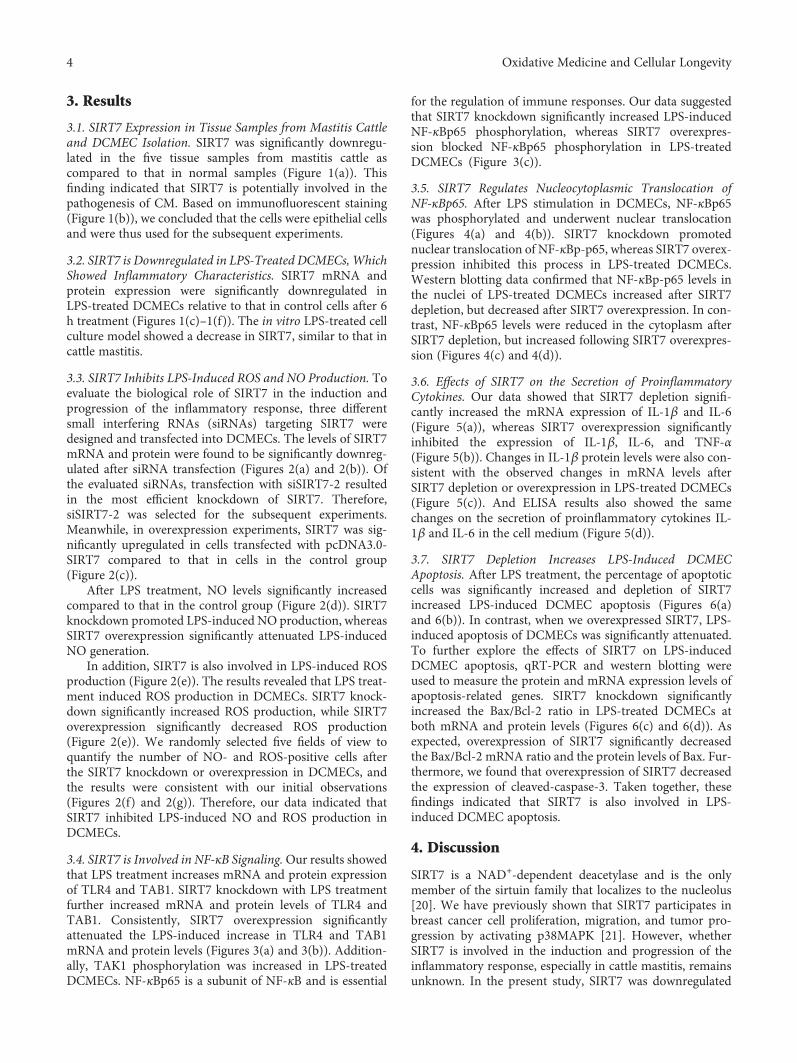
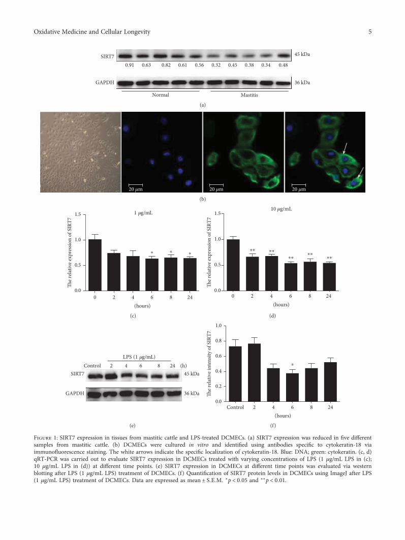
3.1. SIRT7 Expression in Tissue Samples from Mastitis Cattleand DCMEC Isolation. SIRT7 was significantly downregu-lated in the five tissue samples from mastitis cattle ascompared to that in normal samples (Figure 1(a)). Thisfinding indicated that SIRT7 is potentially involved in thepathogenesis of CM. Based on immunofluorescent staining(Figure 1(b)), we concluded that the cells were epithelial cellsand were thus used for the subsequent experiments.
3.2. SIRT7 is Downregulated in LPS-Treated DCMECs, WhichShowed Inflammatory Characteristics. SIRT7 mRNA andprotein expression were significantly downregulated inLPS-treated DCMECs relative to that in control cells after 6h treatment (Figures 1(c)–1(f)). The in vitro LPS-treated cellculture model showed a decrease in SIRT7, similar to that incattle mastitis.
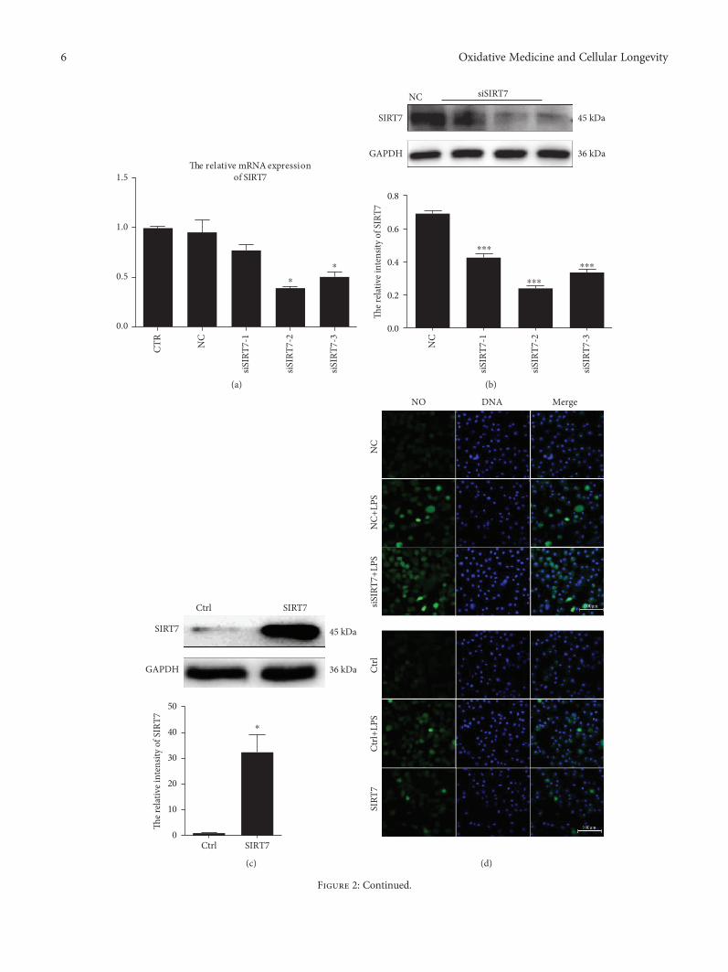
3.3. SIRT7 Inhibits LPS-Induced ROS and NO Production. Toevaluate the biological role of SIRT7 in the induction andprogression of the inflammatory response, three differentsmall interfering RNAs (siRNAs) targeting SIRT7 weredesigned and transfected into DCMECs. The levels of SIRT7mRNA and protein were found to be significantly downreg-ulated after siRNA transfection (Figures 2(a) and 2(b)). Ofthe evaluated siRNAs, transfection with siSIRT7-2 resultedin the most efficient knockdown of SIRT7. Therefore,siSIRT7-2 was selected for the subsequent experiments.Meanwhile, in overexpression experiments, SIRT7 was sig-nificantly upregulated in cells transfected with pcDNA3.0-SIRT7 compared to that in cells in the control group(Figure 2(c)).
After LPS treatment, NO levels significantly increasedcompared to that in the control group (Figure 2(d)). SIRT7knockdown promoted LPS-induced NO production, whereasSIRT7 overexpression significantly attenuated LPS-inducedNO generation.
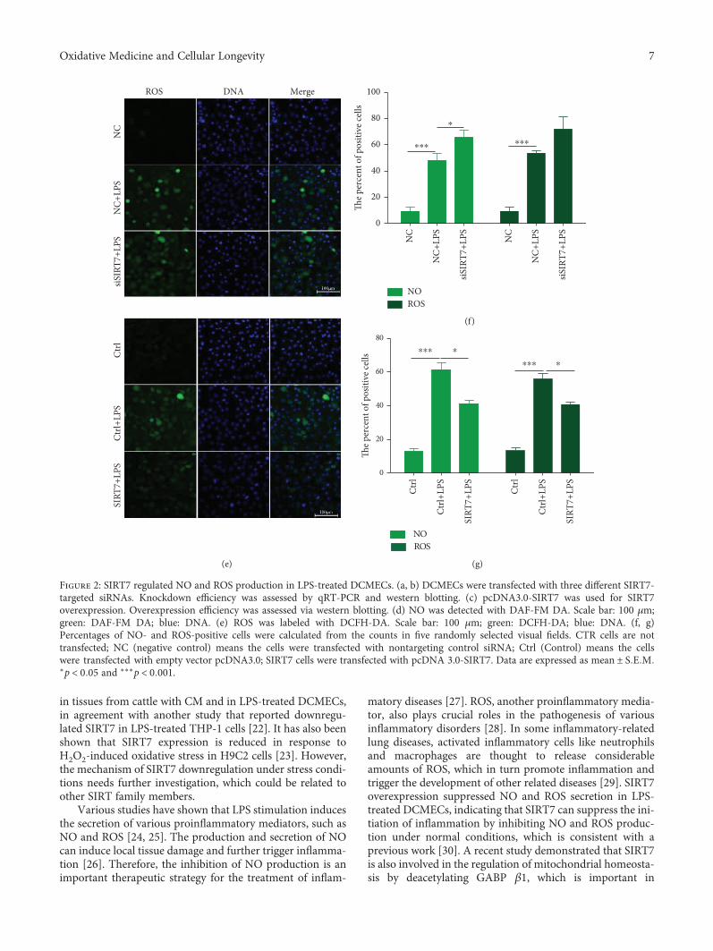
In addition, SIRT7 is also involved in LPS-induced ROSproduction (Figure 2(e)). The results revealed that LPS treat-ment induced ROS production in DCMECs. SIRT7 knock-down significantly increased ROS production, while SIRT7overexpression significantly decreased ROS production(Figure 2(e)). We randomly selected five fields of view toquantify the number of NO- and ROS-positive cells afterthe SIRT7 knockdown or overexpression in DCMECs, andthe results were consistent with our initial observations(Figures 2(f) and 2(g)). Therefore, our data indicated thatSIRT7 inhibited LPS-induced NO and ROS production inDCMECs.
3.4. SIRT7 is Involved in NF-κB Signaling.Our results showedthat LPS treatment increases mRNA and protein expressionof TLR4 and TAB1. SIRT7 knockdown with LPS treatmentfurther increased mRNA and protein levels of TLR4 andTAB1. Consistently, SIRT7 overexpression significantlyattenuated the LPS-induced increase in TLR4 and TAB1mRNA and protein levels (Figures 3(a) and 3(b)). Addition-ally, TAK1 phosphorylation was increased in LPS-treatedDCMECs. NF-κBp65 is a subunit of NF-κB and is essential
for the regulation of immune responses. Our data suggestedthat SIRT7 knockdown significantly increased LPS-inducedNF-κBp65 phosphorylation, whereas SIRT7 overexpres-sion blocked NF-κBp65 phosphorylation in LPS-treatedDCMECs (Figure 3(c)).
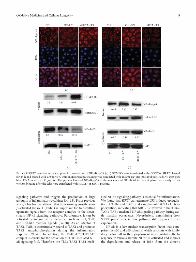
3.5. SIRT7 Regulates Nucleocytoplasmic Translocation ofNF-κBp65. After LPS stimulation in DCMECs, NF-κBp65was phosphorylated and underwent nuclear translocation(Figures 4(a) and 4(b)). SIRT7 knockdown promotednuclear translocation of NF-κBp-p65, whereas SIRT7 overex-pression inhibited this process in LPS-treated DCMECs.Western blotting data confirmed that NF-κBp-p65 levels inthe nuclei of LPS-treated DCMECs increased after SIRT7depletion, but decreased after SIRT7 overexpression. In con-trast, NF-κBp65 levels were reduced in the cytoplasm afterSIRT7 depletion, but increased following SIRT7 overexpres-sion (Figures 4(c) and 4(d)).
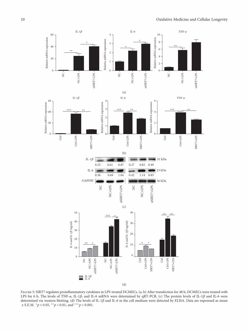
3.6. Effects of SIRT7 on the Secretion of ProinflammatoryCytokines. Our data showed that SIRT7 depletion signifi-cantly increased the mRNA expression of IL-1β and IL-6(Figure 5(a)), whereas SIRT7 overexpression significantlyinhibited the expression of IL-1β, IL-6, and TNF-α(Figure 5(b)). Changes in IL-1β protein levels were also con-sistent with the observed changes in mRNA levels afterSIRT7 depletion or overexpression in LPS-treated DCMECs(Figure 5(c)). And ELISA results also showed the samechanges on the secretion of proinflammatory cytokines IL-1β and IL-6 in the cell medium (Figure 5(d)).
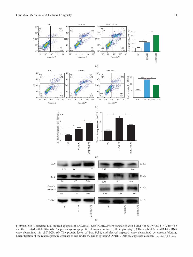
3.7. SIRT7 Depletion Increases LPS-Induced DCMECApoptosis. After LPS treatment, the percentage of apoptoticcells was significantly increased and depletion of SIRT7increased LPS-induced DCMEC apoptosis (Figures 6(a)and 6(b)). In contrast, when we overexpressed SIRT7, LPS-induced apoptosis of DCMECs was significantly attenuated.To further explore the effects of SIRT7 on LPS-inducedDCMEC apoptosis, qRT-PCR and western blotting wereused to measure the protein and mRNA expression levels ofapoptosis-related genes. SIRT7 knockdown significantlyincreased the Bax/Bcl-2 ratio in LPS-treated DCMECs atboth mRNA and protein levels (Figures 6(c) and 6(d)). Asexpected, overexpression of SIRT7 significantly decreasedthe Bax/Bcl-2 mRNA ratio and the protein levels of Bax. Fur-thermore, we found that overexpression of SIRT7 decreasedthe expression of cleaved-caspase-3. Taken together, thesefindings indicated that SIRT7 is also involved in LPS-induced DCMEC apoptosis.
4. Discussion
SIRT7 is a NAD+-dependent deacetylase and is the onlymember of the sirtuin family that localizes to the nucleolus[20]. We have previously shown that SIRT7 participates inbreast cancer cell proliferation, migration, and tumor pro-gression by activating p38MAPK [21]. However, whetherSIRT7 is involved in the induction and progression of theinflammatory response, especially in cattle mastitis, remainsunknown. In the present study, SIRT7 was downregulated
4 Oxidative Medicine and Cellular Longevity
45 kDa
36 kDaGAPDH
SIRT70.63 0.91 0.480.34 0.38 0.45 0.32 0.56 0.61 0.82
Normal Mastitis
(a)
20 �휇m 20 �휇m 20 �휇m
(b)
1 �휇g/mL
0 2 4 6 8 240.0
0.5
1.0
1.5
⁎⁎ ⁎
(hours)
The r
elat
ive e
xpre
ssio
n of
SIR
T7
(c)
10 �휇g/mL
0 2 4 6 8 240.0
0.5
1.0
1.5
⁎⁎ ⁎⁎
⁎⁎⁎⁎
⁎⁎
(hours)
The r
elat
ive e
xpre
ssio
n of
SIR
T7
(d)
SIRT7
GAPDH
45 kDa
LPS (1 �휇g/mL)
36 kDa
6 4 2 Control 8 24 (h)
(e)
Control 2 4 6 8 240.0
0.2
0.4
0.6
0.8
1.0
(hours)
⁎
The r
elat
ive i
nten
sity
of S
IRT7
(f)
Figure 1: SIRT7 expression in tissues from mastitic cattle and LPS-treated DCMECs. (a) SIRT7 expression was reduced in five differentsamples from mastitic cattle. (b) DCMECs were cultured in vitro and identified using antibodies specific to cytokeratin-18 viaimmunofluorescence staining. The white arrows indicate the specific localization of cytokeratin-18. Blue: DNA; green: cytokeratin. (c, d)qRT-PCR was carried out to evaluate SIRT7 expression in DCMECs treated with varying concentrations of LPS (1 μg/mL LPS in (c);10 μg/mL LPS in (d)) at different time points. (e) SIRT7 expression in DCMECs at different time points was evaluated via westernblotting after LPS (1 μg/mL LPS) treatment of DCMECs. (f) Quantification of SIRT7 protein levels in DCMECs using ImageJ after LPS(1 μg/mL LPS) treatment of DCMECs. Data are expressed as mean ± S E M ∗p < 0 05 and ∗∗p < 0 01.
5Oxidative Medicine and Cellular Longevity
0.0
CTR
NC
siSIR
T7-1
siSIR
T7-2
siSIR
T7-3
0.5
1.0
1.5
⁎
⁎
The relative mRNA expressionof SIRT7
(a)
0.0
NC
siSIR
T7-1
siSIR
T7-2
siSIR
T7-3
0.2
0.4
0.6
0.8
⁎⁎⁎
⁎⁎⁎
⁎⁎⁎
The r
elativ
e int
ensit
y of S
IRT7
45 kDa
36 kDaGAPDH
SIRT7
NC siSIRT7
(b)
0Ctrl SIRT7
10
20
30
40
50
⁎
The r
elativ
e int
ensit
y of S
IRT7
SIRT7
GAPDH
45 kDa
36 kDa
SIRT7Ctrl
(c)
NC
NC+
LPS
siSIR
T7+L
PSCt
rlCt
rl+LP
SSI
RT7
NO DNA Merge
(d)
Figure 2: Continued.
6 Oxidative Medicine and Cellular Longevity
in tissues from cattle with CM and in LPS-treated DCMECs,in agreement with another study that reported downregu-lated SIRT7 in LPS-treated THP-1 cells [22]. It has also beenshown that SIRT7 expression is reduced in response toH2O2-induced oxidative stress in H9C2 cells [23]. However,the mechanism of SIRT7 downregulation under stress condi-tions needs further investigation, which could be related toother SIRT family members.
Various studies have shown that LPS stimulation inducesthe secretion of various proinflammatory mediators, such asNO and ROS [24, 25]. The production and secretion of NOcan induce local tissue damage and further trigger inflamma-tion [26]. Therefore, the inhibition of NO production is animportant therapeutic strategy for the treatment of inflam-
matory diseases [27]. ROS, another proinflammatory media-tor, also plays crucial roles in the pathogenesis of variousinflammatory disorders [28]. In some inflammatory-relatedlung diseases, activated inflammatory cells like neutrophilsand macrophages are thought to release considerableamounts of ROS, which in turn promote inflammation andtrigger the development of other related diseases [29]. SIRT7overexpression suppressed NO and ROS secretion in LPS-treated DCMECs, indicating that SIRT7 can suppress the ini-tiation of inflammation by inhibiting NO and ROS produc-tion under normal conditions, which is consistent with aprevious work [30]. A recent study demonstrated that SIRT7is also involved in the regulation of mitochondrial homeosta-sis by deacetylating GABP β1, which is important in
MergeDNAROS
NC
NC+
LPS
siSIR
T7+L
PSCt
rlCt
rl+LP
SSI
RT7+
LPS
(e)
0
NC
NC
NC+
LPS
siSIR
T7+L
PS
NC+
LPS
siSIR
T7+L
PS
20
40
60
80
100
⁎⁎⁎ ⁎⁎⁎
⁎
The p
erce
nt o
f pos
itive
cells
NOROS
(f)
0
Ctrl
Ctrl+
LPS
SIRT
7+LP
S
Ctrl
Ctrl+
LPS
SIRT
7+LP
S
20
40
60
80⁎⁎⁎ ⁎
⁎⁎⁎ ⁎Th
e per
cent
of p
ositi
ve ce
lls
NOROS
(g)
Figure 2: SIRT7 regulated NO and ROS production in LPS-treated DCMECs. (a, b) DCMECs were transfected with three different SIRT7-targeted siRNAs. Knockdown efficiency was assessed by qRT-PCR and western blotting. (c) pcDNA3.0-SIRT7 was used for SIRT7overexpression. Overexpression efficiency was assessed via western blotting. (d) NO was detected with DAF-FM DA. Scale bar: 100 μm;green: DAF-FM DA; blue: DNA. (e) ROS was labeled with DCFH-DA. Scale bar: 100 μm; green: DCFH-DA; blue: DNA. (f, g)Percentages of NO- and ROS-positive cells were calculated from the counts in five randomly selected visual fields. CTR cells are nottransfected; NC (negative control) means the cells were transfected with nontargeting control siRNA; Ctrl (Control) means the cellswere transfected with empty vector pcDNA3.0; SIRT7 cells were transfected with pcDNA 3.0-SIRT7. Data are expressed as mean ± S E M∗p < 0 05 and ∗∗∗p < 0 001.
7Oxidative Medicine and Cellular Longevity
regulating mitochondrial genes [31]. Various studies haveshown that other SIRT family members (SIRT2, SIRT5, andSIRT6) are associated with NF-κB, which plays a critical rolein regulating ROS by targeting enzymes which induce ROS,such as NADPH oxidase, inducible NO synthase, cyclooxy-
genase-2, xanthine oxidoreductase, and cytochrome p450enzymes [32, 33].
TLR4 is a transmembrane protein that is involved in theinnate immune response, which can be activated by LPS.Upon activation, TLR4 stimulates the downstream NF-κB
0
NC
NC+
LPS
siSIR
T7+L
PS NC
NC+
LPS
siSIR
T7+L
PS NC
NC+
LPS
siSIR
T7+L
PS NC
NC+
LPS
siSIR
T7+L
PS
2
4
6
8
⁎⁎
⁎⁎
⁎
⁎
Rela
tive m
RNA
expr
essio
n
Rela
tive m
RNA
expr
essio
n
0
1
2
3
4
⁎⁎ ⁎⁎⁎⁎ ⁎⁎
TLR4TAB1
(a)
60 kDa
67 kDa
TAB1
TLR4 93 kDa
p-TAK1
GAPDH 36 kDa
0.42 0.370.51 0.28 0.88 0.75
0.402.110.76 1.97 0.86 0.34
0.320.85 0.26 0.83 0.65 0.12
NC NC+LPS siSIRT7+LPS Ctrl Ctrl+LPS SIRT7+LPS
(b)
p-p65
p65
65 kDa
65 kDa
GAPDH 36 kDa
NC NC+LPS siSIRT7+LPS Ctrl Ctrl+LPS SIRT7+LPS
0.430.93 0.21 1.21 0.78 0.45
1.98 1.86 2.04 1.39 0.83 1.22
(c)
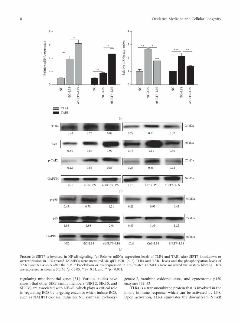
Figure 3: SIRT7 is involved in NF-κB signaling. (a) Relative mRNA expression levels of TLR4 and TAB1 after SIRT7 knockdown oroverexpression in LPS-treated DCMECs were measured via qRT-PCR. (b, c) TLR4 and TAB1 levels and the phosphorylation levels ofTAK1 and NF-κBp65 after the SIRT7 knockdown or overexpression in LPS-treated DCMECs were measured via western blotting. Dataare expressed as mean ± S E M ∗p < 0 05, ∗∗p < 0 01, and ∗∗∗p < 0 001.
8 Oxidative Medicine and Cellular Longevity
signaling pathways and triggers the production of largeamounts of inflammatory cytokines [34, 35]. From previouswork, it has been established that transforming growth factorβ-activated kinase 1 (TAK1) is important for transmittingupstream signals from the receptor complex to the down-stream NF-κB signaling pathways. Furthermore, it can beactivated by inflammatory mediators, such as IL-1, TNF,and Toll-like receptor ligands [36–38]. As an adaptor ofTAK1, TAB1 is constitutively bound to TAK1 and promotesTAK1 autophosphorylation during the inflammatoryresponse [39, 40]. In addition, the TAK1-ECSIT-TRAF6complex is crucial for the activation of TLR4-mediated NF-κB signaling [41]. Therefore, the TLR4-TAK1-TAB1-medi-
ated NF-κB signaling pathway is essential for inflammation.We found that SIRT7 can attenuate LPS-induced upregula-tion of TLR4 and TAB1 and can also inhibit TAK1 phos-phorylation, indicating that SIRT7 is involved in the TLR4-TAK1-TAB1-mediated NF-κB signaling pathway during cat-tle mastitis occurrence. Nevertheless, determining howSIRT7 participates in this pathway still requires furtherexploration.
NF-κB is a key nuclear transcription factor that com-prises the p50 and p65 subunits, which associate with inhib-itory factor IκB in the cytoplasm of unstimulated cells. Inresponse to various stimuli, NF-κB is activated and inducesthe degradation and release of IκBα from the dimeric
NF-�휅
Bp-p
65D
NA
Mer
ge
NC NC+LPS siSIRT7+LPS
(a)
Ctrl Ctrl+LPS SIRT7+LPS
(b)
GAPDH
Histone H3.1
NF-�휅Bp-p65
NF-�휅Bp65
0.650.43 0.2
0.6
NC
NC+
LPS
siSIR
T7+L
PS
0.26 0.13
Nuc
lear
Cyto
plas
mic
(c)
NF-�휅Bp-p65
Histone H3.1
NF-�휅Bp65
GAPDH
0.832.41 0.85
0.87
Ctrl
Ctrl+
LPS
SIRT
7+LP
S0.58 0.82
Nuc
lear
Cyto
plas
mic
(d)
Figure 4: SIRT7 regulates nucleocytoplasmic translocation of NF-κBp-p65. (a, b) DCMECs were transfected with siSIRT7 or SIRT7 plasmidfor 24 h and treated with LPS for 6 h. Immunofluorescence staining was conducted with an anti-NF-κBp-p65 antibody. Red: NF-κBp-p65;blue: DNA; scale bar: 50 μm. (c) The protein levels of NF-κBp-p65 in the nucleus and NF-κBp65 in the cytoplasm were analyzed viawestern blotting after the cells were transfected with siSIRT7 or SIRT7 plasmid.
9Oxidative Medicine and Cellular Longevity
0
NC
NC+
LPS
siSIR
T7+L
PS NC
NC+
LPS
siSIR
T7+L
PS NC
NC+
LPS
siSIR
T7+L
PS
20
40
60
⁎⁎
⁎
IL-1�훽
Rela
tive m
RNA
expr
essio
n
Rela
tive m
RNA
expr
essio
n
Rela
tive m
RNA
expr
essio
n
0
1
2
3
4
⁎
⁎
IL-6
0
2
4
6
8
10
⁎⁎
TNF-�훼
(a)
0
Ctrl
Ctrl+
LPS
SIRT
7+LP
S
Ctrl
Ctrl+
LPS
SIRT
7+LP
S
Ctrl
Ctrl+
LPS
SIRT
7+LP
S
20
40
60
⁎⁎⁎ ⁎⁎ ⁎⁎⁎ ⁎⁎ ⁎⁎⁎ ⁎⁎
IL-1�훽
0
1
2
3
4IL-6
0
2
4
6TNF-�훼
Rela
tive m
RNA
expr
essio
n
Rela
tive m
RNA
expr
essio
n
Rela
tive m
RNA
expr
essio
n(b)
31 kDa
23 kDa
GAPDH
0.870.61 0.25
0.690.36 1.04
NC
NC+
LPS
siSIR
T7+L
PS NC
NC+
LPS
siSIR
T7+L
PS
0.27 0.82 0.49
0.42 1.14 0.85IL-6
IL-1�훽
36 kDa
(c)
0
NC
NC+
LPS
siSIR
T7+L
PS NC
NC+
LPS
siSIR
T7+L
PS
10
20
30
40
50
IL-1�훽IL-6
⁎⁎ ⁎
⁎⁎⁎ ⁎
IL-6
and
IL-1�훽
(ng/
ml)
IL-6
and
IL-1�훽
(ng/
ml)
0
Ctrl
Ctrl+
LPS
SIRT
7+LP
S
Ctrl
Ctrl+
LPS
SIRT
7+LP
S
10
20
30
40
⁎ ⁎
⁎⁎ ⁎⁎
(d)
Figure 5: SIRT7 regulates proinflammatory cytokines in LPS-treated DCMECs. (a, b) After transfection for 48 h, DCMECs were treated withLPS for 6 h. The levels of TNF-α, IL-1β, and IL-6 mRNA were determined by qRT-PCR. (c) The protein levels of IL-1β and IL-6 weredetermined via western blotting. (d) The levels of IL-1β and IL-6 in the cell medium were detected by ELISA. Data are expressed as mean± S E M ∗p < 0 05, ∗∗p < 0 01, and ∗∗∗p < 0 001.
10 Oxidative Medicine and Cellular Longevity
NC
NC+
LPS
siSIR
T7+L
PS
0
5
10
15
20
25
⁎⁎⁎
⁎⁎
Early
apop
totic
(Q2+
Q3)
(%)
NC NC+LPS siSIRT7+LPS104
103
102
101
100
104
103
102
101
100
PIPIPI
100
Q10.54
Q11.39
Q218.9
Q10.90
Q28.05
Q495.7
Q32.06
Q487.9
Q33.17
Q475.7
Q34.03
Q21.68
101 102
Annexin V Annexin V Annexin V103 104 100 101 102 103 104
104
103
102
101
100
100 101 102 103 104
(a)
Ctrl Ctrl+LPS SIRT7+LPS
Ctrl Ctrl+LPS SIRT7+LPS0
5
10
15 ⁎⁎⁎ ⁎
Early
apop
totic
(Q2+
Q3)
(%)
PI PI PI
Q10.30
Q493.2
Q33.75
Q487.9
Q33.39
Q34.94
Q491.1
Q10.84
Q22.72
Q27.92
Q10.17
Q23.78
Annexin V Annexin V Annexin V
104
103
102
101
100
100 101 102 103 104 100 101 102 103 104 100 101 102 103 104
104
103
102
101
100
104
103
102
101
100
(b)
0
NC
NC+
LPS
siSIR
T7+L
PS NC
NC+
LPS
siSIR
T7+L
PS
1
2
3
4
⁎
⁎
The r
elat
ive e
xpre
ssio
n of
Bax
/Bcl
-2
The r
elat
ive e
xpre
ssio
n of
Bax
/Bcl
-2
0
1
2
3 ⁎ ⁎
(c)
20 kDaBAX
Bcl-2
Cleaved-caspase-3
GAPDH
NC
NC-
LPS
siSIR
T7+L
PS
Ctrl
Ctrl+
LPS
SIRT
7+LP
S
26 kDa
17 kDa
36 kDa
0.630.21 1.35
0.630.9 1.2
0.850.77 0.47
0.461.12 0.33
1.33 0.51 0.64
0.650.95 0.35
(d)
Figure 6: SIRT7 alleviates LPS-induced apoptosis in DCMECs. (a, b) DCMECs were transfected with siSIRT7 or pcDNA3.0-SIRT7 for 48 hand then treated with LPS for 6 h. The percentages of apoptotic cells were examined by flow cytometry. (c) The levels of Bax and Bcl-2 mRNAwere determined via qRT-PCR. (d) The protein levels of Bax, Bcl-2, and cleaved-caspase-3 were determined by western blotting.Quantification of the relative protein levels are shown under the bands (protein/GAPDH). Data are expressed as mean ± S E M ∗p < 0 05.
11Oxidative Medicine and Cellular Longevity
complex, followed by the phosphorylation of NF-κBp65 andits subsequent translocation to the nucleus [42]. Phosphory-lation of the NF-κB subunits can either increase or decreasethe transcription of the target genes [43] and thus exert a pro-found effect on NF-κB function. Once NF-κBp-p65 entersthe nucleus, NF-κB initiates the transcription of genes encod-ing proinflammatory cytokines, including IL-1β, IL-6, andTNF-α [44]. Our results revealed that SIRT7 also participatesin NF-κB phosphorylation and nucleocytoplasmic transloca-tion of NF-κBp-p65. SIRT7 was concentrated in the nucleus,while p65 is mainly distributed in the cytosol of unstimulatedcells. Therefore, SIRT7 could bind to p65 in the nucleus andregulate the translocation of p65 between the nucleus andcytoplasm [45]. However, how SIRT7 regulates p65 translo-
cation needs further investigation. The function of SIRT7 ininflammation is similar to that of the other members of thesirtuin family. For example, SIRT1 can suppress inflamma-tion in multiple tissues and macrophages, and SIRT1interacts with NF-κBp65 to inhibit NF-κB-associated tran-scription [46]. In addition, SIRT2 is a deacetylase of NF-κB,which is important for regulating TNF-α-induced NF-κB-dependent gene expression. In unstimulated cells, SIRT2exits in a complex with p65, but under TNF-α stimulation,p65 translocates to the nucleus and is acetylated at K310,K314, and K315 through binding to p300. After stimulation,p65 returned to the cytoplasm and SIRT2 deacetylated p65[47]. Recent work showed that SIRT7 interacts with SIRT1.Therefore, the function of SIRT7 in NF-κB acetylation could
LPS
TAK1 P
NF-�휅Bp65 P
NF-�휅Bp65 P
Nucleus translocation
IL-1�훽 IL-6,TNF-�훼
Inflammatory cytokine transcription
ROS NO
Bax, Bcl-2,caspase-3
Apoptosis
SIRT7
SIRT7
TLR4
TAB1Cytoplasm
Nucleus
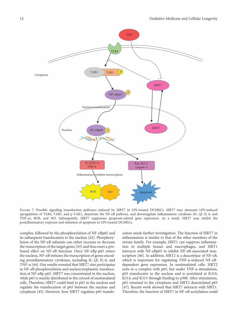
Figure 7: Possible signaling transduction pathways induced by SIRT7 in LPS-treated DCMECs. SIRT7 may attenuate LPS-inducedupregulation of TLR4, TAB1, and p-TAK1, deactivate the NF-κB pathway, and downregulate inflammatory cytokines (IL-1β, IL-6, andTNF-α), ROS, and NO. Subsequently, SIRT7 suppresses apoptosis-related gene expression. As a result, SIRT7 may inhibit theproinflammatory response and initiation of apoptosis in LPS-treated DCMECs.
12 Oxidative Medicine and Cellular Longevity
occur through SIRT1 [48]. Depletion of SIRT7 induced arapid increase in the production of proinflammatory cyto-kines, including TNF-α, IL-1β, and IL-6, which supportedour hypothesis that SIRT7 is involved in NF-κB-mediatedinflammation.
Various studies have shown that increased ROS produc-tion and a release of inflammatory cytokines, such as TNF-α, IL-6, and IL-1β, ultimately lead to apoptotic cell death[49, 50]. The apoptosis-related genes, caspase-3, caspase-8,and Bax, were found to be significantly upregulated in LPS-treated mice [51]. In addition to the function of TLR4 inthe LPS-induced inflammatory response, ROS productioncan trigger the downstream apoptotic pathways, therebyincreasing the amounts of Bax, Bcl-2, and cleaved-caspase-3[52]. Alterations in the expression of both Bcl-2 and Baxare known to trigger the mitochondrial intrinsic apoptosisprogram and then promote cell apoptosis. One studyrevealed that SIRT7 promotes gastric cancer growth andinhibits apoptosis of gastric cancer cells by inhibiting miR-34a activity [53]. Our data indicated that SIRT7 could protectcells from the LPS-induced apoptosis by downregulating Baxand cleaved-caspase-3, which is consistent with one studywhich showed that silencing of SIRT7 decreased antiapopto-tic factor Bcl-2 and NF-κB levels [54]. Because the NF-κB sig-naling pathway is critical in cellular proliferation, apoptosis,and malignant diseases [55], it will be interesting to investi-gate the regulatory mechanism between SIRT7 and NF-κB.
In conclusion, our results suggest that SIRT7 can sup-press LPS-induced inflammation and apoptosis via the NF-κB signaling pathway (Figure 7). Given its anti-inflammatory and antiapoptotic effects, SIRT7 may serve asa potential pharmacological target for CM therapy.
Data Availability
The data used to support the findings of this study are avail-able from the corresponding author upon request.
Conflicts of Interest
The authors declare no conflict of interest.
Acknowledgments
This work was supported by the National Natural ScienceFoundation of China (no. 31772567) and the NationalKey Research and Development Program of China (no.2018YFD0501600).
References
[1] T. Halasa, K. Huijps, O. Østerås, and H. Hogeveen, “Economiceffects of bovine mastitis and mastitis management: a review,”Veterinary Quarterly, vol. 29, no. 1, pp. 18–31, 2007.
[2] N. Kumar, A. Manimaran, A. Kumaresan et al., “Mastitiseffects on reproductive performance in dairy cattle: a review,”Tropical Animal Health and Production, vol. 49, no. 4,pp. 663–673, 2017.
[3] X. Zhao and P. Lacasse, “Mammary tissue damage duringbovine mastitis: causes and control,” Journal of Animal Sci-ence, vol. 86, Supplement 13, pp. 57–65, 2008.
[4] D. D. Bannerman, A. Chockalingam, M. J. Paape, and J. C.Hope, “The bovine innate immune response duringexperimentally-induced Pseudomonas aeruginosa mastitis,”Veterinary Immunology and Immunopathology, vol. 107,no. 3-4, pp. 201–215, 2005.
[5] Y. Wang, D. S. Zarlenga, M. J. Paape, and G. E. Dahl, “Recom-binant bovine soluble CD14 sensitizes the mammary gland tolipopolysaccharide,” Veterinary Immunology and Immunopa-thology, vol. 86, no. 1-2, pp. 115–124, 2002.
[6] M. A. Dobrovolskaia and S. N. Vogel, “Toll receptors, CD14,and macrophage activation and deactivation by LPS,”Microbes and Infection, vol. 4, no. 9, pp. 903–914, 2002.
[7] J. F. Miao, Y. M. Zhu, B. B. Gu, X. B. Wang, S. X. Zou, and Y. E.Deng, “Evaluation of the changes of immune cells duringlipopolysaccharide-induced mastitis in rats,” Cytokine,vol. 40, no. 2, pp. 135–143, 2007.
[8] Y. X. Li, X. H. Li, G. Liu et al., “Fucosterol attenuateslipopolysaccharide-induced acute lung injury in mice,” TheJournal of Surgical Research, vol. 195, no. 2, pp. 515–521,2015.
[9] T. J. Guzik, R. Korbut, and T. Adamek-Guzik, “Nitric oxideand superoxide in inflammation and immune regulation,”Journal of Physiology and Pharmacology, vol. 54, no. 4,pp. 469–487, 2003.
[10] T. Michel and O. Feron, “Nitric oxide synthases: which, where,how, and why?,” Journal of Clinical Investigation, vol. 100,no. 9, pp. 2146–2152, 1997.
[11] W. Droge, “Free radicals in the physiological control of cellfunction,” Physiological Reviews, vol. 82, no. 1, pp. 47–95,2002.
[12] Y. S. Bae, H. Oh, S. G. Rhee, and Y. D. Yoo, “Regulation ofreactive oxygen species generation in cell signaling,”Moleculesand Cells, vol. 32, no. 6, pp. 491–509, 2011.
[13] S. Vallabhapurapu and M. Karin, “Regulation and function ofNF-κB transcription factors in the immune system,” AnnualReview of Immunology, vol. 27, no. 1, pp. 693–733, 2009.
[14] C. Scheidereit, “IκB kinase complexes: gateways to NF-κB acti-vation and transcription,” Oncogene, vol. 25, no. 51, pp. 6685–6705, 2006.
[15] H. Shibuya, K. Yamaguchi, K. Shirakabe et al., “TAB1: An acti-vator of the TAK1 MAPKKK in TGF-β signal transduction,”Science, vol. 272, no. 5265, pp. 1179–1182, 1996.
[16] E. Ford, R. Voit, G. Liszt, C. Magin, I. GrumMt, andL. Guarente, “Mammalian Sir2 homolog SIRT7 is an activatorof RNA polymerase I transcription,” Genes & Development,vol. 20, no. 9, pp. 1075–1080, 2006.
[17] S. Kiran, T. Anwar, M. Kiran, and G. Ramakrishna, “Sirtuin 7in cell proliferation, stress and disease: rise of the seventh sir-tuin!,” Cellular Signalling, vol. 27, no. 3, pp. 673–682, 2015.
[18] O. Vakhrusheva, C. Smolka, P. Gajawada et al., “Sirt7 increasesstress resistance of cardiomyocytes and prevents apoptosis andinflammatory cardiomyopathy in mice,” Circulation Research,vol. 102, no. 6, pp. 703–710, 2008.
[19] V. Saini, J. T. McClure, D. T. Scholl, T. J. DeVries, and H. W.Barkema, “Herd-level relationship between antimicrobial useand presence or absence of antimicrobial resistance in gram-negative bovine mastitis pathogens on Canadian dairy farms,”Journal of Dairy Science, vol. 96, no. 8, pp. 4965–4976, 2013.
13Oxidative Medicine and Cellular Longevity
[20] E. Michishita, J. Y. Park, J. M. Burneskis, J. C. Barrett, andI. Horikawa, “Evolutionarily conserved and nonconserved cel-lular localizations and functions of human SIRT proteins,”Molecular Biology of the Cell, vol. 16, no. 10, pp. 4623–4635,2005.
[21] K. L. Chen, L. Li, F. X. Yang, C. M. Li, Y. R. Wang, and G. L.Wang, “SIRT7 depletion inhibits cell proliferation, migration,and increases drug sensitivity by activating p38MAPK inbreast cancer cells,” Journal of Cellular Physiology, vol. 233,no. 9, pp. 6767–6778, 2018.
[22] Z. Li, B. Bridges, J. Olson, and S. A.Weinman, “The interactionbetween acetylation and serine-574 phosphorylation regulatesthe apoptotic function of FOXO3,” Oncogene, vol. 36, no. 13,pp. 1887–1898, 2017.
[23] W. Yu, Y. C. Fu, X. H. Zhou et al., “Effects of resveratrol onH2O2-induced apoptosis and expression of SIRTs in H9c2cells,” Journal of Cellular Biochemistry, vol. 107, no. 4,pp. 741–747, 2009.
[24] E. Viennois, B. Xiao, S. Ayyadurai et al., “Micheliolide, a newsesquiterpene lactone that inhibits intestinal inflammationand colitis-associated cancer,” Laboratory Investigation,vol. 94, no. 9, pp. 950–965, 2014.
[25] Y. A. Alpizar, B. Boonen, A. Sanchez et al., “TRPV4 activationtriggers protective responses to bacterial lipopolysaccharidesin airway epithelial cells,” Nature Communications, vol. 8,no. 1, p. 1059, 2017.
[26] S. Zidi, F. Bediar-Boulaneb, H. Belguendouz et al., “Local pro-inflammatory cytokine and nitric oxide responses are elevatedin patients with pterygium,” International Journal of Immuno-pathology and Pharmacology, vol. 30, no. 4, pp. 395–405, 2017.
[27] J. N. Sharma, A. Al-Omran, and S. S. Parvathy, “Role of nitricoxide in inflammatory diseases,” Inflammopharmacology,vol. 15, no. 6, pp. 252–259, 2007.
[28] D. Yang, S. G. Elner, Z. M. Bian, G. O. Till, H. R. Petty, andV. M. Elner, “Pro-inflammatory cytokines increase reactiveoxygen species through mitochondria and NADPH oxidasein cultured RPE cells,” Experimental Eye Research, vol. 85,no. 4, pp. 462–472, 2007.
[29] A. Andersson-Sjoland, J. C. Karlsson, and K. Rydell-Torma-nen, “ROS-induced endothelial stress contributes to pulmo-nary fibrosis through pericytes and Wnt signaling,”Laboratory Investigation, vol. 96, no. 2, pp. 206–217, 2016.
[30] M. Gao, X. Li, Y. He et al., “SIRT7 functions in redox homeo-stasis and cytoskeletal organization during oocyte matura-tion,” The FASEB Journal, vol. 32, no. 11, pp. 6228–6238, 2018.
[31] D. Ryu, Y. S. Jo, G. Lo Sasso et al., “A SIRT7-dependent acety-lation switch of GABPβ1 controls mitochondrial function,”Cell Metabolism, vol. 20, no. 5, pp. 856–869, 2014.
[32] C. K. Singh, G. Chhabra, M. A. Ndiaye, L. M. Garcia-Peterson,N. J. Mack, and N. Ahmad, “The role of sirtuins in antioxidantand redox signaling,” Antioxidants & Redox Signaling, vol. 28,no. 8, pp. 643–661, 2018.
[33] M. J. Morgan and Z. G. Liu, “Crosstalk of reactive oxygenspecies and NF-κB signaling,” Cell Research, vol. 21, no. 1,pp. 103–115, 2011.
[34] E. B. Byun, N. Y. Sung, E. H. Byun et al., “The procyanidin tri-mer C1 inhibits LPS-induced MAPK and NF-κB signalingthrough TLR4 in macrophages,” International Immunophar-macology, vol. 15, no. 2, pp. 450–456, 2013.
[35] E. B. Byun, N. Y. Sung, J. N. Park, M. S. Yang, S. H. Park, andE. H. Byun, “Gamma-irradiated resveratrol negatively regu-
lates LPS-induced MAPK and NF-κB signaling throughTLR4 in macrophages,” International Immunopharmacology,vol. 25, no. 2, pp. 249–259, 2015.
[36] T. Kawai and S. Akira, “Signaling to NF-κB by Toll-like recep-tors,” Trends in Molecular Medicine, vol. 13, no. 11, pp. 460–469, 2007.
[37] J. Ninomiya-Tsuji, K. Kishimoto, A. Hiyama, J. Inoue, Z. Cao,and K. Matsumoto, “The kinase TAK1 can activate the NIK-IκB as well as the MAP kinase cascade in the IL-1 signallingpathway,” Nature, vol. 398, no. 6724, pp. 252–256, 1999.
[38] G. Takaesu, R. M. Surabhi, K. J. Park, J. Ninomiya-Tsuji,K. Matsumoto, and R. B. Gaynor, “TAK1 is critical for IκBkinase-mediated activation of the NF-κB pathway,” Journalof Molecular Biology, vol. 326, no. 1, pp. 105–115, 2003.
[39] Z. J. Chen, V. Bhoj, and R. B. Seth, “Ubiquitin, TAK1 and IKK:is there a connection?,” Cell Death and Differentiation, vol. 13,no. 5, pp. 687–692, 2006.
[40] H. Sakurai, H. Miyoshi, J. Mizukami, and T. Sugita, “Phos-phorylation-dependent activation of TAK1 mitogen-activatedprotein kinase kinase kinase by TAB1,” FEBS Letters,vol. 474, no. 2-3, pp. 141–145, 2000.
[41] S. M. Wi, G. Moon, J. Kim et al., “TAK1-ECSIT-TRAF6 com-plex plays a key role in the TLR4 signal to activate NF-κB,” TheJournal of Biological Chemistry, vol. 289, no. 51, pp. 35205–35214, 2014.
[42] M. H. Park and J. T. Hong, “Roles of NF-κB in cancer andinflammatory diseases and their therapeutic approaches,”Cells, vol. 5, no. 2, p. 15, 2016.
[43] F. Christian, E. L. Smith, and R. J. Carmody, “The regulation ofNF-κB subunits by phosphorylation,” Cells, vol. 5, no. 1, p. 12,2016.
[44] V. Fattori, M. S. N. Hohmann, A. C. Rossaneis et al., “Target-ing IL-33/ST2 signaling: regulation of immune function andanalgesia,” Expert Opinion on Therapeutic Targets, vol. 21,no. 12, pp. 1141–1152, 2017.
[45] Y. Miyasato, T. Yoshizawa, Y. Sato et al., “Sirtuin 7 deficiencyameliorates cisplatin-induced acute kidney injury through reg-ulation of the inflammatory response,” Scientific Reports,vol. 8, no. 1, p. 5927, 2018.
[46] Z. Lin and D. Fang, “The roles of SIRT1 in cancer,” Genes &Cancer, vol. 4, no. 3-4, pp. 97–104, 2013.
[47] K. M. Rothgiesser, S. Erener, S. Waibel, B. Lüscher, and M. O.Hottiger, “SIRT2 regulates NF-κB-dependent gene expressionthrough deacetylation of p65 Lys310,” Journal of Cell Science,vol. 123, no. 24, pp. 4251–4258, 2010.
[48] J. Fang, A. Ianni, C. Smolka et al., “Sirt7 promotes adipogen-esis in the mouse by inhibiting autocatalytic activation ofSirt1,” Proceedings of the National Academy of Sciences ofthe United States of America, vol. 114, no. 40, pp. E8352–E8361, 2017.
[49] Y. Zhang, X. Li, J. J. Grailer et al., “Melatonin alleviates acutelung injury through inhibiting the NLRP3 inflammasome,”Journal of Pineal Research, vol. 60, no. 4, pp. 405–414, 2016.
[50] H. Lv, Q. Liu, Z. Wen, H. Feng, X. Deng, and X. Ci, “Xantho-humol ameliorates lipopolysaccharide (LPS)-induced acutelung injury via induction of AMPK/GSK3β-Nrf2 signal axis,”Redox Biology, vol. 12, pp. 311–324, 2017.
[51] W. Kong, K. Kang, Y. Gao et al., “Dexmedetomidine alleviatesLPS-induced septic cardiomyopathy via the cholinergic anti-inflammatory pathway in mice,” American Journal of Transla-tional Research, vol. 9, no. 11, pp. 5040–5047, 2017.
14 Oxidative Medicine and Cellular Longevity
[52] X. Song, M. Guo, T. Wang, W. Wang, Y. Cao, and N. Zhang,“Geniposide inhibited lipopolysaccharide-induced apoptosisby modulating TLR4 and apoptosis-related factors in mousemammary glands,” Life Sciences, vol. 119, no. 1-2, pp. 9–17,2014.
[53] S. Zhang, P. Chen, Z. Huang et al., “Sirt7 promotes gastric can-cer growth and inhibits apoptosis by epigenetically inhibitingmiR-34a,” Scientific Reports, vol. 5, no. 1, article 9787, 2015.
[54] H. L. Wang, R. Q. Lu, S. H. Xie et al., “SIRT7 exhibits onco-genic potential in human ovarian cancer cells,” Asian PacificJournal of Cancer Prevention, vol. 16, no. 8, pp. 3573–3577,2015.
[55] A. B. Alvero, “Recent insights into the role of NF-kappaB inovarian carcinogenesis,” Genome Medicine, vol. 2, no. 8,p. 56, 2010.
15Oxidative Medicine and Cellular Longevity

Stem Cells International
Hindawiwww.hindawi.com Volume 2018
Hindawiwww.hindawi.com Volume 2018
MEDIATORSINFLAMMATION
of
EndocrinologyInternational Journal of
Hindawiwww.hindawi.com Volume 2018
Hindawiwww.hindawi.com Volume 2018
Disease Markers
Hindawiwww.hindawi.com Volume 2018
BioMed Research International
OncologyJournal of
Hindawiwww.hindawi.com Volume 2013
Hindawiwww.hindawi.com Volume 2018
Oxidative Medicine and Cellular Longevity
Hindawiwww.hindawi.com Volume 2018
PPAR Research
Hindawi Publishing Corporation http://www.hindawi.com Volume 2013Hindawiwww.hindawi.com
The Scientific World Journal
Volume 2018
Immunology ResearchHindawiwww.hindawi.com Volume 2018
Journal of
ObesityJournal of
Hindawiwww.hindawi.com Volume 2018
Hindawiwww.hindawi.com Volume 2018
Computational and Mathematical Methods in Medicine
Hindawiwww.hindawi.com Volume 2018
Behavioural Neurology
OphthalmologyJournal of
Hindawiwww.hindawi.com Volume 2018
Diabetes ResearchJournal of
Hindawiwww.hindawi.com Volume 2018
Hindawiwww.hindawi.com Volume 2018
Research and TreatmentAIDS
Hindawiwww.hindawi.com Volume 2018
Gastroenterology Research and Practice
Hindawiwww.hindawi.com Volume 2018
Parkinson’s Disease
Evidence-Based Complementary andAlternative Medicine
Volume 2018Hindawiwww.hindawi.com
Submit your manuscripts atwww.hindawi.com