
Supplemental Data Activity-Induced Protocadherin Arcadlin ...
12
1 Neuron, Volume 56 Supplemental Data Activity-Induced Protocadherin Arcadlin Regulates Dendritic Spine Number by Triggering N-Cadherin Endocytosis via TAO2β and p38 MAP Kinases Shin Yasuda, Hidekazu Tanaka, Hiroko Sugiura, Ko Okamura, Taiki Sakaguchi, Uyen Tran, Takako Takemiya, Akira Mizoguchi, Yoshiki Yagita, Takeshi Sakurai, E.M. De Robertis, and Kanato Yamagata SUPPLEMENTAL EXPERIMENTAL PROCEDURES Construction of Expression Vectors Expression vectors for N-cadherin mutants were constructed by modifying pCXN2-Ncad-myc, in which N-cadherin fused with 5x or 6xmyc-tag is ligated downstream of β-actin promoter (Okamura et al., 2004; Shan et al., 2000). Ncad∆E-myc and Ncad-TM-myc were constructed by replacing the N-cadherin coding sequence between EcoRI and XhoI sites with the N-terminal signal- and pre-sequences fused with the transmembrane-cytoplasmic regions and a transmembrane domain alone of mouse N-cadherin, respectively. The inserted cDNA fragment was amplified with pfu-polymerase (Promega). Point mutations were generated by amplifying the wild-type cDNAs with mutated primers and pfu DNA polymerase. The expression vector for wild-type arcadlin is described (Yamagata et al., 1999). Arcadlin-egfp was constructed by ligating pfu-amplified full-length arcadlin cDNA into pEGFP-N1 (Clontech) between XhoI and EcoRI sites. Acad-CP-EGFP was constructed by ligating the cytoplasmic sequence of arcadlin into pEGFP-C1 (Clontech). The integrity of the constructs was verified by sequencing. For the purification of arcadlin extracellular domain protein (Acad-EC), an arcadlin fragment corresponds to amino acids 31-696 was ligated to BamHI-NotI sites in pET32(c) expression vector (Novagen).
Transcript of Supplemental Data Activity-Induced Protocadherin Arcadlin ...
1
Neuron, Volume 56
Supplemental Data
Activity-Induced Protocadherin Arcadlin Regulates
Dendritic Spine Number by Triggering N-Cadherin
Endocytosis via TAO2β and p38 MAP Kinases
Shin Yasuda, Hidekazu Tanaka, Hiroko Sugiura, Ko Okamura, Taiki Sakaguchi, Uyen Tran,
Takako Takemiya, Akira Mizoguchi, Yoshiki Yagita, Takeshi Sakurai, E.M. De Robertis,
and Kanato Yamagata SUPPLEMENTAL EXPERIMENTAL PROCEDURES Construction of Expression Vectors Expression vectors for N-cadherin mutants were constructed by modifying pCXN2-Ncad-myc, in which N-cadherin fused with 5x or 6xmyc-tag is ligated downstream of
β-actin promoter (Okamura et al., 2004; Shan et al., 2000). Ncad∆E-myc and Ncad-TM-myc were constructed by replacing the N-cadherin coding sequence between EcoRI and XhoI sites with the N-terminal signal- and pre-sequences fused with the transmembrane-cytoplasmic regions and a transmembrane domain alone of mouse N-cadherin, respectively. The inserted cDNA fragment was amplified with pfu-polymerase (Promega). Point mutations were generated by amplifying the wild-type cDNAs with mutated primers and pfu DNA polymerase. The expression vector for wild-type arcadlin is described (Yamagata et al., 1999). Arcadlin-egfp was constructed by ligating pfu-amplified full-length arcadlin cDNA into pEGFP-N1 (Clontech) between XhoI and EcoRI sites. Acad-CP-EGFP was constructed by ligating the cytoplasmic sequence of arcadlin into pEGFP-C1 (Clontech). The integrity of the constructs was verified by sequencing. For the purification of arcadlin extracellular domain protein (Acad-EC), an arcadlin fragment corresponds to amino acids 31-696 was ligated to BamHI-NotI sites in pET32(c) expression vector (Novagen).
2
Immunohistochemistry of Rat Brain Slices Brains of male Sprague Dawley rats (170 g) were freshly dissected 4hr after MECS (30 mA; 60 Hz; pulse width, 1 msec; duration, 0.1 sec), frozen in powdered dry ice and mounted
with OCT compound (-20 ˚C). Sections (25 µm) were cut using a cryostat, mounted on silica-coated slide glasses, treated with graded series of methanol (25%, 50%, 75%, 100%, 75%, 50%, 25%, respectively, in PBS), blocked and permeabilized for 15 min in BL solution (5% normal goat serum, 0.1% Triton X-100, 0.02% sodium azide in PBS). The sections were
then incubated overnight with primary antibodies (mouse anti-N-cadherin, 12.5 µg/ml, Transduction lab., rabbit anti-arcadlin cytoplasmic region, 1:200 (Yamagata et al., 1999)) in BL at 4 ˚C. Immunoreactivity was visualized using species-specific fluorochrome-conjugated secondary antibodies.
Neuron Cultures Neuron cultures were prepared from hippocampi of embryonic day 18 Sprague Dawley rats as described (Banker and Goslin, 1991). Cells were plated at a density of 5,400 cells/cm2 for microscopic analyses and 14,400 cells/cm2 for biochemical analyses. For biotinylation analyses, neurons were plated at density of 5.0 x 10⁴ cells/cm2 onto polyethyleneimine (PEI) -coated dishes. They were maintained for up to 5 weeks in MEM containing N2 supplements (Bottenstein and Sato, 1979), 1 mM sodium pyruvate, 0.1%
ovalbumin, and 5 µM cytosine arabinoside with glial feeder layer. Similar experimental results were obtained with the neurons maintained in Neurobasal medium (GibcoBRL) with B27 supplement (GibcoBRL) without the feeder layer. The neurons from arcadlin/papc mutant mice (acad -/-) (Yamamoto et al., 2000) were collected from brains of P0 pups generated by crossing of acad+/- parents. Neurons collected from individual hippocampi were separately cultured, while determining the genotype of each pool of the neurons. Comparisons of mutant and wild-type neurons were made among cultures derived from a single littermate.
Transfection of cDNA and siRNA into Cultured Neurons Hippocampal neurons (7000 cells/cm2) attached on 18 mm diameter coverslips cultured in 6 cm diameter dishes supplied with Neurobasal/B27 were transfected with cDNAs and siRNAs by using Lipofectamine 2000 (Invitrogen) according to manufacture’s instructions.
3
Preparation of Anti-TAO2β Antibody TAO2 antisera were generated by immunizing two rabbits with HisTrx-TAO2β(398-1056) fusion protein. To obtain TAO2β-specific antibodies, antisera were passed through GST-TAO2β(398-751)-bound beads twice. Immunoblot, immunocytochemistry and immunoprecipitation were performed with this TAO2β-specific flow through serum.
Immunoprecipitation Hippocampi were dissected from control or MECS-treated (4hr) rats (Sprague Dawley, 170 g, male), homogenized in Ca2+-lysis buffer (10 mM Hepes-NaOH [pH7.4], 120 mM NaCl, 2
mM CaCl2, 1% Triton X-100, 10 µg/ml leupeptin, 10 µg/ml pepstatin A, 1 µg/ml aprotinin, 0.2 mM PMSF) with teflon-glass homogenizer, and centrifuged for 1hr to obtain clear protein extracts. Mature neurons cultured for 4-5 weeks (14,400 cells/cm2 x 8 cm2) were depolarized with 25 mM KCl for 1 min followed by recovery in the normal medium for
15-120 min. For the stimulation of cAMP-mediated pathway, 50 µM forskolin and 250 µM IBMX were added to the culture medium simultaneously, and incubated for 15-120 min. The neurons were then harvested in Ca2+-lysis buffer and clarified as above. N-cadherin-associated protein complex was immunoprecipitated from these samples with
10 µl of rabbit anti-pan-cadherin antiserum (Sigma). Although the antiserum is named "pan-cadherin", this is raised against the C-terminal 24 amino acids of chicken N-cadherin, and we confirmed that the antibody recognized mouse N-cadherin exclusively but not E-cadherin or R-cadherin. The arcadlin-associated protein complex was
immunoprecipitated from these samples with 5 µl of rabbit anti-arcadlin antibody. The precipitated protein complex was immunoblotted with anti-N-cadherin (Transduction lab, mouse, 1:100), anti-arcadlin (1:2000), anti-cadherin-11 (Zymed, mouse, 1:800) and anti
β-Catenin (Zymed, rabbit, 1:1000) antibodies. Other immunoblots were probed with anti-synaptophysin (Zymed, rabbit, 1:50), anti-α-catenin (Transduction lab, 1:250), anti-p120 (Transduction lab, 1:1000) and anti-plakoglobin (Transduction lab, 1:2000) antibodies.
COS7 cells (2.0 x 10⁵) were plated on 35 mm diameter dish, cultured for 16 hr,
transfected with the mixture of 0.3 µg of N-cadherin-myc and 0.7 µg of arcadlin (or arcadlin-EGFP) constructs, and 3 µl of FuGENE 6 (Roche), and cultured further for 3 days

4
before harvest. Each sample was immunoprecipitated either with 2 µg of mouse anti-c-myc antibody (Calbiochem) or 2 µl of rabbit anti-GFP serum (Molecular Probe), and the precipitated protein complex was analyzed by immunoblot using anti-c-myc (Calbiochem, 1:1000), anti-GFP, anti-N-cadherin and anti-arcadlin antibodies.
HEK293T cells were maintained as described previously (Sugiura et al., 2004). Cells were plated at 70-80% densities onto polyethyleneimine-coated dishes. Cells were transfected with plasmids using PolyFect or SuperFect (Qiagen) according to
manufacture’s instructions. Immunoprecipitation of TAO2β and arcadlin from HEK293T cells and hippocampi was performed as described previously (Irie et al., 2000; Sugiura et al., 2004). Cells or rat hippocampi were extracted with TNE homogenize buffer (20 mM
Tris-HCl [pH 7.6], 1% Nonidet P-40, 150 mM NaCl, 1 mM EDTA, 1 mM PMSF, 10 μg/mL leupeptin, 10 µg/mL pepstatin A and 10 μg/mL antipain). The extract was precleared by incubation for 1 hr at 4°C with protein A Sepharose and divided into two parts. Antibodies to arcadlin or TAO2β were added to one part, whereas preimmune serum was added to the other part, and the lysates were incubated for 1 hr. The immune complexes were
precipitated with 10 µl of protein A Sepharose (GE Healthcare) for overnight at 4°C and eluted with 20 µl of SDS sample buffer. The precipitated protein complex was immunoblotted with anti-GFP (1:2000, Clontech), anti-arcadlin (1:2000) and anti-TAO2β (1:2000) antibodies.
Measurement of Spine Density Neurons were visualized by transfecting with pEGFP-N1, each coverslip was scanned from one end to another, and every transfected neuron was serially photographed with 40x
objective. Any type of protrusions on the proximal 50-µm region of the thickest dendrite of each neuron were counted. The first 3-10 neurons from one end of the coverslip were counted. The counting was performed with at least three coverslips from independent preparations. Two independent blinded investigators performed photographing and counting, respectively. Quantification of Protein Co-localization
For quantifying co‐localizations of arcadlin with various synaptic markers, Pearson’s correlation coefficient (r) was calculated from a typical image out of at least 3 independent
5
preparations by using the Intensity Correlation Analysis program in ImageJ software.
Animal Care All experiments were approved by the Animal Care and Use Committee of the Tokyo Metropolitan Institute for Neuroscience and the Osaka University Medical School Animal Care Committee, and carried out in accordance with the Guidelines for Care and Use of Animals (Tokyo Metropolitan Institute for Neuroscience, 2000; Osaka University Medical School, 2003). Statistics The data were statistically analyzed by unpaired two-tailed Student’s t test.
6
REFERENCES FOR SUPPLEMENTAL METHODS
Banker, G., and Goslin, K. (1991). Culturing nerve cells (London, The MIT Press).
Bottenstein, J. E., and Sato, G. H. (1979). Growth of a rat neuroblastoma cell line in serum‐free supplemented medium. Proc Natl Acad Sci U S A 76, 514‐517.
Irie, Y., Yamagata, K., Gan, Y., Miyamoto, K., Do, E., Kuo, C. H., Taira, E., and Miki, N. (2000). Molecular cloning and characterization of Amida, a novel protein which interacts with a neuron‐specific immediate early gene product arc, contains novel nuclear localization signals, and causes cell death in cultured cells. J Biol Chem 275, 2647‐2653.
Okamura, K., Tanaka, H., Yagita, Y., Saeki, Y., Taguchi, A., Hiraoka, Y., Zeng, L. H., Colman, D. R., and Miki, N. (2004). Cadherin activity is required for activity‐induced spine remodeling. J Cell Biol 167, 961‐972.
Shan, W. S., Tanaka, H., Phillips, G. R., Arndt, K., Yoshida, M., Colman, D. R., and Shapiro, L. (2000). Functional cis‐heterodimers of N‐ and R‐cadherins. J Cell Biol 148, 579‐590.
Sugiura, H., Iwata, K., Matsuoka, M., Hayashi, H., Takemiya, T., Yasuda, S., Ichikawa, M., Yamauchi, T., Mehlen, P., Haga, T., and Yamagata, K. (2004). Inhibitory role of endophilin 3 in receptor‐mediated endocytosis. J Biol Chem 279, 23343‐23348.
Yamagata, K., Andreasson, K. I., Sugiura, H., Maru, E., Dominique, M., Irie, Y., Miki, N., Hayashi, Y., Yoshioka, M., Kaneko, K., et al. (1999). Arcadlin Is a Neural Activity‐regulated Cadherin Involved in Long Term Potentiation. J Biol Chem 274, 19473‐19479.
Yamamoto, A., Kemp, C., Bachiller, D., Geissert, D., and De Robertis, E. M. (2000). Mouse paraxial protocadherin is expressed in trunk mesoderm and is not essential for mouse development. Genesis 27, 49‐57.
7
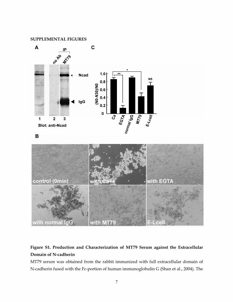
SUPPLEMENTAL FIGURES
Figure S1. Production and Characterization of MT79 Serum against the Extracellular Domain of N-cadherin MT79 serum was obtained from the rabbit immunized with full extracellular domain of N-cadherin fused with the Fc-portion of human immunoglobulin G (Shan et al., 2004). The
8
IgG fraction of the serum was purified for experimental use. (A) Mouse brain homogenate was subjected to immunoblot probed with MT79 (lane 1). A 127 kDa band corresponded to N-cadherin. Brain homogenate was incubated without (lane 2) or with (lane 3) MT79, and the resultant immunoprecipitate was subjected to immunoblot probed with anti-N-cadherin antibody that recognizes the intracellular domain of N-cadherin (Tanaka et al., 2000). Arrow, N-cadherin; arrowhead, immunoglobulin heavy chain. (B) Cell aggregation assay was performed using L929 cells (Takeichi, 1977). N-cadherin transfected cells formed aggregates with the existence of extracellular Ca++ (upper panels). Addition of MT79 specifically inhibited the aggregation of N-cadherin expressing cells (with MT79), but did not the aggregation of E-cadherin expressing cells as control (E-Lcell). Normal IgG as a control did not show inhibitory effect on N-cadherin-mediated cell aggregation (with normal IgG). (C) Cell aggregation was quantified by counting the number of aggregates at time 0 (N0) and 30 min (N30).
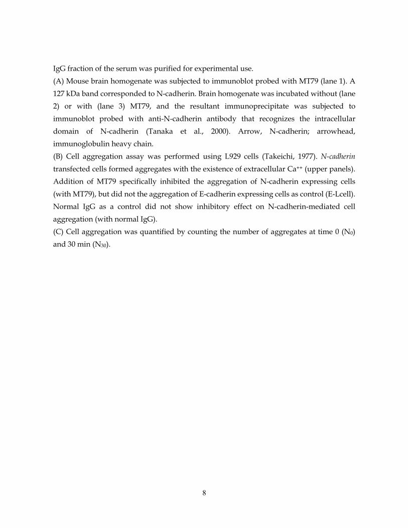
9
Figure S2. Structure of TAO2β Gene (A) (Middle) Organization of the TAO2 coding region exons. Both tao2α and β mRNAs utilize the identical exons 1-14 other than exon 11, of which TAO2β is 18 base longer than that of tao2α (thick line). (Top and bottom) Diagrams of the structure of TAO2α and β mRNAs encoding the C-terminal domains, respectively. The C-terminal region of TAO2β is encoded by a mRNA portion transcribed from a part of exon 15 and entire exons 16-18,
whereas exon 15-derived mRNA covers the whole C-terminal region of TAO2α. (B) Nucleotide and amino acid sequences of TAO2β C‐terminal region. The junction of each exon is indicated.
10
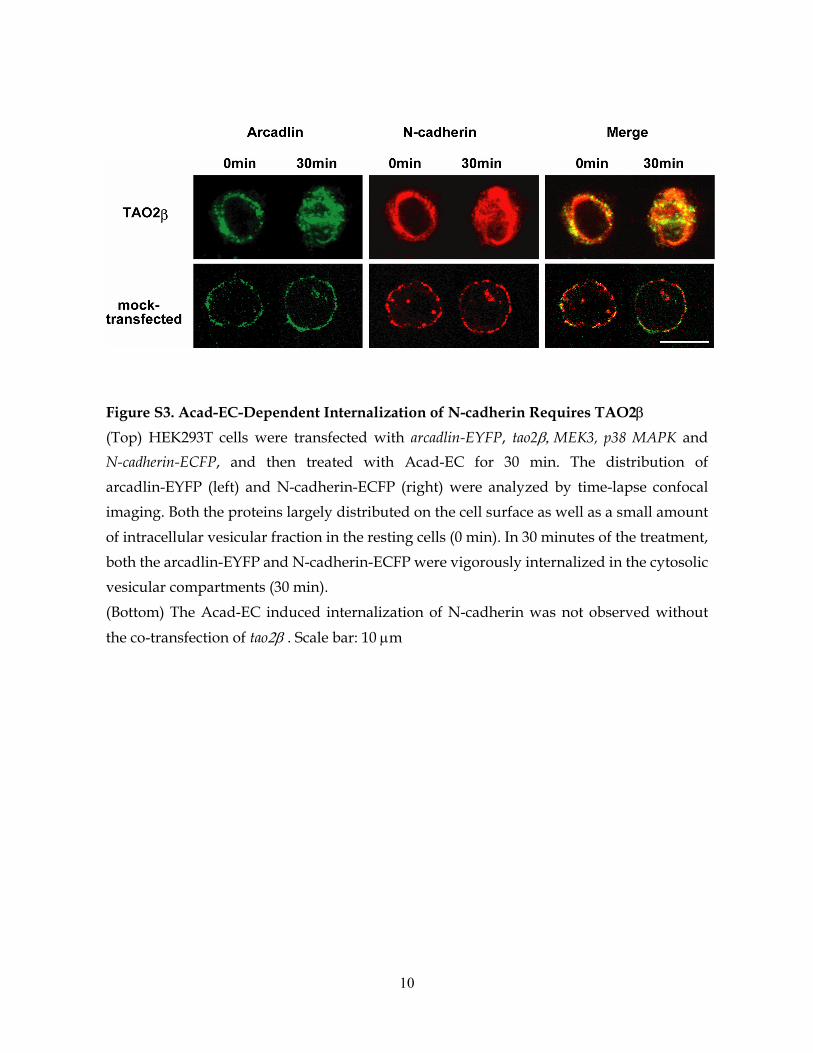
Figure S3. Acad-EC-Dependent Internalization of N-cadherin Requires TAO2β
(Top) HEK293T cells were transfected with arcadlin-EYFP, tao2β, MEK3, p38 MAPK and N-cadherin-ECFP, and then treated with Acad-EC for 30 min. The distribution of arcadlin-EYFP (left) and N-cadherin-ECFP (right) were analyzed by time-lapse confocal imaging. Both the proteins largely distributed on the cell surface as well as a small amount of intracellular vesicular fraction in the resting cells (0 min). In 30 minutes of the treatment, both the arcadlin-EYFP and N-cadherin-ECFP were vigorously internalized in the cytosolic vesicular compartments (30 min). (Bottom) The Acad-EC induced internalization of N-cadherin was not observed without
the co-transfection of tao2β . Scale bar: 10 µm

11
Figure S4. Arcadlin Overexpression and N-cadherin Knockdown to Verify the Role of Arcadlin in the Regulation of Spine Density (A) Cultured acad-/- neurons were doubly transfected with EGFP and mock, arcadlin, or arcadlin-l at 6 DIV, and visualized with GFP at 15 DIV. EGFP positive neurons (green, top) were also positive for transfected arcadlin (middle) or arcadlin-L (right), as examined by immunocytochemistry (red, bottom). Left, mock transfected. (B) HEK293T cells were doubly transfected with mouse N-cadherin-EGFP and N-cadherin-siRNA (lane 1) or scramble-siRNA (lane 2), and subjected to immunoblot for mouse N-cadherin-GFP (GFP epitope). Lane 3, no transfection. Lane 4, N-cadherin-EGFP single transfection. The upper band of the N-cadherin-GFP doublet is supposed to be an immature peptide without truncation of the N-terminal prodomain.
Scale bar: 10 µm
12
REFERENCES FOR SUPPLEMENTAL FIGURE LEGENDS
Shan, W., Yagita, Y., Wang, Z., Koch, A., Svenningsen, A. F., Gruzglin, E., Pedraza, L., and Colman, D. R. (2004). The Minimal Essential Unit for Cadherin‐mediated Intercellular Adhesion Comprises Extracellular Domains 1 and 2. J Biol Chem 279, 55914‐55923.
Takeichi, M. (1977). Functional correlation between cell adhesive properties and some cell surface proteins. J Cell Biol 75, 464‐474.
Tanaka, H., Shan, W., Phillips, G. R., Arndt, K., Bozdagi, O., Shapiro, L., Huntley, G. W., Benson, D. L., and Colman, D. R. (2000). Molecular modification of N‐cadherin in response to synaptic activity. Neuron 25, 93‐107.


![Kurdistan Operator Activity Map[1]](https://static.fdocument.org/doc/165x107/55cf99fc550346d0339ffec6/kurdistan-operator-activity-map1.jpg)