Oligomeric Structure of ²-Glucosidases
7
Journal of Photoscience (2004), Vol. 11(3), pp. 121-127 121 Oligomeric Structure of β-Glucosidases Sang-Yeob Kim and In-Soo Kim * Department of Genetic Engineering, College of Natural Science, Kyungpook National University, Daegu 702-701, South Korea INTRODUCTION β-Glucosidases (E.C. 3.2.1.21) catalyze the hydrolysis of the β-glycosidic bonds of aryl- and alkyl-β-glucoside, and cellobiose. They are widely distributed in all living organisms, as glycoconjugates are one of the most diverse groups of organic molecules in the biosphere. The aglycone of the glucosides confers substrate specificity of the enzyme [1] and the aglycones released from the glucosides play important physiological roles in the fundamental biological processes and in the industrial applications [2-9]. In plants, β-glucosidases have been implicated in regulating various aspects of principal metabolic events and growth- related responses, e.g. phytohormone activation [2-3], cell wall degradation in the endosperm during germination [4], lignification [5], and pathogen defense reaction [6-7]. An oat (Avena sativa) plastidal β-glucosidase is involved in the defense mechanism against fungal infection [8]. Cellulose, our most abundant renewable resource on this planet, breaks down cellulose to cellobiose, and b-glucosidase hydrolyzes the cellobiose to two glucose molecules. In humans, a β- glucosidase hydrolyzes glucosylceramide in the lysosome, and its lack leads to Gauchers disease, with the symptoms of anemia and thinning of the bones [9]. Glycosyl hydrolase (EC 3.2.1.x) have been classified into approximately 60 families on the basis of their amino acid sequence similarities rather than substrate specificity (10-12). These families of glycosyl hydrolase are grouped into 5 larger superfamilies called ‘clan GH A-E’ by significant similarities in tertiary structure along with conservation of the catalytic residues and catalytic mechanism. Family 1 is included in clan GH-A and characterized by an (β/α) 8 barrel structure, retaining in the anomeric configuration of the glycone and two catalytic glutamic acid residues in the β strands number 4 and 7. β-Glucosidases appear in 1, 3 and 4 of these families. In particular, a large number of these enzymes are grouped in family 1. In active site of the family 1 β-glucosidases, the two catalytic glutamic acids in the I/VTENG and TNEP motifs play critical roles in catalysis as a nucleophile or an acid/base catalyst, respectively [13]. However, a few of experimental data on molecular determination of aglycone specificity of β- glucosidases are now available [14-15]. In particular, glycosyl hydrolase family 1 (β-glucosidase) has similar tertiary structure and a strong tendency to form homo- and hetero-oligomers [10-12]. A main difference in their tertiary structure of the monomers lies in the surface loops near active site [16]. The quaternary protein structures of family I β-glucosidases are of great interest because they exist as various forms of oligimers such as dimer, tetramer, octamer or larger aggregates made of different numbers of subunits [16-25]. Nevertheless, the functional implications of such diverse oligomerization schemes remain unclear. Structural studies of the unique oligomeric β-glucosidases are an important requirement toward uncovering their precise role(s) in biological processes. OLIGOMERIZATION CHARACTERISTICS OF β- GLUCOSIDASES The large number of glycoside hydrolase families reflects this diversity of substrates and the need for selective hydrolysis of the glycosidic bond. The three-dimentional crystal structure of several family 1 β-glucosidases (1CBG, white clover (Trifolium repens) linamarase; 1MYR, white mustard (Sinapis alba) myrosinase; 1BGA, Bacillus polymyxa β-glucosidase; 1PBG, Lactococcus lactis phospho-β- galactosidase; 1GOW, Sulfolobus solfataricus β-glucosidase; 1QVB, Thermosphaera aggregans β-glucosidase, 1QOX, Bacillus circulans sp. alkalophilus β-glucosidase; 1ELE, maize β-glucosidase) has recently been elucidated [16-23]. Glycosyl hydrolase family 1 to which the BGA family belongs has similar tertiary structure, catalytic mechanism and a tendency to form homo- and hetero-oligomers [10-12]. Although multimeric enzyme proteins are generally composed of a fixed number or molar ratio of subunits, some BGA family β-glucosidases have a tendency to form discrete homooligomers having different number of subunits. For example, in Hevea β-glucosidase and casava linamarase exist homooligomers of different MW that are composed of different number of single monomers [24-25]. These enzymes *To whom correspondence should be addressed. E-mail : [email protected] Received December 1, 2004; Accepted December 28, 2004
Transcript of Oligomeric Structure of ²-Glucosidases
Journal of Photoscience (2004), Vol. 11(3), pp. 121-127
121
Oligomeric Structure of β-Glucosidases
Sang-Yeob Kim and In-Soo Kim*
Department of Genetic Engineering, College of Natural Science, Kyungpook National University, Daegu 702-701, South Korea
INTRODUCTION
β-Glucosidases (E.C. 3.2.1.21) catalyze the hydrolysis ofthe β-glycosidic bonds of aryl- and alkyl-β-glucoside, andcellobiose. They are widely distributed in all livingorganisms, as glycoconjugates are one of the most diversegroups of organic molecules in the biosphere. The aglycone ofthe glucosides confers substrate specificity of the enzyme [1]and the aglycones released from the glucosides playimportant physiological roles in the fundamental biologicalprocesses and in the industrial applications [2-9].
In plants, β-glucosidases have been implicated in regulatingvarious aspects of principal metabolic events and growth-related responses, e.g. phytohormone activation [2-3], cellwall degradation in the endosperm during germination [4],lignification [5], and pathogen defense reaction [6-7]. An oat(Avena sativa) plastidal β-glucosidase is involved in thedefense mechanism against fungal infection [8]. Cellulose,our most abundant renewable resource on this planet, breaksdown cellulose to cellobiose, and b-glucosidase hydrolyzesthe cellobiose to two glucose molecules. In humans, a β-glucosidase hydrolyzes glucosylceramide in the lysosome,and its lack leads to Gauchers disease, with the symptoms ofanemia and thinning of the bones [9].
Glycosyl hydrolase (EC 3.2.1.x) have been classified intoapproximately 60 families on the basis of their amino acidsequence similarities rather than substrate specificity (10-12).These families of glycosyl hydrolase are grouped into 5 largersuperfamilies called ‘clan GH A-E’ by significant similaritiesin tertiary structure along with conservation of the catalyticresidues and catalytic mechanism. Family 1 is included inclan GH-A and characterized by an (β/α)8 barrel structure,retaining in the anomeric configuration of the glycone andtwo catalytic glutamic acid residues in the β strands number 4and 7. β-Glucosidases appear in 1, 3 and 4 of these families.In particular, a large number of these enzymes are grouped infamily 1. In active site of the family 1 β-glucosidases, the twocatalytic glutamic acids in the I/VTENG and TNEP motifs
play critical roles in catalysis as a nucleophile or an acid/basecatalyst, respectively [13]. However, a few of experimentaldata on molecular determination of aglycone specificity of β-glucosidases are now available [14-15].
In particular, glycosyl hydrolase family 1 (β-glucosidase)has similar tertiary structure and a strong tendency to formhomo- and hetero-oligomers [10-12]. A main difference intheir tertiary structure of the monomers lies in the surfaceloops near active site [16]. The quaternary protein structuresof family I β-glucosidases are of great interest because theyexist as various forms of oligimers such as dimer, tetramer,octamer or larger aggregates made of different numbers ofsubunits [16-25]. Nevertheless, the functional implications ofsuch diverse oligomerization schemes remain unclear.Structural studies of the unique oligomeric β-glucosidases arean important requirement toward uncovering their preciserole(s) in biological processes.
OLIGOMERIZATION CHARACTERISTICS OF β-GLUCOSIDASES
The large number of glycoside hydrolase families reflectsthis diversity of substrates and the need for selectivehydrolysis of the glycosidic bond. The three-dimentionalcrystal structure of several family 1 β-glucosidases (1CBG,white clover (Trifolium repens) linamarase; 1MYR, whitemustard (Sinapis alba) myrosinase; 1BGA, Bacillus polymyxaβ-glucosidase; 1PBG, Lactococcus lactis phospho-β-galactosidase; 1GOW, Sulfolobus solfataricus β-glucosidase;1QVB, Thermosphaera aggregans β-glucosidase, 1QOX,Bacillus circulans sp. alkalophilus β-glucosidase; 1ELE,maize β-glucosidase) has recently been elucidated [16-23].
Glycosyl hydrolase family 1 to which the BGA familybelongs has similar tertiary structure, catalytic mechanismand a tendency to form homo- and hetero-oligomers [10-12].Although multimeric enzyme proteins are generally composedof a fixed number or molar ratio of subunits, some BGAfamily β-glucosidases have a tendency to form discretehomooligomers having different number of subunits. Forexample, in Hevea β-glucosidase and casava linamarase existhomooligomers of different MW that are composed ofdifferent number of single monomers [24-25]. These enzymes
*To whom correspondence should be addressed.E-mail : [email protected] December 1, 2004; Accepted December 28, 2004
122 Sang-Yeob Kim and In-Soo Kim
display several discrete protein bands on native gel electro-phoresis of their extracts as does the oat β-glucosidase. Oat β-glucosidase is a linear assembly of a distinct structure consistedof monomers [26-27], whereas the other enzymes are anaggregate of different numbers of subunits (monomers). Thefunction of such oligomerization has not yet been elucidated,although it is assumed to be required for catalytic activity orstructural stability [16-23].
TECHNIQUES FOR STRUCTURAL STUDY OF OIGOMERIC PROTEINS
Conventional light microscopy and confocal fluorescencescanning light microscopy can not be used for structural studyof individual proteins, because of the low resolution of thesetechniques. Although atomic force microscopy (AFM) has asource of high-resolution information [28], it allows thevisualization only of the surfaces and therefore provideslimited insight into the internal three-dimensional (3-D)structure of macromolecules.
Three different techniques have proven successful forreaching beyond the wavelength limits of visible light andinto high resolution range in which individual molecules andtheir internal structure can be visualized: nuclear magneticresonance spectroscopy (NMR), X-ray crystallography, andhigh-resolution electron microscopy (EM).
Nuclear magnetic resonance spectroscopy (NMR) [29]NMR is particularly well suited for small molecules and for
individual domains of proteins. It allows the study of themacromolecule in solution, and avoids the time-consumingsearch for optimal crystallization conditions. However, thistechnique requires large amounts of protein (several milligrams)and a high solubility in water. NMR can not be successfullyapplied for the structural studies of typical membrane proteinsand filamentous proteins due to a size limit for the proteins tobe studied.
X-ray crystallography [30]X-ray analysis is the first method to solve the structure of
large individual protein and currently the excellent source ofstructural study. It allows the investigation of variousbiological specimens, ranging from peptides to viruses byirradiating a 3-D crystal, recording the spatial distribution andthe intensities of the diffracted X-ray beams and calculatingfrom diffraction patterns. X-ray crystallography is a powerfultechnique to reveal the internal three-dimensional (3-D)structure of macromolecules. But this method has severallimitations to elucidate large multi-component complexes.The critical procedure for an X-ray analysis is to grow largeand well-ordered 3-D crystals. Finding the suitable conditionsfor crystallization is usually a labor-intensive process as theconditions for crystal growth cannot be predicted. Inparticular, proteins of larger molecular weight often resist
attempts at crystallization and crystal may grow after flexiblestructural regions have been removed. In addition, it is moredifficult to crystallize integral membrane proteins andfilamentous proteins, such as actin and tubulin, as they oftenform aggregates rather than well-ordered 3-D crystals.
Transmission electron microscopy (TEM)NMR and X-ray crystallography are used for determining
the structure of proteins at an atomic resolution. However,these methods are limited to the study of small proteins andthe protein must be crystallized first. Electron microscopycannot give high enough resolution to reveal a protein atatomic resolution, but can be especially useful to view largeprotein complexes that cannot be crystallized. EM, combinedwith 3-D image reconstruction techniques of cryo-EM, electrontomography and single-particle analysis, offers some distinctadvantages over the X-ray crystallography and NMR methods:First, large protein assemblies and complexes, with dimensionsof 30-200 nm, are especially suitable for structural study bycryo-EM reconstruction. Second, nonhomogeneous samplescan be effectively studied because a homologous group can bemanually selected from the cryo-EM micrographs. Third,time-resolved studies can be carried out more easily becausebiochemical reactions can be terminated at a preferred timepoint by flash freezing. Forth, phase information for thestructure retained as the diffracted electron beam is refocusedto form a real-space image on an image plane. Finally,reconstruction at medium resolution (~20 Å) can be easilyobtained in a week.
On the other hand, several difficulties must be overcome toobtain the high-resolution information with EM [31-32].First, because the object of interest must be investigated in avacuum, special care must be taken to avoid sample drying.Second, since the object is heavily bombarded with high-energy electrons and biological objects are radiation sensitive,radiation damage must be minimized for recording intelligibleinformation. Finally, a 3-D structure must be reconstructedfrom 2-D projections of the molecule in various orientations,because electron micrographs represent only two dimensional(2-D) projections of a 3-D object.
A brief description for the 3-D image reconstructiontechniques of cryo-EM, electron tomography and single-particle analysis are as follows:
Cryo-electron microscopy The pioneering work of Taylor and Glaeser [33] and of
Dubochet and colleagues [34] paves the way for cryo-electronmicroscopy. Cryo-electron microscopy can be used tovisualise the intermediate structures in the reaction pathwaysof large protein complexes. It has the advantage that the vaporpressure of water becomes negligible at temperatures below -100C. Sample drying can thus be avoided by keeping theprotein at liquid nitrogen temperature. Vitrification of thesample using the plunge-freezing method works particularly
Oligomeric Structure of β-Glucosidases 123
well for individual proteins and multiprotein complexes. Inaddition, there is no distortion of the sample because thesample is always in solution. Also, the electron beam causesless damage to the sample though the low dose methodswhich are normally used in cryo-EM. However, biologicalmacromolecules are normally made up of carbon, hydrogen,oxygen, and nitrogen and the electron absorption of suchmolecules is very low. As a result, image contrast is also verylow and it is hard to detect features when dealing with just afew images. The important procedure for cryo-EM is to formthin vitreous ice. To prepare the sample for cryo-EM, thespecimen grid is rapidly plunged into liquid ethane and thenthe water turns into vitreous ice. The grid is transferred toliquid nitrogen and mounted in the cryoholder of the electronmicroscope.
Electron tomography [35]Electron tomography is particularly well suited for very
large particles such as cell organelles and large multiproteincomplexes. Images of the same specimen are recorded at tiltangle increments of about 1. Since the tolerable electron dosemust be divided over some 120-140 images, and sinceaveraging techniques cannot be applied, the resolution of suchtomograms is fairly low (20-70 Å). However, in comparisonwith large dimensions of such objects, the informationcontent of tomograms can be enormous. The specimen can beeither large individual particles or tissue sections. Usingvitrification of the sample using the plunge-freezing methodin combination with electron tomography allows one toinvestigate very large particles in three dimensions and in finedetail.
Single-particle analysis Single-particle analysis is suited for large proteins or protein
complexes (usually exhibiting a molecular mass higher than 500kDa) that can be obtained in sufficient quantities and homo-geneity. Upon classification and alignment of the particles, a 3-Dstructure can be calculated. Averaging of about 73,000 particlesyielded a resolution of 11.5 Å for the 70S ribosome [36].
QUATERNARY STRUCTURE OF β-GLUCOSIDASES
The members of family 1 β-glycosidase share high similarityin their tertiary structure from plants to archaeal bacteria thathave 17% amino acid sequence homology in the case of maizeand Sulfolobus solfataricus β-glucosidases [23]. The enzyme isglobular in shape, with eight a/b barrel motifs [(α/β)8] and twocatalytic glutamic acid residues in the b strands number 4 and 7[13]. The (α/β)8 fold is stable structural fold and one of thelargest and most regular of all domain structure [37]. It has beenfound in many different kinds of proteins, most of which areenzymes, with completely different amino acid sequences andfunctions. A comparison of the structure of these proteins shows
that the β strands and a helices are connected with loop regionsthat have quite different lengths and conformations in thedifferent proteins. This implies that the β strands and a heliceshave important roles in the structural framework of the enzyme,whereas the loops contain the amino acids responsible for itscatalytic chemistry and substrate binding. In some cases theloops are very long and form independent domains in the overallsubunit structure. The major structural difference among thefamily 1 enzymes lies in the surface loops surrounding the activesite [16].
Although it is common for the family I β-glycosidases toform oligomers, the regions involved in the formation of theoligomers vary widely from one enzyme to the other. A com-parison of critical regions contributing to the dimer formation,among the plant β-glycosidases; maize (Zea mays) β-glucosidase Glu1, white clover (Trifolium repens) linamarase,and white mustard (Sinapis alba) myrosinase shows nosimilarity [17-18, 23]. For example, the important regions ofdimer formation in white clover (Trifolium repens) linamaraseare located at the C-terminal region, whereas all of thedimeric interface residues in white mustard (Sinapis alba)myrosinase are located at the N-terminal region. Also, thedimer interface in the maize (Zea mays) β-glucosidase Glu1 islocated in the mid-section of the polypeptide chain. None ofthe above-mentioned interface residues is conserveduniversally among family 1 glycoside hydrolases, suggestingthat the specific monomer surface sites and domainsinteracting to form a quaternary structure are not critical aslong as they do not block the active site.
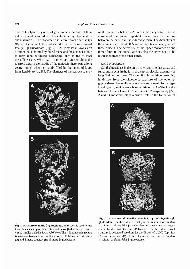
Maize β-glucosidaseIn maize (Zea mays), b-glucosidase (2-O-b-d-
glucopyranosyl-4-hydroxy-7-methoxy-1,4-benzoxazin-3-onehydrolase) occurs in two different forms, ZMGlu1 andZMGlu2 [23]. The deduced protein products of twoisoenzymes cDNAs share 90% sequence identity. Thecatalytically active form of maize β-glucosidase is a 120 kDahomodimer. As with other β-glucosidases, the ZMGlu1monomer folds as a (β/α)8 barrel fold (Fig. 1A). Theconnections between β-strands and a-helices within each β/αrepeat are elaborated further by the occurrence of shortsecondary-structural elements, which have also been observedin the other family 1 β-glycosidase structures. It exists as adimer that has a dimension of 103 Å (long) × 51 Å (wide) × 47Å (high) (Fig. 1B), while each monomer has overall dimensionof about 52 Å (long) × 51 Å (wide) × 47 Å (high). The dimerinterface is formed by residues and peptide spans that arelocated in the mid-section (Thr-270-Pro-400) of thepolypeptide chain.
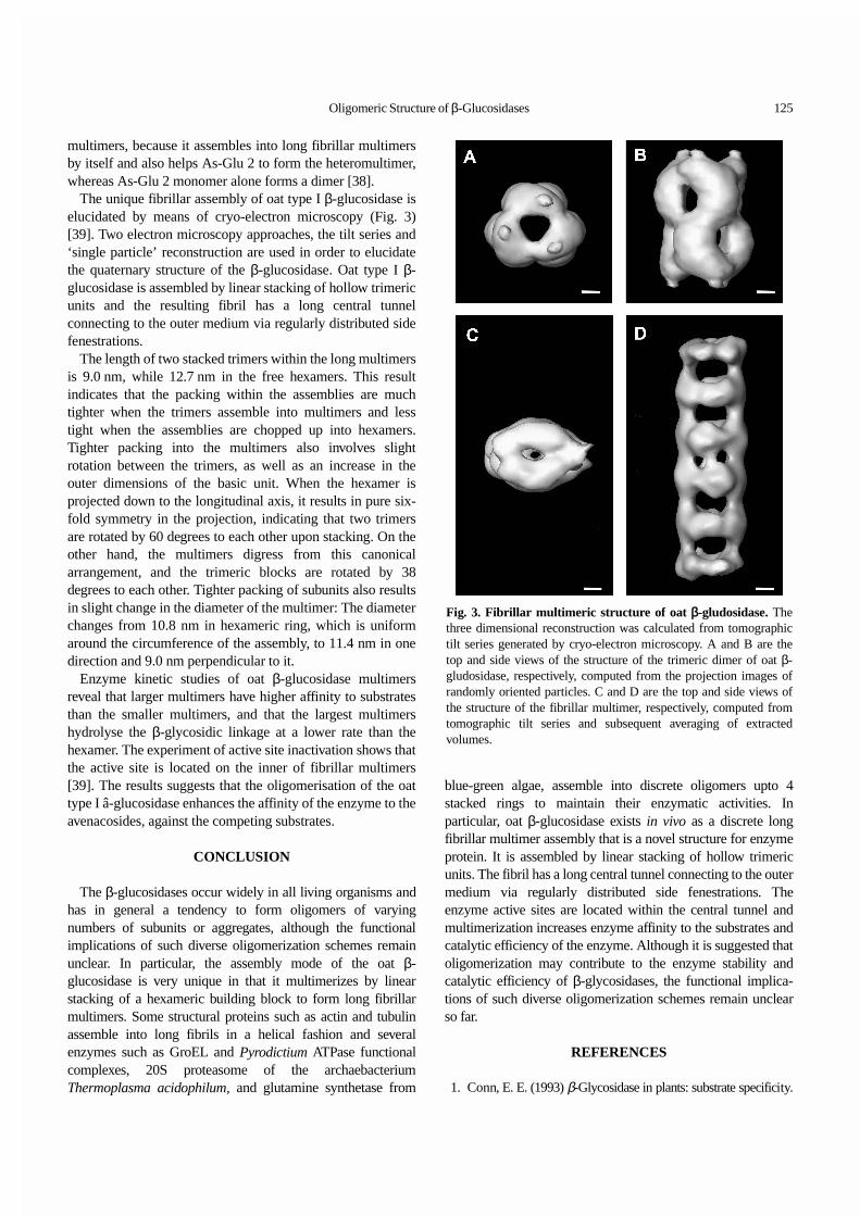
β-Glucosidase of Bacillus circulans sp. alkalophilus β-Glucosidase from B. circulans sp. alkalophilus optimally
growing at pH of above 9 has optimum activity in the pHrange of 6-9 and is fairly stable at temperatures up to 50oC.
124 Sang-Yeob Kim and In-Soo Kim
This cellulolytic enzyme is of great interest because of theirindustrial applications due to the stability at high temperatureand alkaline pH. The monomeric structure shows a similar (β/α)8 barrel structure to those observed within other members offamily 1 β-glucosidase (Fig. 2) [22]. It exists in vivo as anoctamer that is formed by four dimers, and the octamer is ableto form long polymeric assemblies only in the in vitrocrystalline state. When two octamers are viewed along thefourfold axis, in the middle of the molecule there exits a longcentral tunnel which is mainly filled by the layers of loopsfrom Leu364 to Arg369. The diameter of the narrowest entry
of the tunnel is below 5 Å. When the enzymatic functionconsidered, the more important tunnel may be the onebetween the dimers in the octameric form. The diameters ofthese tunnels are about 20 Å and active site cavities open intothese tunnels. The active site of the upper monomer of onedimer faces to the tunnel, as does also the active site of thelower monomer of the other dimer.
Oat β-glucosidaseOat β-glucosidase is the only known enzyme that exists and
functions in cells in the form of a supramolecular assembly oflong fibrillar multimers. The long fibrillar multimer assemblyis distinct from the oligomeric structure of the other β-glycosidases. The multimers exist as two isomeric forms, typeI and type II, which are a homomultimer of As-Glu 1 and aheteromultimer of As-Glu 1 and As-Glu 2, respectively [27].As-Glu 1 monomer plays a crucial role in the formation of
Fig. 1. Structure of maize β-gludosidase. PDB sever is used for thethree dimensional protein structures of maize β-gludosidase. Figurecan be handled with the Swiss-PdbViewer. The 3-dimensional structureis generated based on the coordinates of 1ELE. Monomeric structure(A) and dimeric structure (B) of maize β-gludosidase.
Fig. 2. Structure of Bacillus circulans sp. alkalophilus β-gludosidase. For three dimensional protein structures of Bacilluscirculans sp. alkalophilus β-Gludosidase, PDB sever is used. Figurecan be handled with the Swiss-PdbViewer. The three dimensionalstructure is generated based on the coordinates of 1QOX. Top-view(A) and side-view (B) of the oligomeric structure of Bacilluscirculans sp. alkalophilus β-gludosidase.
Oligomeric Structure of β-Glucosidases 125
multimers, because it assembles into long fibrillar multimersby itself and also helps As-Glu 2 to form the heteromultimer,whereas As-Glu 2 monomer alone forms a dimer [38].
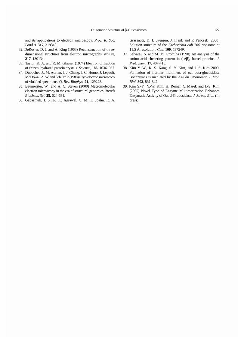
The unique fibrillar assembly of oat type I β-glucosidase iselucidated by means of cryo-electron microscopy (Fig. 3)[39]. Two electron microscopy approaches, the tilt series and‘single particle’ reconstruction are used in order to elucidatethe quaternary structure of the β-glucosidase. Oat type I β-glucosidase is assembled by linear stacking of hollow trimericunits and the resulting fibril has a long central tunnelconnecting to the outer medium via regularly distributed sidefenestrations.
The length of two stacked trimers within the long multimersis 9.0 nm, while 12.7 nm in the free hexamers. This resultindicates that the packing within the assemblies are muchtighter when the trimers assemble into multimers and lesstight when the assemblies are chopped up into hexamers.Tighter packing into the multimers also involves slightrotation between the trimers, as well as an increase in theouter dimensions of the basic unit. When the hexamer isprojected down to the longitudinal axis, it results in pure six-fold symmetry in the projection, indicating that two trimersare rotated by 60 degrees to each other upon stacking. On theother hand, the multimers digress from this canonicalarrangement, and the trimeric blocks are rotated by 38degrees to each other. Tighter packing of subunits also resultsin slight change in the diameter of the multimer: The diameterchanges from 10.8 nm in hexameric ring, which is uniformaround the circumference of the assembly, to 11.4 nm in onedirection and 9.0 nm perpendicular to it.
Enzyme kinetic studies of oat β-glucosidase multimersreveal that larger multimers have higher affinity to substratesthan the smaller multimers, and that the largest multimershydrolyse the β-glycosidic linkage at a lower rate than thehexamer. The experiment of active site inactivation shows thatthe active site is located on the inner of fibrillar multimers[39]. The results suggests that the oligomerisation of the oattype I â-glucosidase enhances the affinity of the enzyme to theavenacosides, against the competing substrates.
CONCLUSION
The β-glucosidases occur widely in all living organisms andhas in general a tendency to form oligomers of varyingnumbers of subunits or aggregates, although the functionalimplications of such diverse oligomerization schemes remainunclear. In particular, the assembly mode of the oat β-glucosidase is very unique in that it multimerizes by linearstacking of a hexameric building block to form long fibrillarmultimers. Some structural proteins such as actin and tubulinassemble into long fibrils in a helical fashion and severalenzymes such as GroEL and Pyrodictium ATPase functionalcomplexes, 20S proteasome of the archaebacteriumThermoplasma acidophilum, and glutamine synthetase from
blue-green algae, assemble into discrete oligomers upto 4stacked rings to maintain their enzymatic activities. Inparticular, oat β-glucosidase exists in vivo as a discrete longfibrillar multimer assembly that is a novel structure for enzymeprotein. It is assembled by linear stacking of hollow trimericunits. The fibril has a long central tunnel connecting to the outermedium via regularly distributed side fenestrations. Theenzyme active sites are located within the central tunnel andmultimerization increases enzyme affinity to the substrates andcatalytic efficiency of the enzyme. Although it is suggested thatoligomerization may contribute to the enzyme stability andcatalytic efficiency of β-glycosidases, the functional implica-tions of such diverse oligomerization schemes remain unclearso far.
REFERENCES
1. Conn, E. E. (1993) β-Glycosidase in plants: substrate specificity.
Fig. 3. Fibrillar multimeric structure of oat β-gludosidase. Thethree dimensional reconstruction was calculated from tomographictilt series generated by cryo-electron microscopy. A and B are thetop and side views of the structure of the trimeric dimer of oat β-gludosidase, respectively, computed from the projection images ofrandomly oriented particles. C and D are the top and side views ofthe structure of the fibrillar multimer, respectively, computed fromtomographic tilt series and subsequent averaging of extractedvolumes.

126 Sang-Yeob Kim and In-Soo Kim
In: β-glucosidase: Biochemistry and Mole-cular Biology. Ed.By Esen, A. American Chemical Society, Washing, DC pp. 15-26.
2. Brzobohaty, B., I. Moore, P. Kristoffersen, L. Bakp, N.Campos, J. Schell and K. Palme (1993) Release of activecytokinin by a β-glucosidase localized to the maize rootmeristem. Science, 262, 1051-1054.
3. Falk, A., and L. Rask (1995) Expression of a zeatin-O-glucoside-degrading beta-glucosidase in Brassica napus.Plant Physiol. 108, 13691377.
4. Leah, R., J. Kigel, I. Svendsen, and J. Mundy (1995) Bio-chemical and molecular characterization of a barley seedbeta-glucosidase. J. Biol. Chem. 270, 1578915797.
5. Dharmawardhana, D. P., B. E. Ellis and J. E. Carlson (1995)A β-glucosidase from lodgepole pine xylem specific for thelignin precursor coniferin. Plant Physiol. 107: 331-339.
6. Zdero, C., F. Bohlmann and H. M. Niemeyer (1990) ent-labdane glycosides from Baccharis pingraea. Phytochemistry,29, 2611 2616.
7. Poulton, J. E. (1990) Cyanogenesis in Plants. Plant Physiol.94, 401-405.
8. Nisius, A. (1988) The stromacentre in Avena plastids: anaggregation of β-glucosidase responsible for the activationof oat-leaf saponins. Planta, 173, 474-481.
9. Beutler, E., G. A. Grabowski (1995) Glucosylceramide lipidoses:Gaucher disease. In: The Metabolic and Molecular Bases ofInherited Disease. Ed. By Scriver, C. R., A. L. Beaudet, W.S. Sly, D. Valle. McGraw-Hill, NY pp. 2641-2670.
10. Henrissat, B. (1991) A classification of glycosyl hydrolasesbased on amino acid sequence similarities. Biochem. J. 280,309-316.
11. Henrissat B., and A. Bairoch (1993) New families in theclassification of glycosyl hydrolases based on amino acidsequence similarities. Biochem. J. 293, 781-788.
12. Henrissat, B. and A. Bairoch (1996) Updating the sequence-based classification of glycosyl hydrolases. Biochem. J. 316,695-696.
13. Henrissat, B., I. Callebaut, S. Fabrega, P. Lehn, J.-E. Mornonand G. Davies (1995) Conserved catalytic machinery and theprediction of a common fold for several families of glycosylhydrolases. Proc. Natl. Acad. USA 92, 7090-7094 .
14. Verdoucq, L., J. Moriniere, D. R. Bevan, A. Esen, A. Vasella,B. Henrissat and M. Czjzek (2004) Structural determinantsof substrate specificity in family 1 beta-glucosidases: novelinsights from the crystal structure of sorghum dhurrinase-1,a plant beta-glucosidase with strict specificity, in complexwith its natural substrate. J. Biol. Chem. 279, 31796-31803.
15. Verdoucq, L., M. Czjzek, J. Moriniere, D. R. Bevan, and A.Esen (2003) Mutational and structural analysis of aglyconespecificity in maize and sorghum beta-glucosidases. J. Biol.Chem. 278, 25055-25062.
16. Sanz-Aparicio, J., J. A. Hermoso, M. Martinez-Ripoll, J. L.Lequerica and J. Polaina (1998) Crystal structure of b-glucosidase A from Bacillus polymyxa: insights into thecatalytic activity in family 1 glycosyl hydrolases. J. Mol.Biol. 275, 491-502.
17. Barrett, T., C. G. Suresh, S. P. Tolley, E. J. Dodson and M. A.
Hughes (1995). The crystal structure of a cyanogenic beta-glucosidase from white clover, a family 1 glycosyl hydrolase.Structure, 3, 951-960.
18. Burmeister, W. P., S. Cottaz, H. Driguez, R. Iori, S. Palmieri andB. Henrissat (1997) The crystal structures of Sinapis albamyrosinase and a covalent glycosyl-enzyme intermediateprovide insights into the substrate recognition and active-sitemachinery of an S-glycosidase. Structure, 5, 663-675.
19. Wiesmann, C., G. Beste, W. Hengstenberg and G. E. Schulz(1995) The three-dimensional structure of 6-phospho-β-galactosidase from Lactococcus lactis. Structure, 3, 961-968.
20. Aguilar, C. F., I. Sanderson, M. Moracci, M. Ciaramella, R.Nucci, M. Rossi and L. H. Pearl (1997) Crystal structure of theβ-glycosidase from the hyperthermophilic archeon Sulfolobussolfataricus: resilience as a key factor in thermostability. J. Mol.Biol. 271, 789-802.
21. Chi, Y.-I., L. A. Martinez-Cruz, k J. Jancari, R. V. Swanson, D.E. Robertson and S.-H. Kim (1999) Crystal structure of the β-glucosidase from hyperthermophile Thermosphaera aggregans:insights into its activity and thermostability. FEBS Lett. 445,375-383.
22. Hakulinen, N., S. Paavilainen, T. Korpela and J. Rouvinen(2000). The crystal structureof beta-glucosidase from Bacilluscirculans sp. alkalophilus: ability to form long polymericassemblies. J. Struct. Biol. 129, 69-79.
23. Czjzek, M., M. Cicek, V. Zamboni, W. P. Burmeister, D. R.Bevan, B. Henrissat and A. Esen (2001), Crystal structure of amonocotyledon (maize ZMGlu1) beta-glucosidase and a modelof its complex with p-nitrophenyl beta-D-thioglucoside.Biochem. J. 354, 37-46.
24. Selmar, D., R. Lieberei, B. Biehl and J. Voigt (1987) Hevealinamarase-α nonspecific β-glycosidase. Plant Physiol. 83,557-563.
25. Sermsuvityawong, K., M. R. Jisnuson, P. S. Svasti, Areetrakui,P. Kisamanonta and M. Chulavatnatol (1995) Aggregation ofcassava linamarase. J. Sci. Soc. Thailand, 21, 283-292.
26. Gus-Mayer, S., H. Brunner, H. A. W. Schneider-Poetsch andW. Rudiger (1994) Avenacosidase from oat: purification,sequence analysis and biochemical characterizaton of a newmember of BGA family of β-glucosidase. Plant Mol. Biol.26, 909-921.
27. Kim, Y.-W., P.-S. Song and I.-S. Kim (1996) Purification andcharacterization of isoenzyme of β-glucosidase from etiolatedoat seedlings. Mol. Cells 6, 773-779.
28. Engel, A., Y. Lyubchenko and D. Müller (1999) Atomicforce microscopy: a powerful tool to observe biomoleculesat work. Trends Cell Biol. 9, 7780.
29. Wuthrich, K. (1995) NMR-this other method for protein andnucleic acid structure determination. Acta. Crystallogr. D.Biol. Crystallogr. 51, 249-270.
30. McPherson, A. (1991) Useful principles for the crystalliza-tion of proteins. In: Crystallization of membrane proteins.Ed. By Michel, H. CRC Press Inc., Boca Rotan, Florida, pp.2-51.
31. Crowther, R. A., D.J. DeRosier and A. Klug (1970) Thereconstruction of a three-dimensional structure from projections
Oligomeric Structure of β-Glucosidases 127
and its applications to electron microscopy. Proc. R. Soc.Lond A. 317, 319340.
32. DeRosier, D. J. and A. Klug (1968) Reconstruction of three-dimensional structures from electron micrographs. Nature,217, 130134.
33. Taylor, K. A. and R. M. Glaeser (1974) Electron diffractionof frozen, hydrated protein crystals. Science, 186, 10361037
34. Dubochet, J., M. Adrian, J. J. Chang, J. C. Homo, J. Lepault,McDowall A. W. and Schultz P. (1988) Cryo-electron microscopyof vitrified specimens. Q. Rev. Biophys. 21, 129228.
35. Baumeister, W., and A. C. Steven (2000) Macromolecularelectron microscopy in the era of structural genomics. TrendsBiochem. Sci. 25, 624-631.
36. Gabashvili, I. S., R. K. Agrawal, C. M. T. Spahn, R. A.
Grassucci, D. I. Svergun, J. Frank and P. Penczek (2000)Solution structure of the Escherichia coli 70S ribosome at11.5 Å resolution. Cell, 100, 537549.
37. Selvaraj, S. and M. M. Gromiha (1998) An analysis of theamino acid clustering pattern in (α/β)8 barrel proteins. J.Prot. chem. 17, 407-415.
38. Kim Y. W., K. S. Kang, S. Y. Kim, and I. S. Kim 2000.Formation of fibrillar multimers of oat beta-glucosidaseisoenzymes is mediated by the As-Glu1 monomer. J. Mol.Biol. 303, 831-842.
39. Kim S.-Y., Y.-W. Kim, H. Reiner, C. Marek and I.-S. Kim(2005) Novel Type of Enzyme Multimerization EnhancesEnzymatic Activity of Oat β-Gludosidase. J. Struct. Biol. (Inpress)


![Elovanoids counteract oligomeric β-amyloid-induced …cognition (Alzheimer’s disease) and sight (age-related macular de-generation [AMD]). How neuroinflammation can be counteracted](https://static.fdocument.org/doc/165x107/5f2eb83dff582622624e3d80/elovanoids-counteract-oligomeric-amyloid-induced-cognition-alzheimeras-disease.jpg)