The production of ²-glucosidases by Fusarium proliferatum NBRC109045 isolated from
13
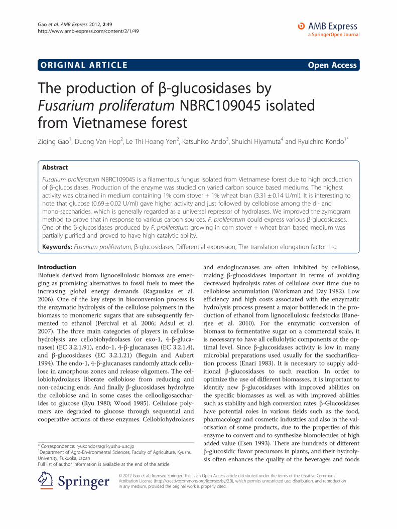
ORIGINAL ARTICLE Open Access The production of β-glucosidases by Fusarium proliferatum NBRC109045 isolated from Vietnamese forest Ziqing Gao 1 , Duong Van Hop 2 , Le Thi Hoang Yen 2 , Katsuhiko Ando 3 , Shuichi Hiyamuta 4 and Ryuichiro Kondo 1* Abstract Fusarium proliferatum NBRC109045 is a filamentous fungus isolated from Vietnamese forest due to high production of β-glucosidases. Production of the enzyme was studied on varied carbon source based mediums. The highest activity was obtained in medium containing 1% corn stover + 1% wheat bran (3.31 ± 0.14 U/ml). It is interesting to note that glucose (0.69 ± 0.02 U/ml) gave higher activity and just followed by cellobiose among the di- and mono-saccharides, which is generally regarded as a universal repressor of hydrolases. We improved the zymogram method to prove that in response to various carbon sources, F. proliferatum could express various β-glucosidases. One of the β-glucosidases produced by F. proliferatum growing in corn stover + wheat bran based medium was partially purified and proved to have high catalytic ability. Keywords: Fusarium proliferatum, β-glucosidases, Differential expression, The translation elongation factor 1-α Introduction Biofuels derived from lignocellulosic biomass are emer- ging as promising alternatives to fossil fuels to meet the increasing global energy demands (Ragauskas et al. 2006). One of the key steps in bioconversion process is the enzymatic hydrolysis of the cellulose polymers in the biomass to monomeric sugars that are subsequently fer- mented to ethanol (Percival et al. 2006; Adsul et al. 2007). The three main categories of players in cellulose hydrolysis are cellobiohydrolases (or exo-1, 4-β-gluca- nases) (EC 3.2.1.91), endo-1, 4-β-glucanases (EC 3.2.1.4), and β-glucosidases (EC 3.2.1.21) (Beguin and Aubert 1994). The endo-1, 4-β-glucanases randomly attack cellu- lose in amorphous zones and release oligomers. The cel- lobiohydrolases liberate cellobiose from reducing and non-reducing ends. And finally β-glucosidases hydrolyze the cellobiose and in some cases the cellooligosacchar- ides to glucose (Ryu 1980; Wood 1985). Cellulose poly- mers are degraded to glucose through sequential and cooperative actions of these enzymes. Cellobiohydrolases and endoglucanases are often inhibited by cellobiose, making β-glucosidases important in terms of avoiding decreased hydrolysis rates of cellulose over time due to cellobiose accumulation (Workman and Day 1982). Low efficiency and high costs associated with the enzymatic hydrolysis process present a major bottleneck in the pro- duction of ethanol from lignocellulosic feedstocks (Bane- rjee et al. 2010). For the enzymatic conversion of biomass to fermentative sugar on a commercial scale, it is necessary to have all cellulolytic components at the op- timal level. Since β-glucosidases activity is low in many microbial preparations used usually for the saccharifica- tion process (Enari 1983). It is necessary to supply add- itional β-glucosidases to such reaction. In order to optimize the use of different biomasses, it is important to identify new β-glucosidases with improved abilities on the specific biomasses as well as with improved abilities such as stability and high conversion rates. β-Glucosidases have potential roles in various fields such as the food, pharmacology and cosmetic industries and also in the val- orisation of some products, due to the properties of this enzyme to convert and to synthesize biomolecules of high added value (Esen 1993). There are hundreds of different β-glucosidic flavor precursors in plants, and their hydroly- sis often enhances the quality of the beverages and foods * Correspondence: [email protected] 1 Department of Agro-Environmental Sciences, Faculty of Agriculture, Kyushu University, Fukuoka, Japan Full list of author information is available at the end of the article © 2012 Gao et al.; licensee Springer. This is an Open Access article distributed under the terms of the Creative Commons Attribution License (http://creativecommons.org/licenses/by/2.0), which permits unrestricted use, distribution, and reproduction in any medium, provided the original work is properly cited. Gao et al. AMB Express 2012, 2:49 http://www.amb-express.com/content/2/1/49
Transcript of The production of ²-glucosidases by Fusarium proliferatum NBRC109045 isolated from
Gao et al. AMB Express 2012, 2:49http://www.amb-express.com/content/2/1/49
ORIGINAL ARTICLE Open Access
The production of β-glucosidases byFusarium proliferatum NBRC109045 isolatedfrom Vietnamese forestZiqing Gao1, Duong Van Hop2, Le Thi Hoang Yen2, Katsuhiko Ando3, Shuichi Hiyamuta4 and Ryuichiro Kondo1*
Abstract
Fusarium proliferatum NBRC109045 is a filamentous fungus isolated from Vietnamese forest due to high productionof β-glucosidases. Production of the enzyme was studied on varied carbon source based mediums. The highestactivity was obtained in medium containing 1% corn stover + 1% wheat bran (3.31 ± 0.14 U/ml). It is interesting tonote that glucose (0.69 ± 0.02 U/ml) gave higher activity and just followed by cellobiose among the di- andmono-saccharides, which is generally regarded as a universal repressor of hydrolases. We improved the zymogrammethod to prove that in response to various carbon sources, F. proliferatum could express various β-glucosidases.One of the β-glucosidases produced by F. proliferatum growing in corn stover + wheat bran based medium waspartially purified and proved to have high catalytic ability.
Keywords: Fusarium proliferatum, β-glucosidases, Differential expression, The translation elongation factor 1-α
IntroductionBiofuels derived from lignocellulosic biomass are emer-ging as promising alternatives to fossil fuels to meet theincreasing global energy demands (Ragauskas et al.2006). One of the key steps in bioconversion process isthe enzymatic hydrolysis of the cellulose polymers in thebiomass to monomeric sugars that are subsequently fer-mented to ethanol (Percival et al. 2006; Adsul et al.2007). The three main categories of players in cellulosehydrolysis are cellobiohydrolases (or exo-1, 4-β-gluca-nases) (EC 3.2.1.91), endo-1, 4-β-glucanases (EC 3.2.1.4),and β-glucosidases (EC 3.2.1.21) (Beguin and Aubert1994). The endo-1, 4-β-glucanases randomly attack cellu-lose in amorphous zones and release oligomers. The cel-lobiohydrolases liberate cellobiose from reducing andnon-reducing ends. And finally β-glucosidases hydrolyzethe cellobiose and in some cases the cellooligosacchar-ides to glucose (Ryu 1980; Wood 1985). Cellulose poly-mers are degraded to glucose through sequential andcooperative actions of these enzymes. Cellobiohydrolases
* Correspondence: [email protected] of Agro-Environmental Sciences, Faculty of Agriculture, KyushuUniversity, Fukuoka, JapanFull list of author information is available at the end of the article
© 2012 Gao et al.; licensee Springer. This is anAttribution License (http://creativecommons.orin any medium, provided the original work is p
and endoglucanases are often inhibited by cellobiose,making β-glucosidases important in terms of avoidingdecreased hydrolysis rates of cellulose over time due tocellobiose accumulation (Workman and Day 1982). Lowefficiency and high costs associated with the enzymatichydrolysis process present a major bottleneck in the pro-duction of ethanol from lignocellulosic feedstocks (Bane-rjee et al. 2010). For the enzymatic conversion ofbiomass to fermentative sugar on a commercial scale, itis necessary to have all cellulolytic components at the op-timal level. Since β-glucosidases activity is low in manymicrobial preparations used usually for the saccharifica-tion process (Enari 1983). It is necessary to supply add-itional β-glucosidases to such reaction. In order tooptimize the use of different biomasses, it is important toidentify new β-glucosidases with improved abilities onthe specific biomasses as well as with improved abilitiessuch as stability and high conversion rates. β-Glucosidaseshave potential roles in various fields such as the food,pharmacology and cosmetic industries and also in the val-orisation of some products, due to the properties of thisenzyme to convert and to synthesize biomolecules of highadded value (Esen 1993). There are hundreds of differentβ-glucosidic flavor precursors in plants, and their hydroly-sis often enhances the quality of the beverages and foods
Open Access article distributed under the terms of the Creative Commonsg/licenses/by/2.0), which permits unrestricted use, distribution, and reproductionroperly cited.
Gao et al. AMB Express 2012, 2:49 Page 2 of 13http://www.amb-express.com/content/2/1/49
produced from them (Gϋnata 2003; Esen 2003). Asidefrom flavor enhancement, foods, feeds, and beverages maybe improved nutritionally by release of vitamins, antioxi-dants, and other beneficial compounds from their glyco-sides (Opassiri et al. 2004). Indeed, β-glucosidase caneither degrade or synthesize small carbohydrate polymers,depending on particular experimental conditions (Croutand Vic 1998). The β-glucosidases can be arranged inthree groups related to localization: intracellular, cell wallassociated, and extracellular. Primarily the extracellularβ-glucosidases are of industrial interest (Soewnsen 2010).The number of fungal species on earth is estimated to1.5 million of which as little as approximately 5% areknown (Hawksworth 1991; 2001). So there is a statementthat calls for all-out effort to unravel the potential ofunknown species found in nature. The identificationand characterization of new fungal species are oftenencountered in literature. Cuc Phuong Park and Ba BePark is the old national one in Vietnam and boasts anengaging cultural and wildlife heritage and enchantingscenery. Covered in a dense forest, these landscapesare rich and diverse tropical and subtropical species ofmicroorganisms for wood and plant degradation. In thepresent study, a potential β-glucosidases-producingfungus NBRC109045 was isolated from Ba Be nationalpark and identified as Fusarium proliferatum. Under opti-mized conditions, F. proliferatum produces β-glucosidaseswith an activity of 3.3 U/ml based on pNPG as substrateand an activity of 426 U/ml based on cellobiose as sub-strate. In this paper, we described ways that (a) isolatingand screening microbes to produce considerable quantitiesof β-glucosidases; (b) modifying the method of zymogramto prove that different carbon sources direct variedβ-glucosidases expression in F. proliferatum; (c) assayingpartial purification to prove high catalytic efficiency ofβ-glucosidase produced by F. proliferatum growing in cornstover + wheat bran based medium.
Materials and methodsMaterialsUnless specified otherwise, all chemicals were of analyt-ical grade. Solubilized crystalline cellulose was obtainedfrom Kyokuto Seiyaku Co., Ltd, Japan. Avicel [(R) RH-101], 4-methylumbelliferyl-β-D-glucoside (MUG) andcarboxymethyl cellulose (CMC) were products of SigmaChemical Co., (St. Louis, Mo, USA). Cellobiose, xylose,glucose, sucrose, galactose and maltose were purchasedfrom Wako Pure Chemical Industries, Ltd, Japan.4-Nitrophenyl-β-D-glucopyranoside monohydrate (pNPG)was purchased from Tokyo Chemical Industry Co., Ltd,Japan. Corn stover was collected from Yingkou city, Liao-ning Province in China. Wheat bran and bagasse wereobtained from private companies.
Strains isolationWood chip of Jatropha carcass, branch and leaves ofJ. carcass, wood chip of Manihot esculenta, branch andleaves of M. esculenta, coconut shell, sugarcane, and ricestraw were used as lignocellulosic sources for degrad-ation in Vietnamese National Park (Ba Be and CucPhuong). One month later, lignocellulosic sources weredug up. All strains that would be screened were isolatedfrom degraded biomass samples and washed soil col-lected. Isolated strains were inoculated on solubilizedcrystalline cellulose (CC) plates and CMC plates to culti-vate for two weeks (Deguchi et al. 2007). The microbesthat could grow on CC and CMC were picked up andinoculated onto malt extract agar (MEA).
Screening of β-glucosidases-producing strainsThe first step of screeningFor primary screening, strains from MEA were platedon potato dextrose agar (PDA) medium in a 9-cm diam-eter Petri dish and incubated at 30°C for 5 days. Thenthe colonies were inoculated on β-glucosidases (EC3.2.1.21) screening agar containing 1% of CMC, 0.5% ofMUG, 1.5% of agar, and Mandels salts (Daenen et al.2008). The cultures were incubated at 30°C for 3 days.Then the plates were observed under UV light. Colonieswhich showed fluorescence were sorted out. It is becausemethylumbelliferyl (MU) which was released from MUGby β-glucosidases can emit fluorescence when inducedby UV light.
The second step of screeningFor secondary screening, the mycelium of the β-glucosidases-producing isolates obtained from the pri-mary screening was transferred to a new PDA mediumin a 9-cm diameter Petri dish and incubated at 30°C.Once the fungus covered most of the PDA plate, agarplates with mycelium were transferred to a sterileblender containing 25 ml of sterile water and homoge-nized for 30 s. Ten ml of the fungal homogenate wasused to inoculate into β-glucosidases secondary screen-ing medium containing 1% corn stover + 1% wheat branin 100 ml, pH 5.0 Mandels salts medium with KH2PO4
2 g l-1, (NH4)2SO4 1.4 g l-1, urea 0.69 g l-1, CaCl2�2H2O0.3 g l-1, MgSO4�7H2O 0.3 g l-1, and 1 ml trace elementssolution composing of MnSO4 1.6 g l-1, ZnSO4 2 g l-1,CuSO4 0.5 g l-1, CoSO4 0.5 g l-1 (Saibi et al. 2011) thenincubated at 30°C, 150 rpm for 5 days. Crude enzymeextract was obtained by centrifuging the liquid mediumat 20 000 g, 4°C for 20 min and collecting the super-natant for confirming the β-glucosidases activity.
Enzyme assayβ-Glucosidases activity towards p-nitrophenyl-β-D-glu-copyranoside (pNPG) was measured with use of amount
Gao et al. AMB Express 2012, 2:49 Page 3 of 13http://www.amb-express.com/content/2/1/49
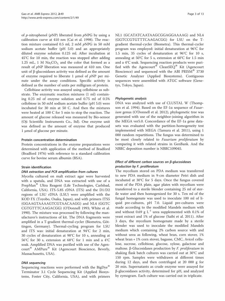
of p-nitrophenol (pNP) liberated from pNPG by using acalibration curve at 410 nm (Cai et al. 1998). The reac-tion mixture contained 0.5 ml, 2 mM pNPG in 50 mMsodium acetate buffer (pH 5.0) and an appropriatelydiluted enzyme solution 0.125 ml. After incubation at45°C for 10 min, the reaction was stopped after adding1.25 mL, 1 M Na2CO3, and the color that formed as aresult of pNP liberation was measured at 410 nm. Oneunit of β-glucosidases activity was defined as the amountof enzyme required to liberate 1 μmol of pNP per mi-nute under the assay conditions. Specific activity isdefined as the number of units per milligram of protein.Cellobiase activity was assayed using cellobiose as sub-
strate. The enzymatic reaction mixtures (1 ml) contain-ing 0.25 ml of enzyme solution and 0.75 ml of 0.5%cellobiose in 50 mM sodium acetate buffer (pH 5.0) wereincubated for 30 min at 50 C. And then the mixtureswere heated at 100 C for 5 min to stop the reaction. Theamount of glucose released was measured by Bio-sensor(Oji Scientific Instruments Co., Itd). One enzyme unitwas defined as the amount of enzyme that produced1 μmol of glucose per minute.
Protein concentration determinationProtein concentrations in the enzyme preparations weredetermined with application of the method of Bradford(Bradford 1976) with reference to a standard calibrationcurve for bovine serum albumin (BSA).
Strain identificationDNA extraction and PCR amplification from culturesMycelia cultured on malt extract agar were harvestedwith a spatula, and DNA was extracted with use of aPrepManW Ultra Reagent (Life Technologies, Carlsbad,California, USA). ITS-5.8S rDNA (ITS) and the D1/D2regions of LSU rDNA (LSU) were amplified with theKOD FX (Toyobo, Osaka, Japan), and with primers ITS5(GGAAGTAAAAGTCGTAACAAGG) and NL4 (GGTCCGTGTTTCAAGACGG) (O'Donnell 1993; White et al.1990). The mixture was processed by following the man-ufacturer’s instructions of kit. The DNA fragments wereamplified in a T-gradient thermal-cycler (Biometra, Göt-tingen, Germany). Thermal-cycling program for LSUand ITS was: initial denaturation at 94°C for 2 min,30 cycles of denaturation at 98°C for 10 s, annealing at56°C for 30 s, extension at 68°C for 1 min and a 4°Csoak. Amplified DNA was purified with use of the Agen-courtW AMPureW Kit (Agencourt Bioscience, Beverly,Massachusetts, USA).
DNA sequencingSequencing reactions were performed with the BigDyeW
Terminator 3.1 Cycle Sequencing Kit (Applied Biosys-tems, Foster City, California, USA), and with primers
NL1 (GCATATCAATAAGCGGAGGAAAAG) and NL4(GGTCCGTGTTTCAAGACGG) for LSU on the T-gradient thermal-cycler (Biometra). This thermal-cyclerprogram was employed: initial denaturation at 96°C for1.5 min, 35 cycles of denaturation at 96°C for 10 s,annealing at 50°C for 5 s, extension at 60°C for 1.5 minand a 4°C soak. Sequencing reaction products were puri-fied with the AgencourtW CleanSEQW Kit (AgencourtBioscience) and sequenced with the ABI PRISMW 3730Genetic Analyzer (Applied Biosystems). Contiguoussequences were assembled with ATGC software (Gene-tyx, Tokyo, Japan).
Phylogenetic analysisDNA was analyzed with use of CLUSTAL W (Thomp-son et al. 1994). Based on the EF-1α sequence of Fusar-ium genus (O'Donnell et al. 2012), phylogenetic tree wasgenerated with use of the neighbor-joining algorithm inthe MEGA ver5.0. Concordance of the EF-1a gene data-sets was evaluated with the partition-homogeneity testimplemented with MEGA (Tamura et al. 2011), using 1000 random repartitions. The fungus was determined tobe most closely related to Fusarium proliferatum bycomparing it with related strains in GenBank. And theNBRC deposition number is NBRC109045.
Effect of different carbon sources on β-glucosidasesproduction by F. proliferatumThe mycelium stored on PDA medium was transferredto new PDA medium in 9-cm diameter Petri dish andincubated at 30°C for 5 days. Once the fungus coveredmost of the PDA plate, agar plates with mycelium weretransferred to a sterile blender containing 25 ml of ster-ile water and then homogenized for 30 s. Ten ml of thefungal homogenate was used to inoculate 100 ml of li-quid pre-cultures, pH 7.0. Liquid pre-cultures weremade according to the modified Mandels medium withand without 0.69 g L-1 urea supplemented with 0.1% ofyeast extract and 1% of glucose (Saibi et al. 2011). After3 days, the mycelium homogenate made by a sterileblender was used to inoculate the modified Mandelsmedium which containing 2% carbon source with andwithout urea as following, wheat bran, corn stover, 1%wheat bran + 1% corn stover, bagasse, CMC, Avicel cellu-lose, sucrose, cellobiose, glucose, xylose, galactose andmaltose. β-Glucosidases production by F. proliferatum inshaking flask batch cultures was carried out at 30°C and150 rpm. Samples were withdrawn at different timesduring 12 days, and then centrifuged at 20 000 g for20 min. Supernatants as crude enzyme were assayed forβ-glucosidases activity, determined for pH, and analyzedby zymogram. Each culture was carried out in triplicate.
Gao et al. AMB Express 2012, 2:49 Page 4 of 13http://www.amb-express.com/content/2/1/49
Electrophoresis and zymogramZymography is an electrophoretic technique for detectionof purified or partly purified β-glucosidase. Zymography isbased on SDS-PAGE that includes a substrate such asMUG or pNPG, which can be degraded by β-glucosidases.The degradation product emits fluorescence or produceschange of color during the reaction period. However, thisis not a practical method to assay β-glucosidases existingin the crude enzyme because various β-glucosidases exist-ing in the crude enzyme caused overlapping fluorescencebands. A modified method that combines effective isola-tion with identification was developed to overcome thelimitation of zymogram in the application on crudeenzyme.
Step1: add the loading buffer for SDS-PAGE to thecrude enzyme solution that was produced byincubating F. proliferatum in corn sotver + wheat branbased medium and glucose based medium, but the mixwas not heated at a temperature of 100°C (Laemmli1970). The mix of the crude enzyme and loading bufferwas injected into the gel. Each sample was injected intofour different wells and then the electrophoresis wasapplied.Step2: After the electrophoresis, the first column ofeach sample was cut out of the gel and then treatedwith Coomassie Brilliant Blue (CBB) staining. The
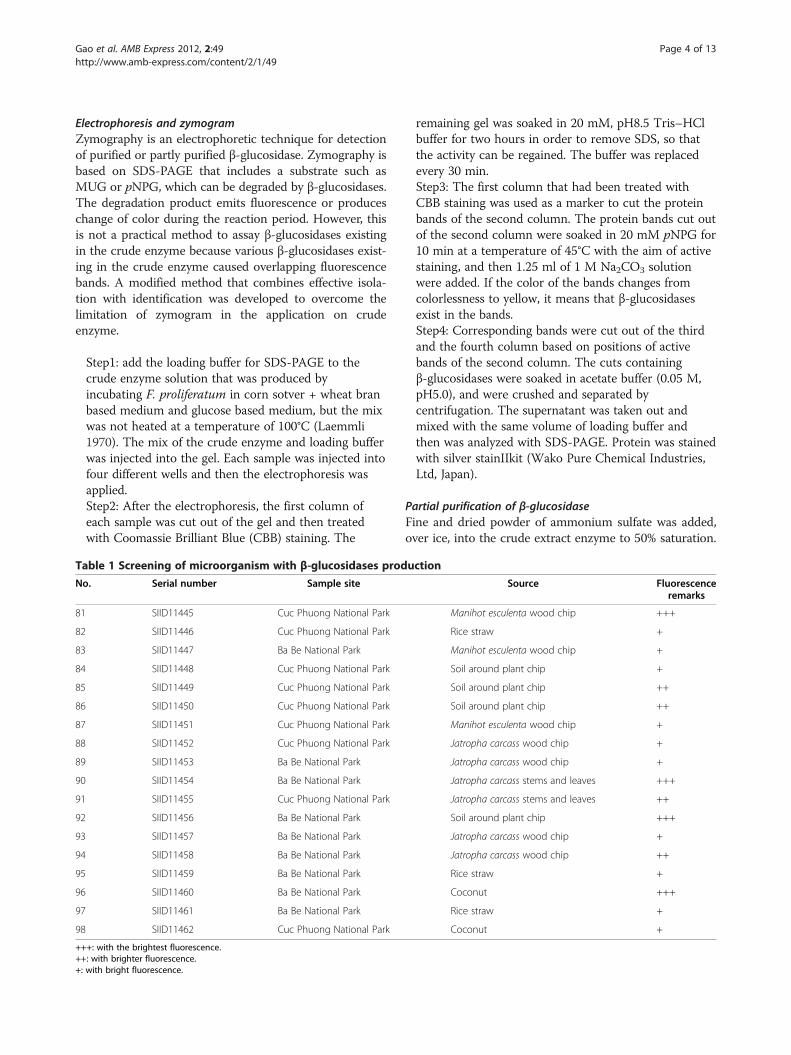
Table 1 Screening of microorganism with β-glucosidases prodNo. Serial number Sample site
81 SIID11445 Cuc Phuong National Park
82 SIID11446 Cuc Phuong National Park
83 SIID11447 Ba Be National Park
84 SIID11448 Cuc Phuong National Park
85 SIID11449 Cuc Phuong National Park
86 SIID11450 Cuc Phuong National Park
87 SIID11451 Cuc Phuong National Park
88 SIID11452 Cuc Phuong National Park
89 SIID11453 Ba Be National Park
90 SIID11454 Ba Be National Park
91 SIID11455 Cuc Phuong National Park
92 SIID11456 Ba Be National Park
93 SIID11457 Ba Be National Park
94 SIID11458 Ba Be National Park
95 SIID11459 Ba Be National Park
96 SIID11460 Ba Be National Park
97 SIID11461 Ba Be National Park
98 SIID11462 Cuc Phuong National Park
+++: with the brightest fluorescence.++: with brighter fluorescence.+: with bright fluorescence.
remaining gel was soaked in 20 mM, pH8.5 Tris–HClbuffer for two hours in order to remove SDS, so thatthe activity can be regained. The buffer was replacedevery 30 min.Step3: The first column that had been treated withCBB staining was used as a marker to cut the proteinbands of the second column. The protein bands cut outof the second column were soaked in 20 mM pNPG for10 min at a temperature of 45°C with the aim of activestaining, and then 1.25 ml of 1 M Na2CO3 solutionwere added. If the color of the bands changes fromcolorlessness to yellow, it means that β-glucosidasesexist in the bands.Step4: Corresponding bands were cut out of the thirdand the fourth column based on positions of activebands of the second column. The cuts containingβ-glucosidases were soaked in acetate buffer (0.05 M,pH5.0), and were crushed and separated bycentrifugation. The supernatant was taken out andmixed with the same volume of loading buffer andthen was analyzed with SDS-PAGE. Protein was stainedwith silver stainIIkit (Wako Pure Chemical Industries,Ltd, Japan).
Partial purification of β-glucosidaseFine and dried powder of ammonium sulfate was added,over ice, into the crude extract enzyme to 50% saturation.
uction
Source Fluorescenceremarks
Manihot esculenta wood chip +++
Rice straw +
Manihot esculenta wood chip +
Soil around plant chip +
Soil around plant chip ++
Soil around plant chip ++
Manihot esculenta wood chip +
Jatropha carcass wood chip +
Jatropha carcass wood chip +
Jatropha carcass stems and leaves +++
Jatropha carcass stems and leaves ++
Soil around plant chip +++
Jatropha carcass wood chip +
Jatropha carcass wood chip ++
Rice straw +
Coconut +++
Rice straw +
Coconut +
Gao et al. AMB Express 2012, 2:49 Page 5 of 13http://www.amb-express.com/content/2/1/49
And then the mix was still stirring at 4°C for 30 min. Aftercentrifugation (42 500 g, 60 min), supernatant was dec-anted and the precipitate was discarded. Ammonium sul-fate was added to bring the supernatant to 80% saturation.The latter was stirred overnight at 4°C and then centri-fuged again. The precipitate was dissolved and dialyzedagainst 20 mM Tris–HCl buffer (pH 8.5). The dialyzed en-zyme solution was centrifuged to remove the insolublecomponent and applied on the DEAE sepharose CL-6Bcolumn (1.5*20 cm) equilibrated with 20 mM Tris–HClbuffer (pH 8.5). The nonadsorbed protein fraction waseluted from the column with starting buffer (100 mL), andthe adsorbed enzyme was collected through 5-stepwiseelution chromatography (sodium chloride concentration:0.1 M, 0.15 M, 0.2 M, 0.25 M and 0.3 M in the same buf-fer). There are two active peaks eluted from DEAE-Sepharose CL-6B at about 0.15 M and 0.25 M NaCl. Theactive fractions (0.15 M NaCl) were pooled and concen-trated by a Centrifugal Filter Devices (Millipore Corpor-ation Billerica, MA, USA), and then chromatographedseparately on a superdex 75 column (1.5*60 cm) equili-brated with 20 mM Tris–HCl buffer (pH 8.5). The pro-teins were eluted with the same buffer at a flow rate of1 mL min-1.
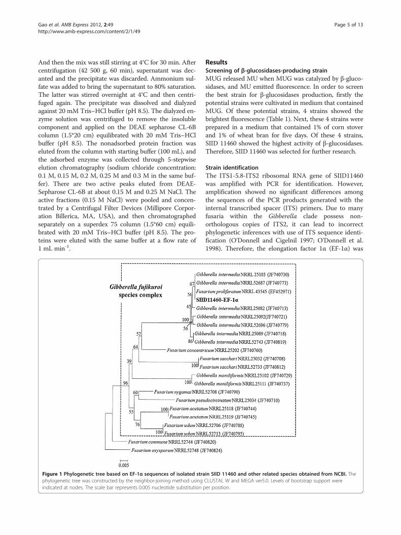
Figure 1 Phylogenetic tree based on EF-1α sequences of isolated straphylogenetic tree was constructed by the neighbor-joining method usingindicated at nodes. The scale bar represents 0.005 nucleotide substitution p
ResultsScreening of β-glucosidases-producing strainMUG released MU when MUG was catalyzed by β-gluco-sidases, and MU emitted fluorescence. In order to screenthe best strain for β-glucosidases production, firstly thepotential strains were cultivated in medium that containedMUG. Of these potential strains, 4 strains showed thebrightest fluorescence (Table 1). Next, these 4 strains wereprepared in a medium that contained 1% of corn stoverand 1% of wheat bran for five days. Of these 4 strains,SIID 11460 showed the highest activity of β-glucosidases.Therefore, SIID 11460 was selected for further research.
Strain identificationThe ITS1-5.8-ITS2 ribosomal RNA gene of SIID11460was amplified with PCR for identification. However,amplification showed no significant differences amongthe sequences of the PCR products generated with theinternal transcribed spacer (ITS) primers. Due to manyfusaria within the Gibberella clade possess non-orthologous copies of ITS2, it can lead to incorrectphylogenetic inferences with use of ITS sequence identi-fication (O'Donnell and Cigelnil 1997; O'Donnell et al.1998). Therefore, the elongation factor 1α (EF-1α) was
in SIID 11460 and other related species obtained from NCBI. TheCLUSTAL W and MEGA ver5.0. Levels of bootstrap support wereer position.
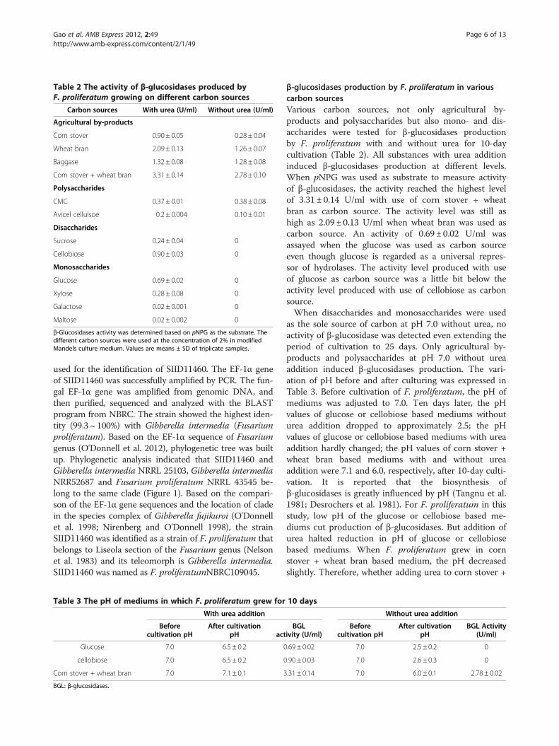
Table 2 The activity of β-glucosidases produced byF. proliferatum growing on different carbon sources
Carbon sources With urea (U/ml) Without urea (U/ml)
Agricultural by-products
Corn stover 0.90 ± 0.05 0.28 ± 0.04
Wheat bran 2.09 ± 0.13 1.26 ± 0.07
Baggase 1.32 ± 0.08 1.28 ± 0.08
Corn stover + wheat bran 3.31 ± 0.14 2.78 ± 0.10
Polysaccharides
CMC 0.37 ± 0.01 0.38 ± 0.08
Avicel cellulsoe 0.2 ± 0.004 0.10 ± 0.01
Disaccharides
Sucrose 0.24 ± 0.04 0
Cellobiose 0.90 ± 0.03 0
Monosaccharides
Glucose 0.69 ± 0.02 0
Xylose 0.28 ± 0.08 0
Galactose 0.02 ± 0.001 0
Maltose 0.02 ± 0.002 0
β-Glucosidases activity was determined based on pNPG as the substrate. Thedifferent carbon sources were used at the concentration of 2% in modifiedMandels culture medium. Values are means ± SD of triplicate samples.
Gao et al. AMB Express 2012, 2:49 Page 6 of 13http://www.amb-express.com/content/2/1/49
used for the identification of SIID11460. The EF-1α geneof SIID11460 was successfully amplified by PCR. The fun-gal EF-1α gene was amplified from genomic DNA, andthen purified, sequenced and analyzed with the BLASTprogram from NBRC. The strain showed the highest iden-tity (99.3 ~ 100%) with Gibberella intermedia (Fusariumproliferatum). Based on the EF-1α sequence of Fusariumgenus (O'Donnell et al. 2012), phylogenetic tree was builtup. Phylogenetic analysis indicated that SIID11460 andGibberella intermedia NRRL 25103, Gibberella intermediaNRR52687 and Fusarium proliferatum NRRL 43545 be-long to the same clade (Figure 1). Based on the compari-son of the EF-1α gene sequences and the location of cladein the species complex of Gibberella fujikuroi (O'Donnellet al. 1998; Nirenberg and O'Donnell 1998), the strainSIID11460 was identified as a strain of F. proliferatum thatbelongs to Liseola section of the Fusarium genus (Nelsonet al. 1983) and its teleomorph is Gibberella intermedia.SIID11460 was named as F. proliferatumNBRC109045.
Table 3 The pH of mediums in which F. proliferatum grew for
With urea addition
Beforecultivation pH
After cultivationpH act
Glucose 7.0 6.5 ± 0.2 0
cellobiose 7.0 6.5 ± 0.2 0
Corn stover + wheat bran 7.0 7.1 ± 0.1 3
BGL: β-glucosidases.
β-glucosidases production by F. proliferatum in variouscarbon sourcesVarious carbon sources, not only agricultural by-products and polysaccharides but also mono- and dis-accharides were tested for β-glucosidases productionby F. proliferatum with and without urea for 10-daycultivation (Table 2). All substances with urea additioninduced β-glucosidases production at different levels.When pNPG was used as substrate to measure activityof β-glucosidases, the activity reached the highest levelof 3.31± 0.14 U/ml with use of corn stover + wheatbran as carbon source. The activity level was still ashigh as 2.09 ± 0.13 U/ml when wheat bran was used ascarbon source. An activity of 0.69 ± 0.02 U/ml wasassayed when the glucose was used as carbon sourceeven though glucose is regarded as a universal repres-sor of hydrolases. The activity level produced with useof glucose as carbon source was a little bit below theactivity level produced with use of cellobiose as carbonsource.When disaccharides and monosaccharides were used
as the sole source of carbon at pH 7.0 without urea, noactivity of β-glucosidase was detected even extending theperiod of cultivation to 25 days. Only agricultural by-products and polysaccharides at pH 7.0 without ureaaddition induced β-glucosidases production. The vari-ation of pH before and after culturing was expressed inTable 3. Before cultivation of F. proliferatum, the pH ofmediums was adjusted to 7.0. Ten days later, the pHvalues of glucose or cellobiose based mediums withouturea addition dropped to approximately 2.5; the pHvalues of glucose or cellobiose based mediums with ureaaddition hardly changed; the pH values of corn stover +wheat bran based mediums with and without ureaaddition were 7.1 and 6.0, respectively, after 10-day culti-vation. It is reported that the biosynthesis ofβ-glucosidases is greatly influenced by pH (Tangnu et al.1981; Desrochers et al. 1981). For F. proliferatum in thisstudy, low pH of the glucose or cellobiose based me-diums cut production of β-glucosidases. But addition ofurea halted reduction in pH of glucose or cellobiosebased mediums. When F. proliferatum grew in cornstover + wheat bran based medium, the pH decreasedslightly. Therefore, whether adding urea to corn stover +
10 days
Without urea addition
BGLivity (U/ml)
Beforecultivation pH
After cultivationpH
BGL Activity(U/ml)
.69 ± 0.02 7.0 2.5 ± 0.2 0
.90 ± 0.03 7.0 2.6 ± 0.3 0
.31 ± 0.14 7.0 6.0 ± 0.1 2.78 ± 0.02
Gao et al. AMB Express 2012, 2:49 Page 7 of 13http://www.amb-express.com/content/2/1/49
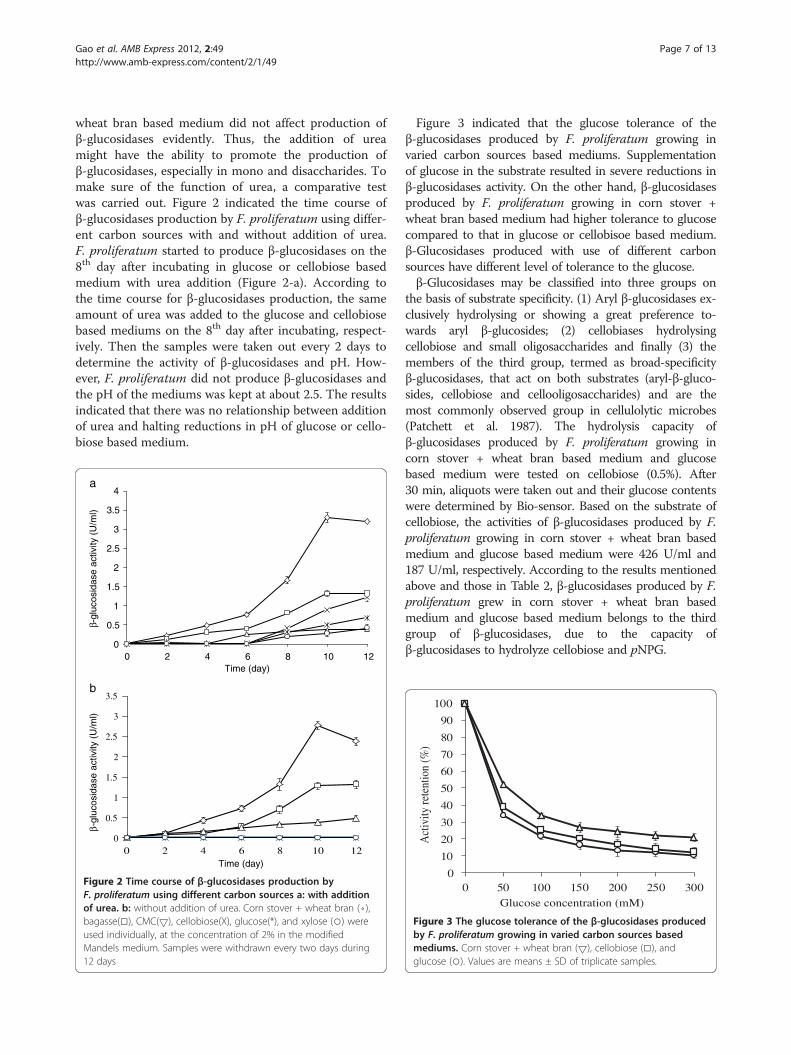
wheat bran based medium did not affect production ofβ-glucosidases evidently. Thus, the addition of ureamight have the ability to promote the production ofβ-glucosidases, especially in mono and disaccharides. Tomake sure of the function of urea, a comparative testwas carried out. Figure 2 indicated the time course ofβ-glucosidases production by F. proliferatum using differ-ent carbon sources with and without addition of urea.F. proliferatum started to produce β-glucosidases on the8th day after incubating in glucose or cellobiose basedmedium with urea addition (Figure 2-a). According tothe time course for β-glucosidases production, the sameamount of urea was added to the glucose and cellobiosebased mediums on the 8th day after incubating, respect-ively. Then the samples were taken out every 2 days todetermine the activity of β-glucosidases and pH. How-ever, F. proliferatum did not produce β-glucosidases andthe pH of the mediums was kept at about 2.5. The resultsindicated that there was no relationship between additionof urea and halting reductions in pH of glucose or cello-biose based medium.
0
0.5
1
1.5
2
2.5
3
3.5
4
0 2 4 6 8 10 12
β-gl
ucos
idas
e ac
tivity
(U
/ml)
Time (day)
a
b
0
0.5
1
1.5
2
2.5
3
3.5
0 2 4 6 8 10 12
β-gl
ucos
idas
e ac
tivity
(U
/ml)
Time (day)
Figure 2 Time course of β-glucosidases production byF. proliferatum using different carbon sources a: with additionof urea. b: without addition of urea. Corn stover + wheat bran (⋄),bagasse(□), CMC(5), cellobiose(Χ), glucose(*), and xylose (○) wereused individually, at the concentration of 2% in the modifiedMandels medium. Samples were withdrawn every two days during12 days
Figure 3 indicated that the glucose tolerance of theβ-glucosidases produced by F. proliferatum growing invaried carbon sources based mediums. Supplementationof glucose in the substrate resulted in severe reductions inβ-glucosidases activity. On the other hand, β-glucosidasesproduced by F. proliferatum growing in corn stover +wheat bran based medium had higher tolerance to glucosecompared to that in glucose or cellobisoe based medium.β-Glucosidases produced with use of different carbonsources have different level of tolerance to the glucose.β-Glucosidases may be classified into three groups on
the basis of substrate specificity. (1) Aryl β-glucosidases ex-clusively hydrolysing or showing a great preference to-wards aryl β-glucosides; (2) cellobiases hydrolysingcellobiose and small oligosaccharides and finally (3) themembers of the third group, termed as broad-specificityβ-glucosidases, that act on both substrates (aryl-β-gluco-sides, cellobiose and cellooligosaccharides) and are themost commonly observed group in cellulolytic microbes(Patchett et al. 1987). The hydrolysis capacity ofβ-glucosidases produced by F. proliferatum growing incorn stover + wheat bran based medium and glucosebased medium were tested on cellobiose (0.5%). After30 min, aliquots were taken out and their glucose contentswere determined by Bio-sensor. Based on the substrate ofcellobiose, the activities of β-glucosidases produced by F.proliferatum growing in corn stover + wheat bran basedmedium and glucose based medium were 426 U/ml and187 U/ml, respectively. According to the results mentionedabove and those in Table 2, β-glucosidases produced by F.proliferatum grew in corn stover + wheat bran basedmedium and glucose based medium belongs to the thirdgroup of β-glucosidases, due to the capacity ofβ-glucosidases to hydrolyze cellobiose and pNPG.
0
10
20
30
40
50
60
70
80
90
100
0 50 100 150 200 250 300
Act
ivity
rete
ntio
n (%
)
Glucose concentration (mM)
Figure 3 The glucose tolerance of the β-glucosidases producedby F. proliferatum growing in varied carbon sources basedmediums. Corn stover + wheat bran (5), cellobiose (□), andglucose (○). Values are means ± SD of triplicate samples.
Gao et al. AMB Express 2012, 2:49 Page 8 of 13http://www.amb-express.com/content/2/1/49
Differential expression of β-glucosidases in response tocarbon sourcesZymogram analysis was used to assay the β-glucosidasesproduced by F. proliferatum that grew in corn stover +wheat bran based medium and glucose based medium.
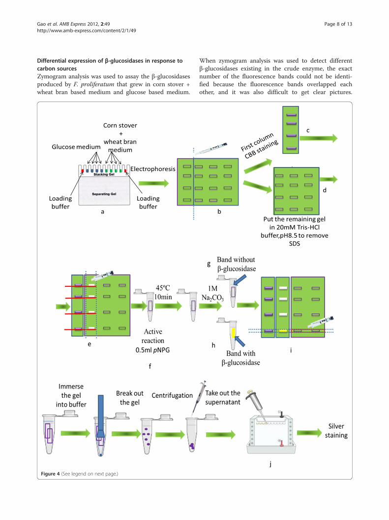
Figure 4 (See legend on next page.)
When zymogram analysis was used to detect differentβ-glucosidases existing in the crude enzyme, the exactnumber of the fluorescence bands could not be identi-fied because the fluorescence bands overlapped eachother, and it was also difficult to get clear pictures.
(See figure on previous page.)Figure 4 Schematic of the modified zymogram. a: add the mix of loading buffer and crude enzyme solution to the gel. But the mix was notheated at 100°C. Blue: crude enzyme from glucose based medium; Green: crude enzyme from corn stover + wheat bran based medium; Red:loading buffer only. b: after electrophoresis, the first column of each sample was cut out. c: the first column of each sample was stained withCBB. d: the remaining gel was soaked in Tris–HCl buffer to remove SDS. e: the first column after CBB staining was used as a marker to cut theprotein bank of the second column. f: the protein bank cut of the second column was soaked in pNPG for active staining. g: after addingNa2CO3, the band coming from the second column kept colorlessness. h: the color of the band from the second column changed fromcolorlessness to yellow following addition of Na2CO3. i: according to the position of active band of the second column, cut the correspondingbands of the third and fourth column. j: protein coming from the bands of the third and fourth column was injected to the gel for SDS-PAGEfollowing a series of treatments.
Gao et al. AMB Express 2012, 2:49 Page 9 of 13http://www.amb-express.com/content/2/1/49
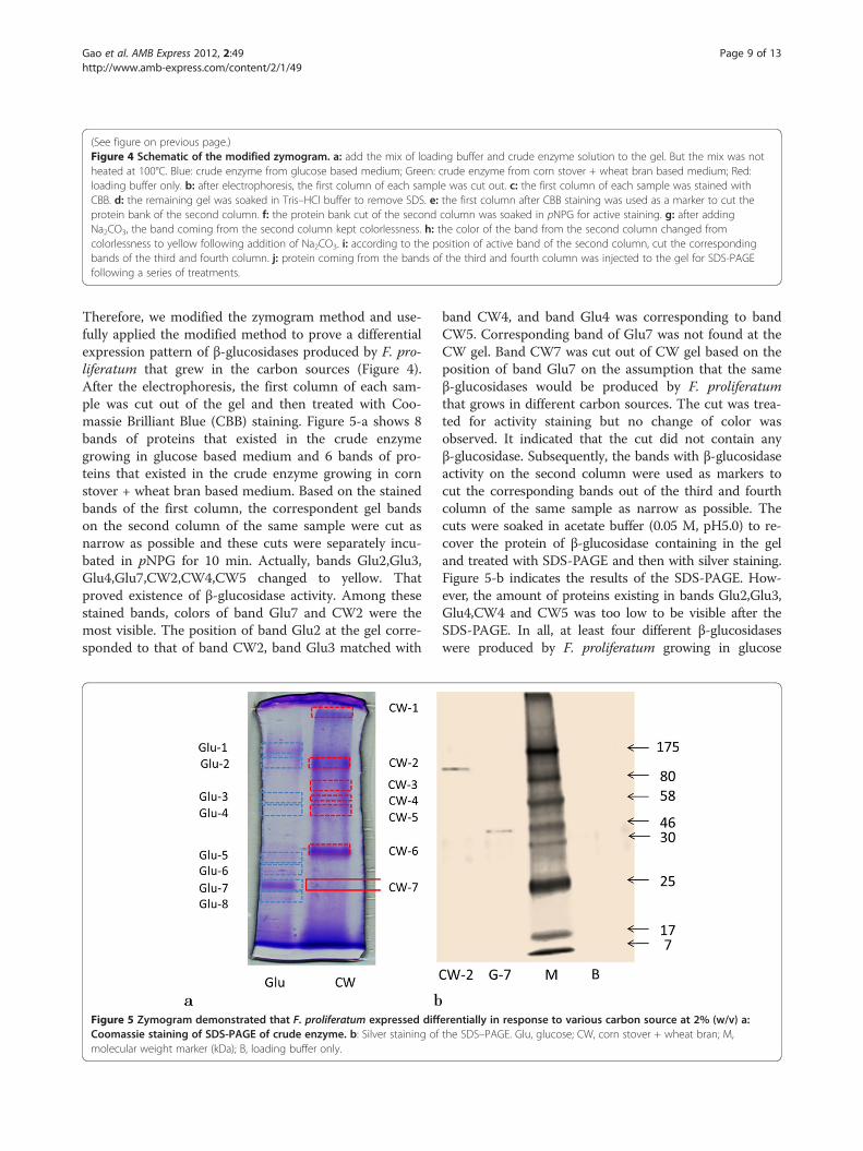
Therefore, we modified the zymogram method and use-fully applied the modified method to prove a differentialexpression pattern of β-glucosidases produced by F. pro-liferatum that grew in the carbon sources (Figure 4).After the electrophoresis, the first column of each sam-ple was cut out of the gel and then treated with Coo-massie Brilliant Blue (CBB) staining. Figure 5-a shows 8bands of proteins that existed in the crude enzymegrowing in glucose based medium and 6 bands of pro-teins that existed in the crude enzyme growing in cornstover + wheat bran based medium. Based on the stainedbands of the first column, the correspondent gel bandson the second column of the same sample were cut asnarrow as possible and these cuts were separately incu-bated in pNPG for 10 min. Actually, bands Glu2,Glu3,Glu4,Glu7,CW2,CW4,CW5 changed to yellow. Thatproved existence of β-glucosidase activity. Among thesestained bands, colors of band Glu7 and CW2 were themost visible. The position of band Glu2 at the gel corre-sponded to that of band CW2, band Glu3 matched with
Figure 5 Zymogram demonstrated that F. proliferatum expressed diffCoomassie staining of SDS-PAGE of crude enzyme. b: Silver staining ofmolecular weight marker (kDa); B, loading buffer only.
band CW4, and band Glu4 was corresponding to bandCW5. Corresponding band of Glu7 was not found at theCW gel. Band CW7 was cut out of CW gel based on theposition of band Glu7 on the assumption that the sameβ-glucosidases would be produced by F. proliferatumthat grows in different carbon sources. The cut was trea-ted for activity staining but no change of color wasobserved. It indicated that the cut did not contain anyβ-glucosidase. Subsequently, the bands with β-glucosidaseactivity on the second column were used as markers tocut the corresponding bands out of the third and fourthcolumn of the same sample as narrow as possible. Thecuts were soaked in acetate buffer (0.05 M, pH5.0) to re-cover the protein of β-glucosidase containing in the geland treated with SDS-PAGE and then with silver staining.Figure 5-b indicates the results of the SDS-PAGE. How-ever, the amount of proteins existing in bands Glu2,Glu3,Glu4,CW4 and CW5 was too low to be visible after theSDS-PAGE. In all, at least four different β-glucosidaseswere produced by F. proliferatum growing in glucose
erentially in response to various carbon source at 2% (w/v) a:the SDS–PAGE. Glu, glucose; CW, corn stover + wheat bran; M,
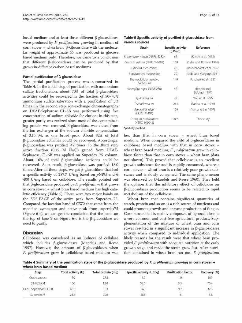
Table 5 Specific activity of purified β-glucosidase fromvarious sources
Strain Specific activity(U/mg)
Reference
Rhizomucor miehei (NRRL 5282) 62 (Krisch et al. 2012)
Candida peltata (NRRL Y-6888) 108 (Saha and Bothast 1996)
Daldinia eschscholzii 78 (Karnchanatat et al. 2007)
Stachybotrys microspora 20 (Saibi and Gargouri 2011)
Thymepkilic anaerobicbacterium
149 (Patchett et al. 1987)
Aspergillus niger (NIAB 280) 42 (Rashid andSiddiqui 1997)
Xylaria regalis 23 (Wei et al. 1996)
Trichoderma sp. 214 (Fadda et al. 1994)
Aspergillus niger(CCRC 31494)
199 (Yan and Lin 1997).
Fusarium proliferatum(NBRC 109045)
288* This study
*partially purified.
Gao et al. AMB Express 2012, 2:49 Page 10 of 13http://www.amb-express.com/content/2/1/49
based medium and at least three different β-glucosidaseswere produced by F. proliferatum growing in medium ofcorn stover + whea bran. β-Glucosidase with the molecu-lar weight of approximate 46 was produced in glucosebased medium only. Therefore, we came to a conclusionthat different β-glucosidases can be produced by thatgrows in different carbon based mediums.
Partial purification of β-glucosidaseThe partial purification process was summarized inTable 4. In the initial step of purification with ammoniumsulfate fractionation, about 70% of total β-glucosidaseactivities could be recovered in the fraction of 50–70%ammonium sulfate saturation with a purification of 3.3times. In the second step, ion-exchange chromatographyon DEAE-Sepharose CL-6B was performed using fiveconcentration of sodium chloride for elution. In this step,greater purity was realized since most of the contaminat-ing protein was removed. β-glucosidase was eluted fromthe ion exchanger at the sodium chloride concentrationof 0.15 M, as one broad peak. About 32% of totalβ-glucosidase activities could be recovered. Accordingly,β-glucosidase was purified 9.2 times. In the third step,active fraction (0.15 M NaCl) gained from DEAE-Sepharose CL-6B was applied on Superdex 75 column.About 16% of total β-glucosidase activities could berecovered. As a result, β-glucosidase was purified 18.0times. After all these steps, we got β-glucosidase that hada specific activity of 287.7 U/mg based on pNPG and 6400 U/mg based on cellobiose. The results pointed outthat β-glucosidase produced by F. proliferatum that growsin corn stover + wheat bran based medium has high cata-lytic efficiency (Table 5). There were two major bands onthe SDS-PAGE of the active peak from Superdex 75.Compared the location band of CW2 that came from themodified zymogram and active peak from superdex75(Figure 6-c), we can get the conclusion that the band onthe top of lane 2 on Figure 6-c is the β-glucosidase weneed to purify.
DiscussionCellobiose was considered as an inducer of cellulasewhich includes β-glucosidases (Mandels and Reese1957). However, the amount of β-glucosidases whenF. proliferatum grew in cellobiose based medium was
Table 4 Summary of the purification steps of the β-glucosidawheat bran based medium
Step Total activity (U) Total protein (mg)
Crude extract 150 9.38
(NH4)2SO4 106 1.98
DEAE Sepharose CL-6B 48.6 0.33
Superdex75 23.8 0.08
less than that in corn stover + wheat bran basedmedium. When compared the yield of β-glucosidases incellobiose based medium with that in corn stover +wheat bran based medium, F. proliferatum grew in cello-biose faster than that in corn stover + wheat bran (datanot shown). This proved that cellobiose is an excellentgrowth substance for and is rapidly consumed, whereascorn stover + wheat bran is a relatively poor growth sub-stance and is slowly consumed. The same phenomenonwas observed by (Mandels and Reese 1960). They heldthe opinion that the inhibitory effect of cellobiose onβ-glucosidases production seems to be related to rapidmetabolism of the cellobiose.Wheat bran that contains significant quantities of
starch, protein and so on is a rich source of nutrients andcould promote growth and enzyme production of fungus.Corn stover that is mainly composed of lignocellulose isa very common and cost-free agricultural product. Sup-plementation of the mixture of wheat bran and cornstover resulted in a significant increase in β-glucosidasesactivity when compared to individual application. Thelikely reasons for the result were that wheat bran pro-vided F. proliferatum with adequate nutrition at the earlygrowth stage and made the strain grow fast. After nutri-tion contained in wheat bran ran out, F. proliferatum
se produced by F. proliferatum growing in corn stover +
Specific activity (U/mg) Purification factor Recovery (%)
16.0 1.0 100
53.5 3.3 70.4
148 9.2 32.3
288 18 15.8
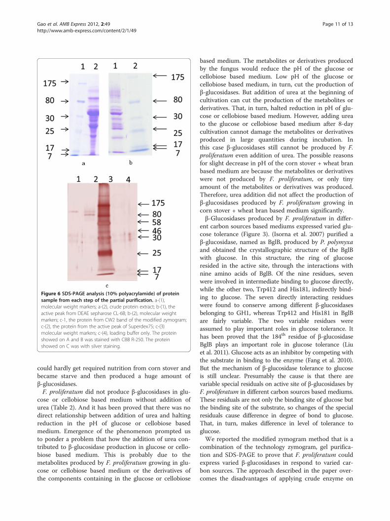
Figure 6 SDS-PAGE analysis (10% polyacrylamide) of proteinsample from each step of the partial purification. a-(1),molecular weight markers; a-(2), crude protein extract; b-(1), theactive peak from DEAE sepharose CL-6B; b-(2), molecular weightmarkers; c-1, the protein from CW2 band of the modified zymogram;c-(2), the protein from the active peak of Superdex75; c-(3)molecular weight markers; c-(4), loading buffer only. The proteinshowed on A and B was stained with CBB R-250. The proteinshowed on C was with silver staining.
Gao et al. AMB Express 2012, 2:49 Page 11 of 13http://www.amb-express.com/content/2/1/49
could hardly get required nutrition from corn stover andbecame starve and then produced a huge amount ofβ-glucosidases.F. proliferatum did not produce β-glucosidases in glu-
cose or cellobiose based medium without addition ofurea (Table 2). And it has been proved that there was nodirect relationship between addition of urea and haltingreduction in the pH of glucose or cellobiose basedmedium. Emergence of the phenomenon prompted usto ponder a problem that how the addition of urea con-tributed to β-glucosidase production in glucose or cello-biose based medium. This is probably due to themetabolites produced by F. proliferatum growing in glu-cose or cellobiose based medium or the derivatives ofthe components containing in the glucose or cellobiose
based medium. The metabolites or derivatives producedby the fungus would reduce the pH of the glucose orcellobiose based medium. Low pH of the glucose orcellobiose based medium, in turn, cut the production ofβ-glucosidases. But addition of urea at the beginning ofcultivation can cut the production of the metabolites orderivatives. That, in turn, halted reduction in pH of glu-cose or cellobiose based medium. However, adding ureato the glucose or cellobiose based medium after 8-daycultivation cannot damage the metabolites or derivativesproduced in large quantities during incubation. Inthis case β-glucosidases still cannot be produced by F.proliferatum even addition of urea. The possible reasonsfor slight decrease in pH of the corn stover + wheat branbased medium are because the metabolites or derivativeswere not produced by F. proliferatum, or only tinyamount of the metabolites or derivatives was produced.Therefore, urea addition did not affect the production ofβ-glucosidases produced by F. proliferatum growing incorn stover + wheat bran based medium significantly.β-Glucosidases produced by F. proliferatum in differ-
ent carbon sources based mediums expressed varied glu-cose tolerance (Figure 3). (Isorna et al. 2007) purified aβ-glucosidase, named as BglB, produced by P. polymyxaand obtained the crystallographic structure of the BglBwith glucose. In this structure, the ring of glucoseresided in the active site, through the interactions withnine amino acids of BglB. Of the nine residues, sevenwere involved in intermediate binding to glucose directly,while the other two, Trp412 and His181, indirectly bind-ing to glucose. The seven directly interacting residueswere found to conserve among different β-glucosidasesbelonging to GH1, whereas Trp412 and His181 in BglBare fairly variable. The two variable residues wereassumed to play important roles in glucose tolerance. Ithas been proved that the 184th residue of β-glucosidaseBglB plays an important role in glucose tolerance (Liuet al. 2011). Glucose acts as an inhibitor by competing withthe substrate in binding to the enzyme (Fang et al. 2010).But the mechanism of β-glucosidase tolerance to glucoseis still unclear. Presumably the cause is that there arevariable special residuals on active site of β-glucosidases byF. proliferatum in different carbon sources based mediums.These residuals are not only the binding site of glucose butthe binding site of the substrate, so changes of the specialresiduals cause difference in degree of bond to glucose.That, in turn, makes difference in level of tolerance toglucose.We reported the modified zymogram method that is a
combination of the technology zymogram, gel purifica-tion and SDS-PAGE to prove that F. proliferatum couldexpress varied β-glucosidases in respond to varied car-bon sources. The approach described in the paper over-comes the disadvantages of applying crude enzyme on
Gao et al. AMB Express 2012, 2:49 Page 12 of 13http://www.amb-express.com/content/2/1/49
zymogram and combines effective isolation with identifi-cation assay.
Competing interestsThe authors declare that they have no competing interests.
Author details1Department of Agro-Environmental Sciences, Faculty of Agriculture, KyushuUniversity, Fukuoka, Japan. 2Center of Biotechnology, Vietnam NationalUniversity, Hanoi, Vietnam. 3Department of Biotechnology, National Instituteof Technology and Evaluation, Tokyo, Japan. 4Advanced TechnologyLaboratories IDEMITSU KOSAN Co., Ltd, Chiba, Japan.
Received: 23 August 2012 Accepted: 29 August 2012Published: 14 September 2012
ReferencesAdsul MG, Bastawde KB, Varma AJ, Gokhale DV (2007) Strain improvement of
Penicillium janthinellum NCIM1171 for increased cellulase production.Bioresour Technol 98:1467–1473
Banerjee S, Mudliar S, Sen R, Giri B, Satpute D, Chakrabarti T, Pandey RA (2010)Commercializing lignocellulosic bioethanol: technology bottlenecks andpossible remedies. Biofuels Bioprod Bioref 4:77–93
Beguin P, Aubert JP (1994) The biological degradation of cellulose. FEMSMicrobiol Rev 13:25–28
Bradford MM (1976) A rapid and sensitive method for the quantitation ofmicrogram quantities of protein utilizing the principle of protein-dyebinding. Anal Biochem 72:248–254
Cai YJ, Buswell JA, Chang ST (1998) β-Glucosidase component of the cellulolyticsystem of the edible straw mushroom, Volvariella volvacea. Enzyme MicrobTechnol 22:122–129
Crout DH, Vic G (1998) Glycosidases and glycosynthetases in glycoside andoligosaccharide synthesis. Curr Opin Chem Biol 2:98–111
Daenen L, Saison D, Sterckx F, Delvaux FR, Verachtert H, Derdelinckx G (2008)Screening and evaluation of the glucoside hydrolase activity inSaccharomyces and Brettanomyces brewing yeasts. J Appl Microbiol104:478–488
Deguchi S, Tsudome M, Shen Y, Konishi S, Tsujii K, Ito S, Horikoshi K (2007)Preparation and characterisation of nanofibrous cellulose plate as a newsolid support for microbial culture. Soft Matter 3:1170–1175
Desrochers M, Jurasek L, Paice MG (1981) High production of β-glucosidase inSchizophyllum commune: isolation of the enzyme and effect of the culturefiltrate on cellulose hydrolysis. Appl Environ Microbiol 41:222–228
Enari TM (1983) Microbial cellulase. In: Fogarty WM, Kelly CT (eds) Microbialenzymes and biotechnology. Applied Science Publishers, London-New York,p p183
Esen A (1993) β-Glycosidases: overview. In: Esen A (ed) β-Glucosidasesbiochemistry and molecular biology, vol 533, ACS Symposium. AmericanChemical Society, Washington, DC, pp 1–14
Esen A (2003) β-Glucosidases. In: Whitaker JR, Voragen AGJ, Wong DWS (eds)Handbook of food enzymology. Marcel Dekker Inc, New York, p 791
Fadda MB, Curreli N, Pompei R, Rescigno A, Runaldi A, Sandust E (1994) A highlyactive fungal β-glucosidase. Appl Biochem Biotechnol 44:263–270
Fang ZM, Fang W, Liu JJ, Hong YZ, Peng H, Zhang XC, Sun BL, Xiao YZ (2010)Cloning and characterization of a β-glucosidase from marine microbialmetagenome with excellent glucose tolerance. J microbial Biotechnol20:1351–1358
Gϋnata Z (2003) Flavor enhancement in fruit juices and derived beverages byexogenous glycosidases and consequences of the use of enzymepreparations. In: Whitaker JR, Voragen AGJ, Wong DWS (eds) Handbook offood enzymology., New York, p 303
Hawksworth DL (1991) The fungal dimension of biodiversity: magnitude,significance, and conservation. Mycol Res 95:641–655
Hawksworth DL (2001) The magnitude of fungal diversity: the 1.5 million speciesestimate revisited. Mycol Res 105:1422–1432
Isorna P, Polaina J, Latorre-García L, Cañada FJ, González B, Sanz-Aparicio J (2007)Crystal structures of Paenibacillus polymyxa β-glucosidase B complexes revealthe molecular basis of substrate specificity and give new insights into the catalytic machinery of family 1 glycosidases. J Mol Biol371:1204–1218
Karnchanatat A, Petsom A, Sangvanich P, Piaphukiew J, Whalley AJS, ReynoldsCD, Sihanonth P (2007) Purification and biochemical characterization of anextracellular β-glucosidase from the wood-decaying fungus Daldiniaeschscholzii (Ehrenb.:Fr.) Rehm. FEMS Microbiol Lett 270:162–170
Krisch J, Bencsik O, Papp T, Vagvolgyi C, Tako M (2012) Characterization of aβ-glucosidase with transgalactosylation capacity from the zygomyceteRhizomucor miehei. Bioresource Technology 114:555–560
Laemmli UK (1970) Cleavage of structural proteins during the assembly of thehead of bacteriophage T4. Nature 227:680–685
Liu J, Zhang X, Fang Z, Fang W, Peng H, Xiao Y (2011) The 184th residue ofβ-glucosidase BglB plays an important role in glucose tolerance. J Biosci andBioeng 112:447–450
Mandels M, Reese ET (1957) Induction of cellulase in Trichoderma viride asinfluenced by carbon source and metals. J Bacteriol 73:269–278
Mandels M, Reese ET (1960) Induction of cellulase in fungi by cellobiose.J Bacteriol 79:816–826
Nelson PE, Toussoun TA, Marasas WFO (1983) Fusarium species: An illustratedmanual for identification. Centre County, Pennsylvania
Nirenberg HI, O'Donnell K (1998) New Fusarium species and combination withthe Gibberella fujikuroi species complex. Mycologia 90:434–458
O'Donnell K (1993) Fusarium and its near relatives. In: Reynolds DR, Taylor JW(eds) The fungal holomorph: mitotic, meiotic and pleomorphic speciation infungal systenatics., Wallingford, p 225
O'Donnell K, Cigelnil E (1997) Two divergent intragenomic rDNA ITS2 typeswithin a monophyletic lineage of the fungus Fusarium are nonorthologous.Mol Phyl Evol 7:103–116
O'Donnell K, Cigelnik E, Nirenberg HI (1998) Molecular systematics andphylogeography of the Gibberella fujikuroi species complex of Fusarium.Mycologia 90:465–493
O'Donnell K, Humber RA, Geiser DM, Kang S, Park B, Robert VARG, Crous PW,Johnston PR, Aoki T, Rooney AP, Rehner SA (2012) Phylogenetic diversity ofinsecticolous fusaria inferred from multilocus DNA sequence data and theirmolecular identification via FUSARIUM-ID and FUSARIUM MLST. Mycologia104:427–445
Opassiri R, Hua Y, Wara-Aswapati O, Akiyama T, Svasti J, Esen A, Ketudat Cairns JR(2004) β-Glucosidase, exo-β-glucanase and pyridoxine transglucosylaseactivities of rice BGlu1. Biochem J 379:125–131
Patchett ML, Daniel RM, Morgan HW (1987) Purification and properties of a stablebeta-glucosidase from an extremely thermophilic anaerobic bacterium.Biochem J 243:779–787
Percival ZYH, Himmel ME, Mielenz JR (2006) Outlook for cellulase improvement:screening and selection strategies. Biotechnol Adv 24:452–481
Ragauskas AJ, Williams CK, Davison BH, Britovsek G, Cairney J, Eckert CA (2006)The path forward for biofuels and biomaterials. Science311:484–489
Rashid MH, Siddiqui KS (1997) Purification and characterization of a β-glucosidasefrom Aspergillus niger. Folia Microbiol 42:544–550
Ryu DDY (1980) Cellulases: biosynthesis and applications. Enzyme Microb Technol2:91–102
Saha BC, Bothast RJ (1996) Production, purification, and characterization of ahighly glucose tolerant novel β-glucosidase from Candida peltata. ApplEnviron Microbiol 62:3165–3170
Saibi W, Gargouri A (2011) Purification and biochemical characterization of anatypical β-glucosidase from Stachybotrys microspore. J Mol Catal B Enzym72:107–115
Saibi W, Abdeljalil S, Gargouri A (2011) Carbon source directs the differentialexpression of β-glucosidases in Stachybotrys microspore. World J MicrobiolBiotechnol 27:1765–1774
Soewnsen A (2010) A new highly efficient beta-glucosidase from the novelspecies Aspergillus Saccharolyticus. Dissertation, Aalborg University
Tamura K, Peterson D, Peterson N, Stecher G, Nei M, Kumar S (2011) MEGA5:Molecular evolutionary genetics analysis using maximum likelihood,evolutionary distance, and maximum parsimony methods. Mol Biol Evol28:2731–2739
Tangnu SK, Blanch HW, Wilke CR (1981) Enhanced production of cellulase,hemicellulase, and β-glucosidase by Trichoderma reesei (Rut C-30). BiotechnolBioeng 23:1837–1849
Thompson JD, Higgins DG, Gibson TJ, CLUSTAL W (1994) Improving thesensitivity of progressive multiple sequence alignment through sequenceweighting, position-specific gap penalties and weight matrix choice. NucleicAcids Res 22:4673–4680
Gao et al. AMB Express 2012, 2:49 Page 13 of 13http://www.amb-express.com/content/2/1/49
Wei DL, Kirimura K, Usami S, Lin TH (1996) Purification and characterization of anextracellular β-glucosidase from the wood- grown fungus Xylaria regalis.Curr Microbiol 33:297–301
White TJ, Bruns T, Lee S, Taylor J (1990) Amplification and direct sequencing offungal ribosomal RNA genes for phylogenetics. In: Innis MA, Gelfand DH,Sninsky JJ, White TJ (eds) PCR Protocols: a guide to methods andapplications., San Diego, pp 315–322
Wood TM (1985) Properties of cellulolytic enzyme systems. Biochem Soc Trans13:407–410
Workman WE, Day DF (1982) Purification and properties of β-glucosidase fromAspergillus terrus. Appl Environ Microbiol 44:1289–1295
Yan TR, Lin CL (1997) Purification and characterization of a glucose-tolerantβ-glucosidase from Aspergillus niger CCRC 31494. Biosci Biotech Biochem61:965–970
doi:10.1186/2191-0855-2-49Cite this article as: Gao et al.: The production of β-glucosidases byFusarium proliferatum NBRC109045 isolated from Vietnamese forest.AMB Express 2012 2:49.
Submit your manuscript to a journal and benefi t from:
7 Convenient online submission
7 Rigorous peer review
7 Immediate publication on acceptance
7 Open access: articles freely available online
7 High visibility within the fi eld
7 Retaining the copyright to your article
Submit your next manuscript at 7 springeropen.com