Nanotechnology 7 vo 2007 - TU Wiengebeshuber/Nanotechnology_7_vo_2007.pdf · 7th lecture Please do...
62
Nanotechnology 7 th lecture Please do give anonymous feedback at http://gmail.google.com Username: lecture.feedback Password: nano123 by just sending from there to there an email with your suggestions: email address: [email protected] . ____________ Sometimes, there are also free gmail accounts available from there – check now and then, and use them as you like. ____________ Download supplementary material for the lecture from www.ille.com
Transcript of Nanotechnology 7 vo 2007 - TU Wiengebeshuber/Nanotechnology_7_vo_2007.pdf · 7th lecture Please do...

Nanotechnology7th lecture
Please do give anonymous feedback at http://gmail.google.com
Username: lecture.feedbackPassword: nano123
by just sending from there to there an email with your suggestions: email address: [email protected].
____________Sometimes, there are also free gmail accounts available from
there – check now and then, and use them as you like.____________
Download supplementary material for the lecture fromwww.ille.com
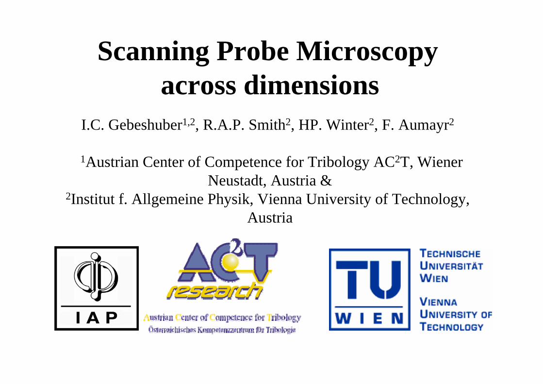
Scanning Probe Microscopy across dimensions
I.C. Gebeshuber1,2, R.A.P. Smith2, HP. Winter2, F. Aumayr2
1Austrian Center of Competence for Tribology AC2T, Wiener Neustadt, Austria &
2Institut f. Allgemeine Physik, Vienna University of Technology, Austria
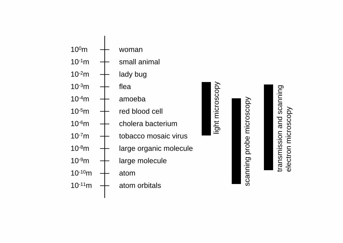
100m
10-1m
10-2m
10-3m
10-4m
10-5m
10-6m
10-7m
10-8m
10-9m
10-10m
10-11m
woman
small animal
lady bug
flea
amoeba
red blood cell
cholera bacterium
tobacco mosaic virus
large organic molecule
large molecule
atom
atom orbitals
light
mic
rosc
opy
scan
ning
pro
be m
icro
scop
y
trans
mis
sion
and
sca
nnin
gel
ectro
n m
icro
scop
y
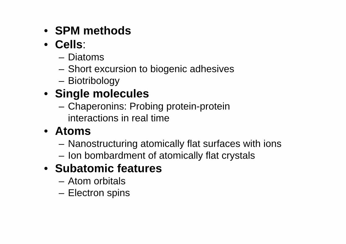
• SPM methods• Cells:
– Diatoms – Short excursion to biogenic adhesives– Biotribology
• Single molecules– Chaperonins: Probing protein-protein
interactions in real time• Atoms
– Nanostructuring atomically flat surfaces with ions– Ion bombardment of atomically flat crystals
• Subatomic features– Atom orbitals– Electron spins
Some SPM methods
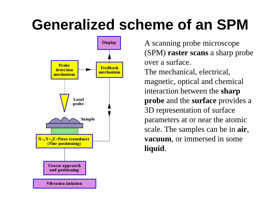
Generalized scheme of an SPMA scanning probe microscope (SPM) raster scans a sharp probe over a surface. The mechanical, electrical, magnetic, optical and chemical interaction between the sharp probe and the surface provides a 3D representation of surface parameters at or near the atomic scale. The samples can be in air, vacuum, or immersed in some liquid.
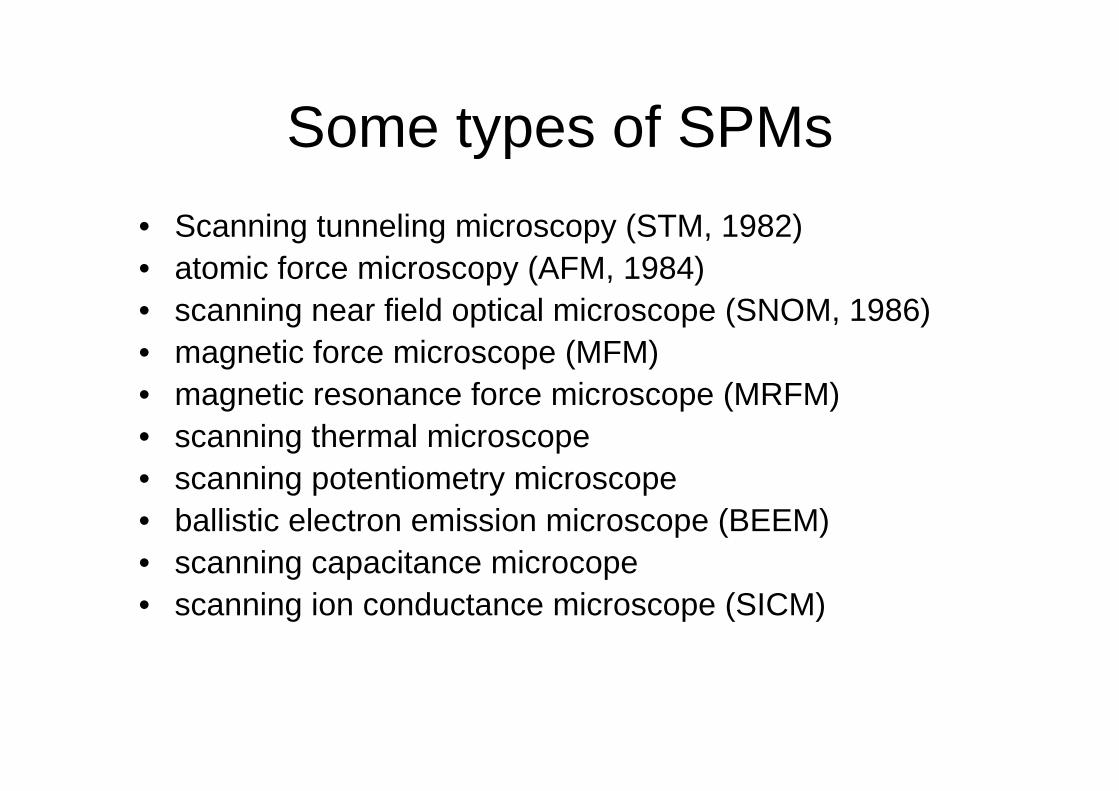
Some types of SPMs• Scanning tunneling microscopy (STM, 1982)• atomic force microscopy (AFM, 1984)• scanning near field optical microscope (SNOM, 1986)• magnetic force microscope (MFM)• magnetic resonance force microscope (MRFM)• scanning thermal microscope• scanning potentiometry microscope• ballistic electron emission microscope (BEEM)• scanning capacitance microcope• scanning ion conductance microscope (SICM)
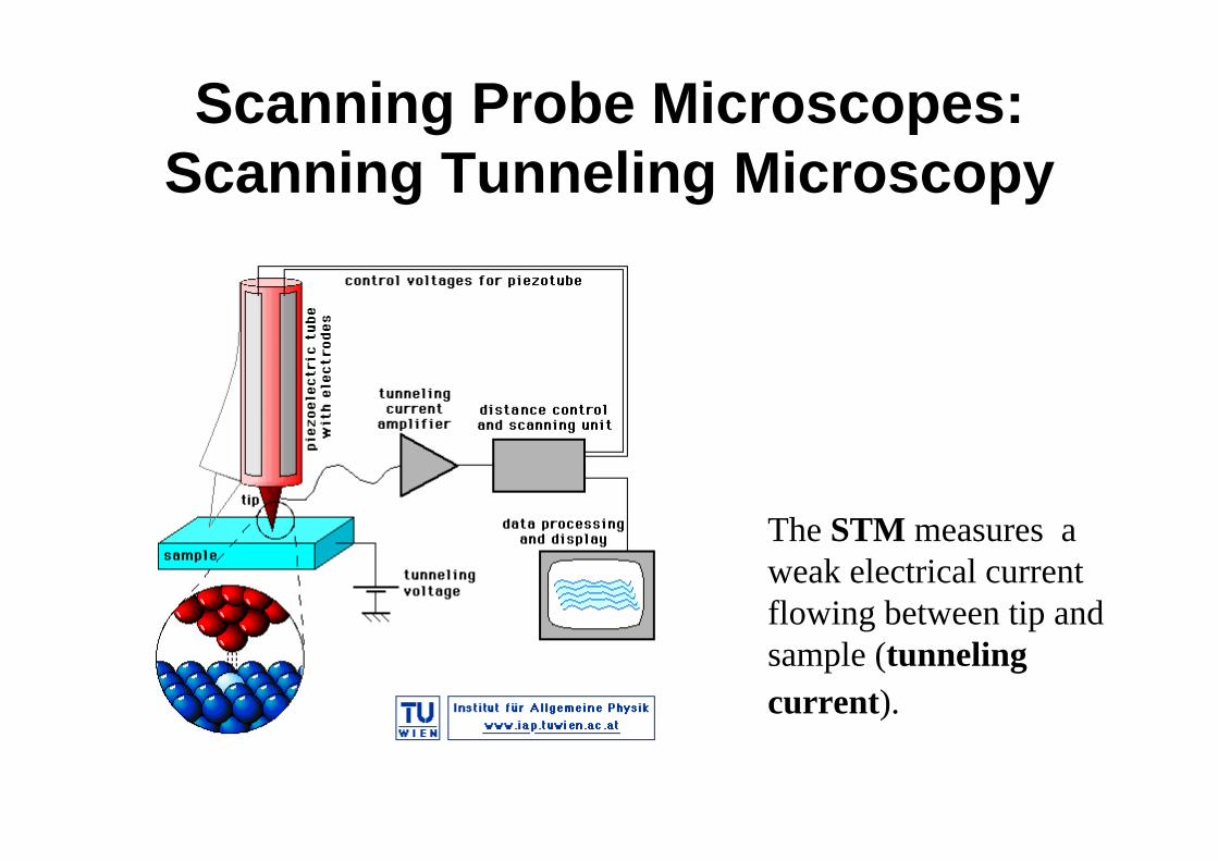
Scanning Probe Microscopes: Scanning Tunneling Microscopy
The STM measures a weak electrical current flowing between tip and sample (tunnelingcurrent).
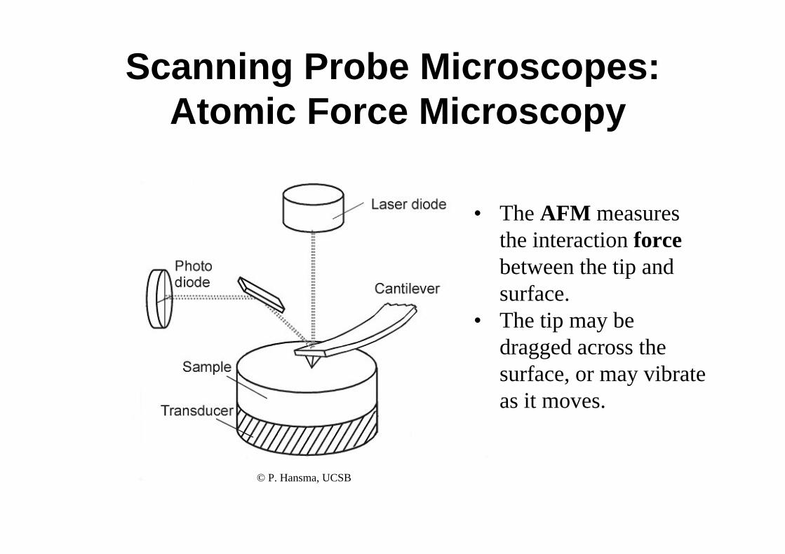
Scanning Probe Microscopes:Atomic Force Microscopy
• The AFM measures the interaction forcebetween the tip and surface.
• The tip may be dragged across the surface, or may vibrate as it moves.
© P. Hansma, UCSB
AFM operational modes
• Contact mode• Non-contact mode• Intermittent mode• Phase imaging
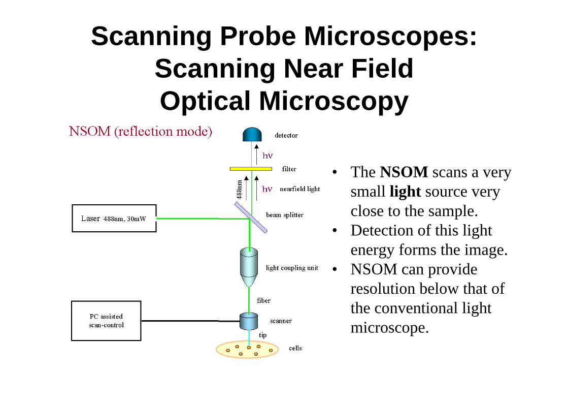
Scanning Probe Microscopes: Scanning Near FieldOptical Microscopy
• The NSOM scans a very small light source very close to the sample.
• Detection of this light energy forms the image.
• NSOM can provide resolution below that of the conventional light microscope.
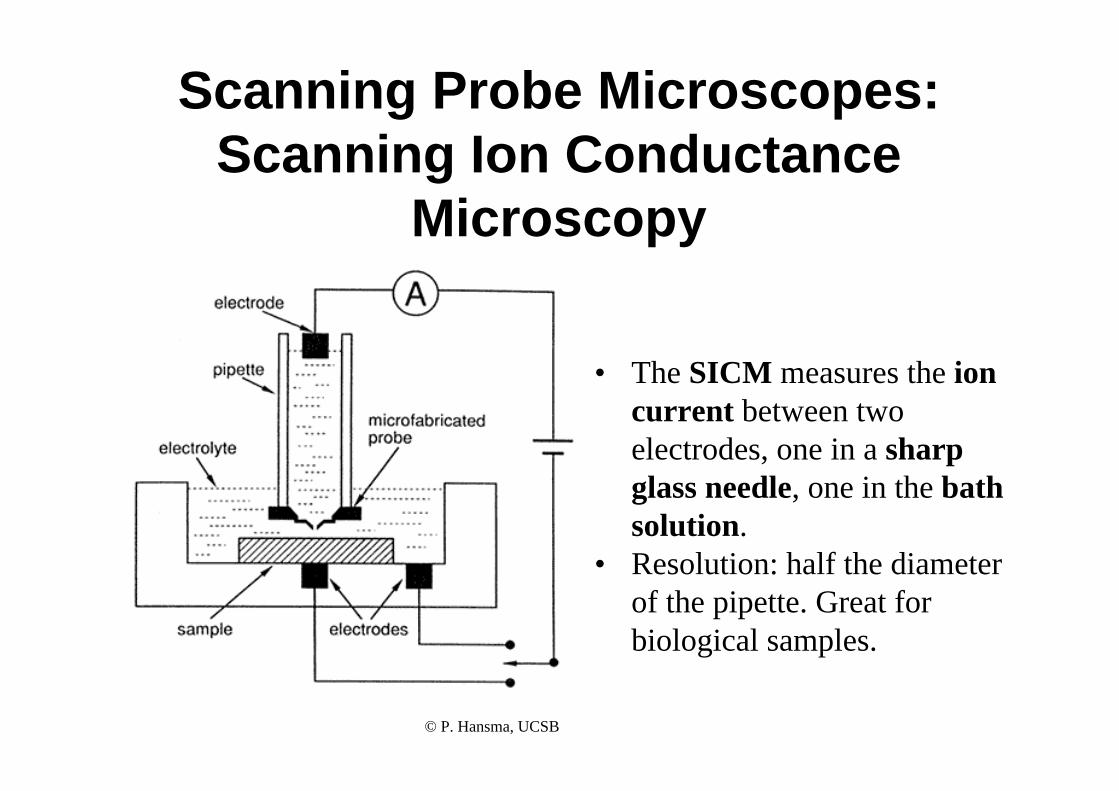
Scanning Probe Microscopes: Scanning Ion Conductance
Microscopy
• The SICM measures the ioncurrent between twoelectrodes, one in a sharpglass needle, one in the bathsolution.
• Resolution: half the diameterof the pipette. Great forbiological samples.
© P. Hansma, UCSB
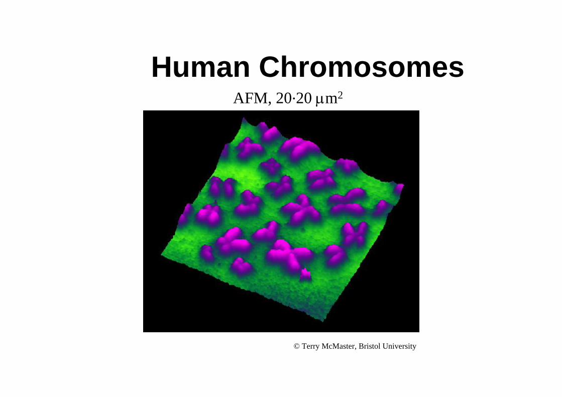
Human Chromosomes
© Terry McMaster, Bristol University
AFM, 20.20 μm2
Cells
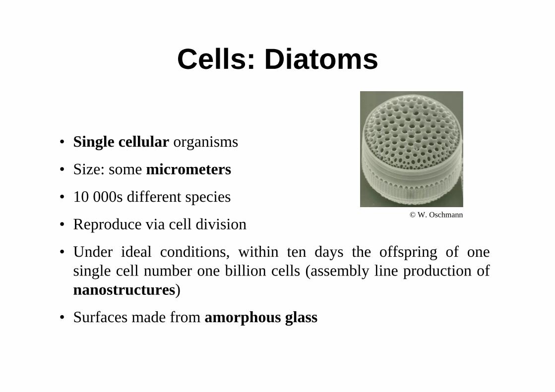
• Single cellular organisms
• Size: some micrometers
• 10 000s different species
• Reproduce via cell division
• Under ideal conditions, within ten days the offspring of onesingle cell number one billion cells (assembly line production of nanostructures)
• Surfaces made from amorphous glass
© W. Oschmann
Cells: Diatoms

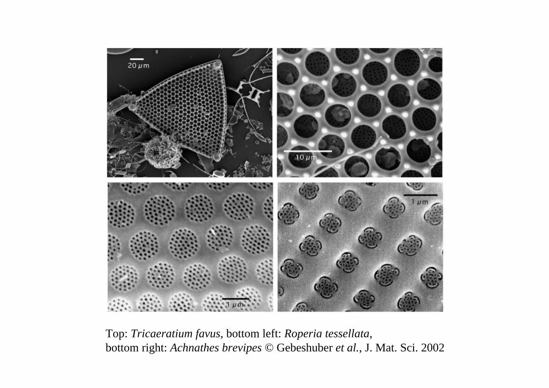
Top: Tricaeratium favus, bottom left: Roperia tessellata, bottom right: Achnathes brevipes © Gebeshuber et al., J. Mat. Sci. 2002
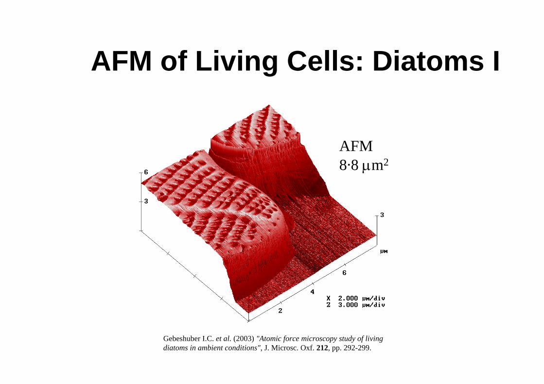
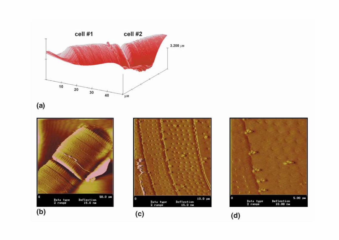
AFM of Living Cells: Diatoms I
Gebeshuber I.C. et al. (2003) "Atomic force microscopy study of living diatoms in ambient conditions", J. Microsc. Oxf. 212, pp. 292-299.
AFM8.8 μm2
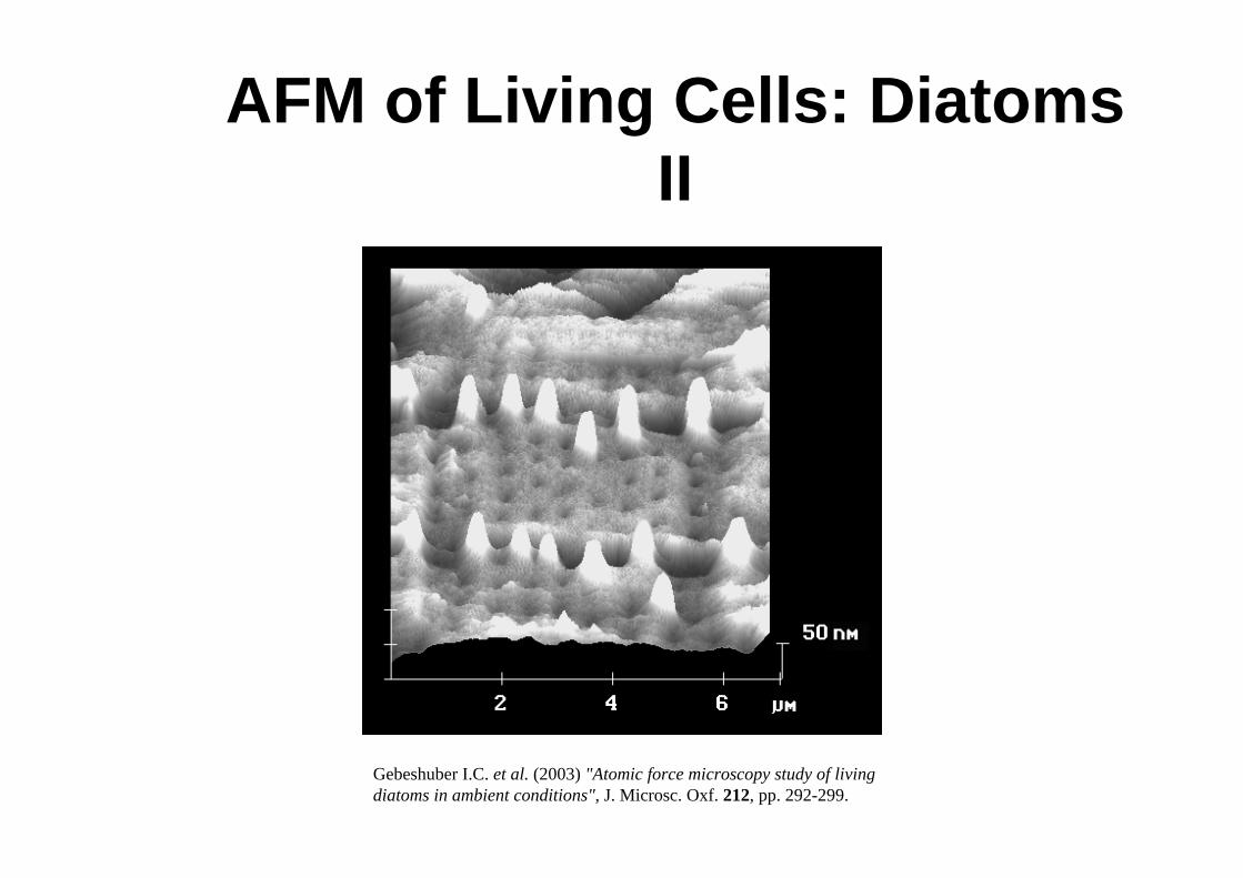
AFM of Living Cells: Diatoms II
Gebeshuber I.C. et al. (2003) "Atomic force microscopy study of living diatoms in ambient conditions", J. Microsc. Oxf. 212, pp. 292-299.
Underwater Adhesives (AFM)
• Most man made adhesives fail to bond in wet conditions, owing to chemical modification of the adhesive or its substrate.
• Engineering strong and robust underwater adhesives that are stable in wet environments are a challenge to current technology.
• Diatoms produce excellent underwater adhesives.• Diatoms living close to the poles of the earth produce ice binding
molecules.
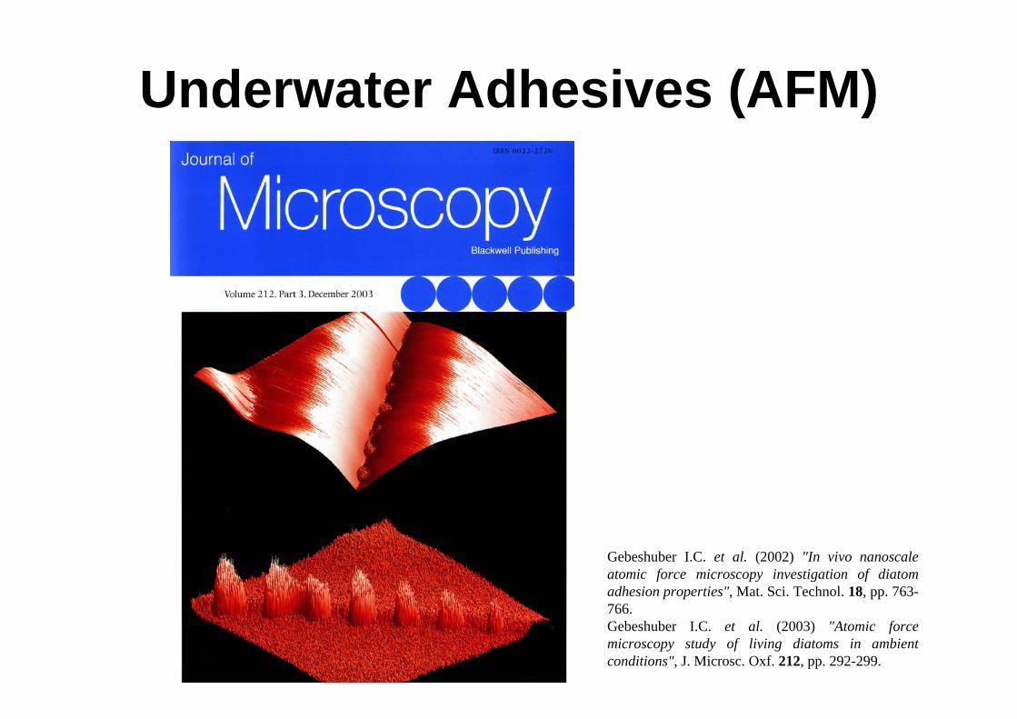
Underwater Adhesives (AFM)
Gebeshuber I.C. et al. (2002) "In vivo nanoscale atomic force microscopy investigation of diatomadhesion properties", Mat. Sci. Technol. 18, pp. 763-766. Gebeshuber I.C. et al. (2003) "Atomic force microscopy study of living diatoms in ambient conditions", J. Microsc. Oxf. 212, pp. 292-299.
To access the adhesive under the diatomremove cell with STM tip!
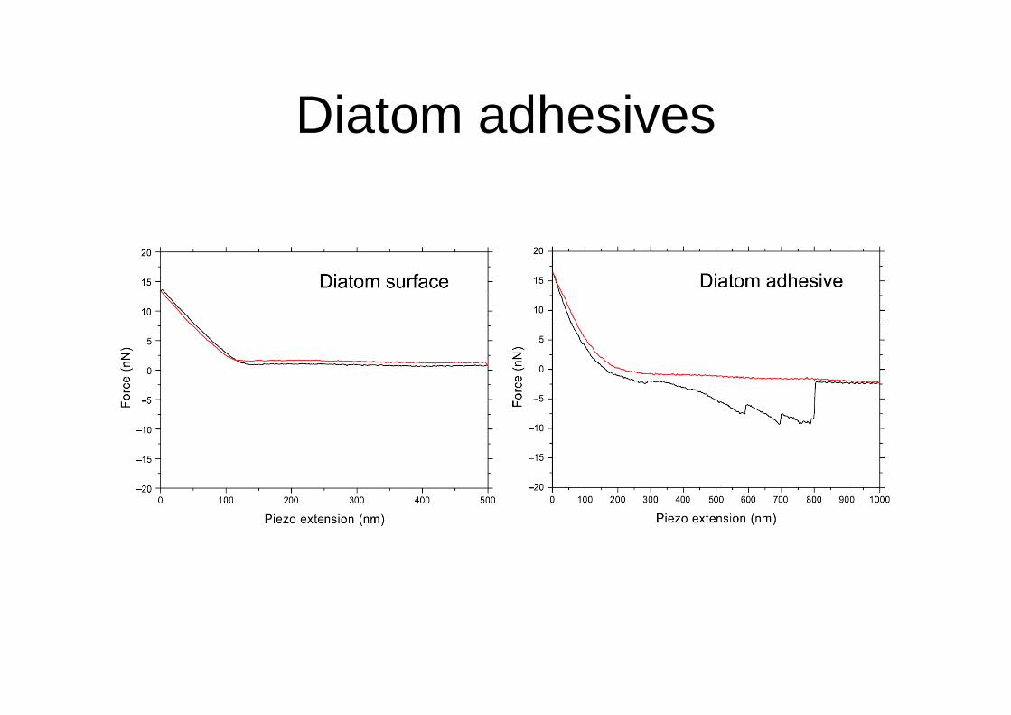
Diatom adhesives
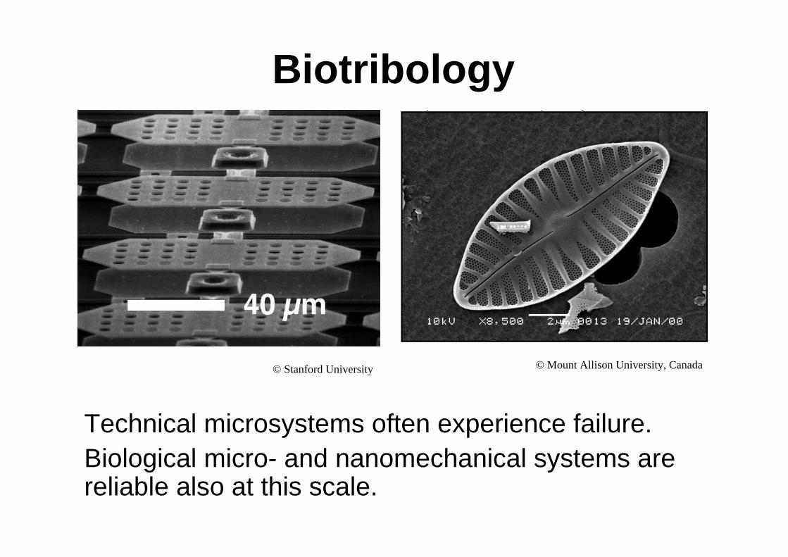
Biotribology
Technical microsystems often experience failure. Biological micro- and nanomechanical systems are reliable also at this scale.
© Stanford University © Mount Allison University, Canada
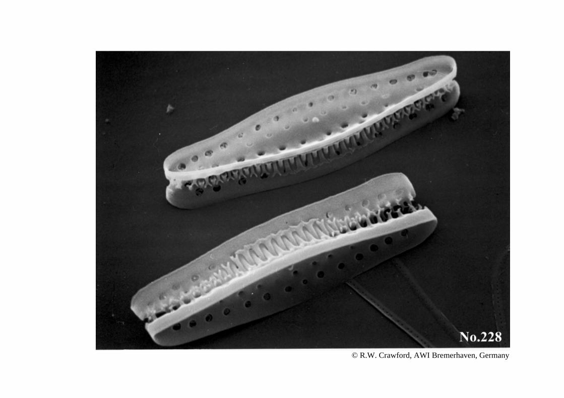
© R.W. Crawford, AWI Bremerhaven, Germany
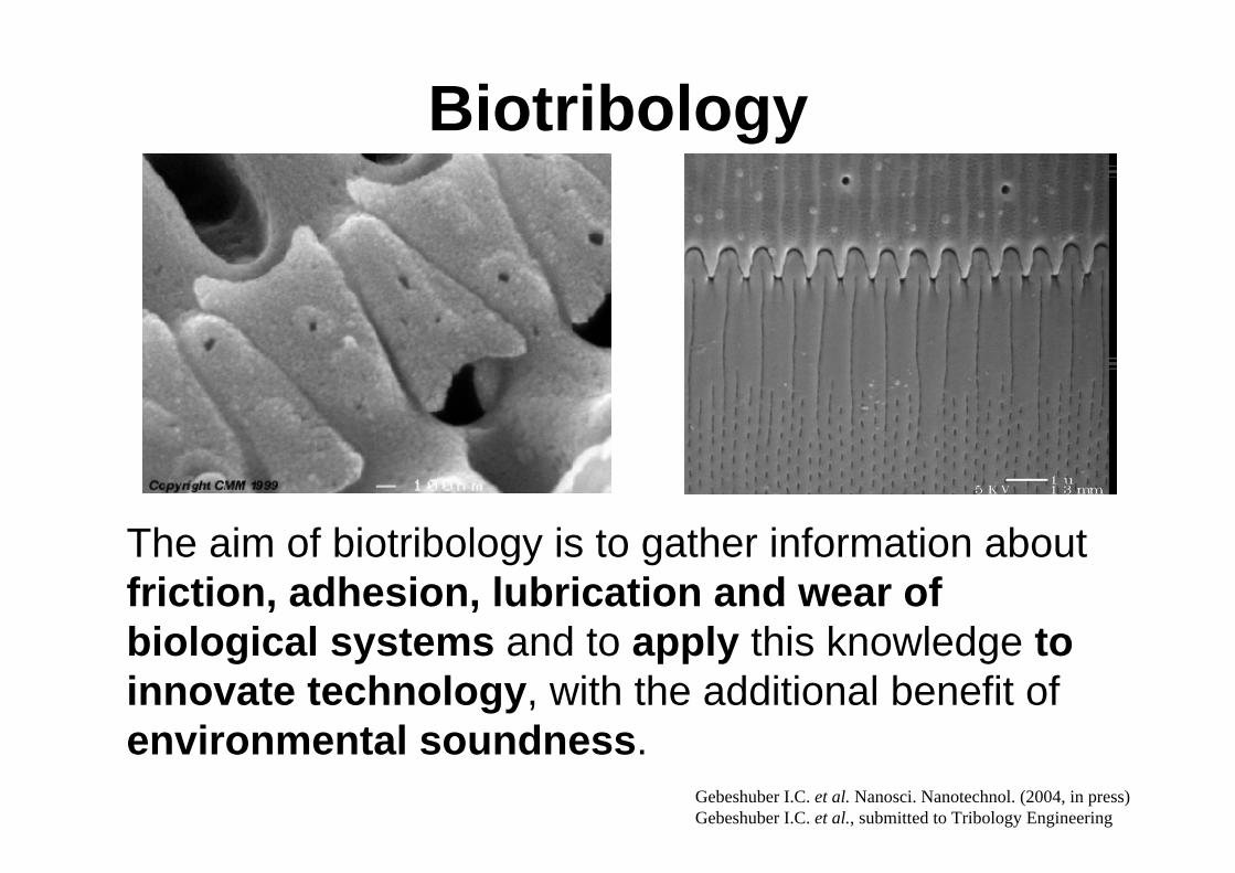
Biotribology
The aim of biotribology is to gather information about friction, adhesion, lubrication and wear of biological systems and to apply this knowledge to innovate technology, with the additional benefit of environmental soundness.
Gebeshuber I.C. et al. Nanosci. Nanotechnol. (2004, in press)Gebeshuber I.C. et al., submitted to Tribology Engineering
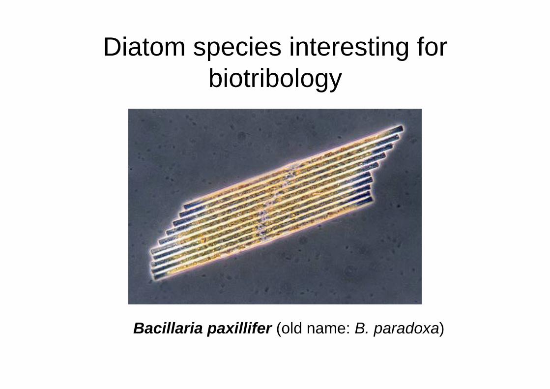
Diatom species interesting forbiotribology
Bacillaria paxillifer (old name: B. paradoxa)
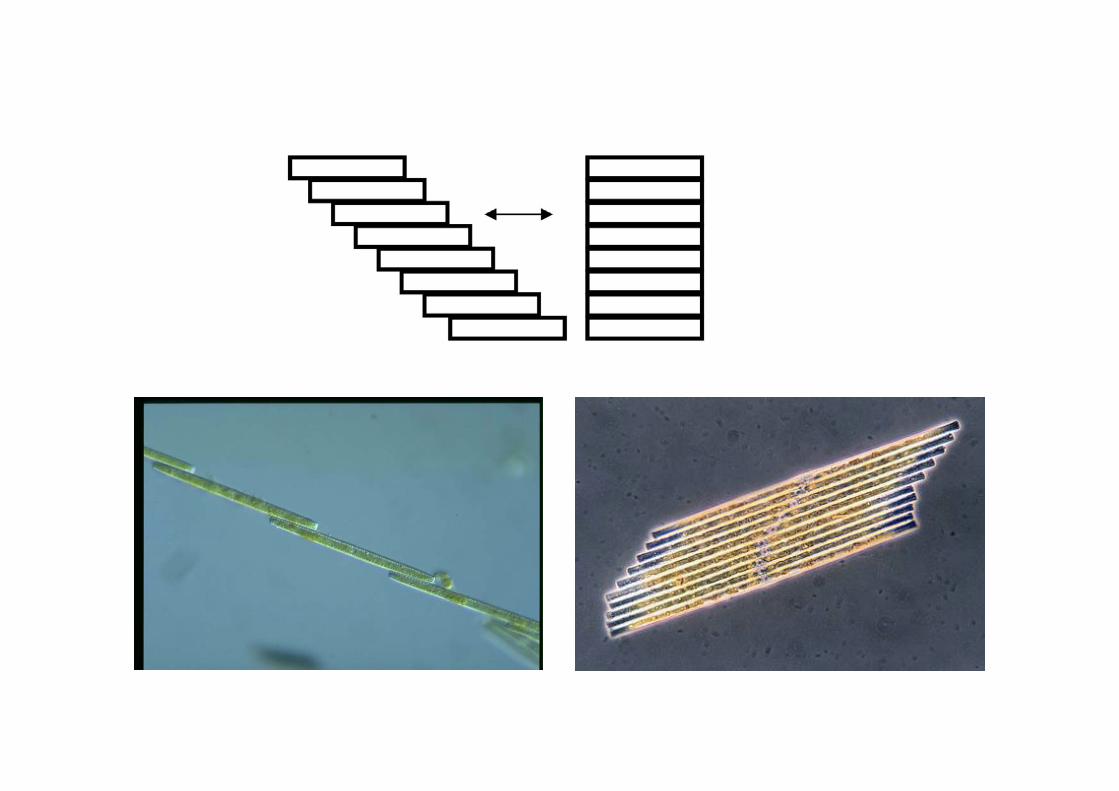

Movie
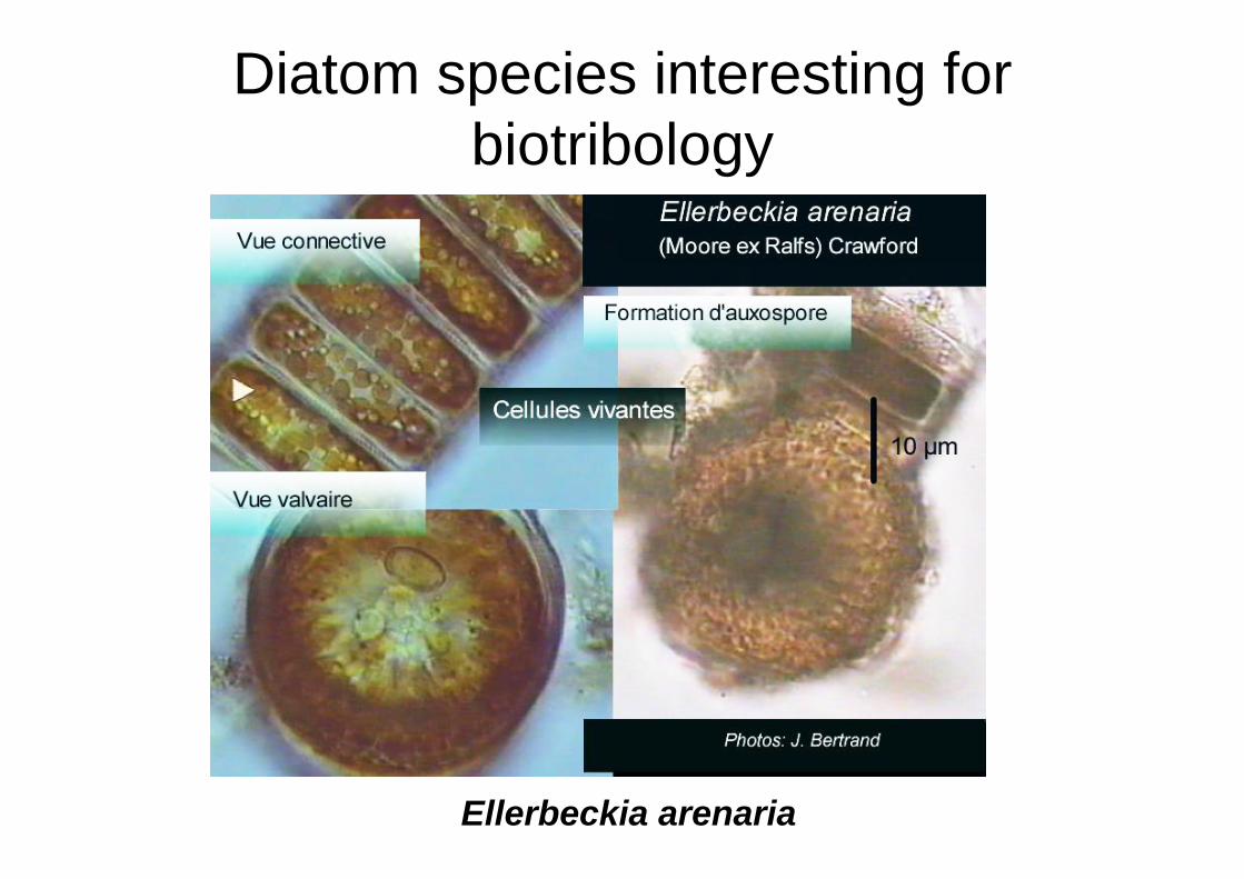
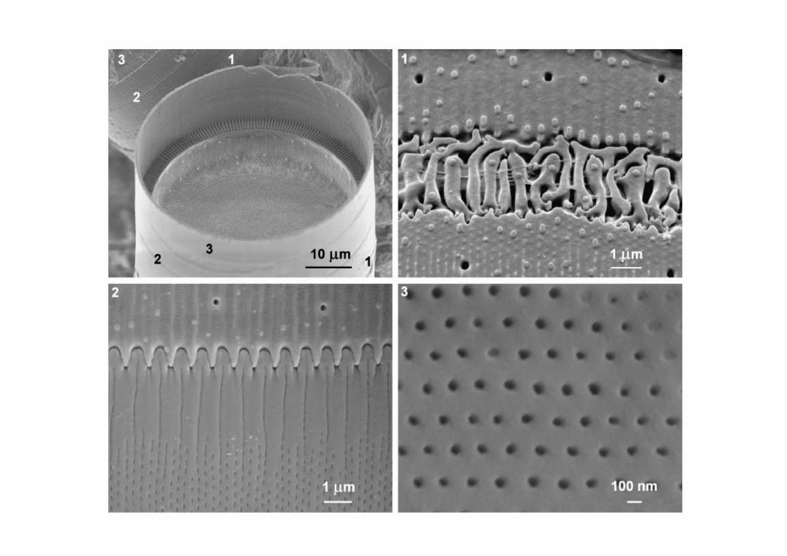
Diatom species interesting forbiotribology
Ellerbeckia arenaria
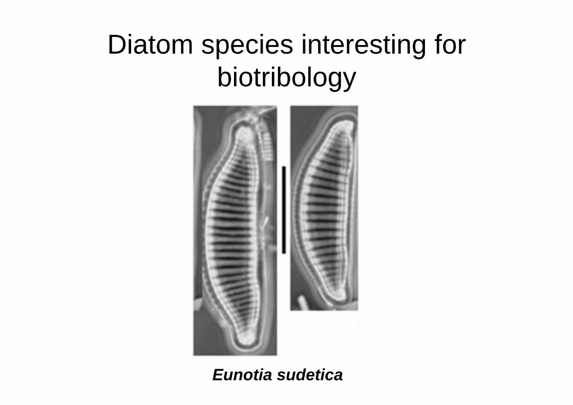
Diatom species interesting forbiotribology
Eunotia sudetica
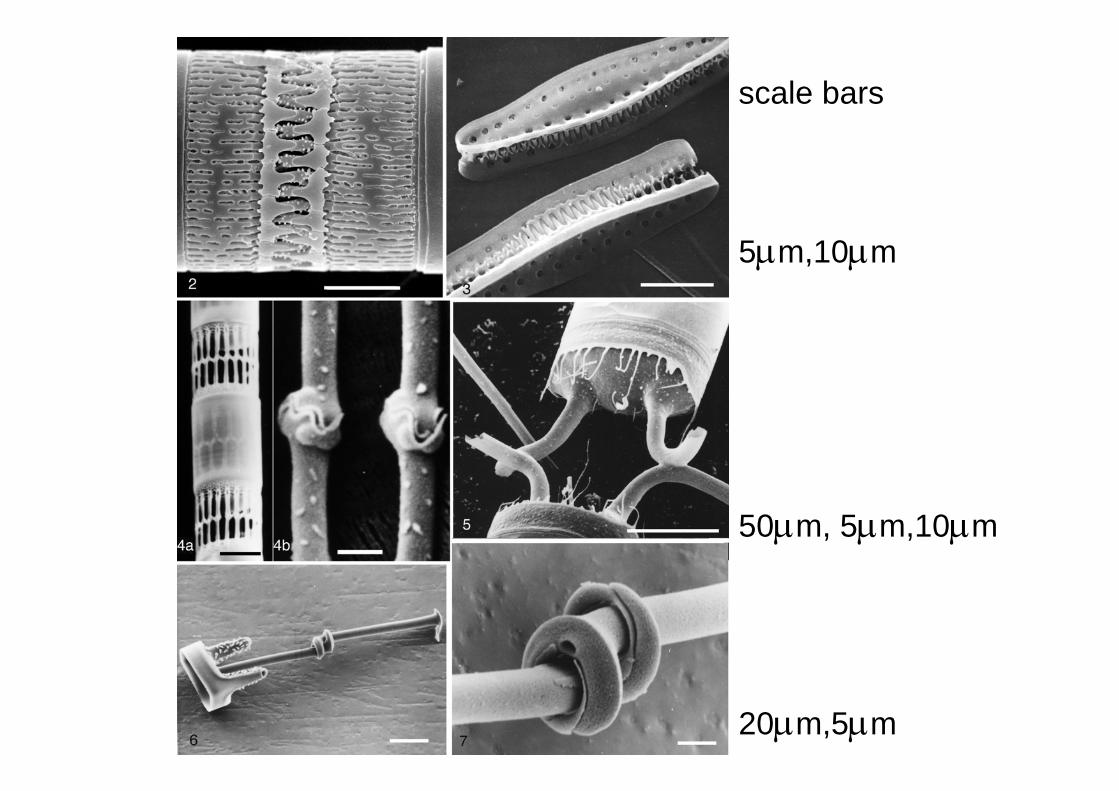
scale bars
5μm,10μm
50μm, 5μm,10μm
20μm,5μm
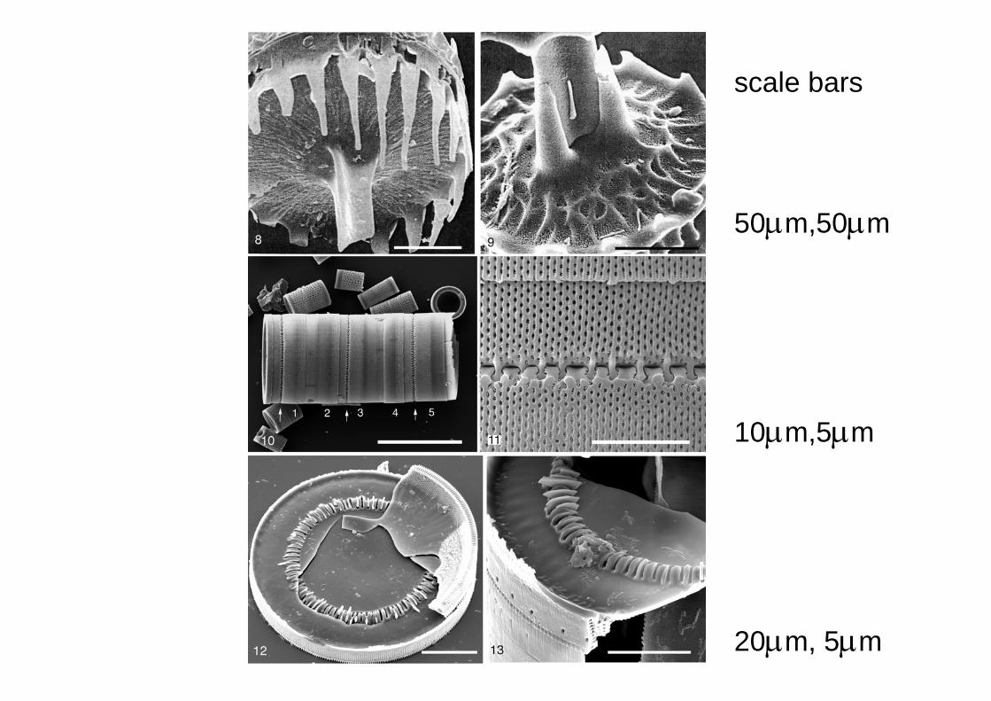
scale bars
50μm,50μm
10μm,5μm
20μm, 5μm
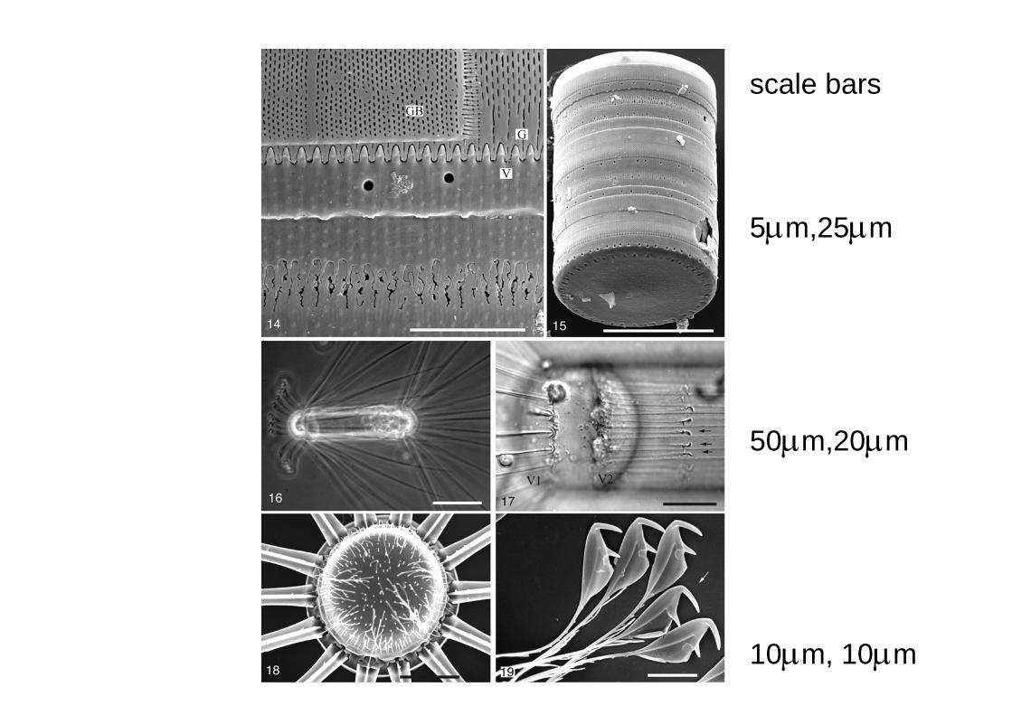
scale bars
5μm,25μm
50μm,20μm
10μm, 10μm
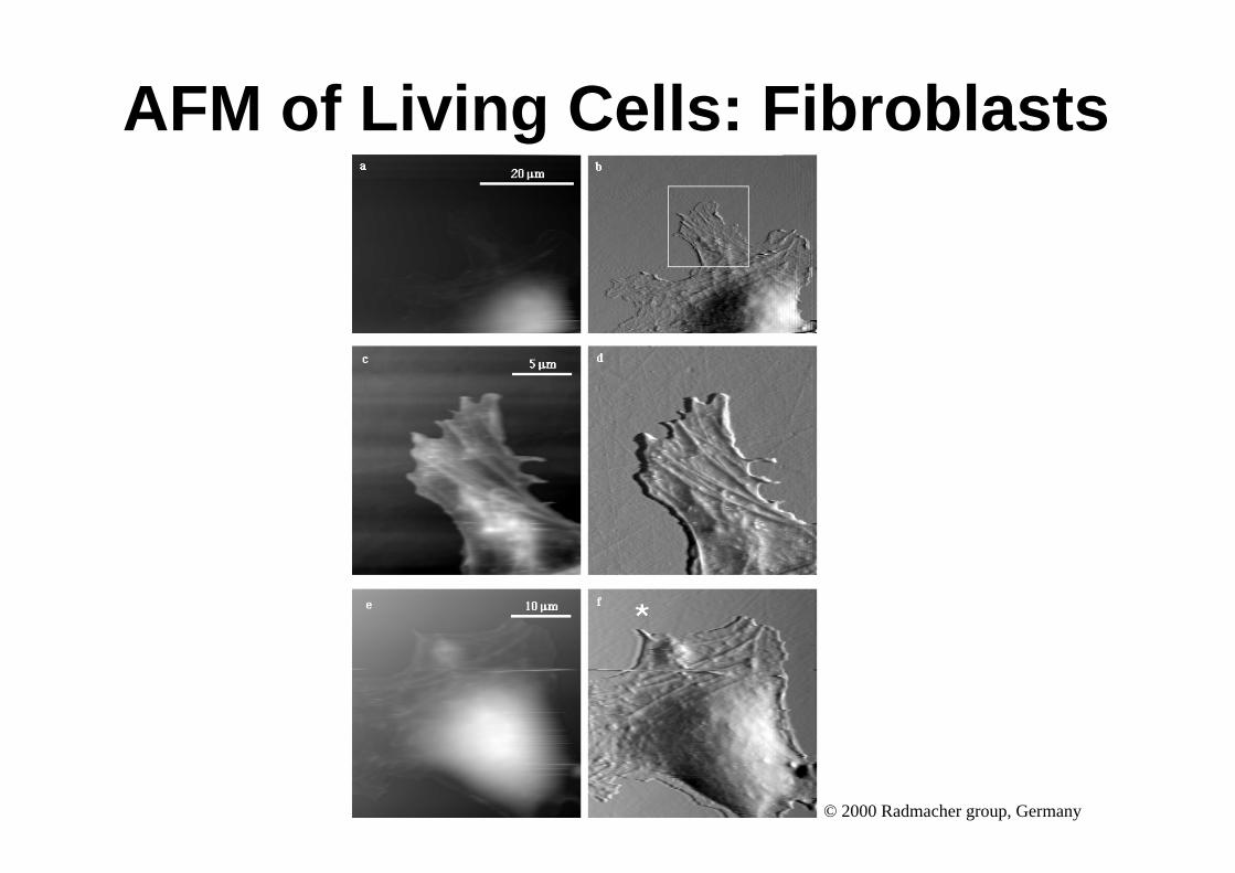
AFM of Living Cells: Fibroblasts
© 2000 Radmacher group, Germany
Single Molecules
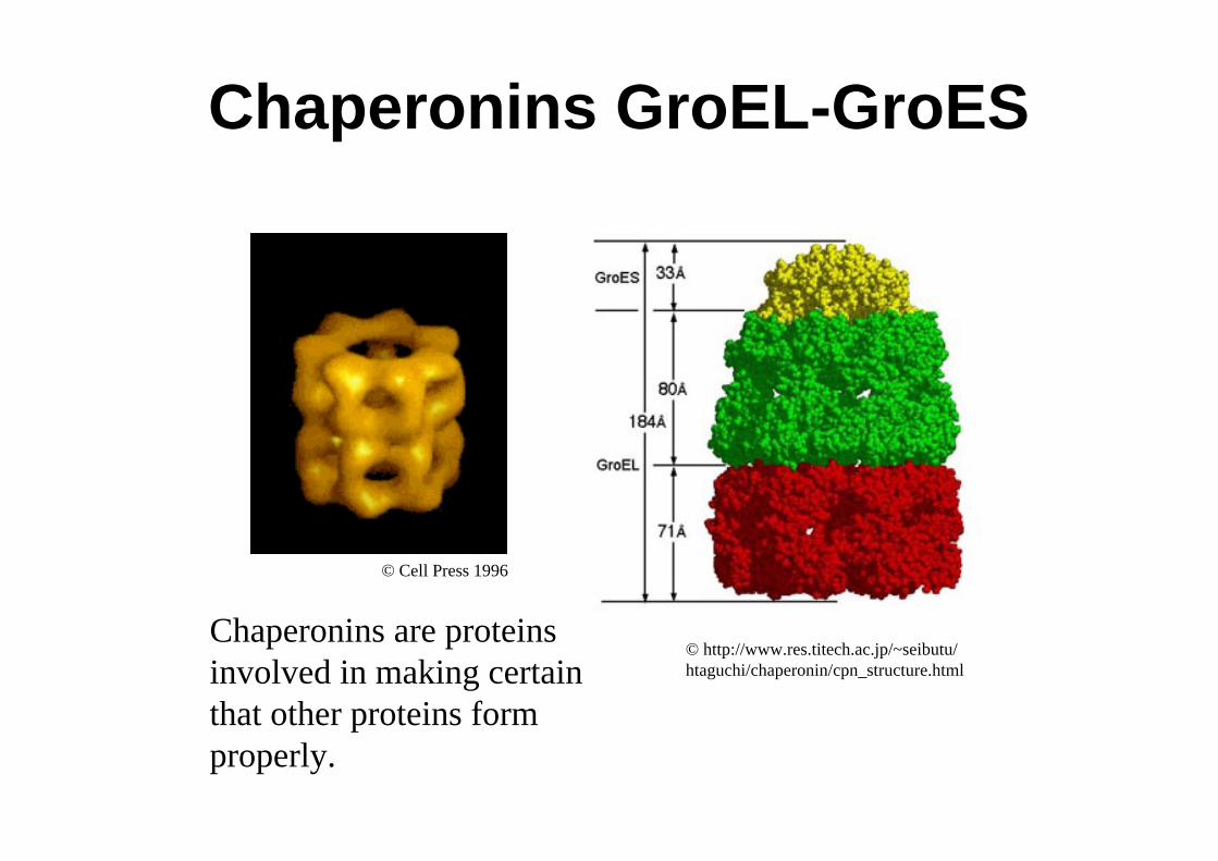
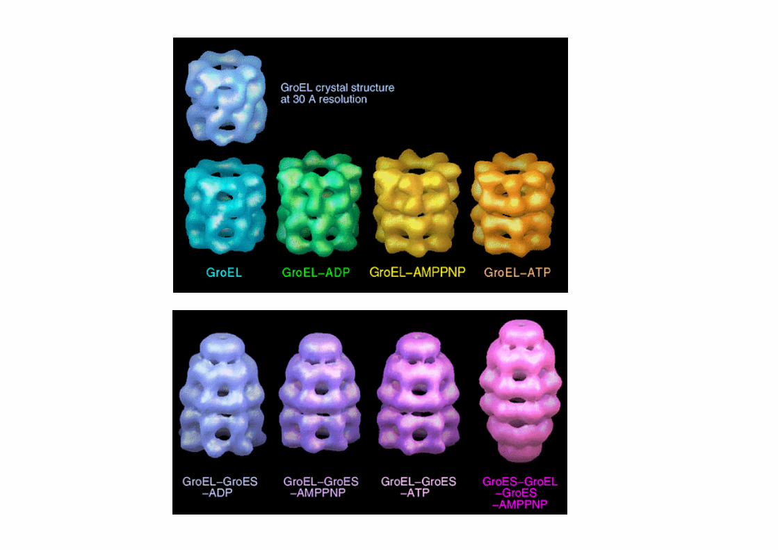
Chaperonins GroEL-GroES
© Cell Press 1996
© http://www.res.titech.ac.jp/~seibutu/htaguchi/chaperonin/cpn_structure.html
Chaperonins are proteins involved in making certain that other proteins form properly.
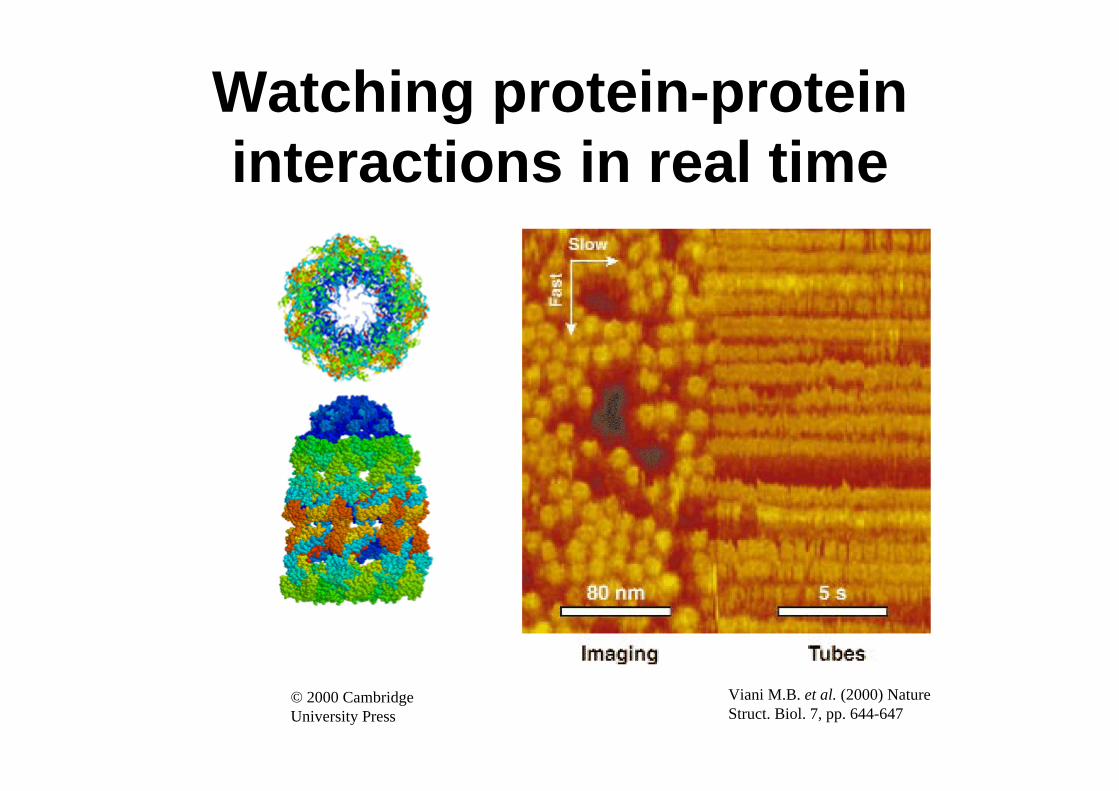
Watching protein-protein interactions in real time
Viani M.B. et al. (2000) Nature Struct. Biol. 7, pp. 644-647
© 2000 Cambridge University Press
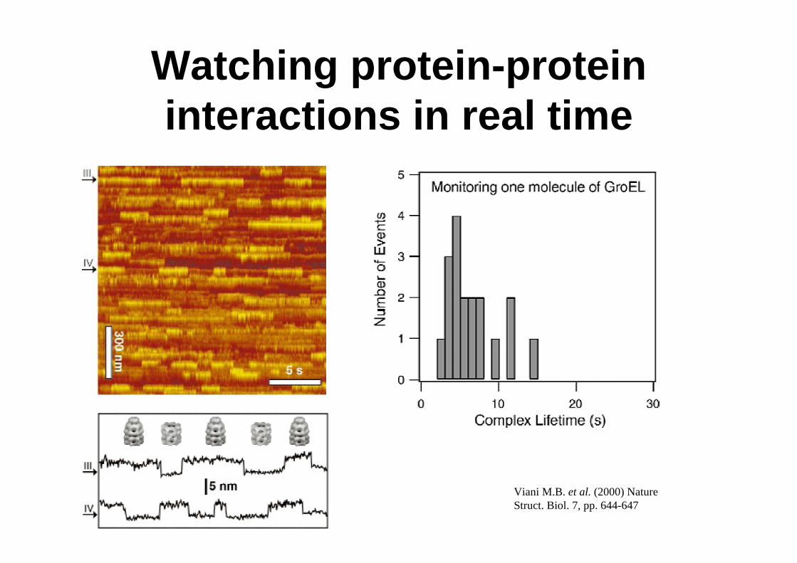
Watching protein-protein interactions in real time
Viani M.B. et al. (2000) Nature Struct. Biol. 7, pp. 644-647
Atoms
Nanostructuring atomically flatsurfaces with ions
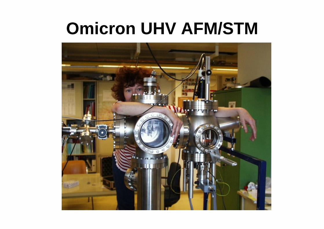
Omicron UHV AFM/STM
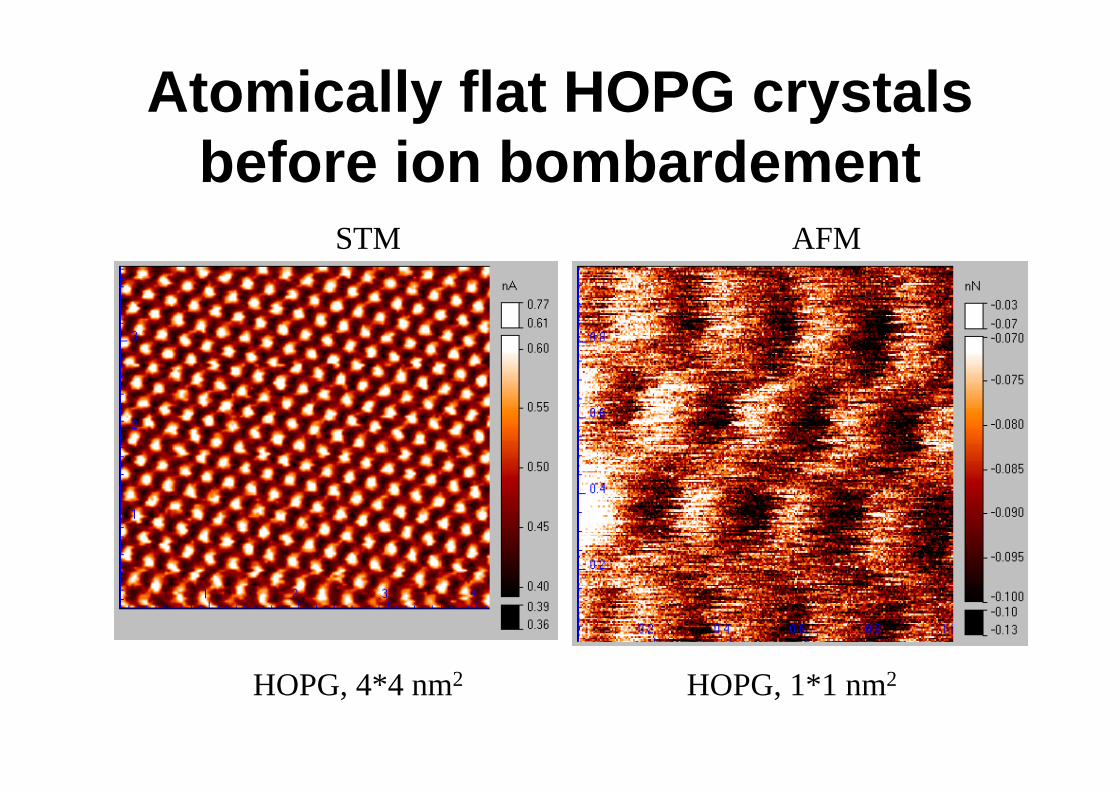
Atomically flat HOPG crystals before ion bombardement
HOPG, 4*4 nm2 HOPG, 1*1 nm2
STM AFM
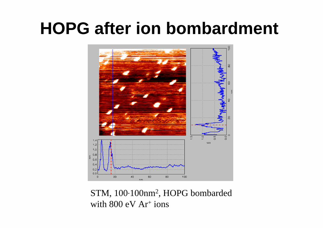
HOPG after ion bombardment
STM, 100.100nm2, HOPG bombarded with 800 eV Ar+ ions
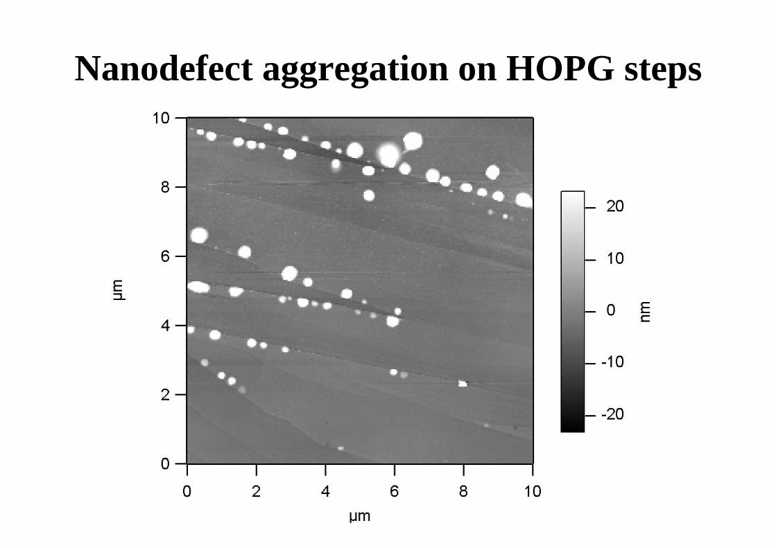
Nanodefect aggregation on HOPG steps
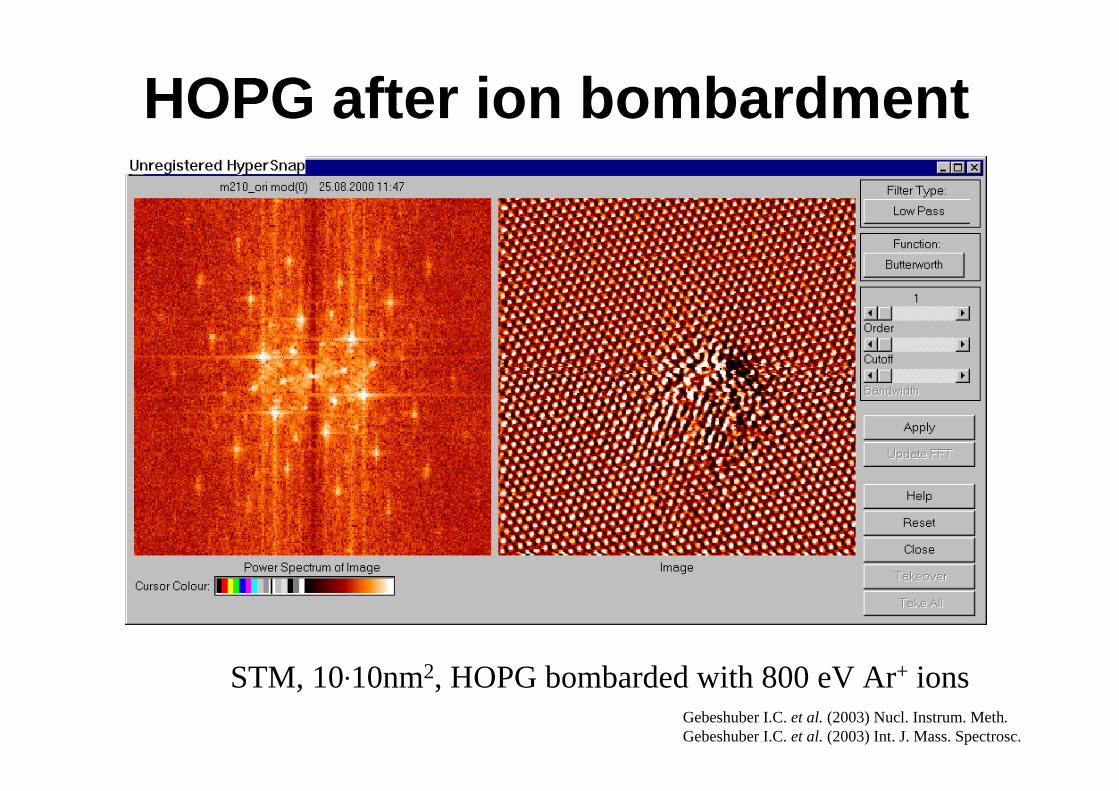
HOPG after ion bombardment
STM, 10.10nm2, HOPG bombarded with 800 eV Ar+ ions Gebeshuber I.C. et al. (2003) Nucl. Instrum. Meth.Gebeshuber I.C. et al. (2003) Int. J. Mass. Spectrosc.
Ion bombardment of atomically flat insulator crystals
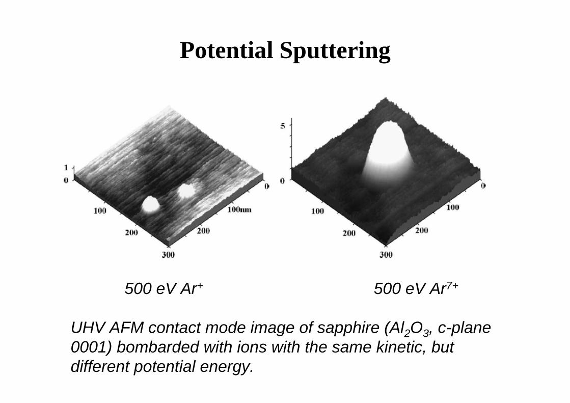
Potential Sputtering
500 eV Ar+ 500 eV Ar7+
UHV AFM contact mode image of sapphire (Al2O3, c-plane 0001) bombarded with ions with the same kinetic, but different potential energy.
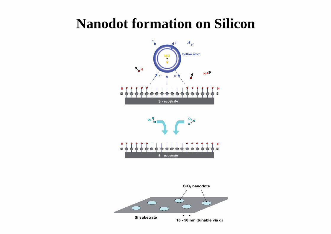
Nanodot formation on Silicon
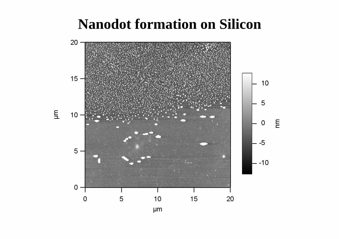
Nanodot formation on Silicon
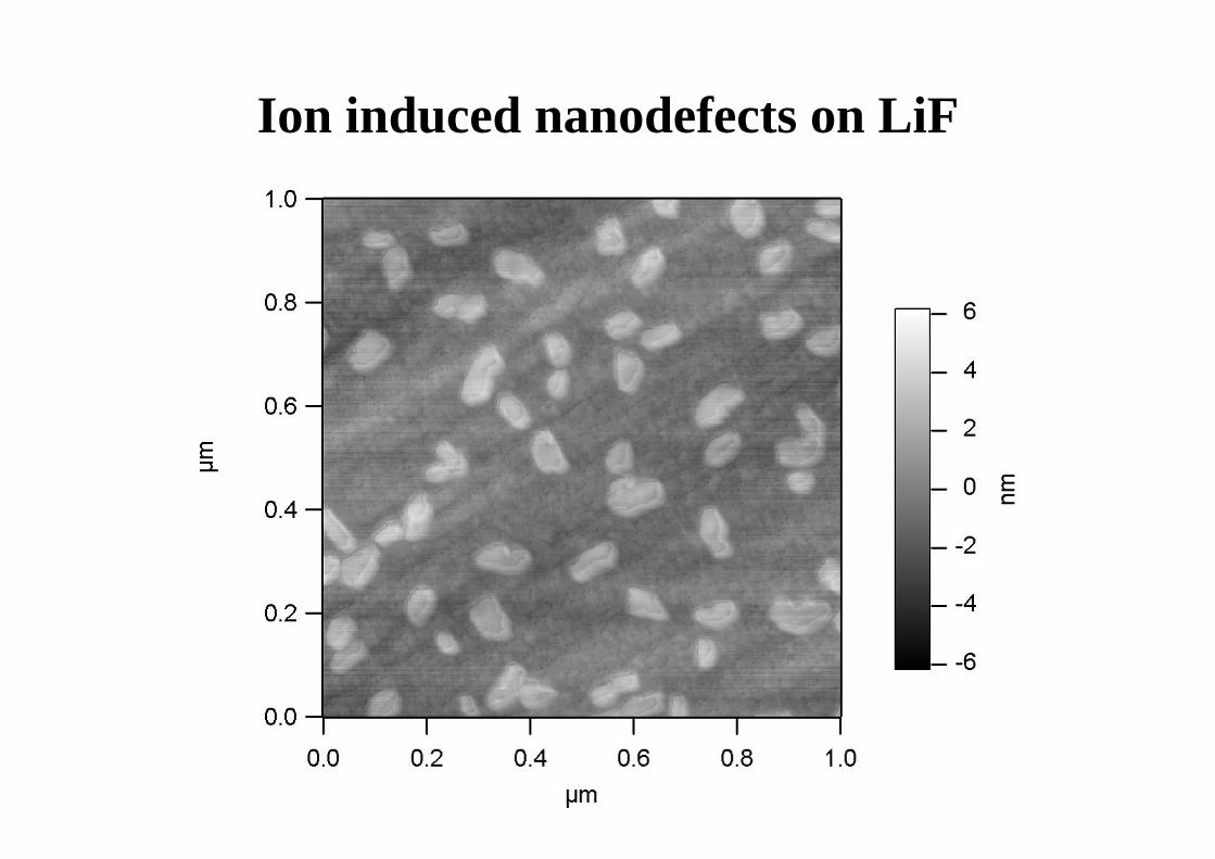
Ion induced nanodefects on LiF
Subatomic features
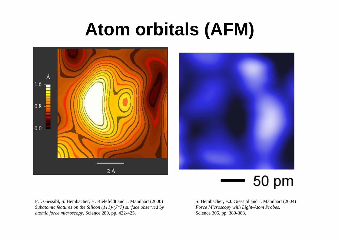
Atom orbitals (AFM)
F.J. Giessibl, S. Hembacher, H. Bielefeldt and J. Mannhart (2000) Subatomic features on the Silicon (111)-(7*7) surface observed by atomic force microscopy. Science 289, pp. 422-425.
S. Hembacher, F.J. Giessibl and J. Mannhart (2004) Force Microscopy with Light-Atom Probes.Science 305, pp. 380-383.
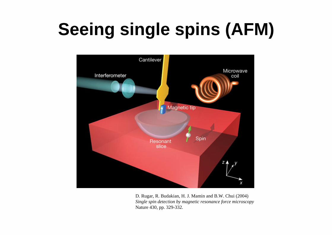
Seeing single spins (AFM)
D. Rugar, R. Budakian, H. J. Mamin and B.W. Chui (2004)Single spin detection by magnetic resonance force microscopyNature 430, pp. 329-332.
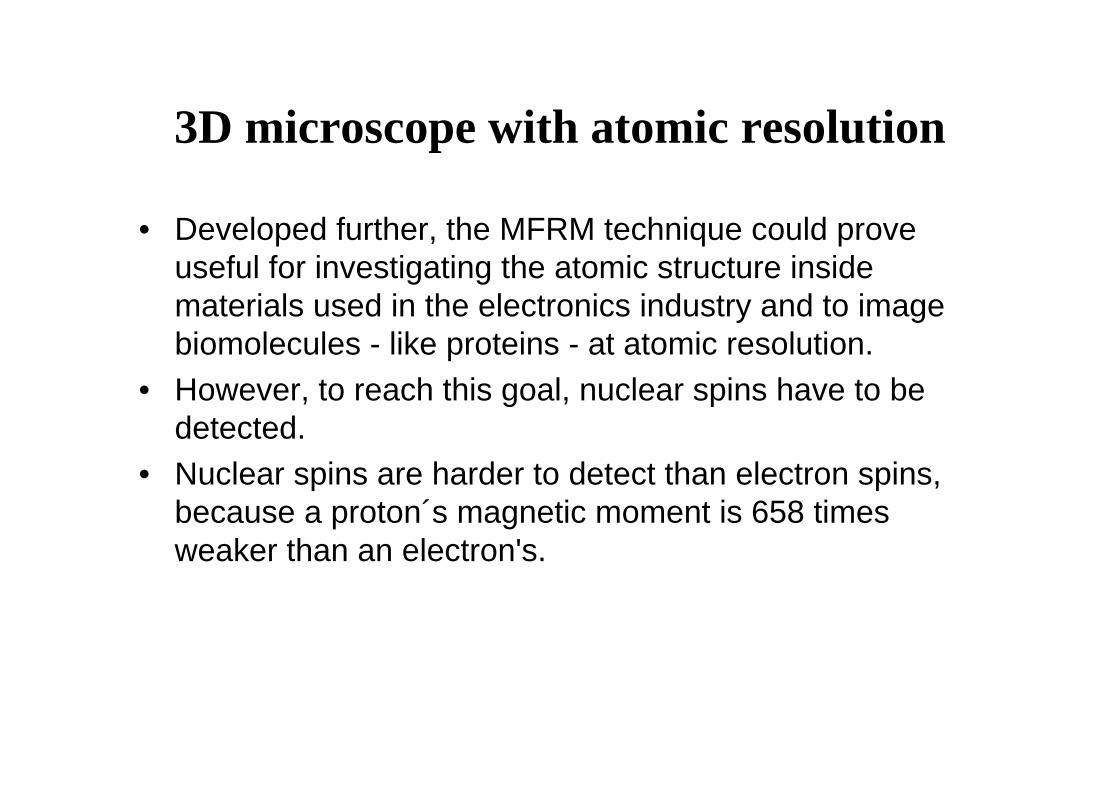
3D microscope with atomic resolution
• Developed further, the MFRM technique could prove useful for investigating the atomic structure inside materials used in the electronics industry and to image biomolecules - like proteins - at atomic resolution.
• However, to reach this goal, nuclear spins have to be detected.
• Nuclear spins are harder to detect than electron spins, because a proton´s magnetic moment is 658 times weaker than an electron's.
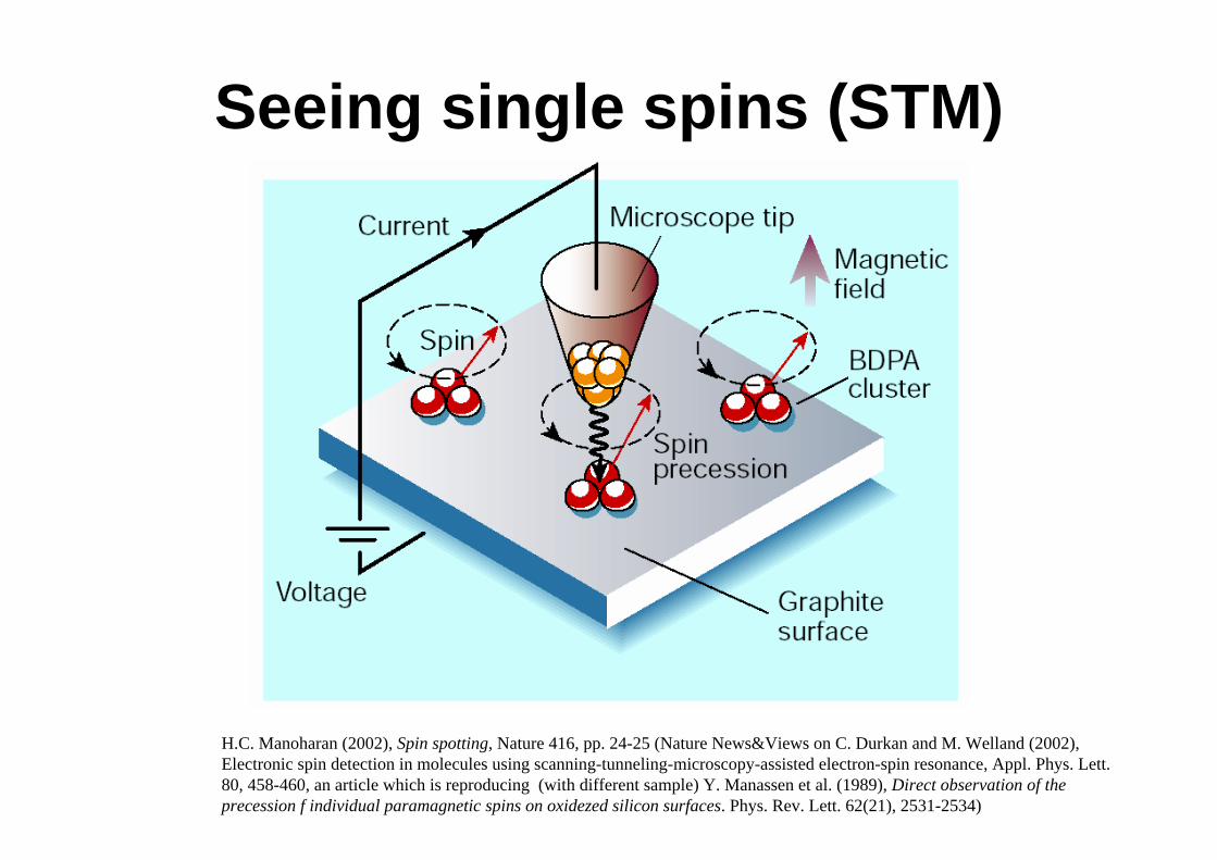
Seeing single spins (STM)
H.C. Manoharan (2002), Spin spotting, Nature 416, pp. 24-25 (Nature News&Views on C. Durkan and M. Welland (2002), Electronic spin detection in molecules using scanning-tunneling-microscopy-assisted electron-spin resonance, Appl. Phys. Lett. 80, 458-460, an article which is reproducing (with different sample) Y. Manassen et al. (1989), Direct observation of the precession f individual paramagnetic spins on oxidezed silicon surfaces. Phys. Rev. Lett. 62(21), 2531-2534)
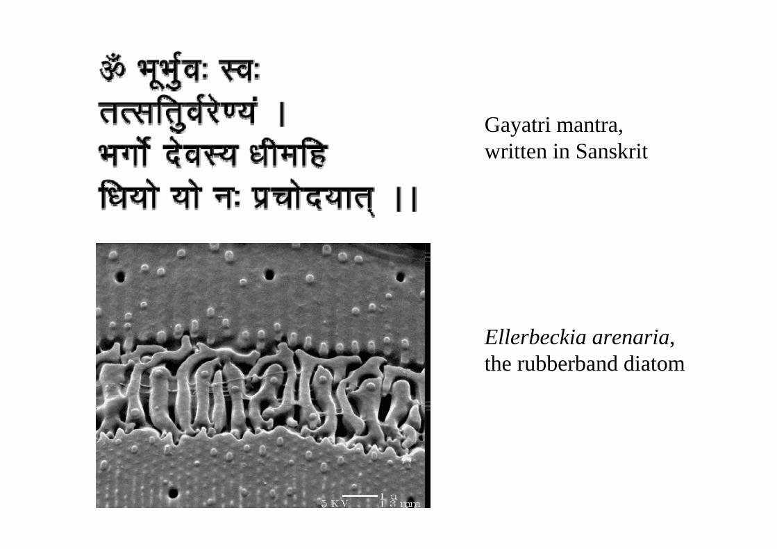
Gayatri mantra, written in Sanskrit
Ellerbeckia arenaria, the rubberband diatom

Thank you for your attention !
NanotechnologyOutlook
30.11.2005: Practical demonstrations at our institute07.12.2005: Clemens Grünberger and Dipl.-Ing. Stefan Schraml will present the SPM. Stefan developed a Scanning Ion Conductance Microscope (SICM) in the course of his diploma thesis and Clemens is working on the photoreceptor of green algae for use in biocomputers.14.12.2005: NO LECTURE11.01.2006: NO LECTUREFirst lecture 2006: January 18, 2006


![α NV5000 1 - Interempresas · 2014. 12. 17. · ISO 10791-9, JIS B6336-9 Max. tool changing time: 8.8 sec. Min. tool changing time: 3.1 sec. ... [ ] Option ISO: International Organization](https://static.fdocument.org/doc/165x107/60e655ad6922254075517bfa/-nv5000-1-interempresas-2014-12-17-iso-10791-9-jis-b6336-9-max-tool-changing.jpg)