17-Estradiol Regulates miR-9-5p and miR-9-3p Stability and ...
3479
Abstract. – OBJECTIVE: To investigate the expression of miR-134 and the change of inflam-matory cytokines in seizure rats and to explore its relationship.
MATERIALS AND METHODS: A rat model of seizures was made by an intraperitoneal in-jection with kainic acid. ELISA kit for detection of seizures in rats was used. The changes of inflammatory cytokines (IL-1, IL-2, IL-6, TNF-α, IFN-γ) and the hippocampal neuronal cell growth were observed. The expression of miR-134 in brain tissue and serum samples of model group and control group was determined by reverse transcriptase-polymerase chain reaction (RT-PCR) quantitative determination.
RESULTS: After the intraperitoneal injection of kainic acid, the rat model of seizure was suc-cessfully established. Compared with the control group, the neuronal cells in the hippocampus of the model group showed evident pathological changes. Inflammatory cytokines in seizures rats showed that the increase of IL-2, IL-6, and TNF-α were larger, but the increase of IL-1 and IFN-γ was not obvious. The results of the RT-PCR quantita-tive analysis showed that the expression of miR-134 in brain tissue and serum of epilepsy rats was lower than that of the control group (p<0.05).
CONCLUSIONS: The expression of miR-134 would be down by the increasing of inflamma-tory cytokines in the epileptic seizure.
Key WordsSeizure fit rats, miR-134, Inflammatory cytokines,
Hippocampal neurons.
Introduction
An epileptic seizure is a kind of brain dysfunc-tion disease triggered by a series of etiologies, which is caused by over discharge due to a neuro-nal abnormality in a certain area of the brain. The main clinical symptoms are a temporary recur-rent disturbance of consciousness and generalized or localized muscle spasms and convulsions. A
large number of studies1-3 have shown that cellu-lar immunity and humoral immune function dis-orders occur during the pathogenesis of epilepsy. The body’s immune mechanisms are involved in the pathogenesis of epilepsy.
Micro-ribonucleic acid (miRNA) is a non-cod-ing conservative RNA with about 20-24 bp in length, which plays an important regulatory role in gene expression. As for gene regulation, one gene could be regulated by multiple miRNAs and mul-tiple genes could be regulated by one miRNA4,5. MiRNA-134 (miR-134) is a highly conservative miRNA that widely exists in animal and human tissues, which is closely related to nervous system diseases and tumor diseases in the animal body.
Clinically, the autoimmune disorder is an im-portant cause of epilepsy. After being injured, en-dothelial cells in brain tissues can release a vari-ety of cytokines, activating T cells, and triggering immune responses in the brain. Then, the expres-sion level of inflammatory regulators is up-regu-lated. This indicates that inflammatory cytokines are closely related to epilepsy.
This study aimed to explore the expression of miR-134 and changes in the serum level of in-flammatory cytokines and to investigate the re-lationship between the changes in inflammatory cytokines and the expression of miR-134 in the rat model of epileptic seizures.
Materials and Methods
Experimental AnimalsIn this study, Sprague-Dawley (SD) rats (male,
20-25 weeks old, n=30) purchased from Hubei Pro-vincial Laboratory Animal Research Center were taken as the experimental animals, and fed in an isolator with the temperature controlled at 25-28°C for preparation of the epileptic seizure rat model. This study was approved by the Animal Ethics Committee of Putian University Animal Center.
European Review for Medical and Pharmacological Sciences 2018; 22: 3479-3484
W.-S. HUANG1, L. ZHU2
1Department of Neurology, The Affiliated Hospital (Group) of Putian University, Putian, China2Department of Neurology, Xuzhou Central Hospital, Xuzhou, China
Corresponding Author: Liang Zhu, MM; e-mail: [email protected]
MiR-134 expression and changes in inflammatory cytokines of rats with epileptic seizures
W.-S. Huang, L. Zhu
3480
Main ReagentsKainic acid (10 mg/bottle) was purchased
from the Cayman Chemical Company (Ann Ar-bor, MI, USA). Enzyme-linked immunosorbent assay (ELISA) kit for inflammatory cytokines was purchased from the Sangon Biotech Co., Ltd. (Wuhan, China); primer synthesis kits, re-verse transcription kits, and (SYBR Green) flu-orescence quantitative kits were purchased from the TSINGKE Biological Technology Co., Ltd. (Kibbutz Beit Haemek, Israel); DY-1-type brain stereotaxic apparatus.
Preparation for the ModelModel group: 15 rats were randomly selected;
kainic acid (10 mg/bottle) was diluted with 10 mL sterile saline; each rat was intraperitoneally in-jected with kainic acid (100 μL/100 g).
Control group: 15 rats were randomly selected; each rat was intraperitoneally injected with saline (100 μL/100 g); behavioral changes of the two groups were observed.
Criteria for successful modeling: epileptic sei-zure symptoms (sluggishness, salivation, tremors, convulsions, etc.) were observed in model rats; rats in the control group behaved normally.
Detection of InflammatoryCytokines
At 5 h after intraperitoneal injection of ka-inic acid or saline, 5 rats were randomly select-ed; 2 mL tail vein blood was taken and cen-trifuged at room temperature for 1 h, and the supernatant was removed. 200 μL serum was taken, and RNA was extracted from it using the TRIzol method. Meanwhile, the ELISA kit was used to detect the serum concentration of in-flammatory cytokines [interleukin-1 (IL-1), IL-2, IL-6, tumor necrosis factor-alpha (TNF-α) and interferon-γ (IFN-γ)].
Pathological Examination5% pentobarbital injection was prepared by
sterile saline. 5 rats in the epileptic seizure model group and 5 rats in the control group were inject-ed with the anesthetic (3 mg/100 g). The thoracic and abdominal cavities of rats were dissected, and their left ventricles were perfused with sa-line at room temperature until the rats died. The cranial cavity of rats was cut open to take brain tissues for preparing slices, while hematoxylin and eosin (HE) staining was performed. At the same time, some brain tissues were taken for RNA extraction.
Reverse Transcription PolymeraseChain Reaction (RT-PCR) and Relatively Quantitative Analysis
According to the instructions of the RT-PCR Kit, RNAs extracted in 1.2.3 and 1.2.4 was reverse-ly transcribed according to the reaction system in Table I. Relatively quantitative detection of miR-NA: U6 RNA was taken as the internal control. The reverse transcription primers of miR-134 and U6: miR-134 (5’-GTCGTATCCAGTGCGTGTC-GTGGAGTCGGCAATTGCACTGGATAGGAC-CCCCTC-3’ and U6 (5’-TGTTGGCGTGGAGT-FG-3’). PCR primers were designed based on the conservative regions of miR-134 and U6, and the relatively quantitative detection of miR-134 was performed according to the instructions of (SYBR Green) fluorescence quantitative kits. Relatively quantitative detection system of miR-134: 25 μL GoTaq qPCR Master Mix (2×); 1 μL miR-134 for-ward primer; 1 μL miR-134 stem-loop RT Prim-er; 2 μL complementary deoxyribonucleic acid (cDNA); 21 μL RNase-Free H2O; 50 μL in total.
Detection by Northern BlotBrain tissues of the rats were homogenized
and added with lysate for lysis, followed by the ultra-centrifugation at 30,000 rpm (4°C, 1.5 h). The precipitate was resuspended using 0.5 mL phosphate-buffered saline (PBS) to determine the total protein concentration. Sodium do-decyl sulfate-polyacrylamide gel electrophoresis (SDS-PAGE) was conducted for samples, which were then transferred to polyvinylidene fluo-ride (PVDF) membrane. The primary antibod-ies were incubated overnight at 4°C, and then, incubated with horseradish peroxidase-labeled secondary antibodies for 1 h after washing. Af-ter the mixed solution of 3,3’-diaminobenzidine (DAB) and hydrogen peroxide were added, fluo-rography technique was used to get the images.
Table I. RT-PCR primer sequences of miR-134 and U6 RNA.
Primer name Primer sequence
miR-134 5’-GGTGTGACTGGTTGACCA-3’ forward chain
miR-134 5’-TGCGTGTCGTGGAGTC-3’ reverse chain
U6 forward 5’-CTCGCTTCGGCAGCACA-3’ chain
U6 reverse 5’-AACGCTTCACGAATTTGCGT-3’ chain
MiR-134 expression and changes in inflammatory cytokines of rats with epileptic seizures
3481
Statistical AnalysisSPSS 17.0 software (SPSS Inc., Chicago, IL,
USA) was used for data analysis. All quantitative data were expressed as mean ± standard deviation (SD). The comparison between groups was done using One-way ANOVA test followed by Post-Hoc Test (Least Significant Difference). p<0.05 represented that the difference was statistically significant.
Results
Pathological Results of Brain Tissues of Rats With Epileptic Seizures
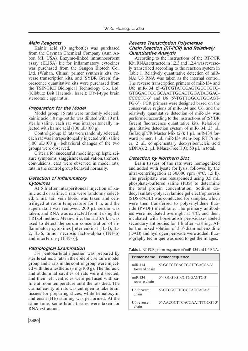
Pathological section results showed that there was a significant difference in the neuronal area of hippocampus between the epileptic seizure model group and the control group. As shown in Figure 1, hippocampal neurons were arranged in disorder in the epileptic seizure model group. Additionally, increased intercellular space, de-creased shrinkage volume, necrotic lysis, un-clear nucleoli and disappearance of cytoplasm were also observed in the epileptic seizure mod-el group.
Inflammatory Cytokine Expressions in Serum of Rats With Epileptic Seizures
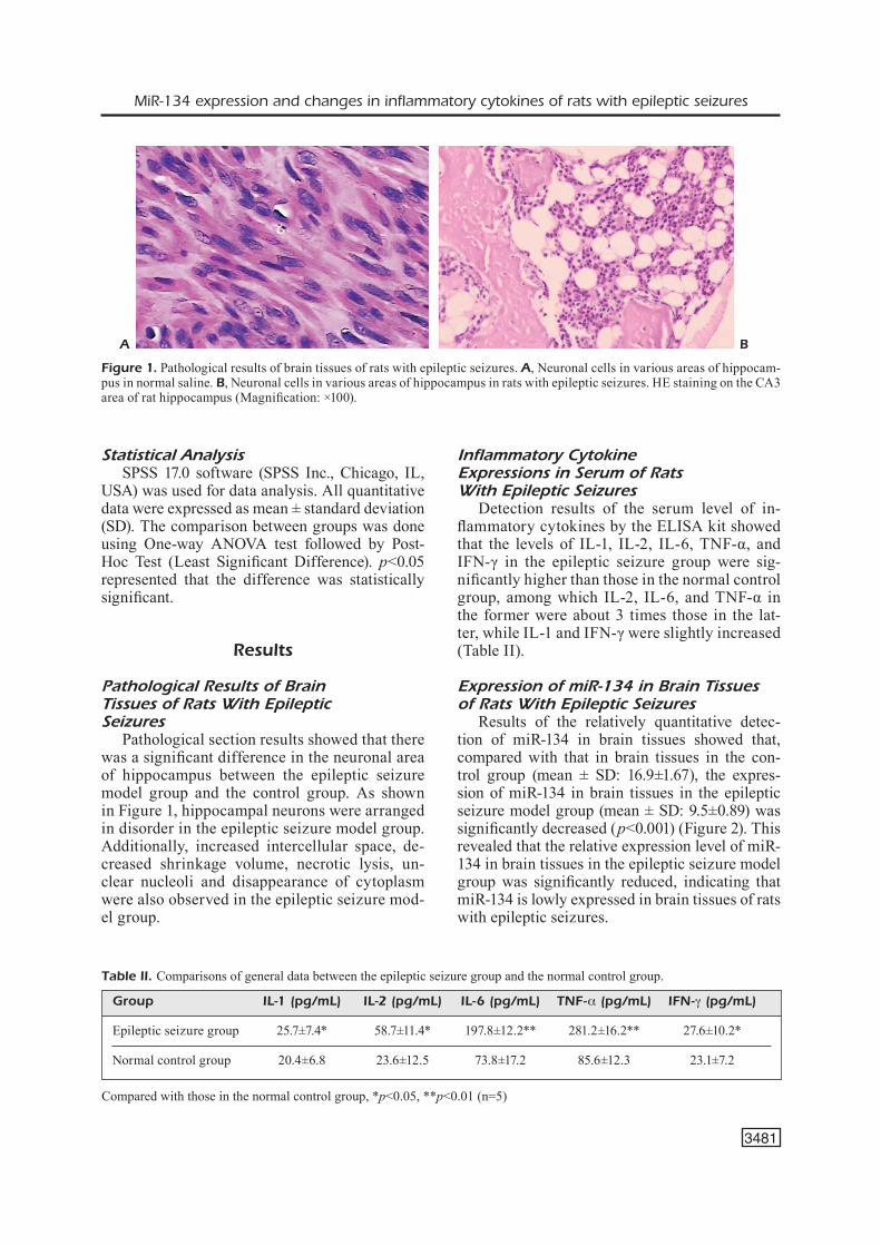
Detection results of the serum level of in-flammatory cytokines by the ELISA kit showed that the levels of IL-1, IL-2, IL-6, TNF-α, and IFN-γ in the epileptic seizure group were sig-nificantly higher than those in the normal control group, among which IL-2, IL-6, and TNF-α in the former were about 3 times those in the lat-ter, while IL-1 and IFN-γ were slightly increased (Table II).
Expression of miR-134 in Brain Tissuesof Rats With Epileptic Seizures
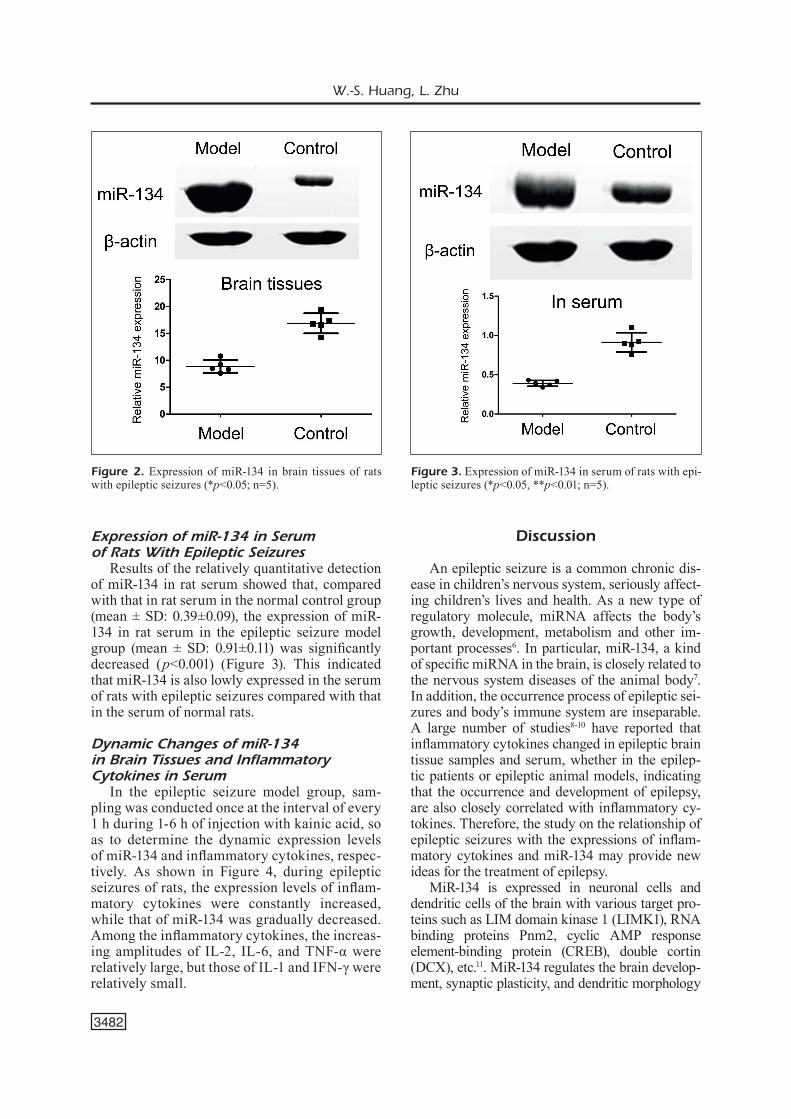
Results of the relatively quantitative detec-tion of miR-134 in brain tissues showed that, compared with that in brain tissues in the con-trol group (mean ± SD: 16.9±1.67), the expres-sion of miR-134 in brain tissues in the epileptic seizure model group (mean ± SD: 9.5±0.89) was significantly decreased (p<0.001) (Figure 2). This revealed that the relative expression level of miR-134 in brain tissues in the epileptic seizure model group was significantly reduced, indicating that miR-134 is lowly expressed in brain tissues of rats with epileptic seizures.
A B
Figure 1. Pathological results of brain tissues of rats with epileptic seizures. A, Neuronal cells in various areas of hippocam-pus in normal saline. B, Neuronal cells in various areas of hippocampus in rats with epileptic seizures. HE staining on the CA3 area of rat hippocampus (Magnification: ×100).
Table II. Comparisons of general data between the epileptic seizure group and the normal control group.
Compared with those in the normal control group, *p<0.05, **p<0.01 (n=5)
Group IL-1 (pg/mL) IL-2 (pg/mL) IL-6 (pg/mL) TNF-α (pg/mL) IFN-γ (pg/mL)
Epileptic seizure group 25.7±7.4* 58.7±11.4* 197.8±12.2** 281.2±16.2** 27.6±10.2*
Normal control group 20.4±6.8 23.6±12.5 73.8±17.2 85.6±12.3 23.1±7.2
W.-S. Huang, L. Zhu
3482
Expression of miR-134 in Serumof Rats With Epileptic Seizures
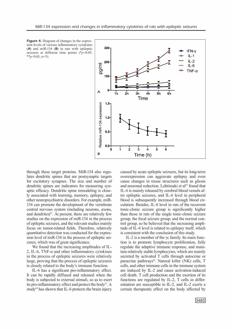
Results of the relatively quantitative detection of miR-134 in rat serum showed that, compared with that in rat serum in the normal control group (mean ± SD: 0.39±0.09), the expression of miR-134 in rat serum in the epileptic seizure model group (mean ± SD: 0.91±0.11) was significantly decreased (p<0.001) (Figure 3). This indicated that miR-134 is also lowly expressed in the serum of rats with epileptic seizures compared with that in the serum of normal rats.
Dynamic Changes of miR-134 in Brain Tissues and Inflammatory Cytokines in Serum
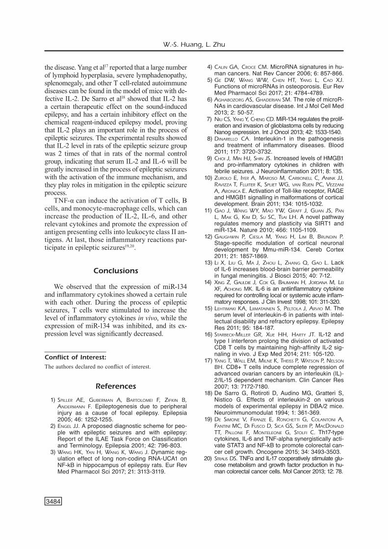
In the epileptic seizure model group, sam-pling was conducted once at the interval of every 1 h during 1-6 h of injection with kainic acid, so as to determine the dynamic expression levels of miR-134 and inflammatory cytokines, respec-tively. As shown in Figure 4, during epileptic seizures of rats, the expression levels of inflam-matory cytokines were constantly increased, while that of miR-134 was gradually decreased. Among the inflammatory cytokines, the increas-ing amplitudes of IL-2, IL-6, and TNF-α were relatively large, but those of IL-1 and IFN-γ were relatively small.
Discussion
An epileptic seizure is a common chronic dis-ease in children’s nervous system, seriously affect-ing children’s lives and health. As a new type of regulatory molecule, miRNA affects the body’s growth, development, metabolism and other im-portant processes6. In particular, miR-134, a kind of specific miRNA in the brain, is closely related to the nervous system diseases of the animal body7. In addition, the occurrence process of epileptic sei-zures and body’s immune system are inseparable. A large number of studies8-10 have reported that inflammatory cytokines changed in epileptic brain tissue samples and serum, whether in the epilep-tic patients or epileptic animal models, indicating that the occurrence and development of epilepsy, are also closely correlated with inflammatory cy-tokines. Therefore, the study on the relationship of epileptic seizures with the expressions of inflam-matory cytokines and miR-134 may provide new ideas for the treatment of epilepsy.
MiR-134 is expressed in neuronal cells and dendritic cells of the brain with various target pro-teins such as LIM domain kinase 1 (LIMK1), RNA binding proteins Pnm2, cyclic AMP response element-binding protein (CREB), double cortin (DCX), etc.11. MiR-134 regulates the brain develop-ment, synaptic plasticity, and dendritic morphology
Figure 2. Expression of miR-134 in brain tissues of rats with epileptic seizures (*p<0.05; n=5).
Figure 3. Expression of miR-134 in serum of rats with epi-leptic seizures (*p<0.05, **p<0.01; n=5).
MiR-134 expression and changes in inflammatory cytokines of rats with epileptic seizures
3483
through these target proteins. MiR-134 also regu-lates dendritic spines that are postsynaptic targets for excitatory synapses. The size and number of dendritic spines are indicators for measuring syn-aptic efficacy. Dendritic spine remodeling is close-ly associated with learning, memory, epilepsy, and other neuropsychiatric disorders. For example, miR-134 can promote the development of the vertebrate central nervous system (including neurons, axons, and dendrites)12. At present, there are relatively few studies on the expression of miR-134 in the process of epileptic seizures, and the relevant studies mainly focus on tumor-related fields. Therefore, relatively quantitative detection was conducted for the expres-sion level of miR-134 in the process of epileptic sei-zures, which was of great significance.
We found that the increasing amplitudes of IL-2, IL-6, TNF-α and other inflammatory cytokines in the process of epileptic seizures were relatively large, proving that the process of epileptic seizures is closely related to the body’s immune function.
IL-6 has a significant pro-inflammatory effect. It can be rapidly diffused and released when the body is subjected to external stimuli, so as to exert its pro-inflammatory effect and protect the body13. A study14 has shown that IL-6 protects the brain injury
caused by acute epileptic seizures, but its long-term overexpression can aggravate epilepsy and even cause changes in tissue structures such as gliosis and neuronal reduction. Lehtimaki et al15 found that IL-6 is mainly released by cerebral blood vessels af-ter epileptic seizures, and IL-6 level in peripheral blood is subsequently increased through blood cir-culation. Besides, IL-6 level in rats of the recurrent tonic-clonic seizure group is significantly higher than those in rats of the single tonic-clonic seizure group, the focal seizure group, and the normal con-trol group, so he believed that the increasing ampli-tude of IL-6 level is related to epilepsy itself, which is consistent with the conclusion of this study.
IL-2 is a member of the γc family. Its main func-tion is to promote lymphocyte proliferation, fully regulate the adaptive immune response, and main-tain relatively stable lymphocytes, which are mainly secreted by activated T cells through autocrine or paracrine pathways16. Natural killer (NK) cells, T cells, and other immune cells in the immune system are induced by IL-2 and cause activation-induced cell death. T cell production and the exertion of its functions are regulated by IL-2, T cells in differ-entiation are susceptible to IL-2, and IL-2 exerts a certain therapeutic effect on the body affected by
Figure 4. Diagram of changes in the expres-sion levels of various inflammatory cytokines (A) and miR-134 (B) in rats with epileptic seizures at different time points (*p<0.05, **p<0.01; n=5).
A
B
W.-S. Huang, L. Zhu
3484
the disease. Yang et al17 reported that a large number of lymphoid hyperplasia, severe lymphadenopathy, splenomegaly, and other T cell-related autoimmune diseases can be found in the model of mice with de-fective IL-2. De Sarro et al18 showed that IL-2 has a certain therapeutic effect on the sound-induced epilepsy, and has a certain inhibitory effect on the chemical reagent-induced epilepsy model, proving that IL-2 plays an important role in the process of epileptic seizures. The experimental results showed that IL-2 level in rats of the epileptic seizure group was 2 times of that in rats of the normal control group, indicating that serum IL-2 and IL-6 will be greatly increased in the process of epileptic seizures with the activation of the immune mechanism, and they play roles in mitigation in the epileptic seizure process.
TNF-α can induce the activation of T cells, B cells, and monocyte-macrophage cells, which can increase the production of IL-2, IL-6, and other relevant cytokines and promote the expression of antigen presenting cells into leukocyte class II an-tigens. At last, those inflammatory reactions par-ticipate in epileptic seizures19,20.
Conclusions
We observed that the expression of miR-134 and inflammatory cytokines showed a certain rule with each other. During the process of epileptic seizures, T cells were stimulated to increase the level of inflammatory cytokines in vivo, while the expression of miR-134 was inhibited, and its ex-pression level was significantly decreased.
Conflict of Interest: The authors declared no conflict of interest.
References
1) Spiller Ae, GubermAn A, bArtolomei F, ZiFkin b, AndermAnn F. Epileptogenesis due to peripheral injury as a cause of focal epilepsy. Epilepsia 2005; 46: 1252-1255.
2) enGel JJ. A proposed diagnostic scheme for peo-ple with epileptic seizures and with epilepsy: Report of the ILAE Task Force on Classification and Terminology. Epilepsia 2001; 42: 796-803.
3) WAnG Hk, YAn H, WAnG k, WAnG J. Dynamic reg-ulation effect of long non-coding RNA-UCA1 on NF-kB in hippocampus of epilepsy rats. Eur Rev Med Pharmacol Sci 2017; 21: 3113-3119.
4) CAlin GA, CroCe Cm. MicroRNA signatures in hu-man cancers. Nat Rev Cancer 2006; 6: 857-866.
5) Ge dW, WAnG WW, CHen Ht, YAnG l, CAo XJ. Functions of microRNAs in osteoporosis. Eur Rev Med Pharmacol Sci 2017; 21: 4784-4789.
6) AGHAboZorG AS, GHAderiAn Sm. The role of microR-NAs in cardiovascular disease. Int J Mol Cell Med 2013; 2: 50-57.
7) niu CS, YAnG Y, CHenG Cd. MiR-134 regulates the prolif-eration and invasion of glioblastoma cells by reducing Nanog expression. Int J Oncol 2013; 42: 1533-1540.
8) dinArello CA. Interleukin-1 in the pathogenesis and treatment of inflammatory diseases. Blood 2011; 117: 3720-3732.
9) CHoi J, min HJ, SHin JS. Increased levels of HMGB1 and pro-inflammatory cytokines in children with febrile seizures. J Neuroinflammation 2011; 8: 135.
10) Zurolo e, iYer A, mAroSo m, CArbonell C, Anink JJ, rAviZZA t, Fluiter k, Spliet WG, vAn riJen pC, veZZAni A, AroniCA e. Activation of Toll-like receptor, RAGE and HMGB1 signalling in malformations of cortical development. Brain 2011; 134: 1015-1032.
11) GAo J, WAnG WY, mAo YW, GrAFF J, GuAn JS, pAn l, mAk G, kim d, Su SC, tSAi lH. A novel pathway regulates memory and plasticity via SIRT1 and miR-134. Nature 2010; 466: 1105-1109.
12) GAuGHWin p, CieSlA m, YAnG H, lim b, brundin p. Stage-specific modulation of cortical neuronal development by Mmu-miR-134. Cereb Cortex 2011; 21: 1857-1869.
13) li X, liu G, mA J, ZHou l, ZHAnG Q, GAo l. Lack of IL-6 increases blood-brain barrier permeability in fungal meningitis. J Biosci 2015; 40: 7-12.
14) XinG Z, GAuldie J, CoX G, bAumAnn H, JordAnA m, lei XF, ACHonG mk. IL-6 is an antiinflammatory cytokine required for controlling local or systemic acute inflam-matory responses. J Clin Invest 1998; 101: 311-320.
15) leHtimAki kA, liimAtAinen S, peltolA J, Arvio m. The serum level of interleukin-6 in patients with intel-lectual disability and refractory epilepsy. Epilepsy Res 2011; 95: 184-187.
16) StArbeCk-miller Gr, Xue HH, HArtY Jt. IL-12 and type I interferon prolong the division of activated CD8 T cells by maintaining high-affinity IL-2 sig-naling in vivo. J Exp Med 2014; 211: 105-120.
17) YAnG t, WAll em, milne k, tHeiSS p, WAtSon p, nelSon bH. CD8+ T cells induce complete regression of advanced ovarian cancers by an interleukin (IL)-2/IL-15 dependent mechanism. Clin Cancer Res 2007; 13: 7172-7180.
18) De Sarro G, Rotiroti D, Audino MG, Gratteri S, Nistico G. Effects of interleukin-2 on various models of experimental epilepsy in DBA/2 mice. Neuroimmunomodulat 1994; 1: 361-369.
19) de Simone v, FrAnZe e, ronCHetti G, ColAntoni A, FAntini mC, di FuSCo d, SiCA GS, Sileri p, mACdonAld tt, pAllone F, monteleone G, StolFi C. Th17-type cytokines, IL-6 and TNF-alpha synergistically acti-vate STAT3 and NF-kB to promote colorectal can-cer cell growth. Oncogene 2015; 34: 3493-3503.
20) StrAuS dS. TNFα and IL-17 cooperatively stimulate glu-cose metabolism and growth factor production in hu-man colorectal cancer cells. Mol Cancer 2013; 12: 78.