The mutated tegument protein UL7 attenuates the virulence of ...
Regular paper
MiR-149 attenuates the proliferation and migration of TGF-β1-induced airway smooth muscle cells by targeting TRPM7 and affecting downstream MAPK signal pathwayZhengyu Zhu1, Liya Zhang2, Ting Jiang3, Yan Qian3, Yun Sun3 and Qian Zhang3✉
1Department of Pediatrics, The Affiliated Changzhou No.2 People’s Hospital of Nanjing Medical University, Changzhou City, Jiangsu Province, 213000, China; 2Department of Emergency, The Affiliated Changzhou No.2 People’s Hospital of Nanjing Medical University, Changzhou City, Jiangsu Province, 213000, China; 3Department of Respiratory and Critical Care Medicine, The Affiliated Changzhou No.2 People’s Hospital of Nanjing Medical University, Changzhou City, Jiangsu Province, 213000, China
Asthma is considered as a general term for various chronic inflammatory diseases of the respiratory tract. Growing evidences have supported that microRNAs were involved in mediating cell proliferation, migration, and other cellular functions. MiR-149 has been found to take part in the development of various cancers. However, whether miR-149 participated in the proliferation and migration of transforming growth factor beta 1 (TGF-β1)-induced airway smooth muscle cells was still unknown. In this study, the expression level of miR-149 in human airway smooth muscle cells (ASMCs) was decreased after TGF-β1 treatment in vitro. Additionally, the over-expres-sion of miR-149 obviously suppressed proliferation and migration in human ASMCs. Besides, we found that over-expression of miR-149 could inhibit the expression of transient receptor potential melastatin 7 (TRPM7) both in protein and gene levels. Furthermore, we demonstrat-ed that miR-149 could inhibit the cell proliferation and migration in human ASMCs by targeting TRPM7 through modulating mitogen-activated protein kinases (MAPKs) signaling pathway. Taken together, we strongly sup-ported that miR-149 might be a key inhibitor of asthma by targeting TRMP7. Therefore, our finding suggests a promising biomarker for the development of further tar-geted therapies for asthma.
Key words: MiR-149, human airway smooth muscle cells (ASMCs), TRPM7, MAPKs pathway
Received: 08 July, 2020; revised: 03 September, 2020; accepted: 11 October, 2020; available on-line: 07 December, 2020
✉e-mail: [email protected] of Financial Support: This study was support-ed by grants from the Jiangsu province social development project (BE2020651 to Q.Z.), and in part from the Jiangsu province “333 talents” project (BRA2020015 to Q.Z.), the Changzhou Sci and Tech Program (CE20205023 to Q.Z.), the Changzhou High-Level Medical Talents Training Project (2016CZLJ017 to Q.Z.).Abbreviations: ASMCs, airway smooth muscle cells; MAPKs, modu-lating mitogen-activated protein kinases; TRPM7, transient recep-tor potential melastatin 7
INTRODUCTION
Bronchial asthma, commonly known as asthma, is a chronic respiratory disease with an increasing incidence rate over decades on a global scale (Kim et al., 2018). Currently, asthma has been considered as the main chronic disease threatening public health in the world. However, the pathogenesis of asthma is complex and has not been well elucidated. Chronic airway inflamma-tion, which was demonstrated to be the essence of asth-
ma, is one of the most important mechanisms leading to airway hyper-responsiveness in asthma (Chapman & Ir-vin, 2015). Airway remodeling is also accompanied with chronic airway obstruction and asthma (Siddiqui et al., 2018). The cell types and airway components involved in the occurrence and development of airway remodeling in asthma are very complex. ASM cells, which acted as the contraction effect cells, are directly related to airway contracture and airflow limitation. The proliferation and migration of ASM cells is an important factor for air-way remodeling in asthma. MicroRNA (miRNA), one type of short non-coding RNA molecules, is an endog-enous non coding single stranded microRNA molecule with a length of 21-25 nucleotides (Bartel, 2004). Mature miRNAs are involved in modulating the cell prolifera-tion, migration, apoptosis, metabolism and other cellu-lar responses by directly degrading target gene mRNA or inhibiting its translation process to negatively regu-late gene expression (Gan & Denecke, 2013). In addi-tion, microRNAs are also closely related to a variety of pathological processes such as inflammation, tumor, and angiogenesis. Meanwhile, microRNAs also participate in the occurrence of various system diseases including respiratory, digestive, nervous and urinary system (Lou et al., 2017). At present, the regulatory role of miRNA in asthma and airway remodeling has been revealed in several literatures (Weidner et al., 2019). However, there is no study to point out its specific role and potential mechanism (Hu et al., 2017). Therefore, it is a promis-ing strategy to explore the function and mechanism of miRNAs to gain insight into the pathogenesis of asthma and to develop new biomarkers for the diagnosis and treatment of asthma.
The family of proteins with transient receptor poten-tial (TRP) that are located in numerous cell membranes contains TRPM (melastatin), TRPV (vanilloid), TRPC (canonical), TRPP (polycystin), TRPML (mucolipin) and TRPA (subfamily A), is responsible for many cel-lular functions (Fleig & Penner, 2004). Among them, the transient receptor potential melastatin 7 (TRPM7) has ion channel and kinase activities (Faouzi et al., 2017). It has been reported that TRPM7 was expressed in a va-riety of cancers (melanoma, human head and neck can-cer, breast cancer, gastric cancer, etc.) and plays pivotal roles in regulating cell survival, apoptosis and cell cycle progression (Yee, 2017). It has also been demonstrated that the binding of TRPM7 to miR-28-5p resulted in the suppression of cell proliferation, migration and invasion in glioma cells (Wan et al., 2019). In addition, miR-543/TRPM7 axis is also involved in the initiation and pro-
Vol. 67, No 4/2020553–560
https://doi.org/10.18388/abp.2020_5417
554 2020Z. Zhu and others
gression of cervical cancer (Liu et al., 2019). Similarly, miR-129-3p is reported to directly target to TRPM7 and act as a negative regulator for TRPM7 expression in a renal cell clear cell carcinoma (Zhao et al., 2018). Al-though various studies have supported the importance of the interaction of TRPM7 and miRNA in cell prolifera-tion, migration, and other cellular functions, whether the binding between TRPM7 and miR-149 plays an impor-tant role in the progression of asthma is still unknown. Transforming growth factor β1 (TGF-β1) has been (Oji-aku et al., 2017) proved to stimulate the cell proliferation of ASMCs in vitro.
In this study, we aimed to explore whether the inter-action between TRPM7 and miR-149 contributed to the proliferation and migration of human ASMCs induced by TGF-β1. At the same time, this study also attempt-ed to clarify the possible relationship between TRPM7 and miR-149. Our results manifested that the expres-sion of miR-149 in human ASMCs was decreased after TGF-β1 treatment. MiR-149 was involved in the regu-lation of proliferation and migration of human ASMCs. TRPM7 was a promising target gene of miR-149 in hu-man ASMCs. Additionally, TRPM7 knockdown could in-hibit the proliferation of human ASMCs, indicating that TRPM7 played an important regulatory role in airway remodeling. Therefore, we proved that miR-149 overex-pression down-regulated the proliferation and migration of human ASMCs after TGF-β1 pre-treatment by target-ing TRPM7 in vitro.
MATERIALS AND METHODS
Experimental reagents. The experimental reagents were all purchased commercially. TGF-β1, RIPA buffer and the experimental antibodies against TRPM7, β-actin, p-AKT, AKT, p-JNK, JNK, p-p38, p38, p-ERK1/2, ERK1/2 and GAPDH were purchased from Thermo Fisher Scientific, USA. The cell culture materials and fe-tal bovine serum (FBS) were obtained from Gibco Life Technologies (Grand Island, NY). 3-(4,5-Dimethylthi-azol-2-yl)-2,5-diphenyltetrazolium bromide (MTT) was purchased from Chemicon (Temecula, CA, USA).
Cell culture. The cell line of ASMCs was purchased from ATCC (Manassas, VA, USA). Human ASMC cells were cultured in Dulbecco’s modified Eagle’s medium (DMEM) (Gibco, Rockville, MD, USA) containing 10% FBS (Gibico, NY, USA Bio-Rad, CA, USA) and 1% penicillin (100 μg/mL) / streptomycin (100 U/mL) at 37°C in an incubator with 5% CO2.
Cell proliferation assay. Cell proliferation was con-ducted by MTT assay according to the standard proto-col. MTT was purchased from Roche (Basel, Switzer-land). Human ASMC cells were plated in a 96-well plate (5×10³ cells per well) and incubated with TGF-β1 (1, 5 or 10 ng/mL). After co-incubation for 24h or 48h each well was added with 20 µL of 5 mg/mL MTT in PBS and co-incubated for another 4 h under 37°C in 5% CO2. Then 150 μL DMSO was added to dissolve the formazan crystals. Viable cells were counted by measur-ing the OD (optical density) value using microreader at a wavelength of 450 nm. Triple detection was performed in all experiments and average results were used to draw the growth curves.
RNA extraction and real-time quantitative reverse transcription-polymerase chain reaction RT-qPCR (RT-qPCR). Total RNAs were extracted from cultured human ASMCs by using Trizol reagent (Thermo Fisher Scientific, USA) then converted into cDNA using the
RevertAid RT Reverse Transcription kit (Thermo Fisher Scientific, USA), which was performed according to the manufacturer’s instructions. mRNA quantification was detected by RT-qPCR by using the RT-PCR kit (Takara Bio, Inc., Otsu, Japan) according to the standard proto-cols and GAPDH was used as the control gene. The rel-ative primer sequences were prepared (Sangon, Shanghai, China) as follows:–MiR-149-forward: 5’-GGCTCTGGCTCCGTGTCTT
-3’ and miR-149-reverse: 5’-CAGTGCAGGGTC-CGAGGTATT-3’.
–TRPM7-forward: 5’-AGTAATTCAACCTGCCTCAA-3’ and TRPM7-reverse: 5’-ATGGGTATCTCTTCTGT-TATGTT-3’.
–U6-forward: 5’-CAAATTCGTGAAGCGTTCCATA-3’ and U6-reverse: 5’-AGTGCAGGGTCCGAGG-TATTC-3’. Each experiment was repeated three times. The fold-
change for mRNA in tested samples relative to U6 was determined by the formula: 2-ΔΔCt.
Cell transfection. MiR-149 mimic, negative control (NC mimic), miR-149 inhibitor (miR-149 inhibitor) and matched negative control (NC inhibitor) were synthe-sized by Shanghai Gena Pharma Corporation (Shang-hai, China). Genomic DNA fragments of TRPM7 were prepared by the amplification of genomic DNA, cloned onto the pcDNA3.1-Luc reporter vector and verified by sequencing. The TRPM7 coding sequence was ob-tained by polymerase chain reaction (PCR) and cloned into the pcDNA3.1(+) plasmid. cDNA3.1-TRPM7 plas-mid was constructed in our lab by inserting the ORF (open reading frame) of TRPM7 into a pcDNA3.1 plasmid. The shRNA sequences targeting TRPM7 gene are #1: 5’-GCATAAATTCCTTAC CATTC-3’ and #2: 5’-GCAAATGGAGTTACCCAAAC-3’, which were syn-thesized by GenePharma in Shanghai, China. All the transfection processes in this study were performed by Lipofectamine 2000 reagents (Invitrogen, Carlsbad, CA, U.S.A.) according to standard instructions.
Cell migration assay. Cell migration was estimated by transwell analysis, which was performed after cells were re-suspended (1×10⁵ cells per mL) in serum-free cell medium. Human ASMCs were co-incubated with TGF-β1(10 ng/mL). After incubation at 37°C for 48 h, the membranes were removed. At the same time, the human ASMCs on the upper side were scraped off. Fi-nally, cells that migrated to the lower side of the mem-brane were fixed with 4% polyoxymethylene and stained with crystal violet. After drying, five visual fields were randomized under a light microscope to calculate the number of cells.
Luciferase reporter assay. To investigate the molec-ular interaction between miR-149 and TRPM7, luciferase reporter assay was performed in HEK-293T cells. The pmirGLO-TRPM7-3′UTR wild type (TRPM7 3′UTR-WT) and pmirGLO-TRPM7-3′UTR mutant (TRPM7 3′UTR-MUT) were designed and purchased from Sangon (Shanghai, China). HEK-293T cells were planted in 12-well plates with an amount of 1.5×10⁵ per well. Then, 30 nM miR-149 mimic or miR-NC was co-transfected with 300 ng plasmid (TRPM7 3′UTR-WT or TRPM7 3′UTR-MUT) into HEK-293T cells by using transfection reagent Lipofectamine 2000 (Invitrogen) according to the standard protocol. After 24 h transfection, the rela-tive luciferase activity was assessed with Dual- Luciferase Reporter Assay System (Promega, Madison, WI, USA).
Western blot analysis. Total proteins from the hu-man ASMCs were lysed and extracted by RIPA Buffer (9800, Cell Signaling, Danvers, MA). The collected pro-
Vol. 67 555MiR-149 inhibits the growth and migration in ASMCs
tein samples were then separated by SDS-PAGE gel electrophoresis. Subsequently, the gel was transferred to a polyvinylidene difluoride (PVDF) membrane and blocked with 5% milk for 1 h at room temperature. Then, the PVDF membranes were incubated overnight with anti-TRPM7 antibody, anti-β-actin antibody, anti-p-AKT/AKT antibodies, anti-p-JNK or JNK antibod-ies, anti-p-p38 or p38 antibodies, anti-p-ERK1/2 or ERK1/2 antibodies (Thermo Fisher Scientific, USA) at4°C. After washed by TBS-T solution for 3–5 times, the membranes were co-incubated with HRP-conjugated secondary antibody for 1h at room temperature. Finally, the ECL detection reagents were used to detect the visu-alized immunocomplexes. Image Pro software was used to calculate the intensity of each blot.
Statistical analysis. All the experiments that performed in this project were repeated at least three times. The data was calculated using means and standard deviations. The software of SPSS 19.0 was used to analyze the data. ANOVA or t-test was applied to assess the significance (p<0.05). Dunnett multiple comparison was used to test the difference between different groups. p<0.05 denotes statistically significant difference. *indicates p<0.05, **indi-cates p<0.01 and ***indicates p<0.001.
RESULTS
MiR-149 was down-regulated in human ASMCs after TGF-β1 treatment
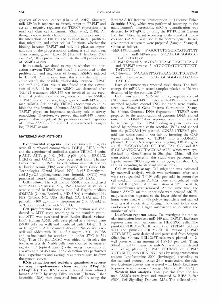
In this study, the correlation between miR-149 expres-sion and the TGF-β1 treatment were investigated in hu-man ASMCs (airway smooth muscle cells) in vitro. Firstly, MTT assay was conducted to determine the cell prolif-eration of human ASMCs after TGF-β1 treatment with various concentrations (1, 5 and 10 ng/mL) for 24 h and 48 h, respectively. We found that human ASMCs treated with TGF-β1 had a higher proliferation rate compared with control group. Moreover, the cell prolif-eration rate in TGF-β1 group treated for 48h was higher than that in 24 h treatment group, which demonstrated a time-dependent increase (Fig. 1A). In addition, the expression of miR-149 was also analyzed by RT-qPCR. The results showed that miR-149 expression was sig-nificantly down-regulated in human ASMCs treated with TGF-β1.The expression level of miR-149 was negatively correlated with the TGF-β1 concentration and the du-ration of stimulation (Fig. 1B). These results manifested
that TGF-β1 can promote cell proliferation and reduce the expression of miR-149 in human ASMCs.
MiR-149 regulated proliferation and migration of human ASMCs induced by TGF-β1 in vitro
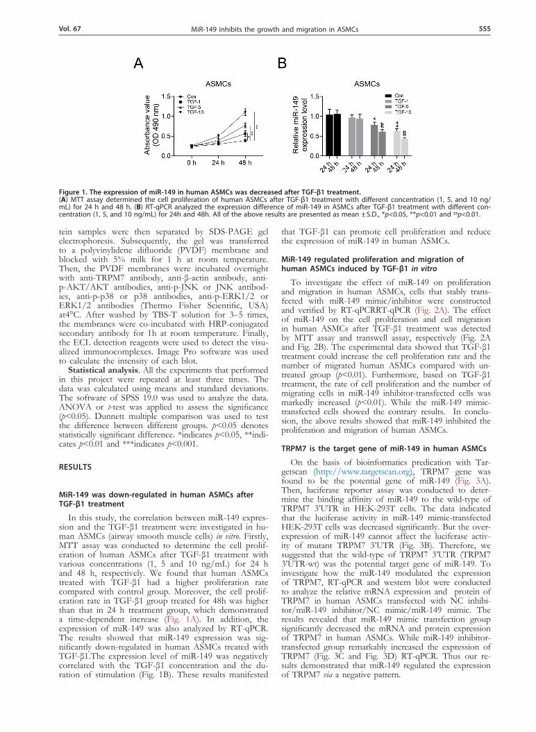
To investigate the effect of miR-149 on proliferation and migration in human ASMCs, cells that stably trans-fected with miR-149 mimic/inhibitor were constructed and verified by RT-qPCRRT-qPCR (Fig. 2A). The effect of miR-149 on the cell proliferation and cell migration in human ASMCs after TGF-β1 treatment was detected by MTT assay and transwell assay, respectively (Fig. 2A and Fig. 2B). The experimental data showed that TGF-β1 treatment could increase the cell proliferation rate and the number of migrated human ASMCs compared with un-treated group (p<0.01). Furthermore, based on TGF-β1 treatment, the rate of cell proliferation and the number of migrating cells in miR-149 inhibitor-transfected cells was markedly increased (p<0.01). While the miR-149 mimic-transfected cells showed the contrary results. In conclu-sion, the above results showed that miR-149 inhibited the proliferation and migration of human ASMCs.
TRPM7 is the target gene of miR-149 in human ASMCs
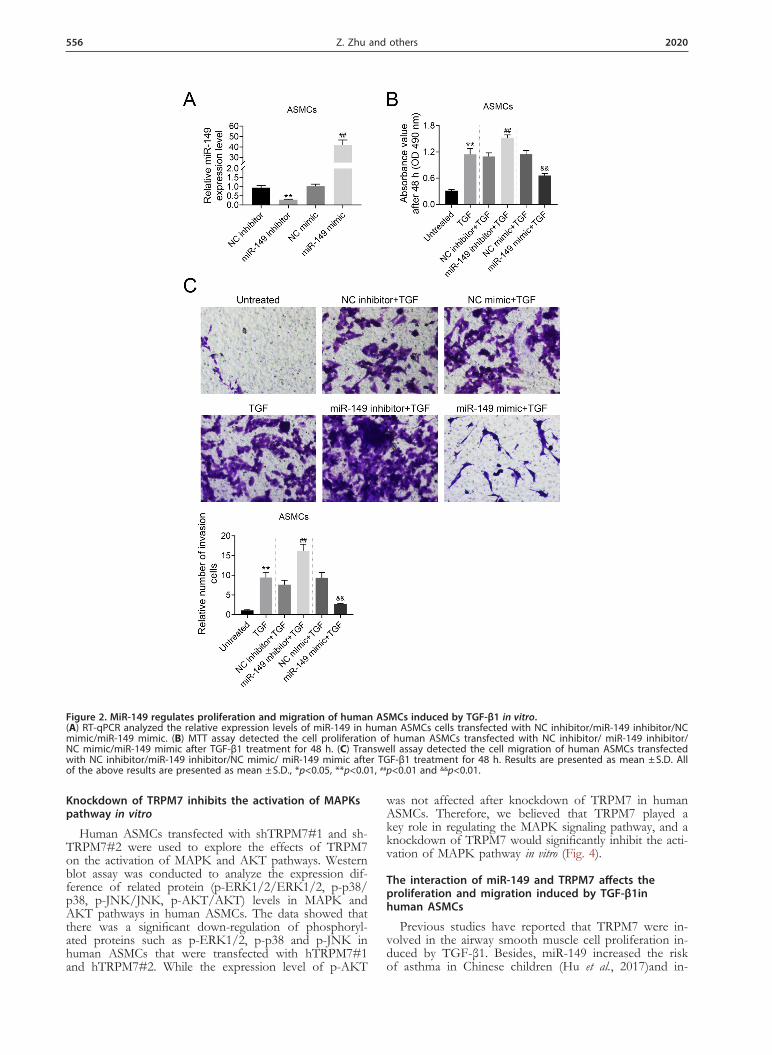
On the basis of bioinformatics predication with Tar-getscan (http://www.targetscan.org), TRPM7 gene was found to be the potential gene of miR-149 (Fig. 3A). Then, luciferase reporter assay was conducted to deter-mine the binding affinity of miR-149 to the wild-type of TRPM7 3’UTR in HEK-293T cells. The data indicated that the luciferase activity in miR-149 mimic-transfected HEK-293T cells was decreased significantly. But the over-expression of miR-149 cannot affect the luciferase activ-ity of mutant TRPM7 3’UTR (Fig. 3B). Therefore, we suggested that the wild-type of TRPM7 3’UTR (TRPM7 3’UTR-wt) was the potential target gene of miR-149. To investigate how the miR-149 modulated the expression of TRPM7, RT-qPCR and western blot were conducted to analyze the relative mRNA expression and protein of TRPM7 in human ASMCs transfected with NC inhibi-tor/miR-149 inhibitor/NC mimic/miR-149 mimic. The results revealed that miR-149 mimic transfection group significantly decreased the mRNA and protein expression of TRPM7 in human ASMCs. While miR-149 inhibitor-transfected group remarkably increased the expression of TRPM7 (Fig. 3C and Fig. 3D) RT-qPCR. Thus our re-sults demonstrated that miR-149 regulated the expression of TRPM7 via a negative pattern.
Figure 1. The expression of miR-149 in human ASMCs was decreased after TGF-β1 treatment. (A) MTT assay determined the cell proliferation of human ASMCs after TGF-β1 treatment with different concentration (1, 5, and 10 ng/mL) for 24 h and 48 h. (B) RT-qPCR analyzed the expression difference of miR-149 in ASMCs after TGF-β1 treatment with different con-centration (1, 5, and 10 ng/mL) for 24h and 48h. All of the above results are presented as mean ± S.D., *p<0.05, **p<0.01 and ##p<0.01.
556 2020Z. Zhu and others
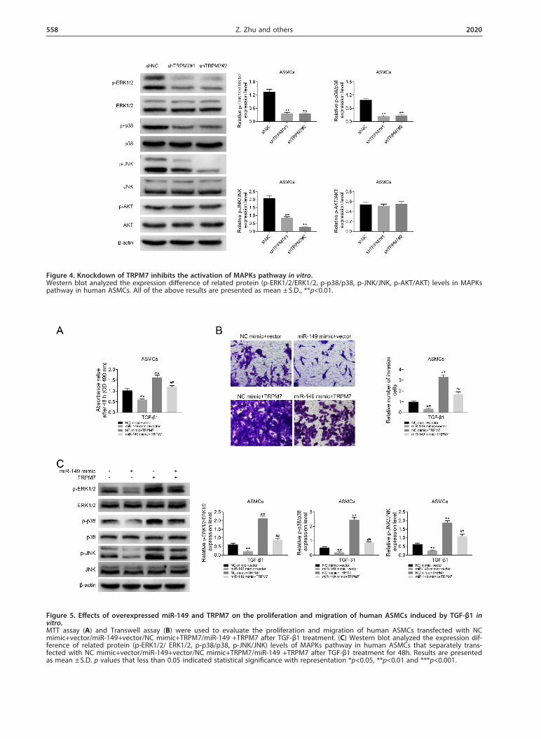
Knockdown of TRPM7 inhibits the activation of MAPKs pathway in vitro
Human ASMCs transfected with shTRPM7#1 and sh-TRPM7#2 were used to explore the effects of TRPM7 on the activation of MAPK and AKT pathways. Western blot assay was conducted to analyze the expression dif-ference of related protein (p-ERK1/2/ERK1/2, p-p38/p38, p-JNK/JNK, p-AKT/AKT) levels in MAPK and AKT pathways in human ASMCs. The data showed that there was a significant down-regulation of phosphoryl-ated proteins such as p-ERK1/2, p-p38 and p-JNK in human ASMCs that were transfected with hTRPM7#1 and hTRPM7#2. While the expression level of p-AKT
was not affected after knockdown of TRPM7 in human ASMCs. Therefore, we believed that TRPM7 played a key role in regulating the MAPK signaling pathway, and a knockdown of TRPM7 would significantly inhibit the acti-vation of MAPK pathway in vitro (Fig. 4).
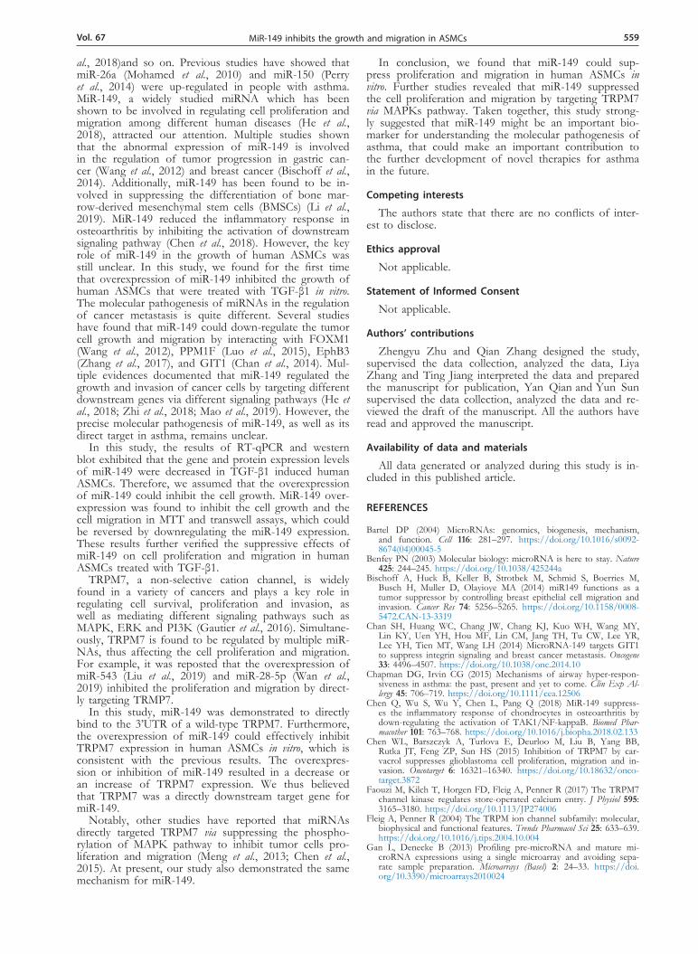
The interaction of miR-149 and TRPM7 affects the proliferation and migration induced by TGF-β1in human ASMCs
Previous studies have reported that TRPM7 were in-volved in the airway smooth muscle cell proliferation in-duced by TGF-β1. Besides, miR-149 increased the risk of asthma in Chinese children (Hu et al., 2017)and in-
Figure 2. MiR-149 regulates proliferation and migration of human ASMCs induced by TGF-β1 in vitro. (A) RT-qPCR analyzed the relative expression levels of miR-149 in human ASMCs cells transfected with NC inhibitor/miR-149 inhibitor/NC mimic/miR-149 mimic. (B) MTT assay detected the cell proliferation of human ASMCs transfected with NC inhibitor/ miR-149 inhibitor/ NC mimic/miR-149 mimic after TGF-β1 treatment for 48 h. (C) Transwell assay detected the cell migration of human ASMCs transfected with NC inhibitor/miR-149 inhibitor/NC mimic/ miR-149 mimic after TGF-β1 treatment for 48 h. Results are presented as mean ± S.D. All of the above results are presented as mean ± S.D., *p<0.05, **p<0.01, ##p<0.01 and &&p<0.01.
Vol. 67 557MiR-149 inhibits the growth and migration in ASMCs
hibited cell proliferation and migration through MAPK pathway in various cancers (He et al., 2018; Zhi et al., 2018). Thus, we further explored whether miR-149 act-ed as a suppressor in MAPK pathway through targeting TRPM7 in asthma. The results of both MTT and tran-swell assays indicated that miR-149 could significantly inhibit proliferation and migration in human ASMCs after TGF-β1 treatment, which could be reversed by transfecting cells with constructed TRPM7 overexpres-sion plasmid (Fig. 5A, B). Western blot analysis also re-vealed a significant down-regulation of phosphorylated proteins such as p-ERK1/2, p-p38 and p-JNK in hu-man ASMCs that were transfected with miR-149 mimics, which was recovered by TRPM7 overexpression. Moreo-ver, an astonishing up-regulation of phosphorylated pro-teins such as p-ERK1/2, p-p38 and p-JNK in human ASMCs that had been transfected with TRPM7 was ob-served (Fig. 5C). We thus considered that miR-149 sup-pressed the MAPKs pathway through targeting TRPM7 in human ASMCs. In summary, we found that miR-149
suppressed the proliferation and migration of human ASMCs by targeting TRPM7 via regulating MAPKs path-way in vitro.
DISCUSSION
As a common chronic disease affecting the airways of the lungs and posing a serious threat to human health, asthma is characterized by remodeling of the airways, in-flammatory reactions and hyperresponsiveness (Holgate, 2008). Although traditional therapies have made great progress, the lack of accurate biomarkers hinders the treatment of asthma (Tiotiu, 2018). Therefore, in order to treat asthma more effectively, people still need to fur-ther understand its pathogenesis to find effective thera-peutic targets.
To date, many studies have shown that miRNAs play an important role in proliferation and migration of vari-ous diseases such as human malignancies (Benfey, 2003), asthma (Heffler et al., 2017), liver fibrosis (Schueller et
Figure 3. TRPM7 is the direct target of miR-149.(A) In this diagram, the sequences of miR-149 binding sites complemented with TRPM7 3’-UTR (625-631). TRPM7 3’-UTR-wt represents the entire 3’UTR sequences of wild-type TRPM7, TRPM7 3’-UTR-mut represented the binding sites were mutated. (B) Luciferase reporter assays was performed in human ASMCs cells to detect the binding affinity of miR-149 to the TRPM7 3’UTR-wt or mut. (C) RT-qPCR ana-lyzed the relative TRPM7 mRNA expression levels in human ASMCs cells transfected with NC inhibitor/miR-149 inhibitor/NC mimic/miR-149 mimic. (D) Western blot analyzed the overexpression or inhibition of miR-149 in human ASMCs cells affected the TRPM7 protein levels. All of the above results are presented as mean ± S.D., *p<0.05, **p<0.01 and ##p<0.01.
558 2020Z. Zhu and others
Figure 4. Knockdown of TRPM7 inhibits the activation of MAPKs pathway in vitro. Western blot analyzed the expression difference of related protein (p-ERK1/2/ERK1/2, p-p38/p38, p-JNK/JNK, p-AKT/AKT) levels in MAPKs pathway in human ASMCs. All of the above results are presented as mean ± S.D., **p<0.01.
Figure 5. Effects of overexpressed miR-149 and TRPM7 on the proliferation and migration of human ASMCs induced by TGF-β1 in vitro. MTT assay (A) and Transwell assay (B) were used to evaluate the proliferation and migration of human ASMCs transfected with NC mimic+vector/miR-149+vector/NC mimic+TRPM7/miR-149 +TRPM7 after TGF-β1 treatment. (C) Western blot analyzed the expression dif-ference of related protein (p-ERK1/2/ ERK1/2, p-p38/p38, p-JNK/JNK) levels of MAPKs pathway in human ASMCs that separately trans-fected with NC mimic+vector/miR-149+vector/NC mimic+TRPM7/miR-149 +TRPM7 after TGF-β1 treatment for 48h. Results are presented as mean ± S.D. p values that less than 0.05 indicated statistical significance with representation *p<0.05, **p<0.01 and ***p<0.001.
Vol. 67 559MiR-149 inhibits the growth and migration in ASMCs
al., 2018)and so on. Previous studies have showed that miR-26a (Mohamed et al., 2010) and miR-150 (Perry et al., 2014) were up-regulated in people with asthma. MiR-149, a widely studied miRNA which has been shown to be involved in regulating cell proliferation and migration among different human diseases (He et al., 2018), attracted our attention. Multiple studies shown that the abnormal expression of miR-149 is involved in the regulation of tumor progression in gastric can-cer (Wang et al., 2012) and breast cancer (Bischoff et al., 2014). Additionally, miR-149 has been found to be in-volved in suppressing the differentiation of bone mar-row-derived mesenchymal stem cells (BMSCs) (Li et al., 2019). MiR-149 reduced the inflammatory response in osteoarthritis by inhibiting the activation of downstream signaling pathway (Chen et al., 2018). However, the key role of miR-149 in the growth of human ASMCs was still unclear. In this study, we found for the first time that overexpression of miR-149 inhibited the growth of human ASMCs that were treated with TGF-β1 in vitro. The molecular pathogenesis of miRNAs in the regulation of cancer metastasis is quite different. Several studies have found that miR-149 could down-regulate the tumor cell growth and migration by interacting with FOXM1 (Wang et al., 2012), PPM1F (Luo et al., 2015), EphB3 (Zhang et al., 2017), and GIT1 (Chan et al., 2014). Mul-tiple evidences documented that miR-149 regulated the growth and invasion of cancer cells by targeting different downstream genes via different signaling pathways (He et al., 2018; Zhi et al., 2018; Mao et al., 2019). However, the precise molecular pathogenesis of miR-149, as well as its direct target in asthma, remains unclear.
In this study, the results of RT-qPCR and western blot exhibited that the gene and protein expression levels of miR-149 were decreased in TGF-β1 induced human ASMCs. Therefore, we assumed that the overexpression of miR-149 could inhibit the cell growth. MiR-149 over-expression was found to inhibit the cell growth and the cell migration in MTT and transwell assays, which could be reversed by downregulating the miR-149 expression. These results further verified the suppressive effects of miR-149 on cell proliferation and migration in human ASMCs treated with TGF-β1.
TRPM7, a non-selective cation channel, is widely found in a variety of cancers and plays a key role in regulating cell survival, proliferation and invasion, as well as mediating different signaling pathways such as MAPK, ERK and PI3K (Gautier et al., 2016). Simultane-ously, TRPM7 is found to be regulated by multiple miR-NAs, thus affecting the cell proliferation and migration. For example, it was reposted that the overexpression of miR-543 (Liu et al., 2019) and miR-28-5p (Wan et al., 2019) inhibited the proliferation and migration by direct-ly targeting TRMP7.
In this study, miR-149 was demonstrated to directly bind to the 3’UTR of a wild-type TRPM7. Furthermore, the overexpression of miR-149 could effectively inhibit TRPM7 expression in human ASMCs in vitro, which is consistent with the previous results. The overexpres-sion or inhibition of miR-149 resulted in a decrease or an increase of TRPM7 expression. We thus believed that TRPM7 was a directly downstream target gene for miR-149.
Notably, other studies have reported that miRNAs directly targeted TRPM7 via suppressing the phospho-rylation of MAPK pathway to inhibit tumor cells pro-liferation and migration (Meng et al., 2013; Chen et al., 2015). At present, our study also demonstrated the same mechanism for miR-149.
In conclusion, we found that miR-149 could sup-press proliferation and migration in human ASMCs in vitro. Further studies revealed that miR-149 suppressed the cell proliferation and migration by targeting TRPM7 via MAPKs pathway. Taken together, this study strong-ly suggested that miR-149 might be an important bio-marker for understanding the molecular pathogenesis of asthma, that could make an important contribution to the further development of novel therapies for asthma in the future.
Competing interests
The authors state that there are no conflicts of inter-est to disclose.
Ethics approval
Not applicable.
Statement of Informed Consent
Not applicable.
Authors’ contributions
Zhengyu Zhu and Qian Zhang designed the study, supervised the data collection, analyzed the data, Liya Zhang and Ting Jiang interpreted the data and prepared the manuscript for publication, Yan Qian and Yun Sun
supervised the data collection, analyzed the data and re-viewed the draft of the manuscript. All the authors have read and approved the manuscript.
Availability of data and materials
All data generated or analyzed during this study is in-cluded in this published article.
REFERENCES
Bartel DP (2004) MicroRNAs: genomics, biogenesis, mechanism, and function. Cell 116: 281–297. https://doi.org/10.1016/s0092-8674(04)00045-5
Benfey PN (2003) Molecular biology: microRNA is here to stay. Nature 425: 244–245. https://doi.org/10.1038/425244a
Bischoff A, Huck B, Keller B, Strotbek M, Schmid S, Boerries M, Busch H, Muller D, Olayioye MA (2014) miR149 functions as a tumor suppressor by controlling breast epithelial cell migration and invasion. Cancer Res 74: 5256–5265. https://doi.org/10.1158/0008-5472.CAN-13-3319
Chan SH, Huang WC, Chang JW, Chang KJ, Kuo WH, Wang MY, Lin KY, Uen YH, Hou MF, Lin CM, Jang TH, Tu CW, Lee YR, Lee YH, Tien MT, Wang LH (2014) MicroRNA-149 targets GIT1 to suppress integrin signaling and breast cancer metastasis. Oncogene 33: 4496–4507. https://doi.org/10.1038/onc.2014.10
Chapman DG, Irvin CG (2015) Mechanisms of airway hyper-respon-siveness in asthma: the past, present and yet to come. Clin Exp Al-lergy 45: 706–719. https://doi.org/10.1111/cea.12506
Chen Q, Wu S, Wu Y, Chen L, Pang Q (2018) MiR-149 suppress-es the inflammatory response of chondrocytes in osteoarthritis by down-regulating the activation of TAK1/NF-kappaB. Biomed Phar-macother 101: 763–768. https://doi.org/10.1016/j.biopha.2018.02.133
Chen WL, Barszczyk A, Turlova E, Deurloo M, Liu B, Yang BB, Rutka JT, Feng ZP, Sun HS (2015) Inhibition of TRPM7 by car-vacrol suppresses glioblastoma cell proliferation, migration and in-vasion. Oncotarget 6: 16321–16340. https://doi.org/10.18632/onco-target.3872
Faouzi M, Kilch T, Horgen FD, Fleig A, Penner R (2017) The TRPM7 channel kinase regulates store-operated calcium entry. J Physiol 595: 3165–3180. https://doi.org/10.1113/JP274006
Fleig A, Penner R (2004) The TRPM ion channel subfamily: molecular, biophysical and functional features. Trends Pharmacol Sci 25: 633–639. https://doi.org/10.1016/j.tips.2004.10.004
Gan L, Denecke B (2013) Profiling pre-microRNA and mature mi-croRNA expressions using a single microarray and avoiding sepa-rate sample preparation. Microarrays (Basel) 2: 24–33. https://doi.org/10.3390/microarrays2010024
560 2020Z. Zhu and others
Gautier M, Perriere M, Monet M, Vanlaeys A, Korichneva I, Dhennin-Duthille I, Ouadid-Ahidouch H (2016) Recent advances in onco-genic roles of the TRPM7 chanzyme. Curr Med Chem 23: 4092–4107. https://doi.org/10.2174/0929867323666160907162002
He Y, Yu D, Zhu L, Zhong S, Zhao J, Tang J (2018) miR-149 in Human cancer: a systemic review. J Cancer 9: 375–388. https://doi.org/10.7150/jca.21044
Heffler E, Allegra A, Pioggia G, Picardi G, Musolino C Gangemi, S (2017) MicroRNA Profiling in asthma: potential biomarkers and therapeutic targets. Am J Respir Cell Mol Biol 57: 642–650. https://doi.org/10.1165/rcmb.2016-0231TR
Holgate ST (2008) Pathogenesis of asthma. Clin Exp Allergy 38: 872–897. https://doi.org/10.1111/j.1365-2222.2008.02971.x
Hu D, Zhang Z, Ke X, Kang H, Hong S (2017) A functional vari-ant of miRNA-149 confers risk for allergic rhinitis and comorbid asthma in Chinese children. Int J Immunogenet 44: 62–70. https://doi.org/10.1111/iji.12307
Kim S, Lee CH, Jin KN, Cho SH, Kang HR (2018) Severe asthma phenotypes classified by site of airway involvement and remodeling via chest CT scan. J Investig Allergol Clin Immunol 28: 312–320. htt-ps://doi.org/10.18176/jiaci.0265
Li Y, Yang F, Gao M, Gong R, Jin M, Liu T, Sun Y, Fu Y, Huang Q, Zhang W, Liu S, Yu M, Yan G, Feng C, He M, Zhang L, Ding F, Ma W, Bi Z, Xu C, Yuan Y, Cai B, Yang L (2019) miR-149-3p Regulates the switch between adipogenic and osteogenic differ-entiation of BMSCs by targeting FTO. Mol Ther Nucleic Acids 17: 590–600. https://doi.org/10.1016/j.omtn.2019.06.023
Liu X, Gan L, Zhang J (2019) miR-543 inhibites cervical cancer growth and metastasis by targeting TRPM7. Chem Biol Interact 302: 83–92. https://doi.org/10.1016/j.cbi.2019.01.036
Lou W, Liu J, Gao Y, Zhong G, Chen D, Shen J, Bao C, Xu L, Pan J, Cheng J, Ding B, Fan W (2017) MicroRNAs in cancer metas-tasis and angiogenesis. Oncotarget 8: 115787–115802. https://doi.org/10.18632/oncotarget.23115
Luo G, Chao YL, Tang B, Li BS, Xiao YF, Xie R, Wang SM, Wu YY, Dong H, Liu XD, Yang SM (2015) miR-149 represses metastasis of hepatocellular carcinoma by targeting actin-regulatory proteins PPM1F. Oncotarget 6: 37808–37823. https://doi.org/10.18632/onco-target.5676
Mao F, Zhang J, Cheng X, Xu Q (2019) miR-149 inhibits cell prolifer-ation and enhances chemosensitivity by targeting CDC42 and BCL2 in neuroblastoma. Cancer Cell Int 19: 357. https://doi.org/10.1186/s12935-019-1082-9
Meng X, Cai C, Wu J, Cai S, Ye C, Chen H, Yang Z, Zeng H, Shen Q, Zou F (2013) TRPM7 mediates breast cancer cell migration and invasion through the MAPK pathway. Cancer Lett 333: 96–102. htt-ps://doi.org/10.1016/j.canlet.2013.01.031
Mohamed JS, Lopez MA, Boriek AM (2010) Mechanical stretch up-regulates microRNA-26a and induces human airway smooth muscle hypertrophy by suppressing glycogen synthase kinase-3beta. J Biol Chem 285: 29336–29347. https://doi.org/10.1074/jbc.M110.101147
Ojiaku CA, Yoo EJ, Panettieri RA Jr (2017) Transforming growth fac-tor beta1 function in airway remodeling and hyperresponsiveness. the missing link? Am J Respir Cell Mol Biol 56: 432–442. https://doi.org/10.1165/rcmb.2016-0307TR
Perry MM, Tsitsiou E, Austin PJ, Lindsay MA, Gibeon DS, Adcock IM, Chung KF (2014) Role of non-coding RNAs in maintaining primary airway smooth muscle cells. Respir Res 15: 58. https://doi.org/10.1186/1465-9921-15-58
Schueller F, Roy S, Vucur M, Trautwein C, Luedde T, Roderburg C (2018) The role of miRNAs in the pathophysiology of liver diseases and toxicity. Int J Mol Sci 19. https://doi.org/10.3390/ijms19010261
Siddiqui S, Shikotra A, Richardson M, Doran E, Choy D, Bell A, Aus-tin CD, Eastham-Anderson J, Hargadon B, Arron JR, Wardlaw A, Brightling CE, Heaney LG, Bradding P (2018) Airway pathological heterogeneity in asthma: Visualization of disease microclusters us-ing topological data analysis. J Allergy Clin Immunol 142: 1457–1468. https://doi.org/10.1016/j.jaci.2017.12.982
Tiotiu A (2018) Biomarkers in asthma: state of the art. Asthma Res Pract 4: 10. https://doi.org/10.1186/s40733-018-0047-4
Wan J, Guo AA, Chowdhury I, Guo S, Hibbert J, Wang G, Liu M (2019) TRPM7 induces mechanistic target of Rap1b through the downregulation of miR-28-5p in glioma proliferation and invasion. Front Oncol 9: 1413. https://doi.org/10.3389/fonc.2019.01413
Wang Y, Zheng X, Zhang Z, Zhou J, Zhao G, Yang J, Xia L, Wang R, Cai X, Hu H, Zhu C, Nie Y, Wu K, Zhang D, Fan D (2012) MicroRNA-149 inhibits proliferation and cell cycle progression through the targeting of ZBTB2 in human gastric cancer. PLoS One 7: e41693. https://doi.org/10.1371/journal.pone.0041693
Weidner J, Malmhäll C, Rådinger M (2019) microRNAs in asthma pathogenesis – from mouse to man. J Transl Genet Genomics. https://doi.org/10.20517/jtgg.2018.30
Yee NS (2017) Role of TRPM7 in cancer: potential as molecular bio-marker and therapeutic target. Pharmaceuticals (Basel) 10. https://doi.org/10.3390/ph10020039
Zhang G, Liu X, Li Y, Wang Y, Liang H, Li K, Li L, Chen C, Sun W, Ren S, Zhu P, Zhang L (2017) EphB3-targeted regulation of miR-149 in the migration and invasion of human colonic carcinoma HCT116 and SW620 cells. Cancer Sci 108: 408–418. https://doi.org/10.1111/cas.13161
Zhao Z, Zhang M, Duan X, Chen Y, Li E, Luo L, Wu W, Peng Z, Qiu H, Zeng G (2018) TRPM7 Regulates AKT/FOXO1-depend-ent tumor growth and is an independent prognostic indicator in renal cell carcinoma. Mol Cancer Res 16: 1013–1023. https://doi.org/10.1158/1541-7786.MCR-17-0767
Zhi Y, Zhou H, Mubalake A, Chen Y, Zhang B, Zhang K, Chu X, Wang R (2018) Regulation and functions of MicroRNA-149 in human cancers. Cell Prolif 51: e12465. https://doi.org/10.1111/cpr.12465


![β at the Intersection of Neuronal Plasticity and ...downloads.hindawi.com/journals/np/2019/4209475.pdf · migration in the cortex [39]. GSK-3 regulates neuronal migration by phosphorylating](https://static.fdocument.org/doc/165x107/5f2bee152cce572aa50fe1ab/-at-the-intersection-of-neuronal-plasticity-and-migration-in-the-cortex-39.jpg)