MiR-149 attenuates the proliferation and migration of TGF ...
Upload
truongthuanCategory
view
216download
0
7735
Abstract. – OBJECTIVE: MicroRNA (miRNA) can widely regulate gene expression. More im-portantly, various miRNA molecules have been found with regulatory functions for tumor cell proliferation or apoptosis. The study showed that miR-21 inhibited apoptosis of cultured can-cer cells, whilst tumor necrosis factor α (TNF-α) plays important roles in the proliferation of tu-mor cells. This study manipulated miR-21 ex-pression in cultured oral cancer cells and aimed to investigate its effects on TNF-α expression, and on proliferation or apoptosis of cancer cells.
MATERIALS AND METHODS: Specific ago-nist and antagonist were synthesized based on miR-21 sequence. In vitro cultured oral cancer cell line, SCC-15 was transfected with agonist or an-tagonist, in parallel with normal cultured cells as negative control group. Quantitative Real-time PCR (qRT-PCR) was used to measure mRNA ex-pression of miR-21 and TNF-α in transfected cells. Western blot was used for measuring TNF-α ex-pression, and 3-(4,5-dimethyl-2-thiazolyl)-2,5-di-phenyl-2-H-tetrazolium bromide (MTT) or Hoechst-33342 staining was used to measure pro-liferation and apoptosis of SCC-15 cells.
RESULTS: MiR-21 expression was potentiat-ed or depressed with transfection of agonist or antagonist, respectively, illustrating the effec-tiveness of synthesized sequence in cultured SCC-15 cells. Moreover, TNF-α expression was positively correlated with miR-21, TNF-α up-reg-ulation significantly potentiated the proliferation potency of SCC-15 cells, and TNF-α down-regu-lation remarkably weakened proliferation poten-cy (p<0.05). The TNF-α expression did not affect apoptosis of SCC-15 cells (p>0.05 compared to the control group).
CONCLUSIONS: MiR-21 could participate in the proliferation of cultured SCC-15 cells via tar-geting TNF-α expression, but without any signif-icant effects on cell apoptosis.
Key Words:Oral cancer, miR-21, SCC-15, TNF-α, Cell prolifera-
tion, Cell apoptosis
Introduction
Oral cancer is a commonly occurred malignant tumor in human head-neck regions, and is one of the malignant tumors occurred within oral cavi-ty. It occupies about 1.9-3.5% of total malignant tumors, and about 4-20% of head-neck cancers, with incidence only lower than nasopharyngeal carcinoma. Mainly consisting of lip carcinoma, gingival carcinoma, and tongue cancer, oral can-cer has relatively higher incidence worldwide. In general males had higher incidence of oral cancer than females, probably due to relatively higher percentage of populations with smoking or alco-hol abuse history. Currently treatment approaches targeting oral cancer are similar as those for oth-er cancers, mainly including surgical resection, radiotherapy, chemotherapy or Chinese medi-cine, although no satisfactory efficiency has been reached1, 2. With the development of biological tar-geting treatment, novel treatment plans including gene therapy, immune therapy, and tumor stem cells have shown major potency. However, these
European Review for Medical and Pharmacological Sciences 2018; 22: 7735-7741
Y.-F. QIU1, M.-X. WANG2, L.-N. MENG3, R. ZHANG4, W. WANG5
1Department of Image Diagnoses, Stomatological College, the First Affiliated Hospital of Harbin Medical University, Harbin, Heilongjiang, China2Department of Stomatology, the Forth Affiliated Hospital of Harbin Medical University, Harbin, Heilongjiang, China3Department of Stomatology, Nangang Branch of Heilongjiang Provincial Hospital, Harbin, Heilongjiang, China4Department of Stomatology, Harbin Children’s Hospital, Harbin, Heilongjiang, China5Department of Oral and Maxillofacial Surgery, the First Affiliated Hospital of Harbin Medical University, Harbin, Heilongjiang, China
Corresponding Author: Yanfen Qiu, MD; e-mail: [email protected]
MiR-21 regulates proliferation and apoptosis of oral cancer cells through TNF-α
Y.-F. Qiu, M.-X. Wang, L.-N. Meng, R. Zhang, W. Wang
7736
methods largely invade body injury taken by clas-sical treatment methods and had better foresights in improving post-op survival and prognosis of patients. Therefore, the identification of valuable treatment target for oral cancer is of critical im-portance for development of specific molecular targeting medicine and prognostic evaluation of oral cancer3. Tumor necrosis factor α (TNF-α) has certain regulatory functions on cell proliferation, apoptosis and differentiation, and has pluripotent biological effects inside the body4. Hoeben et al4 found prominent expression of TNF-α in various tumor tissues including prostate cancer, pancre-atic carcinoma and kidney cancer. It has dual roles in regulating tumor cells, as it can inhibit tumor cell growth and induce tumor necrosis, and improves tumor cell growth, migration and infil-tration potency, thus becoming one critical gene in tumor target gene research5,6. Previous work showed significantly higher TNF-α expression in serum of oral cancer patients compared to normal people7. MicroRNA (miRNA) is widely distrib-uted in eukaryotes, and participates in the reg-ulation of various genes. It exerts the regulatory function mainly via complementary binding with target gene, including direct degradation of target gene mRNA or inhibition of gene translation at post-transcriptional stage to suppress target gene expression level8,9. More importantly, one single protein may be under the regulation of multiple miRNA. Currently the analysis for miRNA has made major progression, and has identified its important regulatory role in various human dis-eases. Certain miRNA molecules have become early signs of tumorigenesis, whilst other miRNA molecules have become target for clinical drug treatment10. Previous researches showed certain relationship between miR expression and alter-nation of abilities in proliferation, apoptosis and migration of oral cancer cells. Multiple miRNA molecules have been found to have up-regulation in oral cancer cells such as miR-134, miR-24 and miR-2111,12. Therefore, the investigation of miR-21 in regulating proliferation and apoptosis of oral
cancer, and downstream targeting TNF-α, have solid theoretical grounds and feasibility. Our work measured changing patterns of TNF-α af-ter miR-21 manipulation, and its correlation with proliferation or apoptosis of oral cancer cells, in order to investigate the function of miR-21 and TNF-α in oral cancer pathogenesis.
Materials and Methods
Major Reagents and EquipmentHuman oral cancer cell line SCC-15 was pur-
chased from Beinuo Biotech. Co. Ltd. (Shanghai, China) and was preserved in liquid nitrogen. Ros-well Park Memorial Institute-1640 (RPMI-1640) medium, option minimum essential media (op-ti-MEM), fetal bovine serum (FBS) and tryp-sin for digestion were purchased from HyClone (South Logan, UT, USA). Phosphate-buffered saline (PBS) powder was purchased from Beyo-time Biotech. (Shanghai, China), and was diluted by ultrapure water for filtering in 0.22 μm pore membrane to sterilize. Dual antibiotics in cell cul-ture were produced by Tiangen Biotech Co. Ltd. (Beijing, China). Liposome cell transfection re-agent, human TNF-α protein assay antibody were produced by Invitrogen/Life Technologies (Carls-bad, CA, USA). RNA extraction kit (TRIzol) and complementary DNA (cNDA) synthesis kit were all purchased from Bio-Tek Inc. (Winooski, VT, USA). Quantitative Real-time PCR (qRT-PCR) reagent was purchased from Omega Bio-Tek. Inc. (Norcross, GA, USA). Microplate reader was purchased from Thermo Fisher Scientific (Waltham, MA, USA). Hoechest-33342 was ob-tained from Suolaibao Tech. (Shanghai, China). Inverted fluorescent microscope was produced by Leica (Frankfurt, Germany). Wet membrane transfer apparatus was purchased from Bio-Rad Laboratories (Hercules, CA, USA). All primers used were synthesized by Sangon Biotech. Co. Ltd. (Shanghai, China). Other common reagents were purchased from Sigma-Aldrich (St. Louis, MO, USA).
SCC-15 Cell Culture and Liposome Transfection
SCC-15 cell line preserved in liquid nitrogen was resuscitated following routine protocols. Cells were firstly cultured in RPMI-1640 medi-um containing 13% fetal bovine serum (FBS), and were continuously cultured. After passage, the third generation of cells was used for further
Table I. Primers used in experiment.
Name Primer sequence (5’-3’)
TNF-α forward CCCGCATCCCAGGACCTCTCTTNF-αreverse CGGGGGACTGGCGAmir-21forward CTAAGACCTGTGGAATGGCmir-21 reverse CTCAAAGATGTCATTGCCGAPDH forward CGGAGTCAACGGATTTGGTCGTAGAPDH reverse AGCCTTCTCCATGGTGGTGAAG
MiR-21 regulates proliferation and apoptosis of oral cancer cells through TNF-α
7737
assay. Before each passage, 10 μl cell re-suspen-sions were used for enumeration. About 10×104 cells were added into each well, and cell transfec-tion was performed after 16 h normal culture. Be-fore transfection, antibiotic-free culture medium was changed. MiR-21 agonist or antagonist was diluted to 100 nM using 50 μl opti-MEM medi-um. Meanwhile, 2 μl liposome solution were di-luted within 50 μl opti-MEM followed by 5 min room temperature incubation. After mixture of two solutions, these were incubated at room tem-perature for 20-30 min. Within each well, 200 μl solution A+B mixture was added. Cells were cul-tured for 4 h and culture medium was changed. Three parallel replicates were performed in each group, using normal cultured cells as the control group. This study was approved by the Ethics Committee of the First Affiliated Hospital of Har-bin Medical University (Harbin, China).
qRT-PCR for mRNA Expressionin Transfected Cells
Following the manual instruction of test kit, total RNA was extracted from cells before tran-sfection, 24 h and 48 h after transfection. cDNA strand was synthesized, and was placed on ice for 5-10 min. A total of 4 μl reverse transcriptase buf-fer, 2 μl dithiothreitol (DTT) and 1 μl deoxy-ribo-nucleoside triphosphate (dNTPs) were added into the mixture, which was incubated at 42°C for 2 min. A total of 1 μl reverse transcriptase was ad-ded for 42°C continuous incubation for 1 h. The reverse transcription reaction was quenched by 65°C. Using miR-21 gene sequence as the tem-plate, testing primers were designed and synthe-sized. Using GAPDH as the internal reference, primers sequences were shown in Table I. The qRT-PCR system consisted of 0.08 μM forward/reverse primer, 10 μl master mix, 2 μl cDNA, 0.4 μl PCR Taq polymerase, and ddH2O up to 20 μl. The sample was briefly centrifuged at 1000 r/min. qRT-PCR conditions were: 94°C for 3 min, followed by 45 cycles each containing 95°C 13 s, and 63°C 40 s. Triplicated wells were set for each sample to reduce the error.
Western Blot for Serum TNF-α Expression in Supernatant from Transfected Cells
Total cellular proteins were collected at 24 h and 48 h after transfection. Culture medium was firstly removed from culture plate. The plate was then washed using pre-cold 1×PBS buffer. After removing all liquids, the plate was placed on
ice, and protein lysis buffer was added (50 μl per well). Well bottom and wall were repeated washed using PBS buffer. Cell suspensions were collected into 1.5 ml enzyme-free Eppendorf (EP) tubes for mixture repeated in 3-4 times until complete ly-sis of cells. All operations were performed on ice. After 4°C centrifugation at 12000 r/min for 15 min, the supernatant was collected into pre-cold 1.5 ml EP tube. After mixture, 1 μl sample was used for bicinchoninic acid (BCA) protein quan-tification, and the remaining samples were kept at -80°C fridge. A total of 20 μg protein samples were used for sodium dodecyl sulfate polyacryla-mide gel electrophoresis (SDS-PAGE) separation using 4% condensing gel and 12% separating gel. Gel electrophoresis was performed under 80 V for 20 min, and 120 V for 35 min. After electrophore-sis, the protein was transferred to polyvinylidene difluoride (PVDF) membrane in a wet transfer-ring apparatus at 385 mA current for 30-60 min. Using β-actin as the internal reference, primary antibody was diluted at 1:100 ratio and was used for 4°C incubation for 8-10 h. After Tris-buffered saline and Tween-20 (TBST) rinsing, 500-fold di-luted secondary antibody was added for 2 h room temperature incubation, followed by 3-5 times of rinsing. Developing and chromogenic substrates were added, and Bio-Rad automatic illuminator was used for analysis of gray values from each group. Using β-actin as the internal reference, expression of TNF-α protein was quantified.
3-(4,5-Dimethyl-2-Thiazolyl)-2,5-Diphenyl- 2-H-Tetrazolium Bromide (MTT) Assay for Effect of TNF-α Expression on Proliferation of SCC-15 Cells
Cells after transfection were enumerated and transferred into 96-well plate (1×10⁵ cells per well). At 36 h or 48 h after transfection, cells were rinsed in serum-free medium. Each well was ad-ded with 20 μl MTT solution for 4 h incubation. Culture medium was then removed, and 150 μl dimethylsulfoxide (DMSO) were added for 10 min room temperature incubation. Optical densi-ty (OD) values of each well were measured in tri-plicates from each group. Averaged values were taken for calculating cell proliferation rate, which was equal to OD of experimental group divided by OD of control group.
Hoechest-33342 Staining for the Effect of TNF-α Expression on Cell Apoptosis
Transfected cells were inoculated into 6-well plate, and were continuously cultured till 24 h or
Y.-F. Qiu, M.-X. Wang, L.-N. Meng, R. Zhang, W. Wang
7738
48 h. After removing culture medium, the culture plate was rinsed in PBS buffer for 3 to 5 times. Each well was fixed by 200 μl paraformaldehyde at 4°C for 20 min, followed by PBS rinsing. About 1 ml staining buffer was then added into each well for 20 min incubation. With repeated PBS rinsing, the plate was observed under an inverted fluorescent microscope for statistical analysis.
Statistical AnalysisSPSS 11.3 software (SPSS Inc., Chicago, IL,
USA) was used for data analysis and statistics. The Student’s t-test was used to compare the dif-ferences between two groups. Tukey’s post-hoc test was used to validate the analysis of varian-ce (ANOVA) for comparing measurement data among groups. A statistical significance was de-fined when p<0.05, and extremely significant dif-ference.
Results
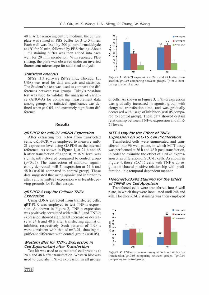
qRT-PCR for miR-21 mRNA ExpressionAfter extracting total RNA from transfected
cells, qRT-PCR was employed to measure miR-21 expression level using GAPDH as the internal reference. As shown in Figure 1, at 24 h and 48 h after transfection of agonist, miR-21 level was significantly elevated compared to control group (p<0.05). The transfection of inhibitor signifi-cantly depressed miR-21 expression at 24 h and 48 h (p<0.01 compared to control group). These data suggested that using agonist and inhibitor to alter cellular miR-21 expression was feasible, pa-ving grounds for further assays.
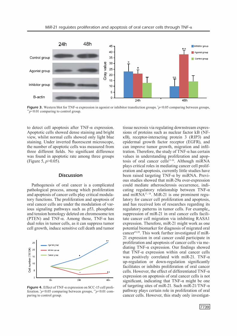
qRT-PCR Assay for Cellular TNF-α Expression
Using cDNA extracted from transfected cells, qRT-PCR was employed to test TNF-α expres-sion. As shown in Figure 2, TNF-α expression was positively correlated with miR-21, and TNF-α expression showed significant increase or decrea-se at 24 h and 48 h after transfecting agonist or inhibitor, respectively. Such patterns of TNF-α were consistent with that of miR-21, showing si-gnificant difference with control group (p<0.05).
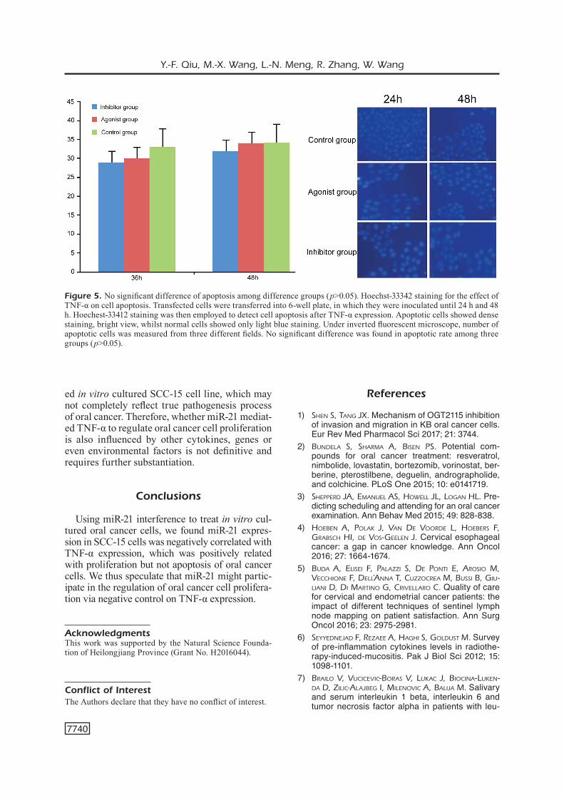
Western Blot for TNF-α Expression in Cell Supernatant after Transfection
Test kit was used to extract total cell proteins at 24 h and 48 h after transfection. Western blot was used to describe TNF-α expression in all groups
of cells. As shown in Figure 3, TNF-α expression was gradually increased in agonist group with elongated transfection time, and was gradually decreased with usage of inhibitor (p<0.05 compa-red to control group). These data showed certain relationship between TNF-α expression and miR-21 levels.
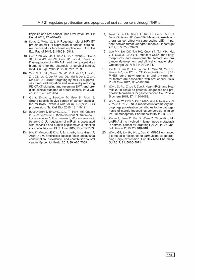
MTT Assay for the Effect of TNF-α Expression on SCC-15 Cell Proliferation
Transfected cells were enumerated and tran-sferred into 96-well palate, in which MTT assay was performed at 36 h and 48 h post-transfection, in order to examine the effect of TNF-α expres-sion on proliferation of SCC-15 cells. As shown in Figure 4, those SCC-15 cells with TNF-α up-re-gulation showed positive relationship with proli-feration, in a temporal dependent manner.
Hoechest-33342 Staining for the Effect of TNF-α on Cell Apoptosis
Transfected cells were transferred into 6-well plate, in which they were inoculated until 24h and 48h. Hoechest-33412 staining was then employed
Figure 1. MiR-21 expression at 24 h and 48 h after tran-sfection.*p<0.05 comparing between groups, **p<0.01 com-paring to control group.
Figure 2. TNF-α expression assay at 36 h and 48 h after transfection. *p<0.05 comparing between groups, **p<0.01 comparing to control group.
MiR-21 regulates proliferation and apoptosis of oral cancer cells through TNF-α
7739
to detect cell apoptosis after TNF-α expression. Apoptotic cells showed dense staining and bright view, whilst normal cells showed only light blue staining. Under inverted fluorescent microscope, the number of apoptotic cells was measured from three different fields. No significant difference was found in apoptotic rate among three groups (Figure 5, p>0.05).
Discussion
Pathogenesis of oral cancer is a complicated pathological process, among which proliferation and apoptosis of cancer cells play critical modula-tory functions. The proliferation and apoptosis of oral cancer cells are under the modulation of var-ious signaling pathways such as p53, phosphate and tension homology deleted on chromosome ten (PTEN) and TNF-α. Among those, TNF-α has dual roles in tumor cells, as it can suppress tumor cell growth, induce sensitive cell death and tumor
tissue necrosis via regulating downstream expres-sions of proteins such as nuclear factor kB (NF-κB), receptor-interacting protein 3 (RIP3) and epidermal growth factor receptor (EGFR), and can improve tumor growth, migration and infil-tration. Therefore, the study of TNF-α has certain values in understanding proliferation and apop-tosis of oral cancer cells13-16. Although miRNA plays critical roles in mediating cancer cell prolif-eration and apoptosis, currently little studies have been raised targeting TNF-α by miRNA. Previ-ous studies showed that miR-29a over-expression could mediate atherosclerosis occurrence, indi-cating regulatory relationship between TNF-α and miRNA17, 18. MiR-21 is one prominent regu-latory for cancer cell proliferation and apoptosis, and has received lots of researches regarding its regulatory patterns in tumor cells. For example,, suppression of miR-21 in oral cancer cells facili-tate cancer cell migration via inhibiting RASA1 expression. Therefore, miR-21 might work as one potential biomarker for diagnosis of migrated oral cancer19,20. This work further investigated if miR-21 expression in oral cancer could participate in proliferation and apoptosis of cancer cells via me-diating TNF-α expression. Our findings showed that TNF-α expression within oral cancer cells was positively correlated with miR-21. TNF-α up-regulation or down-regulation significantly facilitates or inhibits proliferation of oral cancer cells. However, the effect of differentiated TNF-α expression on apoptosis of oral cancer cells is not significant, indicating that TNF-α might be one of targeting sites of miR-21. Such miR-21/TNF-α pathway plays certain role in proliferation of oral cancer cells. However, this study only investigat-
Figure 3. Western blot for TNF-α expression in agonist or inhibitor transfection groups. *p<0.05 comparing between groups, **p<0.01 comparing to control group.
Figure 4. Effect of TNF-α expression on SCC-15 cell proli-feration. *p<0.05 comparing between groups, **p<0.01 com-paring to control group.
Y.-F. Qiu, M.-X. Wang, L.-N. Meng, R. Zhang, W. Wang
7740
ed in vitro cultured SCC-15 cell line, which may not completely reflect true pathogenesis process of oral cancer. Therefore, whether miR-21 mediat-ed TNF-α to regulate oral cancer cell proliferation is also influenced by other cytokines, genes or even environmental factors is not definitive and requires further substantiation.
Conclusions
Using miR-21 interference to treat in vitro cul-tured oral cancer cells, we found miR-21 expres-sion in SCC-15 cells was negatively correlated with TNF-α expression, which was positively related with proliferation but not apoptosis of oral cancer cells. We thus speculate that miR-21 might partic-ipate in the regulation of oral cancer cell prolifera-tion via negative control on TNF-α expression.
AcknowledgmentsThis work was supported by the Natural Science Founda-tion of Heilongjiang Province (Grant No. H2016044).
Conflict of InterestThe Authors declare that they have no conflict of interest.
References
1) Shen S, Tang JX. Mechanism of OGT2115 inhibition of invasion and migration in KB oral cancer cells. Eur Rev Med Pharmacol Sci 2017; 21: 3744.
2) Bundela S, Sharma a, BiSen PS. Potential com-pounds for oral cancer treatment: resveratrol, nimbolide, lovastatin, bortezomib, vorinostat, ber-berine, pterostilbene, deguelin, andrographolide, and colchicine. PLoS One 2015; 10: e0141719.
3) ShePPerd Ja, emanuel aS, howell Jl, logan hl. Pre-dicting scheduling and attending for an oral cancer examination. Ann Behav Med 2015; 49: 828-838.
4) hoeBen a, Polak J, Van de Voorde l, hoeBerS F, graBSch hi, de VoS-geelen J. Cervical esophageal cancer: a gap in cancer knowledge. Ann Oncol 2016; 27: 1664-1674.
5) Buda a, eliSei F, Palazzi S, de PonTi e, aroSio m, Vecchione F, dell’anna T, cuzzocrea m, BuSSi B, giu-liani d, di marTino g, criVellaro c. Quality of care for cervical and endometrial cancer patients: the impact of different techniques of sentinel lymph node mapping on patient satisfaction. Ann Surg Oncol 2016; 23: 2975-2981.
6) SeyyedneJad F, rezaee a, haghi S, golduST m. Survey of pre-inflammation cytokines levels in radiothe-rapy-induced-mucositis. Pak J Biol Sci 2012; 15: 1098-1101.
7) Brailo V, VuciceVic-BoraS V, lukac J, Biocina-luken-da d, zilic-alaJBeg i, milenoVic a, BaliJa m. Salivary and serum interleukin 1 beta, interleukin 6 and tumor necrosis factor alpha in patients with leu-
Figure 5. No significant difference of apoptosis among difference groups (p>0.05). Hoechst-33342 staining for the effect of TNF-α on cell apoptosis. Transfected cells were transferred into 6-well plate, in which they were inoculated until 24 h and 48 h. Hoechest-33412 staining was then employed to detect cell apoptosis after TNF-α expression. Apoptotic cells showed dense staining, bright view, whilst normal cells showed only light blue staining. Under inverted fluorescent microscope, number of apoptotic cells was measured from three different fields. No significant difference was found in apoptotic rate among three groups (p>0.05).
MiR-21 regulates proliferation and apoptosis of oral cancer cells through TNF-α
7741
koplakia and oral cancer. Med Oral Patol Oral Cir Bucal 2012; 17: e10-e15.
8) kong Q, wang w, li P. Regulator role of HPV E7 protein on miR-21 expression in cervical carcino-ma cells and its functional implication. Int J Clin Exp Pathol 2015; 8: 15808-15813.
9) han y, Xu gX, lu h, yu dh, ren y, wang l, huang Xh, hou wJ, wei zh, chen yP, cao yg, zhang r. Dysregulation of miRNA-21 and their potential as biomarkers for the diagnosis of cervical cancer. Int J Clin Exp Pathol 2015; 8: 7131-7139.
10) yan lX, liu yh, Xiang Jw, wu Qn, Xu lB, luo Xl, zhu Xl, liu c, Xu FP, luo dl, mei P, Xu J, zhang kP, chen J. PIK3R1 targeting by miR-21 suppres-ses tumor cell migration and invasion by reducing PI3K/AKT signaling and reversing EMT, and pre-dicts clinical outcome of breast cancer. Int J On-col 2016; 48: 471-484.
11) ge y, zhang l, nikoloVa m, reVa B, FuchS e. Strand-specific in vivo screen of cancer-associa-ted miRNAs unveils a role for miR-21(*) in SCC progression. Nat Cell Biol 2016; 18: 111-121.
12) BumrungThai S, ekalakSananan T, eVanS mF, choPJiTT P, TangSiriwaTThana T, PaTaraPadungkiT n, kleeBkaow P, luanraTanakorn S, kongyingyoeS B, worawichawong S, PienTong c. Up-regulation of miR-21 is associated with cervicitis and human papillomavirus infection in cervical tissues. PLoS One 2015; 10: e0127109.
13) niaz k, maQBool F, khan F, Bahadar h, iSmail haSSan F, aBdollahi m. Smokeless tobacco (paan and gutkha) consumption, prevalence, and contribution to oral cancer. Epidemiol Health 2017; 39: e2017009.
14) yang cy, lin ck, TSao ch, hSieh cc, lin gJ, ma kh, Shieh yS, SyTwu hk, chen yw. Melatonin exerts an-ti-oral cancer effect via suppressing LSD1 in pa-tient-derived tumor xenograft models. Oncotarget 2017; 8: 33756-33769.
15) lien my, lin cw, TSai hc, chen yT, TSai mh, hua ch, yang SF, Tang ch. Impact of CCL4 gene poly-morphisms and environmental factors on oral cancer development and clinical characteristics. Oncotarget 2017; 8: 31424-31434.
16) TSai hT, hSieh mJ, lin cw, Su Sc, miao nF, yang SF, huang hc, lai Fc, liu yF. Combinations of SER-PINB5 gene polymorphisms and environmen-tal factors are associated with oral cancer risks. PLoS One 2017; 12: e0163369.
17) wang d, Fan z, liu F, zuo J. Hsa-miR-21 and Hsa-miR-29 in tissue as potential diagnostic and pro-gnostic biomarkers for gastric cancer. Cell Physiol Biochem 2015; 37: 1454-1462.
18) wu X, Xu w, Feng X, he y, liu X, gao y, yang S, Shao z, yang c, ye z. TNF-a mediated inflammatory ma-crophage polarization contributes to the pathoge-nesis of steroid-induced osteonecrosis in mice. Int J Immunopathol Pharmacol 2015; 28: 351-361.
19) zhang l, zhan X, yan d, wang z. Circulating Mi-croRNA-21 is involved in lymph node metastasis in cervical cancer by targeting RASA1. Int J Gyne-col Cancer 2016; 26: 810-816.
20) wang gB, liu Jh, hu J, Xue k. MiR-21 enhanced glioma cells resistance to carmustine via decrea-sing Spry2 expression. Eur Rev Med Pharmacol Sci 2017; 21: 5065-5071.