The Role of Cytokines TGF-β1 and FGF 1 in the Expression ...
p
REPRODUCTION
© 2017 Society for Reproduction and Fertility DOI: 10.1530/REP-17-0053ISSN 1470–1626 (paper) 1741–7899 (online) Online version via www.reproduction-online.org
RESEARCH
Follicular expression of pro-inflammatory cytokines tumour necrosis factor-α (TNFα), interleukin 6 (IL6) and their receptors in cattle: TNFα, IL6 and macrophages suppress thecal androgen production in vitro
Moafaq Samir†, Claire Glister, Dareen Mattar, Mhairi Laird and Phil G Knight
School of Biological Sciences, University of Reading, Whiteknights, Reading, UK
Correspondence should be addressed to P G Knight; Email: [email protected]
†(M Samir is now at College of Veterinary Medicine, University of Wasit, Wasit, Iraq)
Abstract
Pro-inflammatory cytokines secreted by macrophages and other cell types are implicated as intraovarian factors affecting different aspects of ovarian function including follicle and corpus luteum ‘turnover’, steroidogenesis and angiogenesis. Here, we compared granulosal (GC) and thecal (TC) expression of TNF, IL6 and their receptors (TNFRSF1A, TNFRSF1B and IL6R) during bovine antral follicle development; all five mRNA transcripts were detected in both GC and TC and statistically significant cell-type and follicle stage-related differences were evident. Since few studies have examined cytokine actions on TC steroidogenesis, we cultured TC under conditions that retain a non-luteinized ‘follicular’ phenotype and treated them with TNFα and IL6 under basal and LH-stimulated conditions. Both TNFα and IL6 suppressed androgen secretion concomitantly with CYP17A1 and LHCGR mRNA expression. In addition, TNFα reduced INSL3, HSD3B1 and NOS3 expression but increased NOS2 expression. IL6 also reduced LHCGR and STAR expression but did not affect HSD3B1, INSL3, NOS2 or NOS3 expression. As macrophages are a prominent source of these cytokines in vivo, we next co-cultured TC with macrophages and observed an abolition of LH-induced androgen production accompanied by a reduction in CYP17A1, INSL3, LHCGR, STAR, CYP11A1 and HSD3B1 expression. Exposure of TC to bacterial lipopolysaccharide also blocked LH-induced androgen secretion, an effect reduced by a toll-like receptor blocker (TAK242). Collectively, the results support an inhibitory action of macrophages on thecal androgen production, likely mediated by their secretion of pro-inflammatory cytokines that downregulate the expression of LHCGR, CYP17A1 and INSL3. Bovine theca interna cells can also detect and respond directly to lipopolysaccharide.Reproduction (2017) 154 35–49
Introduction
Cyclic ovarian function involves serial tissue remodelling associated with follicular growth, atresia, ovulation and the generation and regression of corpora lutea. After ovulation, there is a breakdown, repair and orderly regeneration of ovarian tissues. Whilst a resident population of ovarian macrophages exists, during the peri-ovulatory phase, additional macrophages accumulate in the ovary and secret pro-inflammatory cytokines such as tumour necrosis factor-α (TNFα), interleukin-6 (IL6) and interleukin-1b (IL1b) that have local actions on ovarian follicular cells and contribute to the ovulatory process (Cohen et al. 1999, Wu et al. 2004, Turner et al. 2011). Likewise, towards the end of the cycle, macrophages infiltrate the corpus luteum and their inflammatory mediators appear to play a prominent role in luteolysis (Okuda & Sakumoto 2003, Walusimbi & Pate 2013). The cyclic infiltration of macrophages into
ovarian tissue as well as their presence elsewhere in the female reproductive tract is a strong evidence for their multifaceted role in the female reproductive process (Brannstrom et al. 1993, Miller & Hunt 1996, Walusimbi & Pate 2013, Sheldon et al. 2014).
TNFα is a multifunctional pro-inflammatory cytokine belonging to the TNF superfamily. It is mainly secreted by activated macrophages and other immune cells including T lymphocytes although many other cell types including vascular endothelial cells, skeletal muscle cells and ovarian GC have also been shown to express and/or secrete TNFα (Price & Sheldon 2013, Peake et al. 2015). TNFα interacts with signalling receptors TNFRSF1A (TNFR1) and TNFRSF1B (TNFR2) expressed on the surface of many cell types including several associated with the reproductive tract. Given the above-mentioned evidence for the involvement of macrophages, it is perhaps not surprising that TNFα,
10.1530/REP-17-0053
Downloaded from Bioscientifica.com at 03/26/2022 11:06:41PMvia free access
p
M Samir and others36
Reproduction (2017) 154 35–49 www.reproduction-online.org
one of their key cytokine products, has been shown to exert multiple physiological effects on ovarian function including modulatory effects on follicular and luteal steroidogenesis, follicle atresia and luteolysis (Wu et al. 2004, Turner et al. 2011, Walusimbi & Pate 2013, Sheldon et al. 2014).
Similarly, interleukin-6 (IL6), another multifunctional pleiotropic cytokine displaying both pro-inflammatory and anti-inflammatory properties, has been implicated as an intraovarian regulator. Activated macrophages and many other immune cells express IL6, as do most stromal cells (Hunter & Jones 2015). Ovarian TC and GC have both been reported to express IL6 (Taylor & Terranova 1995, Liu et al. 2009, Bromfield & Sheldon 2011, Price & Sheldon 2013). As with TNFα, IL6 production by macrophages is evoked by various stimuli including the activation of toll-like receptors (TLR) by microbial pathogen-associated molecular patterns (PAMPs). Raised levels of other pro-inflammatory cytokines including TNFα and interleukin 1b can further upregulate IL6 production in an autocrine/paracrine manner (Hunter & Jones 2015). In addition to its key role in innate and adaptive immune responses to infection, IL6 has been implicated in various physiological and pathophysiological processes linked with inflammatory responses including metabolic regulation, neuroendocrine control, reproductive dysfunction, insulin resistance and vascular disease (Telleria et al. 1998, Scheller et al. 2011).
In the reproductive system, TNFα and IL6 have been reported to exert regulatory effects on ovarian steroidogenesis, angiogenesis and luteolysis; they are also implicated in regulating pregnancy and parturition (Bornstein et al. 2004, Franczak et al. 2012, Galvao et al. 2013, Sheldon et al. 2014). It is also evident that subfertility commonly associated with postpartum uterine infections in cattle is associated with inflammatory responses to bacterial PAMPs reaching the ovary and adversely affecting follicular oestrogen output and oocyte quality (Sheldon et al. 2014). In recent years, evidence has also accrued to support the concept that chronic ‘low-grade’ inflammation may contribute to the pathogenesis of polycystic ovarian syndrome (PCOS), a common disorder in humans associated with ovarian hyperandrogenism, arrested follicle development and subfertility (Duleba & Dokras 2012, Gonzalez 2012). In this context, there has been a resurgence of interest in exploring the contributions of macrophages and inflammatory mediators as intraovarian regulators.
Despite a considerable body of research on the intraovarian actions of cytokines such as TNFα and IL6, most studies have focused on corpus luteum function and granulosa cells function with relatively little attention directed towards theca cells that play a key role in ovarian androgen production, and hence, granulosal oestrogen output. With this in mind, the present objectives were to (1) generate a transcriptional profile
of theca interna and granulosal expression of TNF, IL6 and their receptors (TNFRSF1A, TNFRSF1B and IL6R) during bovine antral follicle development; (2) compare the follicular expression profile of the endothelial cell ‘marker’ von Willebrand factor (VWF) and the macrophage ‘markers’ (TLR4 and CD68); (3) examine the effects of TNFα and IL6 on steroid production and expression of steroidogenesis-related transcripts by theca interna cells cultured under conditions that retain a non-luteinized ‘follicular’ phenotype; (4) attempt to recapitulate the effects of TNFα and IL6 treatment by co-culturing TC with macrophages, the presumptive source of these cytokines in vivo; (5) determine whether direct exposure to bacterial lipopolysaccharide can modulate thecal androgen secretion.
Materials and methods
Unless stated otherwise, general consumables, chemicals and media were purchased from Sigma UK or Thermo Fisher Scientific.
Ovary collection and isolation of granulosa and theca interna layers for gene expression analysis
As described previously (Glister et al. 2010), bovine ovaries were collected from an abattoir and antral follicles 1–18 mm in diameter were dissected out, sorted according to size and their GC, TC layers and follicular fluid recovered for analysis. Individual follicles in the 1–2 mm (10 follicles per pool, n = 4 pools analysed), 3–4 mm (6 follicles per pool; n = 5 pools analysed) and 5–6 mm (6 follicles per pool; n = 5 pools analysed) size categories were combined for further analysis, while all follicles >7 mm in diameter were processed and analysed individually (n = 7–9 per category). On the basis of oestrogen:progesterone ratio (E:P ratio) in follicular fluid, follicles in the largest 11–18 mm size category were retrospectively subdivided into presumptive healthy large oestrogen-active (LEA) follicles (E:P ratio >1) and large oestrogen-inactive (LEI), most likely undergoing regression (E:P ratio <1) (Glister et al. 2010). After homogenization in 0.5 mL Tri reagent GC and TC extracts were stored at −80°C until RNA purification.
Primary TC culture experiments
For in vitro experiments, GC and TC pooled from 4 to 6 mm follicles from ~10 ovaries (~50 follicles per culture) were collected as above and further processed as described by Glister and coworkers (Glister et al. 2005) to obtain individual cell suspensions. Only results relating to TC are presented in the current paper. The chemically defined, serum-free culture medium used throughout was McCoy’s 5A supplemented with 10 ng/mL insulin (bovine pancreas), 2 mM l-glutamine, 10 mM Hepes, 5 µg/mL apo-transferrin, 5 ng/mL sodium selenite, 0.1% (w/v) BSA and 1% (v/v) antibiotic–antimycotic solution. Cells were plated at 75,000 viable cells/well in 96-well plates (Nunclon, Life Technologies) or 0.5 × 10⁶ viable cells/well in
Downloaded from Bioscientifica.com at 03/26/2022 11:06:41PMvia free access
p
TNFα, IL6 and thecal androgen production 37
www.reproduction-online.org Reproduction (2017) 154 35–49
24-well plates and cultured for 6 days at 38.5°C. Media were removed every 48 h and replaced with fresh media containing treatments (see below). Cell-conditioned medium from the final 48-h culture period was stored at –20°C for analysis of androstenedione (A4) and progesterone (P4) concentrations by ELISA. At the end of culture (96-well plates only), viable cell number was determined using neutral red uptake assay (Glister et al. 2001). In the case of experiments conducted in 24-well plates, cells were lysed using RNeasy lysis buffer (Qiagen) and pooled lysates from 3 replicate wells were combined for RNA extraction.
Monocyte-derived macrophages from peripheral blood mononuclear cells (PBMC)
Peripheral blood monocytes were prepared using a method adapted from Birmingham and Jeska (1980) to incorporate a Histopaque density gradient centrifugation step. Fresh cow blood was collected at a local abattoir in a sterilized 500 mL polypropylene bottle containing 40 mL sterile 4% v/w sodium citrate in ultrapure water. Citrated blood was transferred to the laboratory on ice and was transferred into sterile centrifuge tubes and centrifuged at 1200 g for 25 min at room temperature. The buffy coat was aspirated from the top of the sedimented erythrocyte layer from each tube and pooled into a sterile 50 mL tube. Accuspin tubes (Sigma) were prepared at room temperature by loading with 15 mL Histopaque-1077 according to the manufacturer’s instructions. Briefly, 8 mL of buffy coat-enriched aspirate was poured into each Accuspin tube and centrifuged at 1000 g for 10 min. at room temperature. The top plasma layer was removed, and the retained PBMC layer was aspirated and transferred to a sterile 15 mL conical centrifuge tube. This tube was topped up with PBS and centrifuged at 300 g for 10 min. at room temperature. The supernatant was removed, and the cell pellet was resuspended in 2 mL PBS. Residual erythrocytes were lysed by adding 4 mL sterile water and mixing gently. After 10 s, 4 mL 2× PBS was added and mixed briefly to restore isotonicity. The PBMC suspension was centrifuged at 300 g for 10 min at room temperature, the supernatant was removed, and the cell pellet was resuspended in 2 mL sterile culture medium (McCoys 5A, 10% (v/v) FCS, 2 mM l-glutamine, 1% (v/v) antibiotic–antimycotic solution) for counting (trypan blue; haemocytometer) and plating out in 6-well culture plates at 106 cells/mL. After 24 h, medium was removed and adherent cells (presumptive monocytes) were washed vigorously (×3) with sterile PBS to remove non-attached cells. Thereafter, culture medium was changed every 3 days. After day 7, the adherent cells had a macrophage-like morphology and were immunoreactive for the macrophage markers CD68 and MHCII (data not shown); they also showed a marked (>10-fold) upregulation of TNF, IL6 and TLR4 mRNA expression when treated for 4 h with bacterial lipopolysacharide (LPS) (data not shown). These monocyte-derived cells, hereafter referred to as macrophages, were used in TC co-culture experiments after 7–10 days of culture. After removing media and washing wells (×2) with PBS, trypsin/EDTA was added to detach cells (~5 min) and macrophages were retrieved with the aid of a cell scraper, washed (×2) in
PBS and counted. Macrophages were diluted in serum-free TC culture medium, and 50,000 cells/mL were added to 24-well plates seeded 2 days previously with 0.5 million TC.
Cell culture treatments
Ovine LH (NIADDK oLH-19SIAPP) was provided by the NHPP (Torrance, CA, USA), and human recombinant TNFα and IL6 were purchased from R&D Systems. Lipopolysaccharide (LPS; from E. coli 0111:B4; BioExtra grade) was purchased from Sigma (UK). Treatments were sterilized using 0.2 µm filters before further dilution in sterile culture medium. In a preliminary dose-ranging experiment (data not shown), LH was tested at 0, 10, 100, 500 and 10,000 pg/mL, and 100 pg/mL was shown to give an optimal response in terms of A4 secretion. Cells were treated with TNFα and IL6 at a wide range of concentrations (0.004–50 ng/mL) to evaluate the effects on steroid production. This range includes concentrations of TNFα (100–500 pg/mL) and IL6 (400–900 pg/mL) that have been reported in bovine (buffalo) follicular fluid (Boby et al. 2016) and IL6 concentrations in bovine GC-conditioned media (1–4 ng/mL) (Bromfield & Sheldon 2011). In subsequent experiments to examine the effects on gene expression, maximally effective concentrations of TNFα (10 ng/mL) and IL6 (50 ng/mL) were chosen with the aim of generating robust transcriptional responses. These concentrations are similar to those used in previous in vitro studies on GC (Alpizar & Spicer 1994, Spicer 1998, Salmassi et al. 2001, Glister et al. 2014). Treatments (25 µL/well) were added after 48 and 96 h of culture with an equal volume of blank medium added to control wells. Cultures were terminated at 144 h.
Steroid measurements
Concentrations of P4 and A4 in cell-conditioned media were measured using competitive ELISA (Bleach et al. 2001, Glister et al. 2013). The detection limit of the P4 assay was 20 pg/mL, and intra- and inter-plate CVs were 8% and 10% respectively. The detection limit of the A4 assay was 30 pg/mL, and intra- and inter-plate CVs were 7% and 10% respectively.
RNA isolation, cDNA synthesis and quantitative PCR
Total RNA was isolated from lysates of follicular GC and TC samples using the Tri reagent protocol as described previously (Glister et al. 2010). For cell culture experiments, cell lysates were processed using RNeasy mini kits (Qiagen) according to the manufacturer’s protocol. In both cases, a DNAse treatment step was included to remove potential genomic DNA contamination from RNA preparations. RNA quantity and quality were evaluated by spectrophotometry at 260 and 280 nm, and first-strand cDNA was synthesized from 1 μg of RNA using the AB High-Capacity cDNA synthesis kit (Thermo Fisher Scientific; used according to manufacturer’s protocol) in a 20 μL reaction primed with random hexamers. Primers (Table 1) were designed using Primer-BLAST (http://www.ncbi.nlm.nih.gov/tools/primer-blast) including BLAST specificity checking to exclude potential amplification of
Downloaded from Bioscientifica.com at 03/26/2022 11:06:41PMvia free access
p
M Samir and others38
Reproduction (2017) 154 35–49 www.reproduction-online.org
unintended Bos taurus sequences in the database. Melting curve analysis and agarose gel electrophoresis were used to verify that each primer pair generated a single amplicon of the predicted size. cDNA template log-dilution curves were used to demonstrate satisfactory PCR efficiency and linearity. PCR assays were carried out in a volume of 14 μL, comprising 5 μL cDNA template (1:50 dilution), 1 μL each forward and reverse primers (final concentration 0.36 μM) and 7 μL QuantiTect SYBR Green QPCR 2× Master Mix (Qiagen). Samples were processed on an AB StepOne Plus thermal cycler (Thermo Fisher Scientific) with cycling conditions: 15 min at 95°C (one cycle only) followed by 15 s at 95°C and 1 min at 60°C (40 cycles). The ΔΔCt method (Livak & Schmittgen 2001) was used to compare the relative abundance of each mRNA transcript. Ct values for each transcript in a given sample were first normalized to β-actin Ct value (uniform across experimental all groups; P > 0.1). For follicle GC and TC samples, ΔCt values for each transcript in a given sample were normalized to the mean ΔCt value for that transcript in all tissue samples. For TC culture experiments, the resultant ΔCt values for each treatment replicate were normalized to the mean ΔCt value of the respective vehicle-treated control group. For graphical presentation, ΔΔCt values were converted to fold differences using the formula: fold difference = 2(−ΔΔCt).
Statistical analysis
For statistical analysis of steroid secretion results, data from each batch of cells were normalized to vehicle-treated control values (100%), and results presented are amalgamated from 3 to 4 independent cultures (i.e. 3–4 biological replicates). Quantitative PCR data were analysed as ΔCt values before conversion to fold difference values used for graphical presentation of results. Statistical analysis was done using one- and/or two-way analysis of variance (ANOVA); providing a significant F ratio obtained by ANOVA, post-hoc pairwise comparisons were made using Fisher’s protected least significant difference (PLSD) test.
Results
Expression of TNF, IL6 and their receptors in granulosa and theca interna layers of developing antral follicles
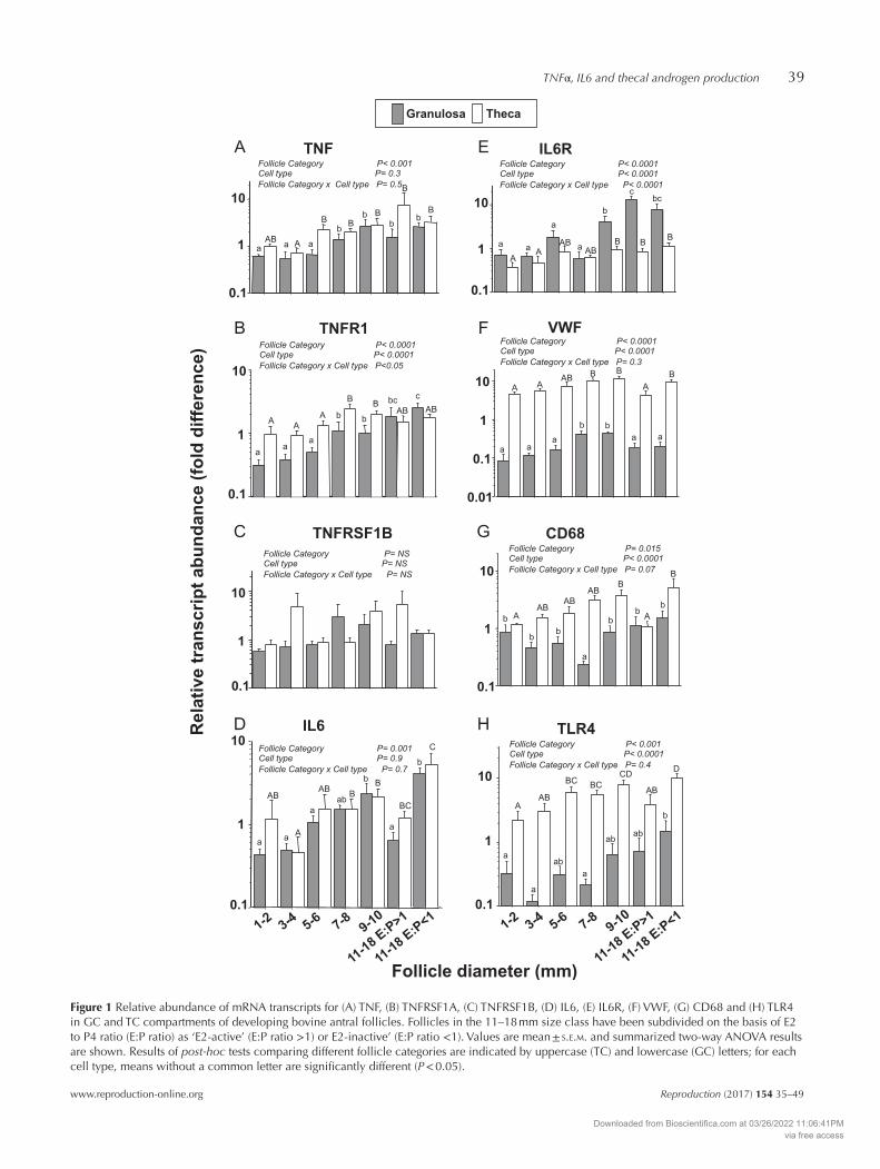
The relative abundance of TNF mRNA increased 3- to 4-fold over the course of antral follicle development in both TC and GC (Fig. 1A). Overall, the relative abundance of TNF mRNA was higher (P < 0.01) in TC than GC (Fig. 1) with greatest expression in TC of LEA follicles (~9-fold higher than in TC of 1–2 mm follicles). The relative abundance of TNFRSF1A mRNA in TC showed a progressive 10-fold increase between 1–2 mm and 11–18 mm follicle size categories (P < 0.0001; Fig. 1B). In contrast, expression level in GC was relatively uniform. There were no significant cell-type or follicle stage-related differences in the abundance of TNFRSF1B mRNA that was only detected at low levels in these samples (Fig. 1C).
The relative abundance of IL6 mRNA was greatest in LEI follicles being ~5-fold higher than that in corresponding LEA follicles of equivalent size (P < 0.001), but there was no significant difference between GC and TC (Fig. 1D). In contrast, the abundance of IL6R mRNA was greater in GC compared with that in TC at all follicle stages, particularly in large (11–18 mm) follicles, regardless of their oestrogen-active status (Fig. 1E). GC expression of IL6R increased ~10-fold between 1–2 mm and 11–18 mm size categories (P < 0.05). TC expression of IL6R was ~2-fold higher in 11–18 mm follicles than that in 1–2 mm follicles.
Expression of putative endothelial cell (VWF) and macrophage (CD68 and TLR4) ‘markers’ in granulosa and theca interna layers of developing antral follicles
Figure 1F shows that VWF mRNA abundance was ~50-fold higher in TC than that in GC. Expression
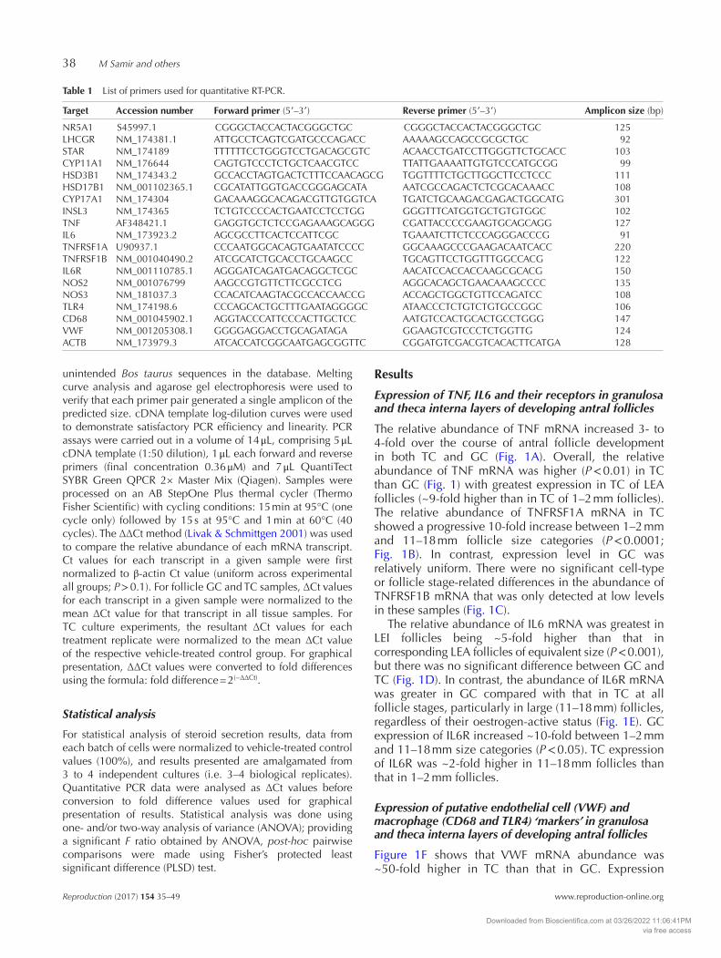
Table 1 List of primers used for quantitative RT-PCR.
Target Accession number Forward primer (5′–3′) Reverse primer (5′–3′) Amplicon size (bp)
NR5A1 S45997.1 CGGGCTACCACTACGGGCTGC CGGGCTACCACTACGGGCTGC 125LHCGR NM_174381.1 ATTGCCTCAGTCGATGCCCAGACC AAAAAGCCAGCCGCGCTGC 92STAR NM_174189 TTTTTTCCTGGGTCCTGACAGCGTC ACAACCTGATCCTTGGGTTCTGCACC 103CYP11A1 NM_176644 CAGTGTCCCTCTGCTCAACGTCC TTATTGAAAATTGTGTCCCATGCGG 99HSD3B1 NM_174343.2 GCCACCTAGTGACTCTTTCCAACAGCG TGGTTTTCTGCTTGGCTTCCTCCC 111HSD17B1 NM_001102365.1 CGCATATTGGTGACCGGGAGCATA AATCGCCAGACTCTCGCACAAACC 108CYP17A1 NM_174304 GACAAAGGCACAGACGTTGTGGTCA TGATCTGCAAGACGAGACTGGCATG 301INSL3 NM_174365 TCTGTCCCCACTGAATCCTCCTGG GGGTTTCATGGTGCTGTGTGGC 102TNF AF348421.1 GAGGTGCTCTCCGAGAAAGCAGGG CGATTACCCCGAAGTGCAGCAGG 127IL6 NM_173923.2 AGCGCCTTCACTCCATTCGC TGAAATCTTCTCCCAGGGACCCG 91TNFRSF1A U90937.1 CCCAATGGCACAGTGAATATCCCC GGCAAAGCCCGAAGACAATCACC 220TNFRSF1B NM_001040490.2 ATCGCATCTGCACCTGCAAGCC TGCAGTTCCTGGTTTGGCCACG 122IL6R NM_001110785.1 AGGGATCAGATGACAGGCTCGC AACATCCACCACCAAGCGCACG 150NOS2 NM_001076799 AAGCCGTGTTCTTCGCCTCG AGGCACAGCTGAACAAAGCCCC 135NOS3 NM_181037.3 CCACATCAAGTACGCCACCAACCG ACCAGCTGGCTGTTCCAGATCC 108TLR4 NM_174198.6 CCCAGCACTGCTTTGAATAGGGGC ATAACCCTCTGTCTGTGCCGGC 106CD68 NM_001045902.1 AGGTACCCATTCCCACTTGCTCC AATGTCCACTGCACTGCCTGGG 147VWF NM_001205308.1 GGGGAGGACCTGCAGATAGA GGAAGTCGTCCCTCTGGTTG 124ACTB NM_173979.3 ATCACCATCGGCAATGAGCGGTTC CGGATGTCGACGTCACACTTCATGA 128
Downloaded from Bioscientifica.com at 03/26/2022 11:06:41PMvia free access
p
TNFα, IL6 and thecal androgen production 39
www.reproduction-online.org Reproduction (2017) 154 35–49
A
B
C
D
E
F
G
H
Figure 1 Relative abundance of mRNA transcripts for (A) TNF, (B) TNFRSF1A, (C) TNFRSF1B, (D) IL6, (E) IL6R, (F) VWF, (G) CD68 and (H) TLR4 in GC and TC compartments of developing bovine antral follicles. Follicles in the 11–18 mm size class have been subdivided on the basis of E2 to P4 ratio (E:P ratio) as ‘E2-active’ (E:P ratio >1) or E2-inactive’ (E:P ratio <1). Values are mean ± s.e.m. and summarized two-way ANOVA results are shown. Results of post-hoc tests comparing different follicle categories are indicated by uppercase (TC) and lowercase (GC) letters; for each cell type, means without a common letter are significantly different (P < 0.05).
Downloaded from Bioscientifica.com at 03/26/2022 11:06:41PMvia free access
p
M Samir and others40
Reproduction (2017) 154 35–49 www.reproduction-online.org
levels increased ~3-fold in both compartments between 1–2 and 9–10 mm in diameter (P < 0.05) but were lower in 11–18 mm follicles than those in 9–10 mm follicles (P < 0.05). Overall, expression levels of CD68 (Fig. 1G) and TLR4 (Fig. 1H) were both higher in TC than those in GC. Thecal CD68 and TLR4 expression increased ~4-fold between 1–2 mm and 9–10 mm (P < 0.05) but was lower in LEA follicles than that in 9–10 mm follicles and LEI follicles (P < 0.05). More variable profiles were seen in GC with CD68 expression being lower in 7–8 mm follicles than those in all other stages (P < 0.05); TLR4 expression in GC was higher in LEI follicles than that in 1–4 mm and 7–8 mm follicles (P < 0.05).
Effect of TNFα on basal and LH-induced production of androstendione and progesterone and on viable cell number
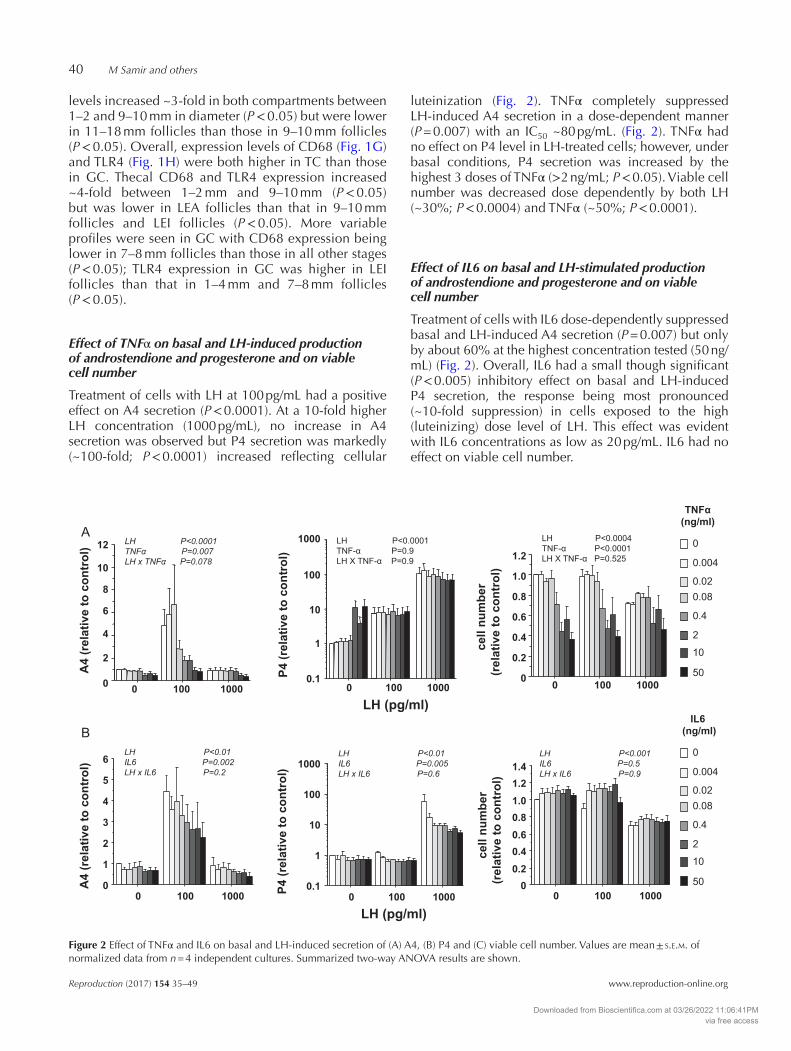
Treatment of cells with LH at 100 pg/mL had a positive effect on A4 secretion (P < 0.0001). At a 10-fold higher LH concentration (1000 pg/mL), no increase in A4 secretion was observed but P4 secretion was markedly (~100-fold; P < 0.0001) increased reflecting cellular
luteinization (Fig. 2). TNFα completely suppressed LH-induced A4 secretion in a dose-dependent manner (P = 0.007) with an IC50 ~80 pg/mL. (Fig. 2). TNFα had no effect on P4 level in LH-treated cells; however, under basal conditions, P4 secretion was increased by the highest 3 doses of TNFα (>2 ng/mL; P < 0.05). Viable cell number was decreased dose dependently by both LH (~30%; P < 0.0004) and TNFα (~50%; P < 0.0001).
Effect of IL6 on basal and LH-stimulated production of androstendione and progesterone and on viable cell number
Treatment of cells with IL6 dose-dependently suppressed basal and LH-induced A4 secretion (P = 0.007) but only by about 60% at the highest concentration tested (50 ng/mL) (Fig. 2). Overall, IL6 had a small though significant (P < 0.005) inhibitory effect on basal and LH-induced P4 secretion, the response being most pronounced (~10-fold suppression) in cells exposed to the high (luteinizing) dose level of LH. This effect was evident with IL6 concentrations as low as 20 pg/mL. IL6 had no effect on viable cell number.
0
1
2
3
4
5
6
0 100 1000
LH P<0.01
IL6 P=0.002
LH x IL6 P=0.2
A4
(re
la
tiv
e to
c
on
tro
l)
0
0.2
0.4
0.6
0.8
1.0
1.2
1.4
0 100 1000
LH P<0.001
IL6 P=0.5
LH x IL6 P=0.9
0.1
1
10
100
1000
0 100 1000
LH P<0.01
IL6 P=0.005
LH x IL6 P=0.6
P4
(re
la
tiv
e to
c
on
tro
l)
ce
ll n
um
be
r
(re
la
tiv
e to
c
on
tro
l)
LH (pg/ml)
LH P<0.0001
TNF P=0.007
LH x TNF P=0.078
0
0.2
0.4
0.6
0.8
1.0
1.2
0 100 1000
LH P<0.0004 TNF- P<0.0001 LH X TNF- P=0.525
ce
ll n
um
be
r
(re
la
tiv
e to
c
on
tro
l)
0
2
4
6
8
10
12
0 100
A
B
1000
A4
(re
la
tiv
e to
c
on
tro
l)
0.1
1
10
100
1000
0 1000100
LH P<0.0001 TNF- P=0.9 LH X TNF- P=0.9
P4
(re
la
tiv
e to
c
on
tro
l)
LH (pg/ml)
50
10
2
0.4
0.080.02
0.004
0
TNF
(ng/ml)
50
10
2
0.4
0.080.02
0.004
0
IL6
(ng/ml)
Figure 2 Effect of TNFα and IL6 on basal and LH-induced secretion of (A) A4, (B) P4 and (C) viable cell number. Values are mean ± s.e.m. of normalized data from n = 4 independent cultures. Summarized two-way ANOVA results are shown.
Downloaded from Bioscientifica.com at 03/26/2022 11:06:41PMvia free access
p
TNFα, IL6 and thecal androgen production 41
www.reproduction-online.org Reproduction (2017) 154 35–49
Effect of TNFα on thecal expression of steroidogenesis-related transcripts and on NOS2 and NOS3
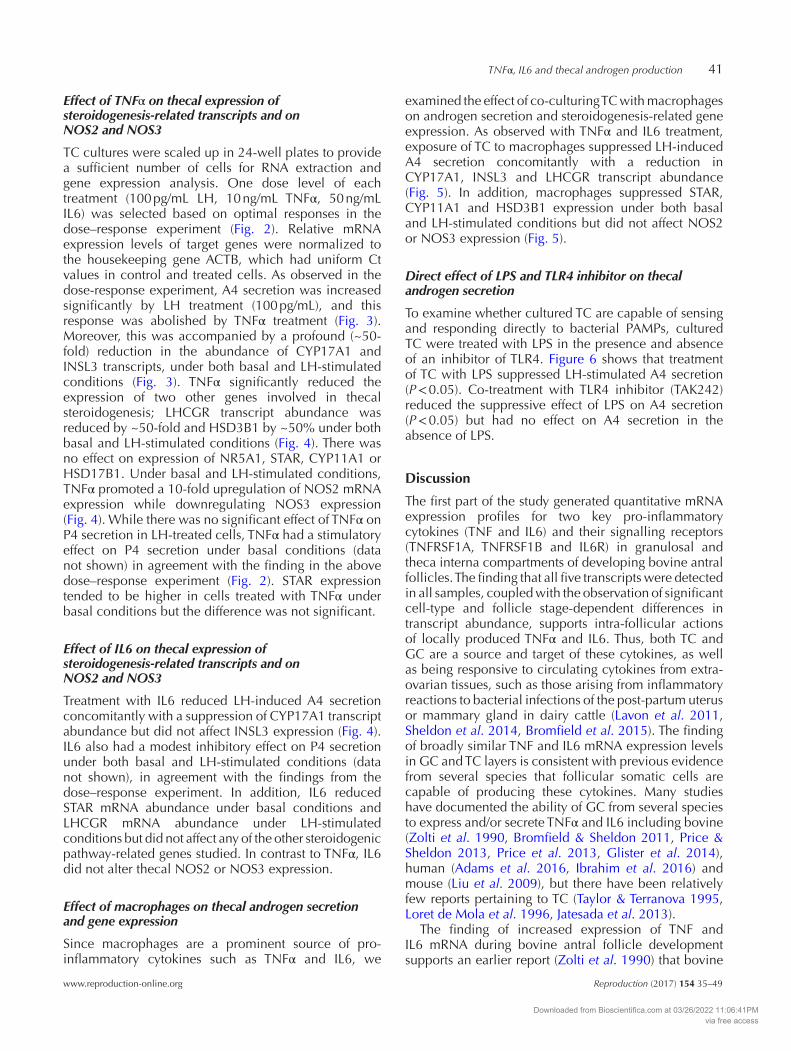
TC cultures were scaled up in 24-well plates to provide a sufficient number of cells for RNA extraction and gene expression analysis. One dose level of each treatment (100 pg/mL LH, 10 ng/mL TNFα, 50 ng/mL IL6) was selected based on optimal responses in the dose–response experiment (Fig. 2). Relative mRNA expression levels of target genes were normalized to the housekeeping gene ACTB, which had uniform Ct values in control and treated cells. As observed in the dose-response experiment, A4 secretion was increased significantly by LH treatment (100 pg/mL), and this response was abolished by TNFα treatment (Fig. 3). Moreover, this was accompanied by a profound (~50-fold) reduction in the abundance of CYP17A1 and INSL3 transcripts, under both basal and LH-stimulated conditions (Fig. 3). TNFα significantly reduced the expression of two other genes involved in thecal steroidogenesis; LHCGR transcript abundance was reduced by ~50-fold and HSD3B1 by ~50% under both basal and LH-stimulated conditions (Fig. 4). There was no effect on expression of NR5A1, STAR, CYP11A1 or HSD17B1. Under basal and LH-stimulated conditions, TNFα promoted a 10-fold upregulation of NOS2 mRNA expression while downregulating NOS3 expression (Fig. 4). While there was no significant effect of TNFα on P4 secretion in LH-treated cells, TNFα had a stimulatory effect on P4 secretion under basal conditions (data not shown) in agreement with the finding in the above dose–response experiment (Fig. 2). STAR expression tended to be higher in cells treated with TNFα under basal conditions but the difference was not significant.
Effect of IL6 on thecal expression of steroidogenesis-related transcripts and on NOS2 and NOS3
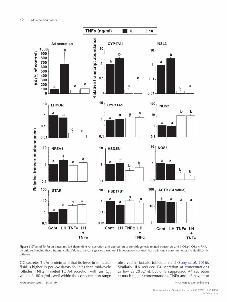
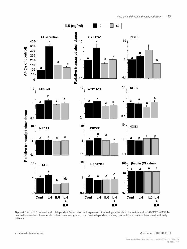
Treatment with IL6 reduced LH-induced A4 secretion concomitantly with a suppression of CYP17A1 transcript abundance but did not affect INSL3 expression (Fig. 4). IL6 also had a modest inhibitory effect on P4 secretion under both basal and LH-stimulated conditions (data not shown), in agreement with the findings from the dose–response experiment. In addition, IL6 reduced STAR mRNA abundance under basal conditions and LHCGR mRNA abundance under LH-stimulated conditions but did not affect any of the other steroidogenic pathway-related genes studied. In contrast to TNFα, IL6 did not alter thecal NOS2 or NOS3 expression.
Effect of macrophages on thecal androgen secretion and gene expression
Since macrophages are a prominent source of pro-inflammatory cytokines such as TNFα and IL6, we
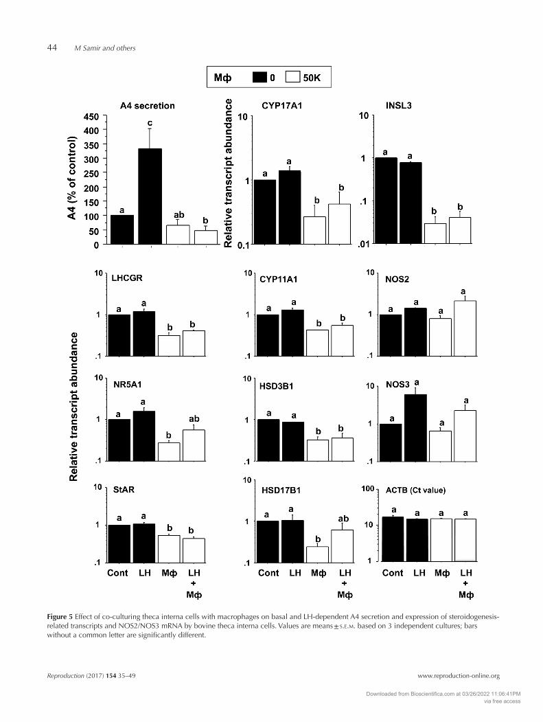
examined the effect of co-culturing TC with macrophages on androgen secretion and steroidogenesis-related gene expression. As observed with TNFα and IL6 treatment, exposure of TC to macrophages suppressed LH-induced A4 secretion concomitantly with a reduction in CYP17A1, INSL3 and LHCGR transcript abundance (Fig. 5). In addition, macrophages suppressed STAR, CYP11A1 and HSD3B1 expression under both basal and LH-stimulated conditions but did not affect NOS2 or NOS3 expression (Fig. 5).
Direct effect of LPS and TLR4 inhibitor on thecal androgen secretion
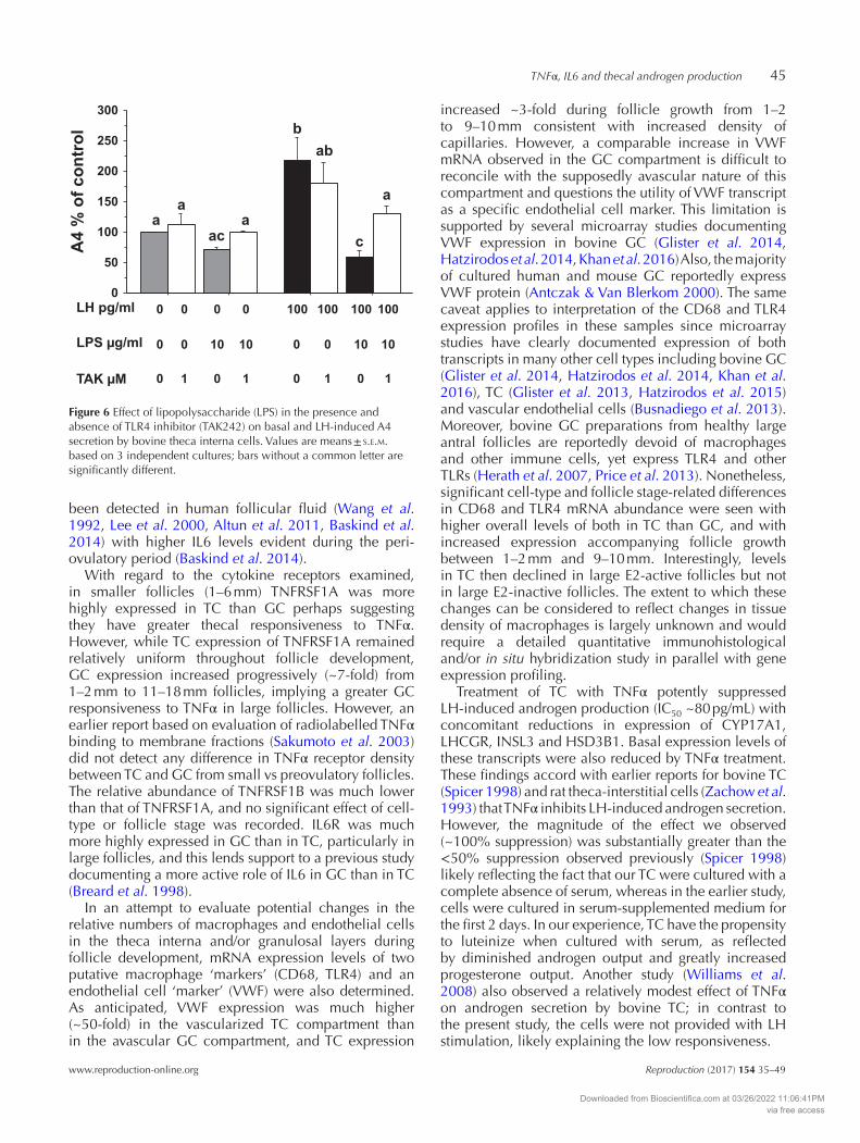
To examine whether cultured TC are capable of sensing and responding directly to bacterial PAMPs, cultured TC were treated with LPS in the presence and absence of an inhibitor of TLR4. Figure 6 shows that treatment of TC with LPS suppressed LH-stimulated A4 secretion (P < 0.05). Co-treatment with TLR4 inhibitor (TAK242) reduced the suppressive effect of LPS on A4 secretion (P < 0.05) but had no effect on A4 secretion in the absence of LPS.
Discussion
The first part of the study generated quantitative mRNA expression profiles for two key pro-inflammatory cytokines (TNF and IL6) and their signalling receptors (TNFRSF1A, TNFRSF1B and IL6R) in granulosal and theca interna compartments of developing bovine antral follicles. The finding that all five transcripts were detected in all samples, coupled with the observation of significant cell-type and follicle stage-dependent differences in transcript abundance, supports intra-follicular actions of locally produced TNFα and IL6. Thus, both TC and GC are a source and target of these cytokines, as well as being responsive to circulating cytokines from extra-ovarian tissues, such as those arising from inflammatory reactions to bacterial infections of the post-partum uterus or mammary gland in dairy cattle (Lavon et al. 2011, Sheldon et al. 2014, Bromfield et al. 2015). The finding of broadly similar TNF and IL6 mRNA expression levels in GC and TC layers is consistent with previous evidence from several species that follicular somatic cells are capable of producing these cytokines. Many studies have documented the ability of GC from several species to express and/or secrete TNFα and IL6 including bovine (Zolti et al. 1990, Bromfield & Sheldon 2011, Price & Sheldon 2013, Price et al. 2013, Glister et al. 2014), human (Adams et al. 2016, Ibrahim et al. 2016) and mouse (Liu et al. 2009), but there have been relatively few reports pertaining to TC (Taylor & Terranova 1995, Loret de Mola et al. 1996, Jatesada et al. 2013).
The finding of increased expression of TNF and IL6 mRNA during bovine antral follicle development supports an earlier report (Zolti et al. 1990) that bovine
Downloaded from Bioscientifica.com at 03/26/2022 11:06:41PMvia free access
p
M Samir and others42
Reproduction (2017) 154 35–49 www.reproduction-online.org
GC secretes TNFα protein and that its level in follicular fluid is higher in peri-ovulatory follicles than mid-cycle follicles. TNFα inhibited TC A4 secretion with an IC50 value of ~80 pg/mL, well within the concentration range
observed in buffalo follicular fluid (Boby et al. 2016). Similarly, IL6 reduced P4 secretion at concentrations as low as 20 pg/mL but only suppressed A4 secretion at much higher concentrations. TNFα and IL6 have also
100TNF (ng/ml)
A4
(%
o
f c
on
tro
l)
0.01
0.1
1
10
b
CYP17A1
c
0.01
0.1
1
10
INSL3
0
100
200
300
400
500
600
700
800
900
1000
a
A4 secretion
Re
la
tiv
e tra
ns
crip
t a
bu
nd
an
ce
a
bb
0.1
1
10 HSD3B1
0.1
1
10 NR5A1
0.1
1
10
100 STAR
0.01
0.1
1
10LHCGR
0.01
0.1
1
10HSD17B1
1
10
100 ACTB (Ct value) Re
la
tiv
e tra
ns
crip
t a
bu
nd
an
ce
)
Cont LH TNF LH
+
TNF
0.1
1
10
a
CYP11A1
0.1
1
10
100
a a
b b
NOS2
0.1
1
10NOS3
Cont LH TNF LH
+
TNF
Cont LH TNF LH
+
TNF
a a
b
c
c
a
b
a c
aa
cc
a
a
a
a a
a a
a
a
a
a
a a
a a
b
aa
b
a aa a
a
Figure 3 Effect of TNFα on basal and LH-dependent A4 secretion and expression of steroidogenesis-related transcripts and NOS2/NOS3 mRNA by cultured bovine theca interna cells. Values are means ± s.e.m. based on 4 independent cultures; bars without a common letter are significantly different.
Downloaded from Bioscientifica.com at 03/26/2022 11:06:41PMvia free access
p
TNFα, IL6 and thecal androgen production 43
www.reproduction-online.org Reproduction (2017) 154 35–49
Figure 4 Effect of IL6 on basal and LH-dependent A4 secretion and expression of steroidogenesis-related transcripts and NOS2/NOS3 mRNA by cultured bovine theca interna cells. Values are means ± s.e.m. based on 4 independent cultures; bars without a common letter are significantly different.
Downloaded from Bioscientifica.com at 03/26/2022 11:06:41PMvia free access
p
M Samir and others44
Reproduction (2017) 154 35–49 www.reproduction-online.org
Figure 5 Effect of co-culturing theca interna cells with macrophages on basal and LH-dependent A4 secretion and expression of steroidogenesis-related transcripts and NOS2/NOS3 mRNA by bovine theca interna cells. Values are means ± s.e.m. based on 3 independent cultures; bars without a common letter are significantly different.
Downloaded from Bioscientifica.com at 03/26/2022 11:06:41PMvia free access
p
TNFα, IL6 and thecal androgen production 45
www.reproduction-online.org Reproduction (2017) 154 35–49
been detected in human follicular fluid (Wang et al. 1992, Lee et al. 2000, Altun et al. 2011, Baskind et al. 2014) with higher IL6 levels evident during the peri-ovulatory period (Baskind et al. 2014).
With regard to the cytokine receptors examined, in smaller follicles (1–6 mm) TNFRSF1A was more highly expressed in TC than GC perhaps suggesting they have greater thecal responsiveness to TNFα. However, while TC expression of TNFRSF1A remained relatively uniform throughout follicle development, GC expression increased progressively (~7-fold) from 1–2 mm to 11–18 mm follicles, implying a greater GC responsiveness to TNFα in large follicles. However, an earlier report based on evaluation of radiolabelled TNFα binding to membrane fractions (Sakumoto et al. 2003) did not detect any difference in TNFα receptor density between TC and GC from small vs preovulatory follicles. The relative abundance of TNFRSF1B was much lower than that of TNFRSF1A, and no significant effect of cell-type or follicle stage was recorded. IL6R was much more highly expressed in GC than in TC, particularly in large follicles, and this lends support to a previous study documenting a more active role of IL6 in GC than in TC (Breard et al. 1998).
In an attempt to evaluate potential changes in the relative numbers of macrophages and endothelial cells in the theca interna and/or granulosal layers during follicle development, mRNA expression levels of two putative macrophage ‘markers’ (CD68, TLR4) and an endothelial cell ‘marker’ (VWF) were also determined. As anticipated, VWF expression was much higher (~50-fold) in the vascularized TC compartment than in the avascular GC compartment, and TC expression
increased ~3-fold during follicle growth from 1–2 to 9–10 mm consistent with increased density of capillaries. However, a comparable increase in VWF mRNA observed in the GC compartment is difficult to reconcile with the supposedly avascular nature of this compartment and questions the utility of VWF transcript as a specific endothelial cell marker. This limitation is supported by several microarray studies documenting VWF expression in bovine GC (Glister et al. 2014, Hatzirodos et al. 2014, Khan et al. 2016) Also, the majority of cultured human and mouse GC reportedly express VWF protein (Antczak & Van Blerkom 2000). The same caveat applies to interpretation of the CD68 and TLR4 expression profiles in these samples since microarray studies have clearly documented expression of both transcripts in many other cell types including bovine GC (Glister et al. 2014, Hatzirodos et al. 2014, Khan et al. 2016), TC (Glister et al. 2013, Hatzirodos et al. 2015) and vascular endothelial cells (Busnadiego et al. 2013). Moreover, bovine GC preparations from healthy large antral follicles are reportedly devoid of macrophages and other immune cells, yet express TLR4 and other TLRs (Herath et al. 2007, Price et al. 2013). Nonetheless, significant cell-type and follicle stage-related differences in CD68 and TLR4 mRNA abundance were seen with higher overall levels of both in TC than GC, and with increased expression accompanying follicle growth between 1–2 mm and 9–10 mm. Interestingly, levels in TC then declined in large E2-active follicles but not in large E2-inactive follicles. The extent to which these changes can be considered to reflect changes in tissue density of macrophages is largely unknown and would require a detailed quantitative immunohistological and/or in situ hybridization study in parallel with gene expression profiling.
Treatment of TC with TNFα potently suppressed LH-induced androgen production (IC50 ~80 pg/mL) with concomitant reductions in expression of CYP17A1, LHCGR, INSL3 and HSD3B1. Basal expression levels of these transcripts were also reduced by TNFα treatment. These findings accord with earlier reports for bovine TC (Spicer 1998) and rat theca-interstitial cells (Zachow et al. 1993) that TNFα inhibits LH-induced androgen secretion. However, the magnitude of the effect we observed (~100% suppression) was substantially greater than the <50% suppression observed previously (Spicer 1998) likely reflecting the fact that our TC were cultured with a complete absence of serum, whereas in the earlier study, cells were cultured in serum-supplemented medium for the first 2 days. In our experience, TC have the propensity to luteinize when cultured with serum, as reflected by diminished androgen output and greatly increased progesterone output. Another study (Williams et al. 2008) also observed a relatively modest effect of TNFα on androgen secretion by bovine TC; in contrast to the present study, the cells were not provided with LH stimulation, likely explaining the low responsiveness.
0
50
100
150
200
250
300
a
a
ac
a
b
ab
c
a
A4 %
o
f co
ntro
l
0LH pg/ml
LPS g/ml
TAK M
0
0
0
0
1
0
10
0
0
10
1
100
0
0
100
0
1
100
10
0
100
10
1
Figure 6 Effect of lipopolysaccharide (LPS) in the presence and absence of TLR4 inhibitor (TAK242) on basal and LH-induced A4 secretion by bovine theca interna cells. Values are means ± s.e.m. based on 3 independent cultures; bars without a common letter are significantly different.
Downloaded from Bioscientifica.com at 03/26/2022 11:06:41PMvia free access
p
M Samir and others46
Reproduction (2017) 154 35–49 www.reproduction-online.org
Interestingly, under both basal and LH-treated conditions, expression of the classical TNFα-responsive gene, NOS2 (iNOS), was markedly upregulated by TNFα treatment while NOS3 (eNOS) expression was downregulated. Treatment of bovine TC with BMP6 was also found to enhance NOS2 expression while suppressing androgen secretion and CYP17A1 and INSL3 expression (Glister et al. 2013). NOS2 mRNA level was reportedly higher in GC of growing dominant bovine follicles compared to subordinate follicles (Zamberlam et al. 2011), and this accords with the present observation that follicular TNF mRNA expression level was maximal in TC of large oestrogen-active follicles, which showed high expression levels of TNFRSF1A in both TC and GC layers. In IVF patients, high levels of nitric oxide in follicular fluid have been associated with reduced E2 production and diminished oocyte quality (Vignini et al. 2008), suggesting a possible association with increased TNFα signalling. TNFα has also been shown to suppress FSH-induced estradiol secretion and CYP19A1 expression by GC (Kaipia et al. 1996, Spicer 1998, Sakumoto et al. 2003, Williams et al. 2008, Glister et al. 2014). With regard to the hypothesis linking chronic low-grade inflammation to the development of PCOS and associated hyperandrogenism (Duleba & Dokras 2012, Gonzalez 2012), the present findings are counter-intuitive since pro-inflammatory cytokines like TNFα might be expected to enhance rather than suppress thecal androgen production as clearly shown here.
Given the observation that IL6 and its receptor are expressed by both TC and GC throughout bovine antral follicle development, we also examined the effect of IL6 on TC steroidogenesis. An inhibitory effect of IL6 was seen on LH-induced secretion of both A4 and P4 although the potency of IL6 in suppressing A4 secretion was much less than that of TNFα. In contrast, IL6 at concentrations as low as 20 pg/mL reduced thecal P4 secretion, indicating effects at concentrations well within the range observed in follicular fluid and GC-conditioned culture medium (Bromfield & Sheldon 2011, Boby et al. 2016). The inhibition of A4 secretion elicited by the much higher (likely supra-physiological) concentration of IL6 (50 ng/mL) used in our gene expression experiment was accompanied by a significant downregulation of CYP17A1, LHCGR and STAR mRNA abundance, but there was no clear effect on expression of the other genes examined including INSL3, NOS2 and NOS3 that were clearly modulated by TNFα. Earlier studies found no effect of IL6 on androgen secretion by rat theca-interstitial cells (Hurwitz et al. 1991) or in vitro perfused rat ovary (Van der Hoek et al. 1998). However, IL6 has been reported to exert an inhibitory action on GC oestrogen secretion (Alpizar & Spicer 1994, Spicer 1998, Tamura et al. 2000, Salmassi et al. 2001). Further studies are needed to unravel these apparently
complex differential actions of TNFα and IL6 on thecal steroidogenesis.
Macrophages, particularly when activated (e.g. by microbial PAMP exposure), are a prominent source of pro-inflammatory cytokines including TNFα, IL6 and IL1b (Plowden et al. 2004). In addition to a resident macrophage population, monocytes are known to infiltrate the ovary in a cyclic manner where they differentiate into macrophages (Wu et al. 2004, Figueroa et al. 2012). Evidence supports a role for macrophages in follicle atresia, follicular-luteal transition and luteal regression (reviews: Wu et al. 2004, Walusimbi & Pate 2013), but their potential involvement in the regulation of orderly follicle growth and steroidogenesis under normal physiological conditions remains unclear. Here, we showed that co-culturing TC with monocyte-derived macrophages blocked LH-induced androgen secretion, accompanied by a reduction in expression of CYP17A1 and several other steroidogenesis-related transcripts including LHCGR, STAR, CYP11A1, HSD3B1 and INSL3. Notably, INSL3 was recently shown to have a positive role in thecal A4 secretion (Glister et al. 2013, Satchell et al. 2013) so the inhibitory effect of both macrophages and TNFα might be mediated, at least in part, by downregulation of INSL3 expression leading to loss of CYP17A1 expression. Alternately, downregulation of LHCGR leading to loss of LH sensitivity could explain the decline in LH-dependent CYP17A1 expression and androgen production since LHCGR expression was inhibited by all three treatments (macrophages, TNFα and IL6).
Both GC (Herath et al. 2007, Bromfield & Sheldon 2011, Shimizu et al. 2012, Price & Sheldon 2013, Price et al. 2013) and TC (Taylor & Terranova 1995, Magata et al. 2014a, Williams et al. 2008) have been shown to express TLR potentially enabling them to detect and respond to bacterial PAMPs (e.g. TLR4 binding to LPS) leading to production of pro-inflammatory cytokines, in a manner akin to macrophages and other immunity-related cells. Indeed, LPS has been shown to promote IL6 and TNF expression/secretion and inhibit CYP19A1 expression and oestradiol secretion by cultured bovine GC (Herath et al. 2007, Price & Sheldon 2013, Price et al. 2013). The present findings confirm TLR4 mRNA expression in follicular theca interna layer and GC layers. Moreover, we show herein a direct inhibitory action of LPS on LH-induced androgen secretion by bovine TC that was attenuated by co-treatment with a TLR4 blocker (TAK242). Magata and coworkers (Magata et al. 2014a) also reported an LPS-induced inhibition of thecal androgen production although another bovine study (Williams et al. 2008) found no effect. Interestingly, LPS and another PAMP from gram-positive bacteria, peptidoglycan, exerted an additive suppressive effect on thecal androgen production (Magata et al. 2014b). LPS was also reported to inhibit
Downloaded from Bioscientifica.com at 03/26/2022 11:06:41PMvia free access
p
TNFα, IL6 and thecal androgen production 47
www.reproduction-online.org Reproduction (2017) 154 35–49
LH-induced androgen and progesterone secretion by rat theca-interstitial cells (Taylor & Terranova 1995).
In summary, cell-type and follicle stage-dependent differences in mRNA expression of TNF, IL6 and their receptors were found in granulosal and theca interna layers of developing bovine antral follicles. The study confirms and extends previous observations regarding the ability of these cytokines to modulate thecal steroidogenesis and demonstrates that macrophage co-culture, like TNFα and IL6 treatment, can suppress thecal androgen secretion and inhibit expression of CYP17A1 and LHCGR. Finally, we provide confirmatory evidence that bovine theca interna cells are directly responsive to bacterial LPS and that this also attenuates LH-dependant androgen secretion. In an in vivo context, the findings provide further supporting evidence for the view that both macrophages and follicular somatic cells contribute to the ‘local’ generation of inflammatory mediators in response to either physiological (i.e. ovarian cycles) or pathophysiological (i.e. bacterial infections) events that can, in turn, exert powerful modulatory actions on ovarian steroidogenesis at both the theca and granulosa cell level.
Declaration of interest
The authors declare that there is no conflict of interest that could be perceived as prejudicing the impartiality of the research reported.
Funding
This work was partially supported by the Biotechnology and Biological Sciences Research Council (award BB/M001369/1 to P G K).
Acknowledgements
The authors thank D Butlin and M Schenk for skilled technical assistance. M S was in receipt of a Doctoral Scholarship from the Iraqi Higher Committee for Education Development.
ReferencesAdams J, Liu Z, Ren YA, Wun WS, Zhou W, Kenigsberg S, Librach C,
Valdes C, Gibbons W & Richards J 2016 Enhanced inflammatory transcriptome in the granulosa cells of women with polycystic ovarian syndrome. Journal of Clinical Endocrinology and Metabolism 101 3459–3468. (doi:10.1210/jc.2015-4275)
Alpizar E & Spicer LJ 1994 Effects of interleukin-6 on proliferation and follicle-stimulating hormone-induced estradiol production by bovine granulosa cells in vitro: dependence on size of follicle. Biology of Reproduction 50 38–43. (doi:10.1095/biolreprod50.1.38)
Altun T, Jindal S, Greenseid K, Shu J & Pal L 2011 Low follicular fluid IL-6 levels in IVF patients are associated with increased likelihood of clinical pregnancy. Journal of Assisted Reproduction and Genetics 28 245–251. (doi:10.1007/s10815-010-9502-8)
Antczak M & Van Blerkom J 2000 The vascular character of ovarian follicular granulosa cells: phenotypic and functional evidence for an
endothelial-like cell population. Human Reproduction 15 2306–2318. (doi:10.1093/humrep/15.11.2306)
Baskind NE, Orsi NM & Sharma V 2014 Follicular-phase ovarian follicular fluid and plasma cytokine profiling of natural cycle in vitro fertilization patients. Fertility and Sterility 102 410–418. (doi:10.1016/j.fertnstert.2014.04.032)
Birmingham JR & Jeska EL 1980 The isolation, long-term cultivation and characterization of bovine peripheral blood monocytes. Immunology 41 807–814. (doi:10.1016/s0580-9517(02)32112-3)
Bleach EC, Glencross RG, Feist SA, Groome NP & Knight PG 2001 Plasma inhibin A in heifers: relationship with follicle dynamics, gonadotropins, and steroids during the estrous cycle and after treatment with bovine follicular fluid. Biology of Reproduction 64 743–752. (doi:10.1095/biolreprod64.3.743)
Boby J, Kumar H, Gupta HP, Jan MH, Singh SK, Patra MK, Nandi S, Abraham A & Krishnaswamy N 2016 Endometritis increases pro-inflammatory cytokines in follicular fluid and cervico-vaginal mucus in the buffalo cow. Animal Biotechnology 1–5. (doi:10.1080/10495398.2016.1244067)
Bornstein SR, Rutkowski H & Vrezas I 2004 Cytokines and steroidogenesis. Molecular and Cellular Endocrinology 215 135–141. (doi:10.1016/j.mce.2003.11.022)
Brannstrom M, Mayrhofer G & Robertson SA 1993 Localization of leukocyte subsets in the rat ovary during the periovulatory period. Biology of Reproduction 48 277–286. (doi:10.1095/biolreprod48.2.277)
Breard E, Benhaim A, Feral C & Leymarie P 1998 Rabbit ovarian production of interleukin-6 and its potential effects on gonadotropin-induced progesterone secretion in granulosa and theca cells. Journal of Endocrinology 159 479–487. (doi:10.1677/joe.0.1590479)
Bromfield JJ & Sheldon IM 2011 Lipopolysaccharide initiates inflammation in bovine granulosa cells via the TLR4 pathway and perturbs oocyte meiotic progression in vitro. Endocrinology 152 5029–5040. (doi:10.1210/en.2011-1124)
Bromfield JJ, Santos JE, Block J, Williams RS & Sheldon IM 2015 Physiology and endocrinology symposium: Uterine infection: linking infection and innate immunity with infertility in the high-producing dairy cow. Journal of Animal Science 93 2021–2033. (doi:10.2527/jas.2014-8496)
Busnadiego O, Gonzalez-Santamaria J, Lagares D, Guinea-Viniegra J, Pichol-Thievend C, Muller L & Rodriguez-Pascual F 2013 LOXL4 is induced by transforming growth factor beta1 through Smad and JunB/Fra2 and contributes to vascular matrix remodeling. Molecular and Cellular Biology 33 2388–2401. (doi:10.1128/MCB.00036-13)
Cohen PE, Nishimura K, Zhu L & Pollard JW 1999 Macrophages: important accessory cells for reproductive function. Journal of Leukocyte Biology 66 765–772.
Duleba AJ & Dokras A 2012 Is PCOS an inflammatory process? Fertility and Sterility 97 7–12. (doi:10.1016/j.fertnstert.2011.11.023)
Figueroa F, Davicino R, Micalizzi B, Oliveros L & Forneris M 2012 Macrophage secretions modulate the steroidogenesis of polycystic ovary in rats: effect of testosterone on macrophage pro-inflammatory cytokines. Life Science 90 733–739. (doi:10.1016/j.lfs.2012.03.019)
Franczak A, Zmijewska A, Kurowicka B, Wojciechowicz B, Petroff BK & Kotwica G 2012 The effect of tumor necrosis factor alpha (TNFalpha), interleukin 1beta (IL1beta) and interleukin 6 (IL6) on endometrial PGF2alpha synthesis, metabolism and release in early-pregnant pigs. Theriogenology 77 155–165. (doi:10.1016/j.theriogenology.2011.07.029)
Galvao AM, Ferreira-Dias G & Skarzynski DJ 2013 Cytokines and angiogenesis in the corpus luteum. Mediators of Inflammation 2013 420186. (doi:10.1155/2013/420186)
Glister C, Tannetta DS, Groome NP & Knight PG 2001 Interactions between follicle-stimulating hormone and growth factors in modulating secretion of steroids and inhibin-related peptides by nonluteinized bovine granulosa cells. Biology of Reproduction 65 1020–1028. (doi:10.1095/biolreprod65.4.1020)
Glister C, Richards SL & Knight PG 2005 Bone morphogenetic proteins (BMP) -4, -6, and -7 potently suppress basal and luteinizing hormone-induced androgen production by bovine theca interna cells in primary culture: could ovarian hyperandrogenic dysfunction be caused by a defect in thecal BMP signaling? Endocrinology 146 1883–1892. (doi:10.1210/en.2004-1303)
Downloaded from Bioscientifica.com at 03/26/2022 11:06:41PMvia free access
p
M Samir and others48
Reproduction (2017) 154 35–49 www.reproduction-online.org
Glister C, Satchell L & Knight PG 2010 Changes in expression of bone morphogenetic proteins (BMPs), their receptors and inhibin co-receptor betaglycan during bovine antral follicle development: inhibin can antagonize the suppressive effect of BMPs on thecal androgen production. Reproduction 140 699–712. (doi:10.1530/REP-10-0216)
Glister C, Satchell L, Bathgate RA, Wade JD, Dai Y, Ivell R, Anand-Ivell R, Rodgers RJ & Knight PG 2013 Functional link between bone morphogenetic proteins and insulin-like peptide 3 signaling in modulating ovarian androgen production. PNAS 110 E1426–E1435. (doi:10.1073/pnas.1222216110)
Glister C, Hatzirodos N, Hummitzsch K, Knight PG & Rodgers RJ 2014 The global effect of follicle-stimulating hormone and tumour necrosis factor alpha on gene expression in cultured bovine ovarian granulosa cells. BMC Genomics 15 72. (doi:10.1186/1471-2164-15-72)
Gonzalez F 2012 Inflammation in Polycystic Ovary Syndrome: underpinning of insulin resistance and ovarian dysfunction. Steroids 77 300–305. (doi:10.1016/j.steroids.2011.12.003)
Hatzirodos N, Irving-Rodgers HF, Hummitzsch K, Harland ML, Morris SE & Rodgers RJ 2014 Transcriptome profiling of granulosa cells of bovine ovarian follicles during growth from small to large antral sizes. BMC Genomics 15 24. (doi:10.1186/1471-2164-15-24)
Hatzirodos N, Hummitzsch K, Irving-Rodgers HF & Rodgers RJ 2015 Transcriptome comparisons identify new cell markers for theca interna and granulosa cells from small and large antral ovarian follicles. PLoS ONE 10 e0119800. (doi:10.1371/journal.pone.0119800)
Herath S, Williams EJ, Lilly ST, Gilbert RO, Dobson H, Bryant CE & Sheldon IM 2007 Ovarian follicular cells have innate immune capabilities that modulate their endocrine function. Reproduction 134 683–693. (doi:10.1530/REP-07-0229)
Hunter CA & Jones SA 2015 IL-6 as a keystone cytokine in health and disease. Nature Immunology 16 448–457. (doi:10.1038/ni.3153)
Hurwitz A, Payne DW, Packman JN, Andreani CL, Resnick CE, Hernandez ER & Adashi EY 1991 Cytokine-mediated regulation of ovarian function: interleukin-1 inhibits gonadotropin-induced androgen biosynthesis. Endocrinology 129 1250–1256. (doi:10.1210/endo-129-3-1250)
Ibrahim LA, Kramer JM, Williams RS & Bromfield JJ 2016 Human granulosa-luteal cells initiate an innate immune response to pathogen-associated molecules. Reproduction 152 261–270. (doi:10.1530/REP-15-0573)
Jatesada J, Elisabeth P & Anne-Marie D 2013 Seminal plasma did not influence the presence of transforming growth factor-beta1, interleukine-10 and interleukin-6 in porcine follicles shortly after insemination. Acta Veterinaria Scandinavica 55 66. (doi:10.1186/1751-0147-55-66)
Kaipia A, Chun SY, Eisenhauer K & Hsueh AJ 1996 Tumor necrosis factor-alpha and its second messenger, ceramide, stimulate apoptosis in cultured ovarian follicles. Endocrinology 137 4864–4870. (doi:10.1210/endo.137.11.8895358)
Khan DR, Fournier E, Dufort I, Richard FJ, Singh J & Sirard MA 2016 Meta-analysis of gene expression profiles in granulosa cells during folliculogenesis. Reproduction 151 R103–R110. (doi:10.1530/REP-15-0594)
Lavon Y, Leitner G, Moallem U, Klipper E, Voet H, Jacoby S, Glick G, Meidan R & Wolfenson D 2011 Immediate and carryover effects of Gram-negative and Gram-positive toxin-induced mastitis on follicular function in dairy cows. Theriogenology 76 942–953. (doi:10.1016/j.theriogenology.2011.05.001)
Lee KS, Joo BS, Na YJ, Yoon MS, Choi OH & Kim WW 2000 Relationships between concentrations of tumor necrosis factor-alpha and nitric oxide in follicular fluid and oocyte quality. Journal of Assisted Reproduction and Genetics 17 222–228. (doi:10.1023/A:1009495913119)
Liu Z, de Matos DG, Fan HY, Shimada M, Palmer S & Richards JS 2009 Interleukin-6: an autocrine regulator of the mouse cumulus cell-oocyte complex expansion process. Endocrinology 150 3360–3368. (doi:10.1210/en.2008-1532)
Livak KJ & Schmittgen TD 2001 Analysis of relative gene expression data using real-time quantitative PCR and the 2(-Delta Delta C(T)) Method. Methods 25 402–408. (doi:10.1006/meth.2001.1262)
Loret de Mola JR, Flores JP, Baumgardner GP, Goldfarb JM, Gindlesperger V & Friedlander MA 1996 Elevated interleukin-6 levels in the ovarian hyperstimulation syndrome: ovarian immunohistochemical localization of interleukin-6 signal. Obstetrics and Gynecology 87 581–587. (doi:10.1016/0029-7844(95)00456-4)
Magata F, Horiuchi M, Miyamoto A & Shimizu T 2014a Lipopolysaccharide (LPS) inhibits steroid production in theca cells of bovine follicles in vitro: distinct effect of LPS on theca cell function in pre- and post-selection follicles. Journal of Reproduction and Development 60 280–287. (doi:10.1262/jrd.2013-124)
Magata F, Horiuchi M, Miyamoto A & Shimizu T 2014b Peptidoglycan inhibits progesterone and androstenedione production in bovine ovarian theca cells. Toxicology In Vitro 28 961–967. (doi:10.1016/j.tiv.2014.04.005)
Miller L & Hunt JS 1996 Sex steroid hormones and macrophage function. Life Science 59 1–14. (doi:10.1016/0024-3205(96)00122-1)
Okuda K & Sakumoto R 2003 Multiple roles of TNF super family members in corpus luteum function. Reproductive Biology and Endocrinology 1 95. (doi:10.1186/1477-7827-1-95)
Peake JM, Della Gatta P, Suzuki K & Nieman DC 2015 Cytokine expression and secretion by skeletal muscle cells: regulatory mechanisms and exercise effects. Exercise Immunology Review 21 8–25.
Plowden J, Renshaw-Hoelscher M, Engleman C, Katz J & Sambhara S 2004 Innate immunity in aging: impact on macrophage function. Aging Cell 3 161–167. (doi:10.1111/j.1474-9728.2004.00102.x)
Price JC & Sheldon IM 2013 Granulosa cells from emerged antral follicles of the bovine ovary initiate inflammation in response to bacterial pathogen-associated molecular patterns via Toll-like receptor pathways. Biology of Reproduction 89 119. (doi:10.1095/biolreprod.113.110965)
Price JC, Bromfield JJ & Sheldon IM 2013 Pathogen-associated molecular patterns initiate inflammation and perturb the endocrine function of bovine granulosa cells from ovarian dominant follicles via TLR2 and TLR4 pathways. Endocrinology 154 3377–3386. (doi:10.1210/en.2013-1102)
Sakumoto R, Shibaya M & Okuda K 2003 Tumor necrosis factor-alpha (TNF alpha) inhibits progesterone and estradiol-17beta production from cultured granulosa cells: presence of TNFalpha receptors in bovine granulosa and theca cells. Journal of Reproduction and Development 49 441–449. (doi:10.1262/jrd.49.441)
Salmassi A, Lu S, Hedderich J, Oettinghaus C, Jonat W & Mettler L 2001 Interaction of interleukin-6 on human granulosa cell steroid secretion. Journal of Endocrinology 170 471–478. (doi:10.1677/joe.0.1700471)
Satchell L, Glister C, Bleach EC, Glencross RG, Bicknell AB, Dai Y, Anand-Ivell R, Ivell R & Knight PG 2013 Ovarian expression of insulin-like peptide 3 (INSL3) and its receptor (RXFP2) during development of bovine antral follicles and corpora lutea and measurement of circulating INSL3 levels during synchronized estrous cycles. Endocrinology 154 1897–1906. (doi:10.1210/en.2012-2232)
Scheller J, Chalaris A, Schmidt-Arras D & Rose-John S 2011 The pro- and anti-inflammatory properties of the cytokine interleukin-6. Biochimica et Biophysica Acta 1813 878–888. (doi:10.1016/j.bbamcr.2011.01.034)
Sheldon IM, Cronin JG, Healey GD, Gabler C, Heuwieser W, Streyl D, Bromfield JJ, Miyamoto A, Fergani C & Dobson H 2014 Innate immunity and inflammation of the bovine female reproductive tract in health and disease. Reproduction 148 R41–R51. (doi:10.1530/REP-14-0163)
Shimizu T, Miyauchi K, Shirasuna K, Bollwein H, Magata F, Murayama C & Miyamoto A 2012 Effects of lipopolysaccharide (LPS) and peptidoglycan (PGN) on estradiol production in bovine granulosa cells from small and large follicles. Toxicology In Vitro 26 1134–1142. (doi:10.1016/j.tiv.2012.06.014)
Spicer LJ 1998 Tumor necrosis factor-alpha (TNF-alpha) inhibits steroidogenesis of bovine ovarian granulosa and thecal cells in vitro. Involvement of TNF-alpha receptors. Endocrine 8 109–115. (doi:10.1385/ENDO:8:2:109)
Tamura K, Kawaguchi T, Hara T, Takatoshi S, Tohei A, Miyajima A, Seishi T & Kogo H 2000 Interleukin-6 decreases estrogen production and messenger ribonucleic acid expression encoding aromatase during in vitro cytodifferentiation of rat granulosa cell. Molecular and Cellular Endocrinology 170 103–111. (doi:10.1016/S0303-7207(00)00334-8)
Taylor CC & Terranova PF 1995 Lipopolysaccharide inhibits rat ovarian thecal-interstitial cell steroid secretion in vitro. Endocrinology 136 5527–5532. (doi:10.1210/endo.136.12.7588304)
Telleria CM, Ou J, Sugino N, Ferguson S & Gibori G 1998 The expression of interleukin-6 in the pregnant rat corpus luteum and its regulation by progesterone and glucocorticoid. Endocrinology 139 3597–3605. (doi:10.1210/endo.139.8.6132)
Downloaded from Bioscientifica.com at 03/26/2022 11:06:41PMvia free access
p
TNFα, IL6 and thecal androgen production 49
www.reproduction-online.org Reproduction (2017) 154 35–49
Turner EC, Hughes J, Wilson H, Clay M, Mylonas KJ, Kipari T, Duncan WC & Fraser HM 2011 Conditional ablation of macrophages disrupts ovarian vasculature. Reproduction 141 821–831. (doi:10.1530/REP-10-0327)
Van der Hoek KH, Woodhouse CM, Brannstrom M & Norman RJ 1998 Effects of interleukin (IL)-6 on luteinizing hormone- and IL-1beta-induced ovulation and steroidogenesis in the rat ovary. Biology of Reproduction 58 1266–1271. (doi:10.1095/biolreprod58.5.1266)
Vignini A, Turi A, Giannubilo SR, Pescosolido D, Scognamiglio P, Zanconi S, Silvi C, Mazzanti L & Tranquilli AL 2008 Follicular fluid nitric oxide (NO) concentrations in stimulated cycles: the relationship to embryo grading. Archives of Gynecology and Obstetrics 277 229–232. (doi:10.1007/s00404-007-0445-y)
Walusimbi SS & Pate JL 2013 Physiology and Endocrinology Symposium: role of immune cells in the corpus luteum. Journal of Animal Science 91 1650–1659. (doi:10.2527/jas.2012-6179)
Wang LJ, Brannstrom M, Robertson SA & Norman RJ 1992 Tumor necrosis factor alpha in the human ovary: presence in follicular fluid and effects on cell proliferation and prostaglandin production. Fertility and Sterility 58 934–940. (doi:10.1016/S0015-0282(16)55438-7)
Williams EJ, Sibley K, Miller AN, Lane EA, Fishwick J, Nash DM, Herath S, England GC, Dobson H & Sheldon IM 2008 The effect of Escherichia coli lipopolysaccharide and tumour necrosis factor alpha on ovarian function. American Journal of Reproductive Immunology 60 462–473. (doi:10.1111/j.1600-0897.2008.00645.x)
Wu R, Van der Hoek KH, Ryan NK, Norman RJ & Robker RL 2004 Macrophage contributions to ovarian function. Human Reproduction Update 10 119–133. (doi:10.1093/humupd/dmh011)
Zachow RJ, Tash JS & Terranova PF 1993 Tumor necrosis factor-alpha attenuation of luteinizing hormone-stimulated androstenedione production by ovarian theca-interstitial cells: inhibition at loci within the adenosine 3′,5′-monophosphate-dependent signaling pathway. Endocrinology 133 2269–2276. (doi:10.1210/endo.133.5.8404680)
Zamberlam G, Portela V, de Oliveira JF, Goncalves PB & Price CA 2011 Regulation of inducible nitric oxide synthase expression in bovine ovarian granulosa cells. Molecular and Cellular Endocrinology 335 189–194. (doi:10.1016/j.mce.2011.01.013)
Zolti M, Meirom R, Shemesh M, Wollach D, Mashiach S, Shore L & Rafael ZB 1990 Granulosa cells as a source and target organ for tumor necrosis factor-alpha. FEBS Letters 261 253–255. (doi:10.1016/0014-5793(90)80565-Z)
Received 27 January 2017First decision 24 February 2017Revised manuscript received 6 April 2017Accepted 21 April 2017
Downloaded from Bioscientifica.com at 03/26/2022 11:06:41PMvia free access