MicroRNA-155 Suppresses Mesangial Cell Proliferation and TGF … · 2019-02-19 · Pathway in Lupus...
9
ORIGINAL ARTICLE MicroRNA-155 Suppresses Mesangial Cell Proliferation and TGF-β1 Production via Inhibiting CXCR5-ERK Signaling Pathway in Lupus Nephritis Jie Kong, 1 Liuxia Li, 1 Zhimin Lu, 1 Jiamin Song, 1 Jiaxin Yan, 1 Junling Yang, 2 Zhifeng Gu, 1,2 and Zhanyun Da 1,3 Abstract—Increasing evidence shows miR-155 plays an important role in regulating inflam- matory processes in systemic lupus erythematosus (SLE), especially in lupus nephritis (LN). Because the chemokine CXCL13 is implicated in the pathogenesis of LN, here we examined whether miR-155 can modulate the activity of CXCL13 or its receptor CXCR5. We deter- mined the expression of CXCL13 in normal and MRL/lpr mice and found elevated levels of CXCL13 in the kidneys of MRL/lpr mice compared with normal kidneys. Besides, CXCL13 expression was mainly detected in the glomerulus, specifically to mesangial areas. We then transfected a miR-155 mimic in human renal mesangial cells (HRMCs) to overexpress miR- 155 and detected decreased protein levels of CXCR5 by western blot analysis. Transfection of the miR-155 mimic into CXCL13-treated HRMCs resulted in a significantly reduced prolif- eration rate of HRMCs as measured by the cell-counting assay and flow cytometry. Moreover, increased intracellular miR-155 also led to decreased phosphorylation of ERK and TGF-β1 production. Together, these results revealed that miR-155 may play a role in the pathogenesis of LN. KEY WORDS: CXCL13; miR-155; mesangial cell; lupus nephritis. INTRODUCTION Systemic lupus erythematosus (SLE) is an autoim- mune disease with immune complexes formation and de- position in multiple organs, among which, the kidney is one of the major target organs [12, 34]. At least 30 to 60% SLE patients have lupus nephritis (LN), and almost all patients have pathological kidney involvement [2, 6]. No- tably, 10% of LN patients progress to end-stage renal disease (ESRD) [16]. Consequently, LN is one of the leading causes of death in SLE [7, 26]. In recent years, early diagnosis, standardized treatment, and new immuno- suppressive agents such as mycophenolate mofetil [19], anti-CD20 monoclonal antibody, belimumab [11], and oth- er drugs have significantly improved the prognosis of LN. However, the mortality rate within 1–5 years in severe refractory LN patients is still high [1]. Therefore, it is of great importance to explore the pathogenesis of LN and search for new therapeutic targets. MicroRNAs (miRNAs) are newly discovered non- coding RNA molecules composed of ~ 22 nucleotides, and they can regulate the expression of target genes by binding to the 3′ untranslated region of the mRNA (3′ Jie Kong and Liuxia Li contributed equally to this work. 1 Department of Rheumatology, Affiliated Hospital of Nantong University, No. 20, XiSi Road, Nantong, 226001, Jiangsu Province, People’ s Re- public of China 2 Research Center of Clinical Medicine, Affiliated Hospital of Nantong University, No. 20, XiSi Road, Nantong, 226001, Jiangsu Province, People’ s Republic of China 3 To whom correspondence should be addressed at Department of Rheu- matology, Affiliated Hospital of Nantong University, No. 20, XiSi Road, Nantong, 226001, Jiangsu Province, People’ s Republic of China. E-mail: [email protected] Inflammation, Vol. 42, No. 1, February 2019 ( # 2018) DOI: 10.1007/s10753-018-0889-1 255 0360-3997/19/0100-0255/0 # 2018 The Author(s)
Transcript of MicroRNA-155 Suppresses Mesangial Cell Proliferation and TGF … · 2019-02-19 · Pathway in Lupus...

ORIGINAL ARTICLE
MicroRNA-155 Suppresses Mesangial Cell Proliferationand TGF-β1 Production via Inhibiting CXCR5-ERK SignalingPathway in Lupus Nephritis
Jie Kong,1 Liuxia Li,1 Zhimin Lu,1 Jiamin Song,1 Jiaxin Yan,1 Junling Yang,2 Zhifeng Gu,1,2 andZhanyun Da1,3
Abstract—Increasing evidence shows miR-155 plays an important role in regulating inflam-matory processes in systemic lupus erythematosus (SLE), especially in lupus nephritis (LN).Because the chemokine CXCL13 is implicated in the pathogenesis of LN, here we examinedwhether miR-155 can modulate the activity of CXCL13 or its receptor CXCR5. We deter-mined the expression of CXCL13 in normal and MRL/lpr mice and found elevated levels ofCXCL13 in the kidneys of MRL/lpr mice compared with normal kidneys. Besides, CXCL13expression was mainly detected in the glomerulus, specifically to mesangial areas. We thentransfected a miR-155 mimic in human renal mesangial cells (HRMCs) to overexpress miR-155 and detected decreased protein levels of CXCR5 bywestern blot analysis. Transfection ofthe miR-155 mimic into CXCL13-treated HRMCs resulted in a significantly reduced prolif-eration rate of HRMCs asmeasured by the cell-counting assay and flow cytometry.Moreover,increased intracellular miR-155 also led to decreased phosphorylation of ERK and TGF-β1production. Together, these results revealed that miR-155 may play a role in the pathogenesisof LN.
KEYWORDS: CXCL13; miR-155; mesangial cell; lupus nephritis.
INTRODUCTION
Systemic lupus erythematosus (SLE) is an autoim-mune disease with immune complexes formation and de-position in multiple organs, among which, the kidney isone of the major target organs [12, 34]. At least 30 to 60%
SLE patients have lupus nephritis (LN), and almost allpatients have pathological kidney involvement [2, 6]. No-tably, 10% of LN patients progress to end-stage renaldisease (ESRD) [16]. Consequently, LN is one of theleading causes of death in SLE [7, 26]. In recent years,early diagnosis, standardized treatment, and new immuno-suppressive agents such as mycophenolate mofetil [19],anti-CD20monoclonal antibody, belimumab [11], and oth-er drugs have significantly improved the prognosis of LN.However, the mortality rate within 1–5 years in severerefractory LN patients is still high [1]. Therefore, it is ofgreat importance to explore the pathogenesis of LN andsearch for new therapeutic targets.
MicroRNAs (miRNAs) are newly discovered non-coding RNA molecules composed of ~ 22 nucleotides,and they can regulate the expression of target genes bybinding to the 3′ untranslated region of the mRNA (3′
Jie Kong and Liuxia Li contributed equally to this work.
1 Department of Rheumatology, AffiliatedHospital of Nantong University,No. 20, XiSi Road, Nantong, 226001, Jiangsu Province, People’s Re-public of China
2 Research Center of Clinical Medicine, Affiliated Hospital of NantongUniversity, No. 20, XiSi Road, Nantong, 226001, Jiangsu Province,People’s Republic of China
3 To whom correspondence should be addressed at Department of Rheu-matology, Affiliated Hospital of Nantong University, No. 20, XiSi Road,Nantong, 226001, Jiangsu Province, People’s Republic of China. E-mail:[email protected]
Inflammation, Vol. 42, No. 1, February 2019 (# 2018)DOI: 10.1007/s10753-018-0889-1
2550360-3997/19/0100-0255/0 # 2018 The Author(s)
UTR) [31]. MiRNAs play important roles in various phys-iological and pathological processes such as cell prolifera-tion, differentiation, and apoptosis by downregulating theexpression of target genes [4, 15]. MiR-155 is a member ofthe miRNA superfamily that mediates innate and adaptiveimmune responses and plays an important role in regulat-ing blood cell generation, inflammation, and immune re-sponses [18]. Previous studies used bioinformatic analysisfor miRNA target prediction of genes and found that miR-155 was involved in the ERK/MAPK signaling pathway[23]. Moreover, miR-155 has been reported to inhibit thesecretion of inflammatory factors by downregulating theERK/MAPK signaling pathway [3]. Recent studies foundthat the serum levels of miR-155 were significantly lowerin SLE patients than those in healthy individuals. Thus,miR-155 may serve as an additional serological marker forSLE [32].
Here, we examined the effects of increased intracel-lular levels of miR-155 activity on the proliferation ofhuman renal mesangial cells (HRMCs) and the expressionof the CXCR5, which were inhibited by the overexpressionof miR-155. Moreover, both protein levels of p-ERK andtransforming growth factor β 1(TGF-β1) production werealso reduced in miR-155-overexpressed HRMCs. There-fore, our results suggest that miR-155 can suppressCXCL13-induced proliferation of HRMCs in LN bydownregulating the CXCR5-ERK signaling pathway.
MATERIALS AND METHODS
Cell Culture
HRMCs were obtained from JENNIO BiologicalTechnology and cultured in RPMI 1640 medium supple-mented with 10% fetal bovine serum (FBS) and 1% pen-icillin/streptomycin, at 37 °C in a 5% CO2-humidifiedatmosphere.
Cell Transfection
HRMCs were placed into 6-well plates at 37 °C under5% CO2 for 24 h before transfection. The miR-155 mimic(Biomics Biotech, China) was transfected into cells usingLipofectamine 2000 (Invitrogen by Life Technologies,USA) at 50% confluency. We used siRNA (Biomics Bio-tech, China) to silence the expression of CXCR5 inHRMCs. The optimal concentration for transfection wasestablished at 100 nmol/mL siRNA. The transfection me-dium contained no FBS or penicillin/streptomycin. After4–6 h of transfection, the medium was replaced by RPMI
1640 with 10% FBS, and the cells were then incubated at37 °C with 5% CO2 for 48–72 h. Total protein wascollected.
Cell Proliferation Assay
Cell proliferation was monitored using the CellCounting Kit-8 (Sangon Biotech, Shanghai, China) ac-cording to the manufacturer’s instructions. Aliquots of100 μL cell suspension were plated into 96-well plates at1 × 103 cells per well and cultured in the growth mediumfor 24 h. Cells were then treated with 0.5 ng/mLrecombinant human CXCL13 (R&D Systems, USA).HRMCs were transfected with either 50 nmol/mL miR-155mimic or 100 nmol/mL siRNA in RPMI 1640 (withoutserum or antibiotics). The number of viable cells wasassessed at 0, 6, 12, 24, and 36 h after treatment. Each wellwas added with 10 μL CCK-8 solution, incubated for 2.5 hin the dark, and measured the absorbance at 450 nm using amicroplate reader (BioTek, USA).
Flow Cytometric Analysis
Cell cycles of HRMCs with CXCL13 treatment ortransfection with siRNA or miRNA were determined byflow cytometry. Cells were digested in 500 μL 0.05%trypsin-EDTA for 5–7 min and added 500 μL RPMI1640 supplemented with 10% FBS to inactivate trypsin.Cells were then centrifuged at 1200 rpm for 5 min, washedand resuspended in cold PBS twice, and incubated at −20 °C in 70% ethanol for at least 24 h. They were thenpermeabilized with 200 μL PBS containing 1% Triton X-100 for 10 min. Finally, the cells were resuspended in500 μL propidium iodide (PI)/RNase staining buffer (BDPharmingen, USA) for 15 min in the dark and analyzed byflow cytometry. MFLT32 Soft was used to calculate thefraction of cells in S phase.
Immunohistochemistry
For immunohistochemistry, kidney samples werefixed in 4% buffered paraformaldehyde and embedded inparaffin. This was followed by deparaffinizing the samplestwice with xylene for 15 min and rehydrating in descend-ing grades of alcohol (100–70%). Then, sections wereheated in a microwave oven at 100 °C with 10 mM citratebuffer (pH 6.0) for 10 min to retrieve the antigen andwashed three times with PBS. Endogenous peroxidasewas inhibited by incubation in 3% H2O2 for 10 min. Sam-ples were then incubated with anti-CXCL13 (Abcam,USA) as primary antibody at 4 °C overnight, followed by
256 Kong, Li, Lu, Song, Yan, Yang, Gu, and Da
a subsequent incubation with peroxidase-labeled goat anti-rabbit lgG for 20 min at room temperature. Peroxidaseactivity was detected using diaminobenzidine (DAB) assubstrate, and the nuclei were counterstained with hema-toxylin. MRL/MPJ mice were used as control. The studywas approved by the Ethics Committees of Affiliated Hos-pital of Nantong University. The approval number is 2017-L096.
Immunofluorescent Staining of Kidney Tissue
The expression and localization of CXCL13 in renaltissues were determined by immunofluorescence assay. Inbrief, kidney samples were deparaffinized, rehydrated, andantigen-retrieved as described. Double-staining wasachieved by incubating the specimen with anti-CXCL13(Abcam, USA) and anti-collagen 4 (Abcam, USA) asprimary antibodies at 4 °C overnight, followed by incuba-tion with daylight594 goat anti-rabbit IgG (Abbkine, USA)and daylight488 goat anti-mouse IgG (Abbkine, USA) assecondary antibodies at room temperature for 2 h in thedark. After three washes with 1 × PBS in the dark, thesamples were mounted with 4 ′ ,6-diamidino-2-phenylindole mounting medium (DAPI, Beyotime Bio-technology, China) for 10min. Images were captured usingan immunofluorescence microscope (Olympus, USA).
Immunofluorescent Staining in Cell Culture
Cells were first fixed in 4% paraformaldehyde for40 min, washed three times with PBS, and then perme-abilized with 1% TritonX-100 (Beyotime Biotechnology,China) at 4 °C for 10 min, before incubating with primaryantibody anti-phospho-p44/42 MAPK(ERK1/2) (Cell Sig-naling Technology, USA), at 4 °C overnight. After washingwith 1 × PBS, cells were incubated with daylight594 goatanti-rabbit IgG (Abbkine, USA) for 2 h in the dark. Thenuclei were stained with DAPI (Beyotime Biotechnology,China). Thereafter, the coverslips were viewed under animmunofluorescence microscope (Olympus, USA).
Enzyme-Linked Immunosorbent Assay
The concentrations of TGF-β1, MCP-1, and IL-1 inthe cell culture supernatants were measured using anELISA kit (R&D Systems, USA) according to the direc-tions of the manufacturer.
Western Blot
Cells were lysed in 5 × SDS-PAGE Sample LoadingBuffer, 100 mM RIPA Lysis Buffer, and 1 mM PMSF
(Beyotime Biotechnology, China) and subsequently heatedto 95 °C for 5 min. Total protein was quantified by BCA(Beyotime Biotechnology, China). The proteins weretransferred to a polyvinylidene difluoride membrane(PVDF) using a Mini Trans-Blot apparatus (Bio-Rad, Her-cules, USA). The filters were incubated in TBST with 5%nonfat dry milk for 1–2 h at room temperature and then at4 °C overnight with anti-CXCR5 (Abcam, USA), and anti-phospho-p44/42 MAPK(ERK1/2) (Cell Signaling Tech-nology, USA) or anti-p44/42 MAPK(ERK1/2) (Cell Sig-naling Technology, USA) and GAPDH antibody(Proteintech, USA) and then washed with TBST. Afterfurther incubation with secondary antibody conjugatedwith horseradish peroxidase (HRP) for 1 h at room tem-perature, relative expression levels of protein were quanti-fied using Quantity One software by ECL.
Statistical Analysis
Data were collected from three independent experi-ments and shown as the means ± standard deviation (SD).Statistical analysis was performed using GraphPad Prism.Oneway analysis of variance (ANOVA) was used to indi-cate the differences. p < 0.05 was defined as significant.
RESULTS
CXCL13 Is Highly Expressed in the Glomerular of LNPatients
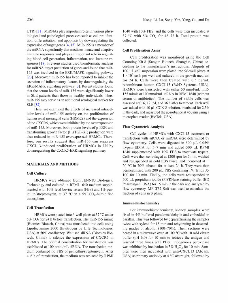
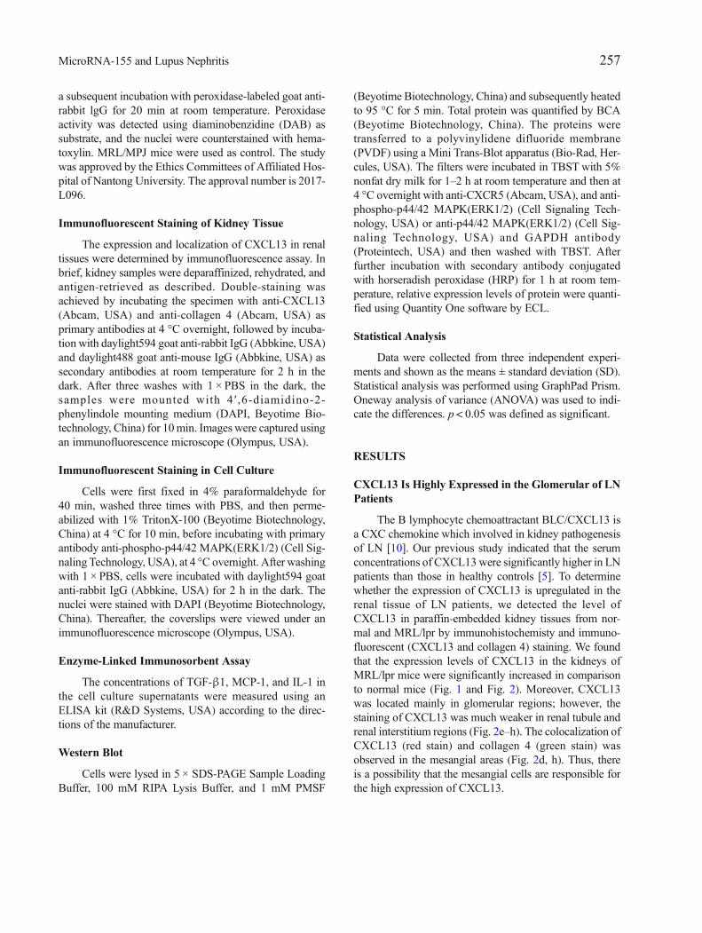
The B lymphocyte chemoattractant BLC/CXCL13 isa CXC chemokine which involved in kidney pathogenesisof LN [10]. Our previous study indicated that the serumconcentrations of CXCL13were significantly higher in LNpatients than those in healthy controls [5]. To determinewhether the expression of CXCL13 is upregulated in therenal tissue of LN patients, we detected the level ofCXCL13 in paraffin-embedded kidney tissues from nor-mal and MRL/lpr by immunohistochemisty and immuno-fluorescent (CXCL13 and collagen 4) staining. We foundthat the expression levels of CXCL13 in the kidneys ofMRL/lpr mice were significantly increased in comparisonto normal mice (Fig. 1 and Fig. 2). Moreover, CXCL13was located mainly in glomerular regions; however, thestaining of CXCL13 was much weaker in renal tubule andrenal interstitium regions (Fig. 2e–h). The colocalization ofCXCL13 (red stain) and collagen 4 (green stain) wasobserved in the mesangial areas (Fig. 2d, h). Thus, thereis a possibility that the mesangial cells are responsible forthe high expression of CXCL13.
257MicroRNA-155 and Lupus Nephritis
Increasing the Abundance of miR-155 ReducesCXCL13-Induced Proliferation of HRMCs
The level of miR-155 in the serum of SLE patientswas significantly lower than that of healthy individuals[32], suggesting a protective role of miR-155 in reducingthe proliferation of mesangial cells, endothelial cells, orpodocytes in LN patients.We raised the abundance ofmiR-155 in HRMCs by transfection with a miR-155 mimic andthen used the CCK8 kit to examine the effects of theelevated miR-155 on the growth of the cells. The HRMCstransfected with a miR-155 mimic or transfected withsiRNA to silence CXCR5were then treated with or withoutCXCL13, and cell variability was then detected after 0, 6,12, 24, and 36 h.
We observed that CXCL13 treatment could signifi-cantly increase the cell proliferation in HRMCs (Fig. 3a);however, overexpression of miR-155 could significantlyabrogate CXCL13-stimulated cell proliferation. No
significant difference in the growth rates was observedbetween non-treated cells and miR-155 mimic-treated cells(Fig. 3a). Moreover, transfection of siRNA to silenceCXCR5 could also reverse CXCL13-stimulated HMRCproliferation (Fig. 3b). The results led us to hypothesizethat miR-155 may decrease the ability of CXCL13-treatedHRMCs to proliferate by downregulating CXCR5.
Flow cytometric analysis was used to determine thefraction of cells in S phase (Fig. 3c–g) during cell prolifer-ation. Consistently, we found that CXCL13 could signifi-cantly increase the percentage of cells in S phase comparedwith non-treated controls (76.09 ± 1.44 vs. 68.88 ± 0.70)(Fig. 3e, c). Increasing the abundance of miR-155 inCXCL13-treated HRMCs reduced the percentage of Sphase from 76.09 ± 1.44 to 62.03 ± 0.3, which was similarto that observed after CXCR5-silencing (65.46 ± 0.05), seeFig. 3(e–g) and Table 1. No significant difference in thegrowth rates was observed between non-treated cells andmiR-155 mimic-treated cells (Fig. 3c, d). Briefly, the
Fig. 1. Detection of CXCL13 in mouse kidney tissues using immunochemical staining. Staining of CXCL13 in MRL/lpr mice (a) and control (b). Originalmagnification, × 400.
Fig. 2. Detection of CXCL13 and collagen 4 expression in renal tissues by immunofluorescent microscopy. Colocalization of CXCL13 (red) and collagen 4(green) merged to yellow in images. (a–d) Low expression of CXCL13 in glomerulus, renal tubules, and renal interstitium in control mice. (e–h) Highexpression of CXCL13 in glomerulus of MRL/lpr mice. (d, h) The indicated areas in Figs. C and D showing the location of CXCL13 and collagen 4 in theglomerulus. Original magnification, × 400.
258 Kong, Li, Lu, Song, Yan, Yang, Gu, and Da
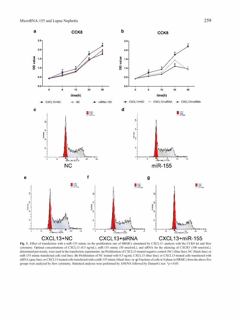
Fig. 3. Effect of transfection with a miR-155 mimic on the proliferation rate of HRMCs stimulated by CXCL13: analysis with the CCK8 kit and flowcytometry. Optimal concentrations of CXCL13 (0.5 ng/mL), miR-155 mimic (50 nmol/mL), and siRNA for the silencing of CXCR5 (100 nmol/mL),determined previously, were used in the transfection experiments. (a) Proliferation of CXCL13-treated negative control (NC) (blue line); NC (black line); ormiR-155 mimic-transfected cells (red line). (b) Proliferation of NC treated with 0.5 ng/mL CXCL13 (blue line); or CXCL13-treated cells transfected withsiRNA (gray line); or CXCL13-treated cells transfected with a miR-155 mimic (black line). (c–g) Fractions of cells in S phase in HRMCs from the above fivegroups were analyzed by flow cytometry. Statistical analyses were performed by ANOVA followed by Dunnett’s test. *p < 0.05.
259MicroRNA-155 and Lupus Nephritis
results of the CCK8 tests and flow cytometric analysisshowed that miR-155 could reduce the proliferation ofHRMCs by downregulating CXCR5.
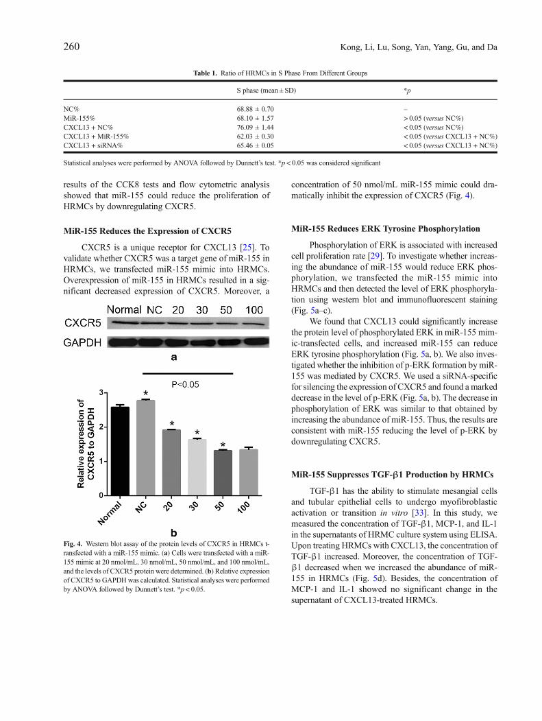
MiR-155 Reduces the Expression of CXCR5
CXCR5 is a unique receptor for CXCL13 [25]. Tovalidate whether CXCR5 was a target gene of miR-155 inHRMCs, we transfected miR-155 mimic into HRMCs.Overexpression of miR-155 in HRMCs resulted in a sig-nificant decreased expression of CXCR5. Moreover, a
concentration of 50 nmol/mL miR-155 mimic could dra-matically inhibit the expression of CXCR5 (Fig. 4).
MiR-155 Reduces ERK Tyrosine Phosphorylation
Phosphorylation of ERK is associated with increasedcell proliferation rate [29]. To investigate whether increas-ing the abundance of miR-155 would reduce ERK phos-phorylation, we transfected the miR-155 mimic intoHRMCs and then detected the level of ERK phosphoryla-tion using western blot and immunofluorescent staining(Fig. 5a–c).
We found that CXCL13 could significantly increasethe protein level of phosphorylated ERK in miR-155 mim-ic-transfected cells, and increased miR-155 can reduceERK tyrosine phosphorylation (Fig. 5a, b). We also inves-tigated whether the inhibition of p-ERK formation by miR-155 was mediated by CXCR5. We used a siRNA-specificfor silencing the expression of CXCR5 and found a markeddecrease in the level of p-ERK (Fig. 5a, b). The decrease inphosphorylation of ERK was similar to that obtained byincreasing the abundance of miR-155. Thus, the results areconsistent with miR-155 reducing the level of p-ERK bydownregulating CXCR5.
MiR-155 Suppresses TGF-β1 Production by HRMCs
TGF-β1 has the ability to stimulate mesangial cellsand tubular epithelial cells to undergo myofibroblasticactivation or transition in vitro [33]. In this study, wemeasured the concentration of TGF-β1, MCP-1, and IL-1in the supernatants of HRMC culture system using ELISA.Upon treating HRMCs with CXCL13, the concentration ofTGF-β1 increased. Moreover, the concentration of TGF-β1 decreased when we increased the abundance of miR-155 in HRMCs (Fig. 5d). Besides, the concentration ofMCP-1 and IL-1 showed no significant change in thesupernatant of CXCL13-treated HRMCs.
Table 1. Ratio of HRMCs in S Phase From Different Groups
S phase (mean ± SD) *p
NC% 68.88 ± 0.70 –MiR-155% 68.10 ± 1.57 > 0.05 (versus NC%)CXCL13 + NC% 76.09 ± 1.44 < 0.05 (versus NC%)CXCL13 + MiR-155% 62.03 ± 0.30 < 0.05 (versus CXCL13 + NC%)CXCL13 + siRNA% 65.46 ± 0.05 < 0.05 (versus CXCL13 + NC%)
Statistical analyses were performed by ANOVA followed by Dunnett’s test. *p < 0.05 was considered significant
Fig. 4. Western blot assay of the protein levels of CXCR5 in HRMCs t-ransfected with a miR-155 mimic. (a) Cells were transfected with a miR-155 mimic at 20 nmol/mL, 30 nmol/mL, 50 nmol/mL, and 100 nmol/mL,and the levels of CXCR5 protein were determined. (b) Relative expressionof CXCR5 to GAPDHwas calculated. Statistical analyses were performedby ANOVA followed by Dunnett’s test. *p < 0.05.
260 Kong, Li, Lu, Song, Yan, Yang, Gu, and Da

Fig. 5. Phosphorylation of ERK in HMRCs and measurement of the concentration of TGF-β1 in supernatants of HRMCs using ELISA. (a–c) Western blotand immunofluorescent analysis in HRMCs transfected with siRNA or miR-155 mimic with or without CXCL13 treatment. (b) Relative expression ofCXCR5 toGAPDHwas calculated. (c) Immunofluorescentmeasurement of the tyrosine phosphorylation level in ERK (red). (d) The HRMCswere incubatedfor 12 h after treatment, and the supernatants were then collected for detecting TGF-β1 levels. Statistical analyses were performed by ANOVA followed byDunnett’s test. *p < 0.05 (NC versus CXCL13 + NC, miR-155, or siRNA); **p < 0.05 (CXCL13 + NC versus CXCL13 + miR-155 or CXCL13 + siRNA).
261MicroRNA-155 and Lupus Nephritis

DISCUSSION
MiR-155 is an important effector in immune systemwhich could regulate innate and adaptive immune responses[9, 17]. Increasing evidence indicates that many miRNAssuch as miR-146, miR-21, and miR-155 have the ability tonegatively regulate the activation of inflammatory pathwaysin myeloid cells, suggesting they have anti-inflammatoryeffects [20]. Previous studies have shown that mice withmiR-155 gene deficiency have functional defects in T and Blymphocytes and other immune cells [30] Dysregulated miR-155 expression can cause serious complications in the im-mune system. The expression of miR-155 is induced inmultiple sclerosis (MS), rheumatoid arthritis (RA), andSjögren’s syndrome [28]. In contrast, in SLE patients, serummiR-155 levels were found to be decreased compared withthat in healthy controls and positively correlated with estimat-ed glomerular filtration rate (eGFR) [32]. Up to date, very fewstudies focused on the role of miR-155 in inflammatoryresponses in HRMCs. Mesangial cells, endothelial cells, andpodocytes are three main cell types in the glomerulus [35].Mesangial cells and their matrix form the central stalk of theglomerulus and interact closely with endothelial cells andpodocytes [14]. Aberrant proliferation, apoptosis, and activa-tion of mesangial cells are frequently observed in LN [27]. Inthis study, we found that CXCL13 increased HRMCs prolif-eration, while overexpression of miR-155 could abrogate theCXCL13-stimulated cell proliferation.
MiRNA-mediated gene regulation usually reduces theamount of target proteins [22]. To find the target of miR-155, we transfectedmiR-155mimic into HRMCs and foundthe decreased levels of CXCR5.MiR-155 is a component ofthe inflammatory response and regulates the ERK-MAPKsignaling pathway in T cells [24]. However, thus far therehas been no information regarding the relationship betweenERK and miR-155 in HRMCs. P-ERK can phosphorylate awide range of cellular substrate, modulates the transcription-al activity of the cell, and triggers cell growth and differen-tiation [13, 21]. We transfected a miR-155 mimic or siRNAin HRMCs and found decreased phosphorylation of ERKand inhibited cell proliferation rate. These results suggestthat the decrease in p-ERK formation was due to the inhi-bition of CXCR5. But CXCR5 expression was only partial-ly suppressed in miR-155 mimic-transfected HRMCs, sug-gesting that there may be an alternative pathway indepen-dent of miR-155 and CXCR5.
TGF-β1 belongs to a family of cytokines involved inmany physiological processes, including growth, differentia-tion, proliferation, tissue remodeling, and wound healing [8].In LN, TGF-β1 induces mesangial cells to undergo
myofibroblastic activation or transition. When we treatedHRMCs with CXCL13, the concentration of TGF-β1 in-creased; conversely, when we transfected the miR-155 mimicinto HRMCs, the level of TGF-β1 decreased. These resultsindicated that CXCL13 may promote myofibroblastic activa-tion of HRMCs, but miR-155 mimic abrogates this process.
In summary, our study has shown that miR-155 canreduce the proliferation of HRMCs and the production ofTGF-β1 by downregulating the expression of the CXCR5-ERK signaling pathway upon CXCL13 stimulation. Ourfindings suggest that miR-155 is involved in the diseasepathogenesis of LN and may be further validated as a newtherapeutic target for treating LN.
FUNDING INFORMATION
This work was supported by the National Natural Sci-ence Foundation of China (81601410); the Jiangsu ProvincialCommission of Health and Family Planning (H201623); andthe Special Fund for Clinical Medical Science and Technol-ogy of Nantong (HS2014071, HS2016003).
COMPLIANCEWITH ETHICAL STANDARDS
Conflict of Interest. The authors declare that there are noconflicts of interest.
Ethical Approval. The study was approved by the EthicsCommittees of Affiliated Hospital of Nantong University.The approval number is 2017-L096.
Open Access This article is distributed under the terms ofthe Creative Commons Attribution 4.0 International License(http://creativecommons.org/licenses/by/4.0/), which per-mits unrestricted use, distribution, and reproduction in anymedium, provided you give appropriate credit to the originalauthor(s) and the source, provide a link to the CreativeCommons license, and indicate if changes were made.
REFERENCES
1. Almaani, S., A. Meara, and B.H. Rovin. 2017. Update on lupusnephritis. Clinical Journal of the American Society of Nephrology12 (5): 825–835. https://doi.org/10.2215/cjn.05780616.
2. Anders, H.J., and A.B. Fogo. 2014. Immunopathology of lupusnephritis. Seminars in Immunopathology 36 (4): 443–459. https://doi.org/10.1007/s00281-013-0413-5.
3. Chen, S., and B.A. Smith. 2015. MicroRNA-155-deficient dendriticcells cause less severe GVHD through reduced migration and
262 Kong, Li, Lu, Song, Yan, Yang, Gu, and Da
defective inflammasome activation. 126 (1): 103–112. https://doi.org/10.1182/blood-2014-12-617258.
4. Connerty, P., A. Ahadi, and G. Hutvagner. 2015. RNA bindingproteins in the miRNA pathway. International Journal of MolecularSciences 17 (1). https://doi.org/10.3390/ijms17010031.
5. Da, Z., and L. Li. 2016. CXCL13 promotes proliferation of mesangialcells by combination with CXCR5 in SLE. 2016: 2063985.
6. Davidson, A., and C. Aranow. 2010. Lupus nephritis: lessons frommurine models. Nature Reviews Rheumatology 6 (1): 13–20. https://doi.org/10.1038/nrrheum.2009.240.
7. Devarapu, S.K., and H.J. Anders. 2018. Toll-like receptors in lupusnephritis. 25 (1): 35. https://doi.org/10.1186/s12929-018-0436-2.
8. Fujio, K., T. Komai, M. Inoue, K. Morita, T. Okamura, and K.Yamamoto. 2016. Revisiting the regulatory roles of the TGF-betafamily of cytokines. Autoimmunity Reviews 15 (9): 917–922. https://doi.org/10.1016/j.autrev.2016.07.007.
9. Garo, L.P., and G. Murugaiyan. 2016. Contribution of microRNAsto autoimmune diseases. Cellular and Molecular Life Sciences 73(10): 2041–2051. https://doi.org/10.1007/s00018-016-2167-4.
10. Hafez, S.S., S. Saad Wel, and N.H. Shedid. 2014. B-cell-attractingchemokine-1 (BCA-1/CXCL13) in systemic lupus erythematosus,its correlation to disease activity and renal involvement. The Egyp-tian Journal of Immunology 21 (2): 23–32.
11. Hahn, B.H., M.A. McMahon, A. Wilkinson, W.D. Wallace, D.I.Daikh, J.D. Fitzgerald, G.A. Karpouzas, et al. 2012. AmericanCollege of Rheumatology guidelines for screening, treatment, andmanagement of lupus nephritis.Arthritis Care Res (Hoboken) 64 (6):797–808. https://doi.org/10.1002/acr.21664.
12. Han, X., Y. Wang, X. Zhang, Y. Qin, B. Qu, L. Wu, J. Ma, Z. Zhou, J.Qian,M. Dai, Y. Tang, E.K.L. Chan, J.B. Harley, S. Zhou, and N. Shen.2016. MicroRNA-130b ameliorates murine lupus nephritis throughtargeting the type I interferon pathway on renal mesangial cells. Arthritis& Rhematology 68 (9): 2232–2243. https://doi.org/10.1002/art.39725.
13. Imperial, R., O.M. Toor, A. Hussain, J. Subramanian, and A.Masood. 2017. Comprehensive pancancer genomic analysis reveals(RTK)-RAS-RAF-MEK as a key dysregulated pathway in cancer:its clinical implications. Seminars in Cancer Biology. https://doi.org/10.1016/j.semcancer.2017.11.016.
14. Kurihara,H., andT. Sakai. 2017.Cell biology ofmesangial cells: the thirdcell that maintains the glomerular capillary. Anatomical Science Interna-tional 92 (2): 173–186. https://doi.org/10.1007/s12565-016-0334-1.
15. Lorenzen, J.M., H. Haller, and T. Thum. 2011. MicroRNAs asmediators and therapeutic targets in chronic kidney disease. NatureReviews. Nephrology 7 (5): 286–294. https://doi.org/10.1038/nrneph.2011.26.
16. Luan, J., C. Jiao, W. Kong, J. Fu, W. Qu, Y. Chen, X. Zhu, et al.2018. circHLA-C plays an important role in lupus nephritis bysponging miR-150. Molecular Therapy-Nucleic Acids 10: 245–253. https://doi.org/10.1016/j.omtn.2017.12.006.
17. Ma, Xiaodong, Lindsey E. Becker Buscaglia, Juanita R. Barker, andYong Li. 2011. MicroRNAs in NF-kB signaling. Journal of Molec-ular Cell Biology 3 (3): 159–166.
18. Mashima, R. 2015. Physiological roles of miR-155. Immunology145 (3): 323–333. https://doi.org/10.1111/imm.12468.
19. Mok, C.C. 2015. Mycophenolate mofetil for lupus nephritis: anupdate. Expert Review of Clinical Immunology 11 (12): 1353–1364. https://doi.org/10.1586/1744666x.2015.1087314.
20. O’Connell, R.M., D.S. Rao, A.A. Chaudhuri, and D. Baltimore.2010. Physiological and pathological roles for microRNAs in theimmune system. Nature Reviews. Immunology 10 (2): 111–122.https://doi.org/10.1038/nri2708.
21. Oh, S.E., and M.M. Mouradian. 2018. Cytoprotective mechanismsof DJ-1 against oxidative stress through modulating ERK1/2 and
ASK1 signal transduction. Redox Biology 14: 211–217. https://doi.org/10.1016/j.redox.2017.09.008.
22. Patel, V., and L. Noureddine. 2012. MicroRNAs and fibrosis. Cur-rent Opinion in Nephrology and Hypertension 21 (4): 410–416.https://doi.org/10.1097/MNH.0b013e328354e559.
23. Qayum, A.A., A. Paranjape, and D. Abebayehu. 2016. IL-10-inducedmiR-155 targets SOCS1 to enhance IgE-mediated mast cell function.196 (11): 4457–4467. https://doi.org/10.4049/jimmunol.1502240.
24. Rouquette-Jazdanian, A.K., R.L. Kortum, W. Li, R.K. Merrill, P.H.Nguyen, L.E. Samelson, and C.L. Sommers. 2015. miR-155 controlslymphoproliferation in LATmutant mice by restraining T-cell apoptosisvia SHIP-1/mTOR and PAK1/FOXO3/BIM pathways. PLoS One 10(6): e0131823. https://doi.org/10.1371/journal.pone.0131823.
25. Schiffer, L., K. Worthmann, H. Haller, and M. Schiffer. 2015.CXCL13 as a new biomarker of systemic lupus erythematosus andlupus nephritis - from bench to bedside? Clinical and ExperimentalImmunology 179 (1): 85–89. https://doi.org/10.1111/cei.12439.
26. Schwartz, N., B. Goilav, and C. Putterman. 2014. The pathogenesis,diagnosis and treatment of lupus nephritis. Current Opinion inRheumatology 26 (5): 502–509. https://doi.org/10.1097/bor.0000000000000089.
27. Seret, G., Y. Le Meur, Y. Renaudineau, and P. Youinou. 2012.Mesangial cell-specific antibodies are central to the pathogenesisof lupus nephritis. Clinical and Developmental Immunology 2012:579670. https://doi.org/10.1155/2012/579670.
28. Singh, R.P., I. Massachi, S. Manickavel, S. Singh, N.P. Rao, S.Hasan, D.K. Mc Curdy, S. Sharma, D. Wong, B.H. Hahn, and H.Rehimi. 2013. The role of miRNA in inflammation and autoimmu-nity. Autoimmunity Reviews 12 (12): 1160–1165. https://doi.org/10.1016/j.autrev.2013.07.003.
29. Sun, Y., W.Z. Liu, T. Liu, X. Feng, N. Yang, and H.F. Zhou. 2015.Signaling pathway of MAPK/ERK in cell proliferation, differentia-tion, migration, senescence and apoptosis. Journal of Receptor andSignal Transduction Research 35 (6): 600–604. https://doi.org/10.3109/10799893.2015.1030412.
30. Thai, T.H., H.C. Patterson, D.H. Pham, K. Kis-Toth, D.A. Kaminski,and G.C. Tsokos. 2013. Deletion of microRNA-155 reduces auto-antibody responses and alleviates lupus-like disease in the Fas(lpr)mouse. Proceedings of the National Academy of Sciences of theUnited States of America 110 (50): 20194–20199. https://doi.org/10.1073/pnas.1317632110.
31. Towler, B.P., C.I. Jones, and S.F. Newbury. 2015. Mechanisms ofregulation of mature miRNAs. Biochemical Society Transactions 43(6): 1208–1214. https://doi.org/10.1042/bst20150157.
32. Wang, G., L.S. Tam, E.K. Li, B.C. Kwan, K.M. Chow, C.C. Luk,P.K. Li, and C.C. Szeto. 2010. Serum and urinary cell-freemiR-146aand miR-155 in patients with systemic lupus erythematosus. TheJournal of Rheumatology 37 (12): 2516–2522. https://doi.org/10.3899/jrheum.100308.
33. Wang, B., J.C. Jha, S. Hagiwara, A.D. McClelland, K. Jandeleit-Dahm, M.C. Thomas, M.E. Cooper, and P. Kantharidis. 2014.Transforming growth factor-beta1-mediated renal fibrosis is depen-dent on the regulation of transforming growth factor receptor 1expression by let-7b. Kidney International 85 (2): 352–361.https://doi.org/10.1038/ki.2013.372.
34. Wang, H., J. Wang, and Y. Xia. 2017. Defective suppressor ofcytokine signaling 1 signaling contributes to the pathogenesis ofsystemic lupus erythematosus. Frontiers in Immunology 8: 1292.https://doi.org/10.3389/fimmu.2017.01292.
35. Yung, S., and T.M. Chan. 2017. Anti-dsDNA antibodies and resi-dent renal cells - their putative roles in pathogenesis of renal lesionsin lupus nephritis. Clinical Immunology 185: 40–50. https://doi.org/10.1016/j.clim.2016.09.002.
263MicroRNA-155 and Lupus Nephritis