Expansion, restimulation, and analysis of multivirus ... · final T cell product for the in vitro...
6
Material for feeder cell preparation PepTivator high-throughput (HT) plates Fluorescent antibody kits Product Order no. 1 mL Leukapheresis MACSprep PBMC Isolation Kit 130-115-169 Introduction Virus-specific T cells can be enriched based on their production of IFN-γ after restimulation with the appropriate antigen by using, for example, the CliniMACS® Cytokine Capture System (IFN-gamma). This technology easily allows the enrichment of singlevirus- and multivirus-specific T cells. However, the final analysis of the reactivity of each individual virus-specific T cell population out of a multivirus-specific T cell product is very challenging, especially when the final target cell number is very low. To address these challenges, we have developed this protocol (for research use only), describing the whole workflow starting from T cell in vitro expansion to increase the cell number for analysis, restimulation with the appropriate individual antigen to trigger a specific IFN-γ response, and final flow cytometry analysis, e.g., using the MACSQuant® Analyzer 10 in combination with the Express Mode RCI_CD4CD8_h_02. Materials Buffers, cell culture media, and reagents Expansion, restimulation, and analysis of multivirus-specific T cells Product Components Order no. Rapid Cytokine Inspector (RCI) (CD4/CD8) Kit 1 mL CD4/CD8 T Cell Detection Cocktail, human: • CD3-VioBlue® (clone: BW264-56) • CD4-APC (clone: VIT4) • CD8-FITC (clone BW135/80) • CD14-PerCP (clone TÜK4) • CD20-PerCP (clone LT20) • FcR Blocking Reagent 2×200 µL Brefeldin A (100 µg/mL) 2 mL Inside Fix 2 mL Inside Perm 130-097-343 Rapid Cytokine Inspector (RCI) Anti-IFN-γ-PE 1 mL monoclonal Anti-IFN-γ-PE antibody (clone 45-15) 130-097-600 Product Comments Referred to as CliniMACS PBS/ EDTA Buffer CliniMACS buffer • TexMACS medium • Human IL-2 IS, premium grade Prepare 500 mL TexMACS medium including 50 IU/mL IL-2 TexMACS incl. IL-2 PBS (pH 7.2) containing 0.5% BSA and 2 mM EDTA Dilute MACS BSA Stock Solution (# 130-091-376) 1:20 with autoMACS® Rinsing Solution (# 130-091-222). Keep buffer cold (2–8 °C) PBS/BSA/EDTA buffer Product Order no. PepTivator CMV pp65 (HT) – premium grade, human 130-097-727 PepTivator AdV5 Hexon (HT) – premium grade, human 130-098-237 PepTivator EBV consensus (HT) – premium grade, human 130-119-472 Control plate (8×12) 130-098-235
Transcript of Expansion, restimulation, and analysis of multivirus ... · final T cell product for the in vitro...

Material for feeder cell preparation
PepTivator high-throughput (HT) plates
Fluorescent antibody kits
Product Order no.
1 mL Leukapheresis
MACSprep PBMC Isolation Kit 130-115-169
IntroductionVirus-specific T cells can be enriched based on their production of IFN-γ after restimulation with the appropriate antigen by using, for example, the CliniMACS® Cytokine Capture System (IFN-gamma). This technology easily allows the enrichment of singlevirus- and multivirus-specific T cells. However, the final analysis of the reactivity of each individual virus-specific T cell population out of a multivirus-specific T cell product is very challenging, especially when the final target cell number is very low. To address these challenges, we have developed this protocol (for research use only), describing the whole workflow starting from T cell in vitro expansion to increase the cell number for analysis, restimulation with the appropriate individual antigen to trigger a specific IFN-γ response, and final flow cytometry analysis, e.g., using the MACSQuant® Analyzer 10 in combination with the Express Mode RCI_CD4CD8_h_02.
Materials
Buffers, cell culture media, and reagents
Expansion, restimulation, and analysis of multivirus-specific T cells
Product Components Order no.
Rapid Cytokine Inspector (RCI) (CD4/CD8) Kit
1 mL CD4/CD8 T Cell Detection Cocktail, human:• CD3-VioBlue®
(clone: BW264-56)• CD4-APC (clone: VIT4)• CD8-FITC (clone BW135/80)• CD14-PerCP (clone TÜK4)• CD20-PerCP (clone LT20)• FcR Blocking Reagent2×200 µL Brefeldin A (100 µg/mL)2 mL Inside Fix2 mL Inside Perm
130-097-343
Rapid Cytokine Inspector (RCI) Anti-IFN-γ-PE
1 mL monoclonal Anti-IFN-γ-PE antibody (clone 45-15)
130-097-600
Product Comments Referred to as
CliniMACS PBS/EDTA Buffer
CliniMACS buffer
• TexMACS medium
• Human IL-2 IS, premium grade
Prepare 500 mL TexMACS medium including 50 IU/mL IL-2
TexMACS incl. IL-2
PBS (pH 7.2) containing 0.5% BSA and 2 mM EDTA
Dilute MACS BSA Stock Solution(# 130-091-376) 1:20 with autoMACS® Rinsing Solution (# 130-091-222). Keep buffer cold (2–8 °C)
PBS/BSA/EDTA buffer
Product Order no.
PepTivator CMV pp65 (HT) – premium grade, human
130-097-727
PepTivator AdV5 Hexon (HT) – premium grade, human
130-098-237
PepTivator EBV consensus (HT) – premium grade, human
130-119-472
Control plate (8×12) 130-098-235

2
Equipment and disposables
Material Order no.
MACSQuant Analyzer 10 130-096-343
MACSQuantify Software (Software Version 2.11 patch 2-4 or higher (required for the Virus-Specif-ic T Cell - CCS - Express Mode Package))
Virus-Specific T Cell - CCS - Express Mode Package or Express Mode RCI_CD4CD8_h_02
160-002-372
Chill 5 Rack 130-092-951
Pipette tips, appropriate sizes
Combitips, appropriate sizes
12×75 mm FACS tubes
15 mL conical tubes
24-well plate
Micropipettes
Vortex mixer
Refrigerator
CO2 incubator
Orbital shaker
* Contact your Miltenyi Biotec technical representative for any question.
MethodsAfter enrichment of the multivirus-specific T cells using the CliniMACS® Cytokine Capture System (IFN-gamma), a sample of 1×10⁵ viable antigen-specific T cells has to be taken from the final T cell product for the in vitro expansion assay. The in vitro expansion of multivirus-specific T cells is performed together with irradiated feeder cells. The preparation of feeder cells, the co-culture conditions with the T cells and the fluorescent analysis using the MACSQuant® 10 Analyzer and the Express Mode RCI_CD4CD8_h_02 are described in the following section.
In vitro expansion of virus-specific T cells
Preparation of PBMCs from leukapheresis (LP) using the MACSprep™ PBMC Isolation Kit, humanFeeder cells are prepared from irradiated PBMCs out of 1 mL of LP material.
1. Wash 1 mL of LP twice with 20 mL CliniMACS buffer (300×g, 10 min).
2. Resuspend cells in 5 mL CliniMACS buffer.
3. Pipette 4 mL of PBMC Isolation Buffer into a 15 mL tube.
4. Add 200 µL of MACSprep™ PBMC Isolation Cocktail.
5. Add 400 µL of MACSprep Anti-Biotin MicroBeads and mix by vortexing.
6. Add washed LP to the suspension.
7. Close tube tightly and invert gently three times.
8. Place the tube in an upright position in, e.g., a tube rack and incubate for 3 min at room temperature.
9. Centrifuge for 3 min at 50×g for erythrocyte sedimentation.
10. Prepare LS Column by rinsing with 2 mL of buffer. Discard effluent and change collection tube. For details refer to the LS Column data sheet.
11. After erythrocytes have sedimented, carefully collect the supernatant and apply instantly onto the prepared LS Column. Collect flow-through containing unlabeled target cells.
Note: Leave a residual volume of supernatant (approximately 1–2 mm above erythrocyte pellet) to avoid unintended aspiration of erythrocytes.
12. Wash column with 1×3 mL of CliniMACS buffer. Collect unlabeled cells that pass through and combine with the effluent from step 11.
Note: Perform washing steps by adding buffer aliquots as soon as the column reservoir is empty.
13. Wash cells with TexMACS™ medium (200×g, 10min).
14. Discard supernatant and resuspend cells in 2 mL TexMACS medium.
15. Take an aliquot to determine cell number.
Irradiation of feeder cells1. Irradiate cells with 40–60 Gy (3500–5000 rads) using a
cesium 137 source. Individual irradiators may vary in performance, and the optimum degree of irradiation has to be determined.
2. Resuspend irradiated feeder cells in TexMACS medium in a concentration of 1×10⁷/mL.
Co-culture setup and maintenanceIn the following step, the virus-specific T cells are co-cultured with the irradiated feeder cells for 10–14 days in order to expand the T cells.
1. Determine the number of viable CD3+ T cells of the final T cell product. For a detailed protocol please refer to the respective Application Note.
2. Co-culture the T cells and irradiated feeder cells in a 1:100 ratio, e.g., resuspend the T cells in TexMACS medium incl. IL-2 in a concentration of 1×10⁵/mL and the irradiated feeder cells in TexMACS medium incl. IL-2 in a concentration of 1×10⁷/mL. Seed 1 mL of T cell suspension and 1 mL of irradiated feeder cell suspension per well in a 24 well plate.
Day 5–6: Split cells 1:2.
Day 7–8: Medium exchange: Remove 50% and add 50% new TexMACS medium incl. IL-2. If cells grow fast, split 1:2.
Day 9–10: Split cells 1:2.
Day 12–13: Harvest the cells and determine cell number. Re-seed cells in a concentration of 5×10⁶–1×10⁷/mL into a 24-well plate in TexMACS medium WITHOUT addition of IL-2.
Day 13 or 14 in the morning: Restimulation of T cells: refer to the next section for details.
Restimulation of T cells using the PepTivator® HT peptide poolsPepTivator® HT Peptide Pools are lyophilized peptide pools in the wells of a 96-well plate, allowing stimulation and analysis of cells directly on the plate. The convenient
96-well format, composed of 12 individually removable strips of eight wells each allows for easy and flexible experimental set-up. Different antigens can be combined by assembling different strips. A control can also be added easily by using strips from the control plate (12×8). The capacity is 0.06 nmol (0.01 μg) per peptide, per well. One well is for stimulation of up to 10⁶ total cells in 100 μL.
1. Depending on the used MACS® GMP PepTivator Peptide Pools for the generation of virus-specific T cells, prepare the respective PepTivator® HT Peptide Pools for the restimulation protocol. The following protocol describes the restimulation of CMV, AdV and EBV-specific T cells.

3
2. Prepare a PepTivator HT plate with one strip of
- PepTivator HT CMV pp65
- PepTivator HT Adv5 Hexon
- PepTivator HT EBV Consensus
- PepTivator HT Control plate
Before removing the plate from the aluminum pouch, warm it up to room temperature. Remove the necessary amount of strips. Store the remaining ones at –20 °C in the tightly sealed aluminum pouch protected from humidity.
3. Carefully resuspend the cells by pipetting up and down and transfer 100 µL of the overnight stored expanded T cells (5×10⁵–1×10⁶ T cells/100 µL) into one well of each of the strips. Perform duplicates or triplicates in case of enough cell material.
4. Incubate the cells in an incubator for 2 h at 37 °C and 5% CO₂.
5. Prepare a 1:10 dilution of the Brefeldin A solution (taken from the Rapid Cytokine Inspector (CD4/CD8) T cell Kit) in cultivation medium.
6. Add 10 µl of the diluted Brefeldin A solution to each of the wells and mix gently.
7. Incubate the cells in an incubator for another 4 h.
Flow cytometry staining of T cells using the Rapid Cytokine Inspector (RCI) (CD4/CD8 T cell) Kit and the RCI Anti-IFN-γ-PE1. Remove 80 μL of cell culture supernatant from each well.
2. Prepare a fluorescent antibody staining mix. You need 50 μL of the staining mix for each well: add 10 μL of CD4/CD8 T Cell Detection Cocktail and 10 μL of RCI Anti-IFN-γ-PE antibody to 30 μL of PBS/BSA/EDTA buffer (e. g. for four wells: 40 μL CD4/CD8 Detection Cocktail + 40 μL Anti-IFN-γ-PE + 120 μL buffer).
3. Add 50 μL of staining mix to each well.
4. Mix well for 2 min on orbital shaker.
5. Incubate for 8 min in the dark at RT.
6. Add 25 μL Inside Fix to each well.
7. Mix well for 2 min on orbital shaker.
8. Incubate for 18 min in the dark at RT.
9. Add 25 μL of 10× Inside Perm.
10. Mix well for 2 min on orbital shaker.
11. Incubate for 8 min in the dark at RT.
12. Add 100 μL of PBS/BSA/EDTA buffer.
13. Centrifuge cells at 300×g for 5 min and aspirate supernatant.
14. Resuspend cells in 200 μL PBS/BSA/EDTA buffer.
15. Analyze cells using the MACSQuant® Analyzer 10.
Data acquisition, gating strategy and resultsThe following analysis is performed using the MACSQuant Analyzer 10 and the Express Mode RCI_CD4CD8_h_02. A detailed protocol on sample acquisition and analysis using the Express Mode is described in the following section.
Fully automated flow acquisition with the MACSQuant® Analyzer 10 using the Express Mode RCI_CD4CD8_h_02
Note: Perform calibration and compensation (if necessary) of the MACSQuant Analyzer 10. Note: Do not change any instrument settings during acquisition of one experiment series.
1. Click on the Open icon and on the Instrument setting button. Choose the currently valid instrument setting from the public tab.
2. Verify that Height is switched on (click on the Advanced button in the Channels tab: Height must be selected under Features).
3. Select Chill 96 rack from the rack drop-down menu in the Experiment tab.
4. In the rack dialog box, the Chill 96 rack will be displayed. Select the appropriate number of sample positions to match the number of samples that will be used.
Note: For adaptation of experiment settings make sure that samples are activated for editing (indicated by orange rim).
5. Select the Express Mode RCI_CD4CD8_h_02: In the Settings tab, click on the Express button, then select Analysis from the Type drop-down menu and choose RCI_CD4CD8_h_02 from the Mode drop-down list. All experiment settings except sample mixing are loaded automatically. The loaded values are shown in the respective fields in the Experiment tab. These can be adapted manually if needed, for example, uptake volume. An information pop-up reminds you to select an appropriate mixing of your samples. The mixing can be chosen from the Mix sample drop-down menu at the left hand-side. Select mix medium from the Mix sample drop-down menu, as the Express Mode RCI_CD4CD8_h_02 requires a mixing of samples.
6. In the Annotations tab, replace the placeholder annotation Anti-Cyt-PE by the correct annotation Anti-IFN-g-PE in the B2 channel.
7. Fill in the Description and Sample ID for each well.
8. Check in the experiment table that the correct Sample ID matches to your samples: Select View and Experiment table… and check in the Acquisition tab if the assigned Sample IDs fit to your samples in the rack. In addition to that, check in the Settings tab that the Express Mode RCI_CD4CD8_h_02 is assigned. In case of inconsistencies, please correct.
9. Start the acquisition.
Analysis of data from samples acquired with the Express Mode RCI_CD4CD8_h_02The analysis of the data files can be performed on the MACSQuant Instrument itself or on a PC with installed MACSQuantify™ Software version 2.11 or higher.
Note: The MACSQuantify software version and the Express Mode package version must be the same on the MACSQuant instrument and on the analysis PC.
4
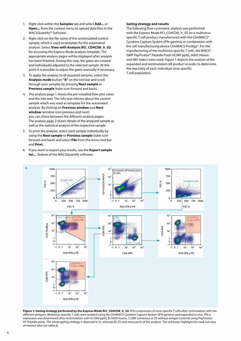
1. Right click within the Samples tab and select Add... or Open... from the context menu to upload data files to the MACSQuantify™ Software.
2. Right click on the file name of the unstimulated control sample, which is used as template for the automated analysis. Select View with Analysis.RCI_CD4CD8_h_02 for accessing the Express Mode analysis template. The appropriate analysis pages will be displayed after analysis has been finished. During this step, the gates are created and individually adjusted to the selected sample. At this point it is possible to adjust the gates manually if necessary.
3. To apply the analysis to all acquired samples, select the Analysis mode button “A” on the tool bar and scroll through your samples by pressing Next sample or Previous sample (tube icon forward and back).
4. The analysis page 1 shows the pre-installed flow plot views and the info text. The info text informs about the control sample which was used as template for the automated analysis. By clicking on Previous window and Next window (window icon previous and next) you can chose between the different analysis pages. The analysis page 2 shows details of the analyzed sample as well as the statistical analysis of the respective sample.
5. To print the analysis, select each sample individually by using the Next sample or Previous sample (tube icon forward and back) and select File from the menu tool bar and Print.
6. If you want to export your results, use the Export sample list… feature of the MACSQuantify software.
Gating strategy and resultsThe following flow cytometric analysis was performed with the Express Mode RCI_CD4CD8_h_02 on a multivirus-specific T cell product manufactured with the CliniMACS® Cytokine Capture System (IFN-gamma) in combination with the cell manufacturing device CliniMACS Prodigy®. For the manufacturing of the multivirus-specific T cells, the MACS® GMP PepTivator® Peptide Pools HCMV pp65, AdV5 Hexon and EBV Select were used. Figure 1 depicts the analysis of the expanded and restimulated cell product in order to determine the reactivity of each individual virus-specific T cell population.
A
FSC-A
FSC
-H
100000
500 750
250
500
750
1000
250 1e³-101
1e¹ 1e²0
1e³
1e²
1e¹
Anti-IFN-γ-PE
CD14
/CD20
-PerCP
-1 1
FSC-A
SSC
-A
100000
500 750
250
500
750
1000
250
1e³-101
1e¹ 1e²0
1e³
1e²
1e¹
Anti-IFN-γ-PE
CD4-APC
-1 11e³-101
1e¹ 1e²0
1e³
1e²
1e¹
Anti-IFN-γ-PE
CD3-VioBlue
-1 1
1e³-101
1e¹ 1e²0
1e³
1e²
1e¹
Anti-IFN-γ-PE
CD8-FITC
-1 1
1e³-101
1e¹ 1e²0
1e³
1e²
1e¹
CD4-APC
CD8-FITC
-1 1
CD3+ T cells CD8+ T cells
CD4+ T cells
Single cells Lymphocytes
Exclusion of monocytes/ B cells
CD3+IFN-γ+ T cells (P6)
CD8+IFN-γ+ T cells (UR8)
CD4+IFN-γ+ T cells (UR7)
Figure 1: Gating strategy performed by the Express Mode RCI_CD4CD8_h_02. IFN-γ expression of virus-specific T cells after restimulation with the different antigens. Multivirus-specific T cells were isolated using the CliniMACS Cytokine Capture System (IFN-gamma) and expanded in vitro. IFN-γ expression was determined after restimulation with A) CMV pp65, B) AdV5 hexon, C) EBV consensus or D) without antigen (control) using PepTivator HT Peptide pools. The whole gating strategy is depicted in A), whereas B)–D) only show parts of the analysis. The red boxes highlight the read-out area of interest (also see table 6).
5
B
1e³-101
1e¹ 1e²0
1e³
1e²
1e¹
Anti-IFN-γ-PE
CD3-VioBlue
-1 1 1e³-101
1e¹ 1e²0
1e³
1e²
1e¹
Anti-IFN-γ-PE
CD4-APC
-1 1
1e³-101
1e¹ 1e²0
1e³
1e²
1e¹
Anti-IFN-γ-PE
CD8-FITC
-1 1
1e³-101
1e¹ 1e²0
1e³
1e²
1e¹
CD4-APC
CD8-FITC
-1 1
CD3+ T cells CD8+ T cells
CD4+ T cells
CD3+IFN-γ+ T cells (P6)
CD8+IFN-γ+ T cells (UR8)
CD4+IFN-γ+ T cells (UR7)
1e³-101
1e¹ 1e²0
1e³
1e²
1e¹
CD4-APC
CD8-FITC
-1 1
C
1e³-101
1e¹ 1e²0
1e³
1e²
1e¹
Anti-IFN-γ-PE
CD3-VioBlue
-1 1 1e³-101
1e¹ 1e²0
1e³
1e²
1e¹
Anti-IFN-γ-PECD4-APC
-1 1
1e³-101
1e¹ 1e²0
1e³
1e²
1e¹
Anti-IFN-γ-PE
CD8-FITC
-1 1
CD3+ T cells CD3+IFN-γ+ T cells (P6)
CD8+IFN-γ+ T cells (UR8)
CD8+ T cells
CD4+IFN-γ+ T cells (UR7)
CD4+ T cells
D
CMV_FSCA vs FSCHCMV_FSCA vs FSCHCMV_FSCA vs FSCH 1e³-101
1e¹ 1e²0
1e³
1e²
1e¹
Anti-IFN-γ-PE
CD3-VioBlue
-1 1 CMV_FSCA vs FSCHCMV_FSCA vs FSCHCMV_FSCA vs FSCH 1e³-101
1e¹ 1e²0
1e³
1e²
1e¹
Anti-IFN-γ-PE
CD4-APC
-1 1
CMV_FSCA vs FSCHCMV_FSCA vs FSCHCMV_FSCA vs FSCH 1e³-101
1e¹ 1e²0
1e³
1e²
1e¹
Anti-IFN-γ-PE
CD8-FITC
-1 1
CMV_FSCA vs FSCHCMV_FSCA vs FSCHCMV_FSCA vs FSCH 1e³-101
1e¹ 1e²0
1e³
1e²
1e¹
CD4-APC
CD8-FITC
-1 1
CD3+ T cells CD3+IFN-γ+ T cells (P6)
CD8+IFN-γ+ T cells (UR8)
CD8+ T cells
CD4+IFN-γ+ T cells (UR7)
CD4+ T cells
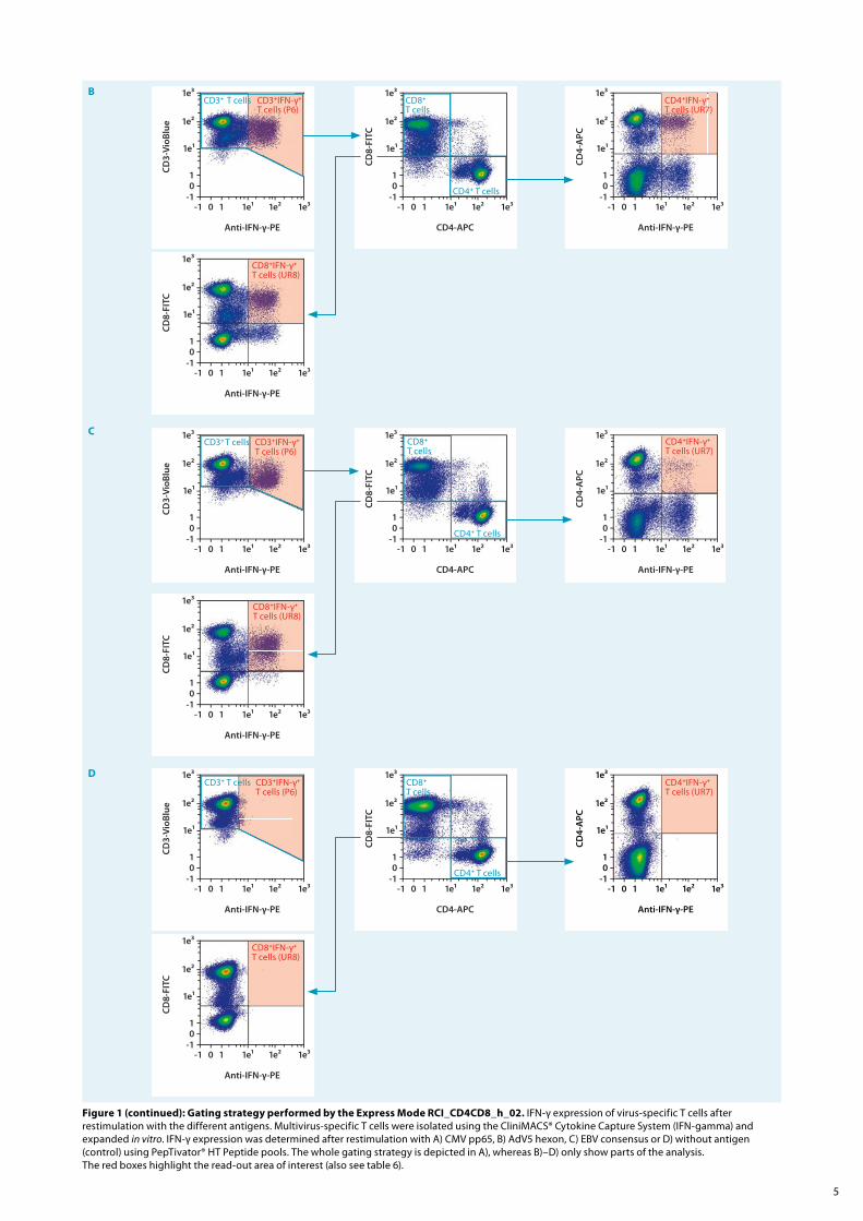
Figure 1 (continued): Gating strategy performed by the Express Mode RCI_CD4CD8_h_02. IFN-γ expression of virus-specific T cells after restimulation with the different antigens. Multivirus-specific T cells were isolated using the CliniMACS® Cytokine Capture System (IFN-gamma) and expanded in vitro. IFN-γ expression was determined after restimulation with A) CMV pp65, B) AdV5 hexon, C) EBV consensus or D) without antigen (control) using PepTivator® HT Peptide pools. The whole gating strategy is depicted in A), whereas B)–D) only show parts of the analysis. The red boxes highlight the read-out area of interest (also see table 6).
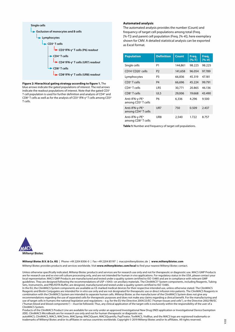
Automated analysisThe automated analysis provides the number (Count) and frequency of target cell populations among total (Freq. [%-T]) and parent cell population (Freq. [%-#]), here exemplary shown for CMV. A detailed statistical analysis can be exported as Excel format.
Single cells
Exclusion of monocytes and B cells
Lymphocytes
CD3+ T cells
CD3+IFN-γ+ T cells (P6) readout
CD4+ T cells
CD4+IFN-γ+ T cells (UR7) readout
CD8+ T cells
CD8+IFN-γ+ T cells (UR8) readout
Figure 2: Hierachical gating strategy according to figure 1. The blue arrows indicate the gated populations of interest. The red arrows indicate the readout populations of interest. Note that the gated CD3+ T cell population is used for further definition and analysis of CD4+ and CD8+ T cells as well as for the analysis of CD3+ IFN-γ+ T cells among CD3+ T cells.
Population Definition Count Freq. [%-T]
Freq. [%-#]
Single cells P1 144,861 98.225 98.225
CD14–CD20– cells P2 141,658 96.054 97.789
Lymphocytes P3 66,836 45.319 47.181
CD3+ T cells P4 66,696 45.224 99.791
CD4+ T cells LR5 30,771 20.865 46.136
CD8+ T cells UL5 29,006 19.668 43.490
Anti-IFN-γ-PE+
among CD3+ T cellsP6 6,336 4.296 9.500
Anti-IFN-γ-PE+ among CD4+ T cells
UR7 750 0.509 2.437
Anti-IFN-γ-PE+ among CD8+ T cells
UR8 2,540 1.722 8.757
Table 1: Number and frequency of target cell populations.
V.02
Miltenyi Biotec B.V. & Co. KG | Phone +49 2204 8306-0 | Fax +49 2204 85197 | [email protected] | www.miltenyibiotec.com
Miltenyi Biotec provides products and services worldwide. Visit www.miltenyibiotec.com/local to find your nearest Miltenyi Biotec contact.
Unless otherwise specifically indicated, Miltenyi Biotec products and services are for research use only and not for therapeutic or diagnostic use. MACS GMP Products are for research use and ex vivo cell culture processing only, and are not intended for human in vivo applications. For regulatory status in the USA, please contact your local representative. MACS GMP Products are manufactured and tested under a quality system certified to ISO 13485 and are in compliance with relevant GMP guidelines. They are designed following the recommendations of USP <1043> on ancillary materials. The CliniMACS® System components, including Reagents, Tubing Sets, Instruments, and PBS/EDTA Buffer, are designed, manufactured and tested under a quality system certified to ISO 13485.In the EU, the CliniMACS System components are available as CE-marked medical devices for their respective intended use, unless otherwise stated. The CliniMACS Reagents and Biotin Conjugates are intended for in vitro use only and are not designated for therapeutic use or direct infusion into patients. The CliniMACS Reagents in combination with the CliniMACS System are intended to separate human cells. Miltenyi Biotec as the manufacturer of the CliniMACS System does not give any recommendations regarding the use of separated cells for therapeutic purposes and does not make any claims regarding a clinical benefit. For the manufacturing and use of target cells in humans the national legislation and regulations – e.g. for the EU the Directive 2004/23/EC (“human tissues and cells”), or the Directive 2002/98/EC (“human blood and blood components”) – must be followed. Thus, any clinical application of the target cells is exclusively within the responsibility of the user of a CliniMACS System.Products of the CliniMACS Product Line are available for use only under an approved Investigational New Drug (IND) application or Investigational Device Exemption (IDE). CliniMACS MicroBeads are for research use only and not for human therapeutic or diagnostic use. autoMACS, CliniMACS, MACS, MACSmix, MACSprep, MACSQuant, MACSQuantify, PepTivator, TexMACS, VioBlue, and the MACS logo are registered trademarks or trademarks of Miltenyi Biotec and/or its affiliates in various countries worldwide. Copyright © 2019 Miltenyi Biotec and/or its affiliates. All rights reserved.