MODULATING EFFECT OF ESTRADIOL, IFN-γ AND T-CELLS ON ESTROGEN RECEPTORS EXPRESSION AND SENSETIVITY...
1
MODULATING EFFECT OF ESTRADIOL, IFN-γ AND T-CELLS ON ESTROGEN RECEPTORS EXPRESSION AND SENSETIVITY TO TAMOXIFEN OF BREAST CANCER CELLS IN VITRO. Perepelytsina E.M1 , Sidorenko M.V1 , Gergeliuk T.S.1 , Skachkova O.V.2 1-Department of biotechnical problems of diagnostic IPCC, Kiev, Ukraine. 2-National Cancer Institute, Kiev, Ukraine. Background Study Objectives Methods Results The fight against malignant neoplasms is one of the most pressing problems in biology and medicine. Breast cancer (BC) takes the first place in the structure of cancer incidence and cause of death of thousands women in Ukraine every year. One of the main treatments for breast cancer along with chemotherapy and radiotherapy, is a hormonal influence on sensitive cells of the tumor. 75% of all breast tumors is hormone sensitive, because express estrogen (ER) and progesterone receptors (PR). The most effective drugs are anti-estrogen substances, the most famous of them - tamoxifen (TAM). The level of PR and ER is an important parameter of effectiveness of TAM. Loss of sensitivity of tumor to TAM over time may be associated with decreasing of the number of steroid receptors. In this regard, it is necessary to find methods of influence on receptor status of cells BC. The aim of this work was to study the influence of factors of cellular microenvironment, such as estradiol (E),, interferon gamma, T-lymphocytes on expression estrogen and epidermal growth factor receptors and sensitiveness to anti-estrogen treatment. MCF-7 cells were cultured under standard conditions. For obtaining C-medium a cell line MT-4 (human cell chronic lymphocytic leukemia) was used. Recombinant IFN-γ was added to MCF-7 in concentration of 10 IU/ml, TAM - 100 nM, E –10 nM, conditioned medium (C-medium) from T-lymphocytes – dilution 1:1 with culture medium. The next samples were studied: MCF-7+C-medium, MCF-7+IFN-γ, MCF-7+E2, MCF-7+C-medium+TAM, MCF- 7+IFN-γ+TAM, MCF-7+E2+TAM. Cell survival was determined by MTT test. The distribution of the cell population between the cell cycle stages was measured using flow cytometry. Expression of ER and EGF-R was visualized by immunocytochemistry (DAKO, USA). Study design. MCF-7 cells in 2-D culture were sowing and cultured two days in full culture medium. After reseeding four groups were formed with different condition of microenvironment: 1 - control, 2 - cultivation with estradiol (10 -9 M); 3 - cultivation with C-medium from T-lymphocytes (dilution 1:1 culture medium ); 4 - incubation in pure IFN-γ (10IU / ml ). They ware substences of “the first line”: estradiol, C- mediun from T-lymphocytes, IFN gamma, tamoxifen. After two days of incubation the “second line” drugs were added to culture : tamoxifen, estradiol. After two days of incubation all samples were analyzed. Cell proliferate activity were measured by counting live and dead cells colored with trypan blue, cell survival was determined by MTT-test [Mosmann T., 1983]. Distribution of cell population by cell cycle was calculated by flow cytometry "Becton Dickinson" at λir = 488 nm and λem = 585/40 nm.. For statistical analysis processing of results and evaluation have been carried out by Student`s test. Level of ER and EGFR expression was demonstrated by immuno-cytochemistry staining (Dako, USA). Reported p values was *p ≤ 0,05 or **p ≤ 0,01. Perepelytsina O. [email protected] GO VBTPD IPCC http://www.morpho-cell.org Incubation with IFN-γ increased percent G2|M cells in suspension fraction. This tendency has been enhanced after adding TAM. C-medium stimulated the decreasing of cell percent in S phase in adhesion as well as in suspension fraction. C-medium + TAM leaded to decreasing percent of cells in G2|M in favor of G0|G1 in adhesion and S phase in suspension. And the most interesting that TAM demonstrated antiproliferation influence only on adhesion subpopulation of tumor cells. Suspension fraction in that time has G0|G1 cell subpopulation from 36%-52% and G2|M – from 9%-35%. In accordance with our data of immunocytologycal analysis IFN-γ, E2 and C-medium stimulated expression of ER and did not increased expression of EGFR. TAM had the opposite effect. It suppressed the expression of ER and stimulated EGFR. Conclusion Joint action of TAM and IFN-γ caused the decreasing in survival of MCF-7 cells in adhesive fraction by 80%. As a result: TAM increased the expression of genes, regulated by IFN-γ, on the one hand, the enhancement effect of TAM by IFN-γ in breast cancer cell culture - on the other hand. In suspension fraction we observed a slight decreasing in the percentage of alive cells. This may be due to the ability of TAM to influence the adhesive fraction to a greater extent and activation of cell proliferation in suspension fraction by interferon through amplifying the expression of ER in the cells of breast cancer. The data obtained show the influence of TAM on the adhesive fraction of cells to a greater degree than in suspension. This difference of influence may be explained by different organization tumor cell cytoskeleton, and therefore different effects of TAM on cell differentiation and proliferation. TAM induces a restructuring of β-katenin / E-cadherin complexes that are characteristic of epithelial tissues. Our data have demonstrated that cell microenvironmental conditions (hormonal and humoral) have a strong influence on ER and EGFR expression in breast cancer cell and as a result can modulate sensitiveness to treatment. Combination of antiestrogen therapy with balanced approach of IFN-γ, T-cells and level of E2/Pr may provide cumulative effect in antitumor treatment. C-medium Tamoxifen In accordance with our data C- medium (Fig. 3 A ) has demonstrated stimulation effect on cell proliferation and migration, as a result percentage of alive cells in suspension fraction increased by 40%. On the proliferation of cells in suspension fraction affects not only interferon, but also a range of proinflammatory cytokines, which are in the microenvironment. IFN-γ has a weak cytostatic effect on MCF-7 cells (Fig.3 B) due to its dual action. On the one hand, it increased the titer of ER, on the other - inhibited proliferation of tumor cells by regulating apoptosis and cell cycle. The mechanism of action of IFN-γ signaling through transduction and transcriptional activity of the JAK and STAT pathways. IFN-γ regulates the induction of cytokines (IL-s, TNF, CSF) and activates NF-κB in cells of the breast cancer. TAM has a cytotoxic effect on MCF-7 cells (Fig. 3 C ). In a suspension fraction TAM reduced the percentage of alive cells by 19%, and adhesive - 80%. Cytotoxic effect of TAM is associated with selective binding to the ER and inhibition of proliferation of ER "+" of MCF-7 cells. Incubation MCF-7 with TAM, E2+TAM and C-medium + TAM leaded to decreasing alive cells number in adhesion fraction. Control (suspension) 45% 5% 50% G0 /G1 G2/M S INF gamma (adhesion) 42% 11% 47% G0/G1 G2/M S INFgamma (suspension) 4 3% 2 1% 3 6% G0 /G1 G2 /M S Control (adhesion) 43% 14% 43% G0/G1 G2/M S C-medium+TAM (adhesion) 46% 2% 52% G0 /G 1 G2/M S C-medium+TAM (suspension) 47% 9% 44% G0/G1 G2/M S C-medium+E (adhesion) 50 % 8% 42 % G0/ G1 G2/ M S C-medium+E (suspension) 4 6% 10 % 44 % G0 /G1 G2/M S C-medium (adhesion) 6 4% 7% 29 % G 0/G 1 G 2/ M S INFgamma+TAM (adhesion) 56 % 1% 43% G0/G1 G2 /M S C-medium (suspension) 18% 24% 58% G0/G1 G2/M S IFNgamma+TAM (suspension) 29% 35% 36% G0/G1 G2/M S TAM (suspension) 22% 26% 52% G0/G1 G2/M S ТАМ (adhes ion) 50% 0% 50% G0/G1 G2/M S Figure 1 shows the number of alive and dead cells in whole culture (adhesive and suspension fractions) in total. In samples where MCF-7 cells was cultured with recombinant INFγ (sample 2) the number of alive cells increased 2.65 times in comparison with “0 point”. This level was less than control by 24%, thus IFN-γ has a weak cytostatic effect on MCF-7 cells. TAM demonstrated cytotoxic influence (Fig. 1, sample 3) and increased the percentage of dead cells in both fractions (Fig.2). C-medium + E and IFN γ + E stimulated cell proliferation, but a lot of cells died (Samples 4, 5). Samples 6, 7, 8, 9 (TAM with C- medium, IFN γ and estradiol) indicated the reduction of the alive cells number in comparison with control samples (Fig.1). To determine the cause of cell death, we analyzed the distribution of cells between the suspension and adhesion factions. In adhesion fraction we noticed decreasing alive cells number in all samples which were cultured with substance of the “first line” (Fig. 2). On the basis of data on the number of living cells ( Fig. 1) it could be assumed that actively proliferating tumor cells migrate to the suspension. That`s why in suspension fraction we observed increasing number of alive cells for samples: E2 and C-medium. But later on, part of cells possibly died by anoikis pathway due to lack of the necessary paracrine stimuli and cell-cell contacts. Decreased number of the cells in suspension fraction was demonstrated for samples with IFN-γ and TAM. The investigation of apoptosis in 2-D culture of breast cancer cells was carried out by cytofluorometry (Fig. 6). It was discovered that the percentage of cells in apoptosis has increased in suspension fraction of cells samples which were incubated with IFN-γ by 10% (sample 2), IFN-γ + E – by 21% (sample 5), TAM + E – by 20% (sample 6), IFN-γ + TAM – by 16% (sample 8), and TAM + C-medium - by 13% (sample 9). C-medium with E redused the percentage of cells in apoptosis by 12% (sample 4). In adhesive fraction TAM increased the percentage of cells in apoptosis by 10% (sample 3), C- medium + E – by 35% (sample 4), IFN-γ + E reduced the percentage of cells in apoptosis by 19% (sample 5). IFN γ Fig. 4 Expression of Estrogen receptor in samples of the MCF-7 culture. Control C-medium IFN γ Tamoxifen Fig. 5 Expression of Epidermal growth factor receptor in samples of the MCF-7 culture. * * * * * * # # # -50 -40 -30 -20 -10 0 10 20 30 40 1 2 3 4 5 6 7 8 9 % cell in apoptosis to control Suspension Adhesion Fig.6 Percent cell in apoptosis in different samples in adhesion and suspension fraction, campare with control. 1- control; 2- INFgamma; 3- ТАМ; 4- C-medium+E; 5- INFgamma+E; 6- TAM+E; 7- C-medium+TAM; 8- INFgamma+TAM; 9- TAM+C-medium * * * # # # # -100 -80 -60 -40 -20 0 20 40 60 control INF gamma TAM C- medium Estradiol Cells number changing , % to control Suspension fraction Adhesion fraction * * * * * * * * 0 200 400 600 800 1000 1200 1400 1600 0 point 1 2 3 4 5 6 7 8 9 Cells number, E03 cells /ml alive cells dead cells Fig.1. Alive\dead cells number in different condition of microenvironment. 1-control, 2- IFNγ, 3 -TAM, 4- C-medium + E, 5- IFNγ + E, 6- TAM + E, 7 - C-medium + TAM, 8 - IFNγ + TAM, 9 - TAM + C-medium. Fig.2. Percent alive cells in suspension and adhesion fraction under different condition of microenvironment compare with control. * * # # # 0 20 40 60 80 100 120 140 K 0 TAM TAM+Estradiol TAM+C- medium Cell survival, % Suspension Adhesion * # # # 0 20 40 60 80 100 120 K 0 INF gamma INF+Estradiol INF+TAM Cell survival, % Suspension Adhesion * * # # # 0 20 40 60 80 100 120 140 160 K 0 C-medium C-medium+E2 C- medium+TAM Cell survival, % Suspension Adhesion Fig.3. Percent alive cells in suspension and adhesion fraction under different condition of microenvironment compare with control. A B C Control Fig.7 Distribution cell population on cell cycle in different condition of microenvironment.
-
Upload
- -
Category
Health & Medicine
-
view
109 -
download
2
Transcript of MODULATING EFFECT OF ESTRADIOL, IFN-γ AND T-CELLS ON ESTROGEN RECEPTORS EXPRESSION AND SENSETIVITY...
MODULATING EFFECT OF ESTRADIOL, IFN-γ AND T-CELLS ON ESTROGEN RECEPTORS EXPRESSION AND SENSETIVITY TO TAMOXIFEN
OF BREAST CANCER CELLS IN VITRO.
Perepelytsina E.M1 , Sidorenko M.V1 , Gergeliuk T.S.1 , Skachkova O.V.21-Department of biotechnical problems of diagnostic IPCC, Kiev, Ukraine.2-National Cancer Institute, Kiev, Ukraine.
Background
Study Objectives
Methods
Results
The fight against malignant neoplasms is one of the most pressing problems in biology and medicine. Breast cancer (BC) takes the first place in the structure of cancer incidence and cause of death of thousands women in Ukraineevery year. One of the main treatments for breast cancer along with chemotherapy and radiotherapy, is a hormonal influence on sensitive cells of the tumor. 75% of all breast tumors is hormone sensitive, because express estrogen (ER)and progesterone receptors (PR). The most effective drugs are anti-estrogen substances, the most famous of them - tamoxifen (TAM). The level of PR and ER is an important parameter of effectiveness of TAM. Loss of sensitivity oftumor to TAM over time may be associated with decreasing of the number of steroid receptors. In this regard, it is necessary to find methods of influence on receptor status of cells BC.
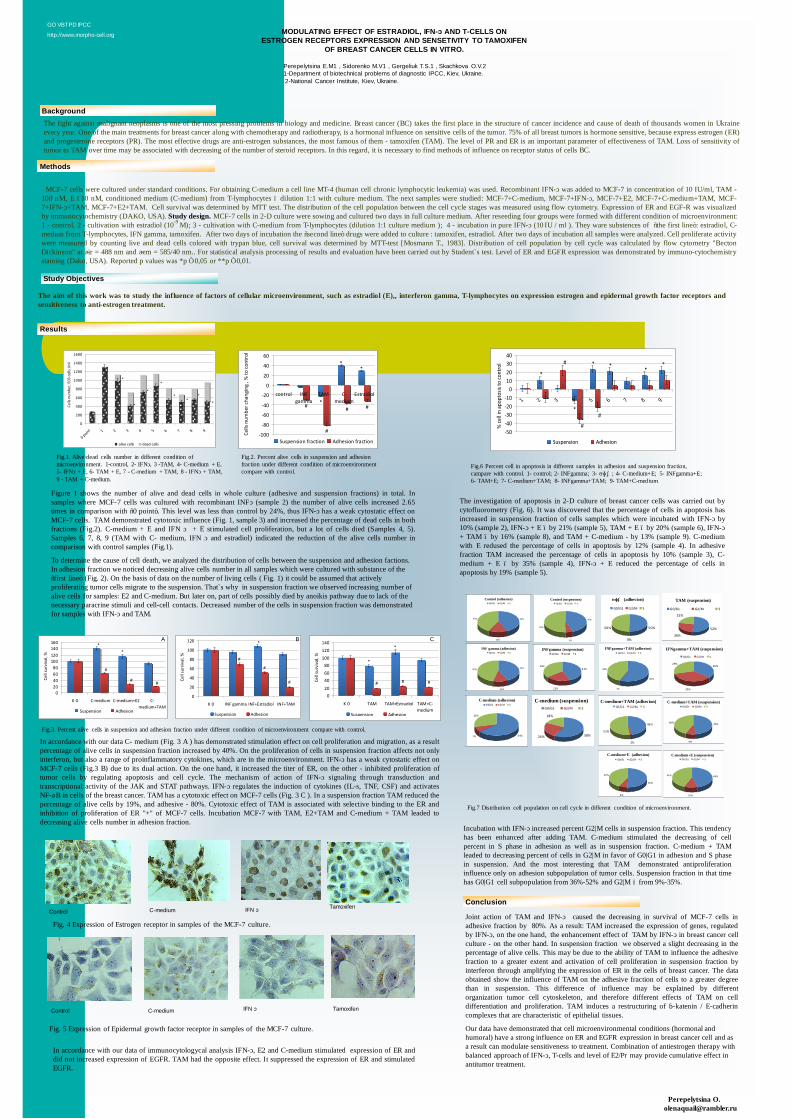
The aim of this work was to study the influence of factors of cellular microenvironment, such as estradiol (E),, interferon gamma, T-lymphocytes on expression estrogen and epidermal growth factor receptors andsensitiveness to anti-estrogen treatment.
MCF-7 cells were cultured under standard conditions. For obtaining C-medium a cell line MT-4 (human cell chronic lymphocytic leukemia) was used. Recombinant IFN-γ was added to MCF-7 in concentration of 10 IU/ml, TAM -100 nM, E –10 nM, conditioned medium (C-medium) from T-lymphocytes – dilution 1:1 with culture medium. The next samples were studied: MCF-7+C-medium, MCF-7+IFN-γ, MCF-7+E2, MCF-7+C-medium+TAM, MCF-7+IFN-γ+TAM, MCF-7+E2+TAM. Cell survival was determined by MTT test. The distribution of the cell population between the cell cycle stages was measured using flow cytometry. Expression of ER and EGF-R was visualizedby immunocytochemistry (DAKO, USA). Study design. MCF-7 cells in 2-D culture were sowing and cultured two days in full culture medium. After reseeding four groups were formed with different condition of microenvironment:1 - control, 2 - cultivation with estradiol (10-9 M); 3 - cultivation with C-medium from T-lymphocytes (dilution 1:1 culture medium ); 4 - incubation in pure IFN-γ (10 IU / ml ). They ware substences of “the first line”: estradiol, C-mediun from T-lymphocytes, IFN gamma, tamoxifen. After two days of incubation the “second line” drugs were added to culture : tamoxifen, estradiol. After two days of incubation all samples were analyzed. Cell proliferate activitywere measured by counting live and dead cells colored with trypan blue, cell survival was determined by MTT-test [Mosmann T., 1983]. Distribution of cell population by cell cycle was calculated by flow cytometry "BectonDickinson" at λir = 488 nm and λem = 585/40 nm.. For statistical analysis processing of results and evaluation have been carried out by Student`s test. Level of ER and EGFR expression was demonstrated by immuno-cytochemistrystaining (Dako, USA). Reported p values was *p ≤ 0,05 or **p ≤ 0,01.
Perepelytsina O. [email protected]
GO VBTPD IPCC
http://www.morpho-cell.org
Incubation with IFN-γ increased percent G2|M cells in suspension fraction. This tendencyhas been enhanced after adding TAM. C-medium stimulated the decreasing of cellpercent in S phase in adhesion as well as in suspension fraction. C-medium + TAMleaded to decreasing percent of cells in G2|M in favor of G0|G1 in adhesion and S phasein suspension. And the most interesting that TAM demonstrated antiproliferationinfluence only on adhesion subpopulation of tumor cells. Suspension fraction in that timehas G0|G1 cell subpopulation from 36%-52% and G2|M – from 9%-35%.
In accordance with our data of immunocytologycal analysis IFN-γ, E2 and C-medium stimulated expression of ER anddid not increased expression of EGFR. TAM had the opposite effect. It suppressed the expression of ER and stimulatedEGFR.
Conclusion
Joint action of TAM and IFN-γ caused the decreasing in survival of MCF-7 cells inadhesive fraction by 80%. As a result: TAM increased the expression of genes, regulatedby IFN-γ, on the one hand, the enhancement effect of TAM by IFN-γ in breast cancer cellculture - on the other hand. In suspension fraction we observed a slight decreasing in thepercentage of alive cells. This may be due to the ability of TAM to influence the adhesivefraction to a greater extent and activation of cell proliferation in suspension fraction byinterferon through amplifying the expression of ER in the cells of breast cancer. The dataobtained show the influence of TAM on the adhesive fraction of cells to a greater degreethan in suspension. This difference of influence may be explained by differentorganization tumor cell cytoskeleton, and therefore different effects of TAM on celldifferentiation and proliferation. TAM induces a restructuring of β-katenin / E-cadherincomplexes that are characteristic of epithelial tissues.
Our data have demonstrated that cell microenvironmental conditions (hormonal and humoral) have a strong influence on ER and EGFR expression in breast cancer cell and as a result can modulate sensitiveness to treatment. Combination of antiestrogen therapy with balanced approach of IFN-γ, T-cells and level of E2/Pr may provide cumulative effect in antitumor treatment.
C-medium Tamoxifen
In accordance with our data C- medium (Fig. 3 A ) has demonstrated stimulation effect on cell proliferation and migration, as a resultpercentage of alive cells in suspension fraction increased by 40%. On the proliferation of cells in suspension fraction affects not onlyinterferon, but also a range of proinflammatory cytokines, which are in the microenvironment. IFN-γ has a weak cytostatic effect onMCF-7 cells (Fig.3 B) due to its dual action. On the one hand, it increased the titer of ER, on the other - inhibited proliferation oftumor cells by regulating apoptosis and cell cycle. The mechanism of action of IFN-γ signaling through transduction andtranscriptional activity of the JAK and STAT pathways. IFN-γ regulates the induction of cytokines (IL-s, TNF, CSF) and activatesNF-κB in cells of the breast cancer. TAM has a cytotoxic effect on MCF-7 cells (Fig. 3 C ). In a suspension fraction TAM reduced thepercentage of alive cells by 19%, and adhesive - 80%. Cytotoxic effect of TAM is associated with selective binding to the ER andinhibition of proliferation of ER "+" of MCF-7 cells. Incubation MCF-7 with TAM, E2+TAM and C-medium + TAM leaded todecreasing alive cells number in adhesion fraction.
Control (suspension)
45%
5%
50%
G0 /G1 G2/M S
INF gamma (adhesion)
42%
11%
47%
G0/G1 G2/M SINFgamma (suspension)
43%
21%
36%
G0/G1 G2/M S
Control (adhesion)
43%
14%
43%
G0/G1 G2/M S
C-medium+TAM (adhesion)
46%
2%
52%
G0/G1 G2/M SC-medium+TAM (suspension)
47%
9%
44%
G0/G1 G2/M S
C-medium+E (adhesion)
50%
8%
42%
G0/G1 G2/M SC-medium+E (suspension)
46%
10%
44%
G0/G1 G2/M S
C-medium (adhesion)
64%7%
29%
G0/G1 G2/M S
INFgamma+TAM (adhesion)
56 %
1%
43%
G0/G1 G2 /M S
C-medium (suspension)
18%
24% 58%
G0/G1 G2/M S
IFNgamma+TAM (suspension)
29%
35%
36%
G0/G1 G2/M S
TAM (suspension)
22%
26%
52%
G0/G1 G2/M S
ТАМ (adhesion)
50%
0%
50%
G0/G1 G2/M S
Figure 1 shows the number of alive and dead cells in whole culture (adhesive and suspension fractions) in total. Insamples where MCF-7 cells was cultured with recombinant INFγ (sample 2) the number of alive cells increased 2.65times in comparison with “0 point”. This level was less than control by 24%, thus IFN-γ has a weak cytostatic effect onMCF-7 cells. TAM demonstrated cytotoxic influence (Fig. 1, sample 3) and increased the percentage of dead cells in bothfractions (Fig.2). C-medium + E and IFN γ + E stimulated cell proliferation, but a lot of cells died (Samples 4, 5).Samples 6, 7, 8, 9 (TAM with C- medium, IFN γ and estradiol) indicated the reduction of the alive cells number incomparison with control samples (Fig.1).
To determine the cause of cell death, we analyzed the distribution of cells between the suspension and adhesion factions. In adhesion fraction we noticed decreasing alive cells number in all samples which were cultured with substance of the “first line” (Fig. 2). On the basis of data on the number of living cells ( Fig. 1) it could be assumed that actively proliferating tumor cells migrate to the suspension. That`s why in suspension fraction we observed increasing number of alive cells for samples: E2 and C-medium. But later on, part of cells possibly died by anoikis pathway due to lack of the necessary paracrine stimuli and cell-cell contacts. Decreased number of the cells in suspension fraction was demonstrated for samples with IFN-γ and TAM.
The investigation of apoptosis in 2-D culture of breast cancer cells was carried out bycytofluorometry (Fig. 6). It was discovered that the percentage of cells in apoptosis hasincreased in suspension fraction of cells samples which were incubated with IFN-γ by10% (sample 2), IFN-γ + E – by 21% (sample 5), TAM + E – by 20% (sample 6), IFN-γ+ TAM – by 16% (sample 8), and TAM + C-medium - by 13% (sample 9). C-mediumwith E redused the percentage of cells in apoptosis by 12% (sample 4). In adhesivefraction TAM increased the percentage of cells in apoptosis by 10% (sample 3), C-medium + E – by 35% (sample 4), IFN-γ + E reduced the percentage of cells inapoptosis by 19% (sample 5).
IFN γ
Fig. 4 Expression of Estrogen receptor in samples of the MCF-7 culture.
Control C-medium IFN γ Tamoxifen
Fig. 5 Expression of Epidermal growth factor receptor in samples of the MCF-7 culture.
*
*
* **
*#
#
#
-50-40-30-20-10
010203040
1 2 3 4 5 6 7 8 9
% ce
ll in
apop
tosis
to co
ntro
l
Suspension Adhesion
Fig.6 Percent cell in apoptosis in different samples in adhesion and suspension fraction, campare with control. 1- control; 2- INFgamma; 3- ТАМ; 4- C-medium+E; 5- INFgamma+E; 6- TAM+E; 7- C-medium+TAM; 8- INFgamma+TAM; 9- TAM+C-medium
**
*##
#
#
-100
-80
-60
-40
-20
0
20
40
60
control INFgamma
TAM C-medium
Estradiol
Cells
num
ber c
hang
ing
, % to
cont
rol
Suspension fraction Adhesion fraction
*
*
*
*
**
*
*
0
200
400
600
800
1000
1200
1400
1600
0 point 1 2 3 4 5 6 7 8 9
Cells
num
ber,
E03
cells
/ml
alive cells dead cells
Fig.1. Alive\dead cells number in different condition of microenvironment. 1-control, 2- IFNγ, 3 -TAM, 4- C-medium + E, 5- IFNγ + E, 6- TAM + E, 7 - C-medium + TAM, 8 - IFNγ + TAM, 9 - TAM + C-medium.
Fig.2. Percent alive cells in suspension and adhesion fraction under different condition of microenvironment compare with control.
*
*
###
020406080
100120140
K 0 TAM TAM+Estradiol TAM+C-medium
Cell
surv
ival
, %
Suspension Adhesion
*
#
#
#
0
20
40
60
80
100
120
K 0 INF gamma INF+Estradiol INF+TAM
Cell
surv
ival
, %
Suspension Adhesion
**
##
#
020406080
100120140160
K 0 C-medium C-medium+E2 C-medium+TAM
Cell
surv
ival
, %
Suspension Adhesion
Fig.3. Percent alive cells in suspension and adhesion fraction under different condition of microenvironment compare with control.
A B C
Control
Fig.7 Distribution cell population on cell cycle in different condition of microenvironment.