Supplementary Information Employing in vitro metabolism to ...
Upload
shih-hsin-kanCategory
view
154download
0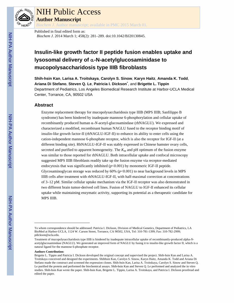
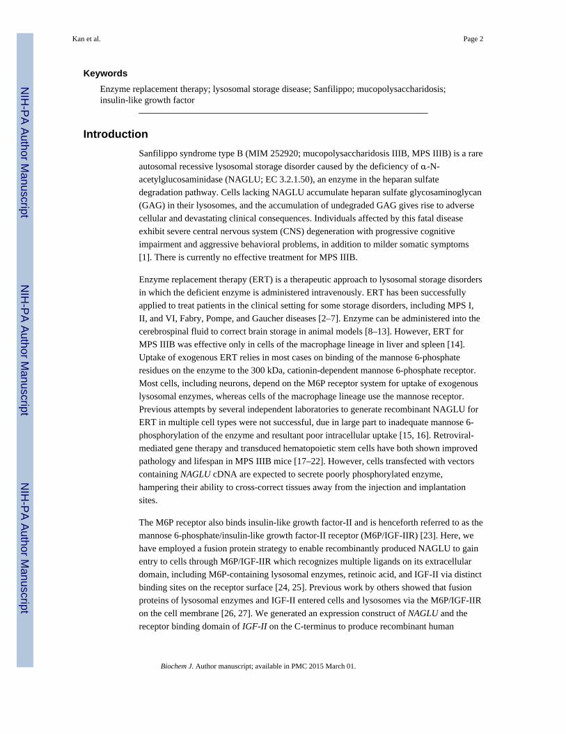
Insulin-like growth factor II peptide fusion enables uptake andlysosomal delivery of α-N-acetylglucosaminidase tomucopolysaccharidosis type IIIB fibroblasts
Shih-hsin Kan, Larisa A. Troitskaya, Carolyn S. Sinow, Karyn Haitz, Amanda K. Todd,Ariana Di Stefano, Steven Q. Le, Patricia I. Dickson*, and Brigette L. TippinDepartment of Pediatrics, Los Angeles Biomedical Research Institute at Harbor-UCLA MedicalCenter, Torrance, CA, 90502 USA
Abstract
Enzyme replacement therapy for mucopolysaccharidosis type IIIB (MPS IIIB; Sanfilippo B
syndrome) has been hindered by inadequate mannose 6-phosphorylation and cellular uptake of
recombinantly produced human α-N-acetyl-glucosamindase (rhNAGLU). We expressed and
characterized a modified, recombinant human NAGLU fused to the receptor binding motif of
insulin-like growth factor-II (rhNAGLU-IGF-II) to enhance its ability to enter cells using the
cation-independent mannose 6-phosphate receptor, which is also the receptor for IGF-II (at a
different binding site). RhNAGLU-IGF-II was stably expressed in Chinese hamster ovary cells,
secreted and purified to apparent homogeneity. The Km and pH optimum of the fusion enzyme
was similar to those reported for rhNAGLU. Both intracellular uptake and confocal microscopy
suggested MPS IIIB fibroblasts readily take up the fusion enzyme via receptor-mediated
endocytosis that was significantly inhibited (p<0.001) by monomeric IGF-II peptide.
Glycosaminoglycan storage was reduced by 60% (p<0.001) to near background levels in MPS
IIIB cells after treatment with rhNAGLU-IGF-II, with half-maximal correction at concentrations
of 3–12 pM. Similar cellular uptake mechanism via the IGF-II receptor was also demonstrated in
two different brain tumor-derived cell lines. Fusion of NAGLU to IGF-II enhanced its cellular
uptake while maintaining enzymatic activity, supporting its potential as a therapeutic candidate for
MPS IIIB.
To whom correspondence should be addressed: Patricia I. Dickson, Division of Medical Genetics, Department of Pediatrics, LABioMed at Harbor-UCLA, 1124 W. Carson Street, Torrance, CA 90502, USA, Tel: 310-781-1399; Fax: 310-782-2999;[email protected].
Treatment of mucopolysaccharidosis type IIIB is hindered by inadequate intracellular uptake of recombinantly-produced alpha-N-acetylglucosamindase (NAGLU). We generated an improved form of NAGLU by fusing it to insulin-like growth factor II, which is anatural ligand for the mannose 6-phosphate receptor.
Authors Contribution:Brigette L. Tippin and Patricia I. Dickson developed the original concept and supervised the project. Shih-hsin Kan and Larisa A.Troitskaya conceived and designed the experiments. Shihhsin Kan, Carolyn S. Sinow, Karyn Haitz, Amanda K. Todd and Ariana DiStefano made the construct and screened the expression clones. Shih-hsin Kan, Larisa A. Troitskaya, Carolyn S. Sinow and Steven Q.Le purified the protein and performed the biochemical assays. Shih-hsin Kan and Steven Q. Le performed and analyzed the in vitrostudies. Shih-hsin Kan wrote the paper. Shih-hsin Kan, Brigette L. Tippin, Larisa A. Troitskaya, and Patricia I. Dickson proofread andedited the paper.
NIH Public AccessAuthor ManuscriptBiochem J. Author manuscript; available in PMC 2015 March 01.
Published in final edited form as:Biochem J. 2014 March 1; 458(2): 281–289. doi:10.1042/BJ20130845.
NIH
-PA
Author M
anuscriptN
IH-P
A A
uthor Manuscript
NIH
-PA
Author M
anuscript
Keywords
Enzyme replacement therapy; lysosomal storage disease; Sanfilippo; mucopolysaccharidosis;insulin-like growth factor
Introduction
Sanfilippo syndrome type B (MIM 252920; mucopolysaccharidosis IIIB, MPS IIIB) is a rare
autosomal recessive lysosomal storage disorder caused by the deficiency of α-N-
acetylglucosaminidase (NAGLU; EC 3.2.1.50), an enzyme in the heparan sulfate
degradation pathway. Cells lacking NAGLU accumulate heparan sulfate glycosaminoglycan
(GAG) in their lysosomes, and the accumulation of undegraded GAG gives rise to adverse
cellular and devastating clinical consequences. Individuals affected by this fatal disease
exhibit severe central nervous system (CNS) degeneration with progressive cognitive
impairment and aggressive behavioral problems, in addition to milder somatic symptoms
[1]. There is currently no effective treatment for MPS IIIB.
Enzyme replacement therapy (ERT) is a therapeutic approach to lysosomal storage disorders
in which the deficient enzyme is administered intravenously. ERT has been successfully
applied to treat patients in the clinical setting for some storage disorders, including MPS I,
II, and VI, Fabry, Pompe, and Gaucher diseases [2–7]. Enzyme can be administered into the
cerebrospinal fluid to correct brain storage in animal models [8–13]. However, ERT for
MPS IIIB was effective only in cells of the macrophage lineage in liver and spleen [14].
Uptake of exogenous ERT relies in most cases on binding of the mannose 6-phosphate
residues on the enzyme to the 300 kDa, cationin-dependent mannose 6-phosphate receptor.
Most cells, including neurons, depend on the M6P receptor system for uptake of exogenous
lysosomal enzymes, whereas cells of the macrophage lineage use the mannose receptor.
Previous attempts by several independent laboratories to generate recombinant NAGLU for
ERT in multiple cell types were not successful, due in large part to inadequate mannose 6-
phosphorylation of the enzyme and resultant poor intracellular uptake [15, 16]. Retroviral-
mediated gene therapy and transduced hematopoietic stem cells have both shown improved
pathology and lifespan in MPS IIIB mice [17–22]. However, cells transfected with vectors
containing NAGLU cDNA are expected to secrete poorly phosphorylated enzyme,
hampering their ability to cross-correct tissues away from the injection and implantation
sites.
The M6P receptor also binds insulin-like growth factor-II and is henceforth referred to as the
mannose 6-phosphate/insulin-like growth factor-II receptor (M6P/IGF-IIR) [23]. Here, we
have employed a fusion protein strategy to enable recombinantly produced NAGLU to gain
entry to cells through M6P/IGF-IIR which recognizes multiple ligands on its extracellular
domain, including M6P-containing lysosomal enzymes, retinoic acid, and IGF-II via distinct
binding sites on the receptor surface [24, 25]. Previous work by others showed that fusion
proteins of lysosomal enzymes and IGF-II entered cells and lysosomes via the M6P/IGF-IIR
on the cell membrane [26, 27]. We generated an expression construct of NAGLU and the
receptor binding domain of IGF-II on the C-terminus to produce recombinant human
Kan et al. Page 2
Biochem J. Author manuscript; available in PMC 2015 March 01.
NIH
-PA
Author M
anuscriptN
IH-P
A A
uthor Manuscript
NIH
-PA
Author M
anuscript
NAGLU-IGF-II (rhNAGLUIGF-II). In this study, rhNAGLU-IGF-II was expressed and
purified from Chinese hamster ovary (CHO) cells for biochemical characterization and
further tested for functional delivery to MPS IIIB cells and brain tumor-derived cell lines,
and correction of GAG storage in vitro.
Experimental Procedures
Molecular cloning of NAGLU-IGF-II
An expression cassette containing the full-length cDNA coding for 743 amino acids of
human NAGLU (NM_000263.3), a short unstructured linker and the c-myc epitope
(EQKLISEED), followed by a portion of the IGF-II cDNA (NM_001007139.4) encoding
amino acids 32–91 was synthesized using codon optimization for expression in CHO cells
by Genscript USA Inc (Piscataway, NJ) and provided in the cloning vector pUC57. The
DNA fragment was subcloned into pCI-neo (Promega Corporation, Madison, WI) at the
XhoI and XbaI sites and transformed into XL1-Blue Supercompetent cells (Agilent
Technologies, Santa Clara, CA) for amplification. The resulting expression vector was
named pCI-NagGScIGF. Full length human NAGLU cDNA was provided in pCMV-
NAGLU by Dr. E. F. Neufeld (University of California, Los Angeles, CA) and was
subcloned into pCI-neo at the EcoRI and XbaI sites to form pCI-NAGLU without any other
epitopes.
Cell culture and expression lines in Chinese Hamster Ovary cells
All chemical reagents were obtained from Sigma-Aldrich (St. Louis, MO), unless otherwise
specified. CHO dhfr− cells were cultured in Ham’s F12/DME (Irvine Scientific, Irvine, CA)
supplemented with 10% fetal bovine serum (SAFC Biosciences, Lenexa, KS), 1 mM non-
essential amino acids, 1 mM sodium pyruvate, 2 mM L-glutamine, and antibiotics (100
units/ml penicillin G, 100 µg/ml streptomycin sulfate, Irvine Scientific, Irvine, CA) and 50
µg/ml gentamicin sulfate (EMD Chemicals Inc., Gibbstown, NJ) at 37°C in a 5% CO2 air
atmosphere. pCI-NagGScIGF (or pCINAGLU) was linearized with Ahd I (New England
BioLabs, Ipswich, MA) and transfected into CHO dhfr− cells using PolyFect Transfection
Reagent (Qiagen Inc., Valencia, CA). The stable lines were selected by their resistance to
700 µg/ml G-418 (EMD Chemicals Inc., Gibbstown, NJ), and colonies were formed after 7–
14 days. Individual colonies were isolated and the highestyielding expressors of secreted
rhNAGLU-IGF-II (5H10) or rhNAGLU (117-1.511) were identified by NAGLU activity
assay. Stable CHO cell clones were maintained in Ham’s F12/DME with supplements
containing 250 µg/ml G-418.
For protein production, rhNAGLU-IGF-II clone 5H10 or rhNAGLU clone 117-1.511 were
seeded into roller bottles and grown to confluence, at which time the medium was replaced
with EX-CELL PF CHO serum-free medium supplemented with 4 mM L-glutamine,
nucleosides (10 mg/l each of guanosine, adenosine, uridine, cytosine, hypoxanthine, and
thymidine), 50 µg/ml gentamicin sulfate and 250 µg/ml G-418. Secreted NAGLU activity
was monitored daily for 7–14 days until NAGLU expression reached a plateau, before the
conditioned medium was harvested for enzyme purification.
Kan et al. Page 3
Biochem J. Author manuscript; available in PMC 2015 March 01.
NIH
-PA
Author M
anuscriptN
IH-P
A A
uthor Manuscript
NIH
-PA
Author M
anuscript
Purification of modified recombinant NAGLU enzymes
rhNAGLU-IGF-II and rhNAGLU enzymes were purified from culture medium. Conditioned
medium was filtered (0.2 µm), supplemented with methyl-α-D-glucopyranoside (10 mM) and
stored at 4°C prior to purification (below).
Medium containing rhNAGLU-IGF-II was initially concentrated using an Amicon
ultrafiltration concentrator with a YM30 membrane (EMD Millipore Corp., Billerica, MA)
to its 20% volume, dialyzed against PBS, and loaded onto an 80 ml Concavalin A (Con A)
Sepharose column (GE Healthcare Bio-Sciences Corp, Piscataway, NJ), pre-equilibrated
with binding buffer (20 mM sodium phosphate pH 6.8; 300 mM NaCl; 10 mM methyl-α-D-
glucopyranoside; 1 mM β- mercaptoethanol). The column was washed with one column
volume of binding buffer followed by two column volumes of wash buffer (20 mM sodium
phosphate pH 5.8; 10 mM methyl-α-D-glucopyranoside; 10 mM methyl α-D-
mannopyranoside). RhNAGLU-IGF-II was eluted (20 mM sodium phosphate pH 5.8, 300
mM NaCl, 10 mM methyl-α-D-glucopyranoside, 500 mM methyl α-D-mannopyranoside) and
collected in 15 ml fractions. NAGLU was identified in the fractions by enzymatic activity
assay.
The fractions containing enzyme activity were pooled and concentrated to a final volume of
6 ml. The concentrate was mixed with 300 µl of c-myc affinity beads (50% slurry; Medical
& Biological Laboratories, Nagoya, Japan) and incubated overnight at 4°C with gentle
tumbling. The beads were washed with PBS (3 times, 6 ml each) and rhNAGLU-IGF-II was
eluted by incubating the beads at 4°C with 1 ml of PBS containing 0.1 mM c-myc peptide in
3 rounds: 1 hour, 2 hours, and overnight. Purified rhNAGLU-IGF-II was sterile filtered (0.2
µm) and stored in aliquots at 4°C.
The purification of rhNAGLU was performed as described by Weber et al. [16] with some
modifications. All column and reagents were from GE Healthcare Bio-Sciences Corp
(Piscataway, NJ), unless specified otherwise. Production medium was concentrated,
followed by buffer exchange (25 mM Tris-HCl, pH 8.4) using a PD10 column. The sample
was then loaded on a 1 ml HiTrap Q Sepharose column and eluted with a NaCl step gradient
(100-200-300-400-500 mM). Fractions with the highest NAGLU activity were pooled,
buffer-exchanged (50 mM sodium acetate, pH 5.5; 50 mM NaCl), and loaded onto a 5 ml
HiTrap Heparin HP column. A linear sodium chloride gradient (100–600 mM NaCl in 50
mM sodium acetate, pH 5.5) was applied for resin wash and protein elution. After
concentrating, pooled fractions with the highest NAGLU activity were loaded onto a
Sephacryl S-200 column (25×600 mm) and eluted with S buffer (25 mM sodium phosphate,
pH 5.8; 200 mM NaCl). The purity of the enzyme was analyzed using SDS-PAGE stained
with Coomassie Brilliant Blue R-250 (Bio-Rad Laboratories; Hercules, CA).
Activity assay and protein determination for rhNAGLU enzymes
Fluorometric measurements of NAGLU activity were performed essentially as described
elsewhere with minor modifications [15, 28]. Briefly, medium or purified enzyme was
incubated with 0.1 mM 4-methylumbelliferyl-2-acetamido-2-deoxy-α-D-glucopyranoside (4-
MUNG; Toronto Research Chemicals Inc., North York, ON, Canada) in 50 µl of reaction
Kan et al. Page 4
Biochem J. Author manuscript; available in PMC 2015 March 01.
NIH
-PA
Author M
anuscriptN
IH-P
A A
uthor Manuscript
NIH
-PA
Author M
anuscript
buffer (0.1 M sodium acetate, pH 4.3; 0.5 mg/ml BSA) at 37°C for 1 hour. Reactions were
quenched by the addition of 1 ml of glycine carbonate buffer, pH 10.5. Fluorescence
measurements were obtained using an RF-1501 spectrofluorophotometer (Shimadzu
Scientific Instruments, Columbia, MD) at excitation and emission wavelengths of 360 nm
and 450 nm, respectively. One activity unit equaled 1 nmol converted substrate per hour.
Protein concentration was estimated using the Bradford method and bovine serum albumin
was used as a standard (Bio-Rad Laboratories, Hercules, CA). Specific activity was defined
as units of activity per mg of protein.
SDS-PAGE and Western blot
The purity of rhNAGLU-IGF-II was determined by 4–12 % Bis-Tris SDS-PAGE followed
by staining with SYPRO Ruby (Life Technologies, Carlsbad, CA). The approximate
molecular mass of the enzyme was estimated by comparison to Precision Plus Protein All
Blue Standards (Bio-Rad Laboratories; Hercules, CA). Standard immunoblotting was
performed with rabbit anti-NAGLU polycolonal antibody (1:2,500; a gift from Dr. E.F.
Neufeld, UCLA), mouse anti-c-myc monoclonal antibody (1:2,500; Santa Cruz
Biotechnology, Inc., Santa Cruz, CA), or rabbit anti-human IGF-II antibody (1:1,250;
Abcam, Boston, MA), diluted in 5% nonfat milk and incubated for 1–2 hours at room
temperature. HRP conjugated secondary antibodies, goat anti-rabbit IgG or goat anti-mouse
IgG (Southern Biotechnology, Birmingham, AL) was diluted at 1:5,000 in 5% nonfat milk,
incubated for 1 hour, and detected with SuperSignal West Dura Substrate (Thermo
Scientific, Waltham, MA).
Glycosylation analysis
Deglycosylation of the enzymes was performed by first denaturing in 0.5% SDS/40 mM
dithiothreitol for 10 minutes at 100°C. Denatured rhNAGLU-IGF-II, 50 units, was incubated
for 1 hour at 37°C with either 200 units of endoglycosidase Hf (Endo Hf; New England
Biolabs; Ipswich, MA) in 50 mM sodium citrate (pH 5.5), or 2 units of glycopeptidase F
(PNGase F; Sigma-Aldrich, St. Louis, MO) in 50 mM sodium phosphate (pH 7.5) plus 1%
nonidet P-40. Recombinant α-L-iduronidase (Biomarin Pharmaceutical, Novato, CA) was
used as an internal control for Endo Hf and PNGase F digestion (not shown). Digested
products were resolved with SDS-PAGE as described above.
Substrate kinetics
Km was determined at standard reaction conditions (50 µl reaction volume, pH 4.2, 1 hour
incubation at 37°C) with 1.6 units (12.6 ng) of rhNAGLU-IGF-II. 4-MUNG substrate
concentration ranging from 0.5 µM to 1 mM. Triplicate reactions were performed at each
substrate concentration. Data were plotted as substrate concentration versus the average
value for product formed and were best fit to a rectangular hyperbola using Sigma Plot 10.0
(Systat Software Inc, Chicago, IL) and analyzed by non-linear regression. Time course
experiments were performed as described above except using a fixed 0.8 mM 4-MUNG
substrate (8 times the Km) concentration and varied incubations times (1 min to 6 hours). To
assess the optimal pH range for the enzymatic activity of rhNAGLU-IGF-II, assays were
Kan et al. Page 5
Biochem J. Author manuscript; available in PMC 2015 March 01.
NIH
-PA
Author M
anuscriptN
IH-P
A A
uthor Manuscript
NIH
-PA
Author M
anuscript
performed with 1.6 units of enzyme in 50 µl of 200 mM/100 mM sodium phosphate-citrate
buffers over the pH range of 2.6–7.9 with 0.1 mM 4-MUNG at 37°C for 1 hour.
Uptake kinetics
Human MPS IIIB skin fibroblasts (GM1426; Coriell Institute Camden, NJ) were seeded in
6-well plates in 2 ml of Eagle’s Minimum Essential Medium (EMEM) with supplements and
antibiotics as described above at the concentration of 3.33×10⁵ cells/well. After incubation
for 24 hours or when confluence was reached, the medium was removed, and cells were
incubated with 1 ml of EMEM without serum containing increasing amounts (10 to 320
units/ml) of purified rhNAGLU-IGF-II or rhNAGLU. Following incubation for 4 hours at
37°C and 5% CO2, cells were harvested by trypsinization, and cell pellets were washed in
PBS and resuspended in 60 µl PAD buffer (10 mM sodium phosphate, pH 5.8, 0.02 %
sodium azide, 0.1 mM dithiothreitol, 0.1% Triton X-100). Cell lysates were then sonicated
for 20 seconds and centrifuged at 13,000 rpm for 15 min. 25 µl of each supernatant was used
to measure NAGLU activity as described above. Data were analyzed in Sigma Plot 12.0
using non-linear regression.
Uptake inhibition assay
Uptake inhibition assays were performed in MPS IIIB fibroblasts and two brain tumor-
derived cell lines, Dao Y and Es (cerebellar medulloblastoma and glioblastoma,
respectively, kindly provided by Dr. J Lasky, Los Angeles Biomedical Research Institute at
Harbor-UCLA, Torrance, CA). Cells were seeded in 12-well plates and grown to
confluence. Culture medium was replaced with 0.5 ml of EMEM without serum prior to the
addition of uptake inhibitors. 5 µg/ml recombinant human IGF-II (R&D Systems;
Minneapolis, MN) or 5 mM D-mannose 6-phosphate (Sigma-Aldrich; St. Louis, MO) was
applied to the cells for 10 minutes prior to applying purified rhNAGLU-IGF-II at a final
concentration of 160 units/ml. Following 4 hours incubation at 37°C and 5% CO2, cells
were harvested and intracellular NAGLU activity was measured as described above. The
enzyme activity measured in MPS IIIB fibroblasts with rhNAGLU-IGF-II treatment alone
was defined as 100% and the intracellular enzyme activity observed in the presence of
inhibitor(s) was normalized to rhNAGLU-IGF-II activity. The endogenous intracellular
enzyme activity measured in brain tumor-derived cell lines was defined as 100% and the
enzyme uptake with or without inhibitor was normalized to this value.
GAG storage reduction
GAG in cultured fibroblasts were labeled with H2 35SO4 as described elsewhere [29, 30].
Briefly, MPS IIIB fibroblasts were grown in six-well plates until they reached confluence.
The medium was removed and replaced with 1.5 ml serum-free EMEM supplemented with
1 mM pyruvate, 1 mM non-essential amino acids, and 25 µCi/ml H2 35SO4 (Perkin-Elmer,
Waltham, MA). Purified rhNAGLU-IGF-II was applied to the cells at different
concentrations (0, 0.05, 0.1, 0.5 and 1 units per ml), and cells were labeled for 72 hours at
37°C and 5% CO2. The medium was removed and cells were rinsed with PBS twice before
harvesting by trypsinization and centrifugation. GAG extraction was performed twice from
the cell pellets by boiling briefly in 80% ethanol and centrifugation in a clinical centrifuge
(15 min, at the highest speed). Pellets were resuspended in 10% sodium hydroxide and
Kan et al. Page 6
Biochem J. Author manuscript; available in PMC 2015 March 01.
NIH
-PA
Author M
anuscriptN
IH-P
A A
uthor Manuscript
NIH
-PA
Author M
anuscript
neutralized with 2 M acetic acid, and radiolabeled GAG was measured via scintilliation
counting (Tri-Carb 2800TR, Perkin-Elmer, Waltham, MA). Radioactive counts per minute
were normalized to protein concentrations as determined using a Bio-Rad protein assay
described above. Data were plotted as enzyme applied in the reaction versus the average 35S
cpm per mg protein detected and were best fit to an exponential decay using Sigma Plot.
Half-maximal concentration for correction was calculated using the pharmacokinetics
module (IC50) in GraphPad Prism (GraphPad Software Inc., La Jolla, CA).
Confocal microscopy
For qualitative determination of enzyme uptake by confocal microscopy, MPS IIIB
fibroblasts were grown on Millicell EZ slides (EMD Millipore Corp., Billerica, MA) in 200
µl EMEM. Cells were treated with 160 units/ml purified NAGLU-IGF-II with or without
inhibitor (5 µg/ ml rhIGF-II peptide or 5 mM M6P) for 4 hours at 37°C and 5% CO2. To
determine whether purified enzyme could be delivered to lysosomes, 0.5 mg/mL dextran-
Texas red (Life Technologies, Carlsbad, CA) was incubated with cells in the presence of
160 units/ml purified NAGLU-IGF-II for 4 h [31]. Cells were washed 3 times with PBS,
fixed (4% paraformaldehyde in PBS), permeabilized (0.2% Triton X 100 in PBS), and
blocked (1 hour, 10% goat serum in PBS). Rabbit polyclonal antibody against NAGLU
(1:2,500) was applied and incubated overnight at 4°C. Cells were rinsed in PBS and
incubated for 1 hour at room temperature with Alexa Fluor 488-conjugated goat anti-rabbit
IgG at 1:2,500 (Life Technologies, Carlsbad, CA). After rinsing in PBS, cells were mounted
onto glass microscope slides using Vectashield with DAPI (Vector Laboratories,
Burlingame, CA). Uptake images of anti-NAGLU were captured using a Leica TCS SP2 or
SP8 confocal microscope (Leica Microsystems, Bannockburn, IL).
Statistics
For each sample assayed biochemically, the mean of triplicate experiments was calculated
along with standard deviations for each experimental point. Treatment groups were
compared using ANOVA with post-hoc Tukey-Kramer test (SYSTAT13, Systat Corp.,
Chicago, IL).
Results
Characterization of NAGLU-IGF-II expressed in a CHO cell line
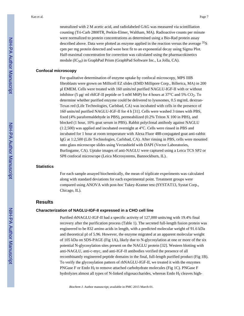
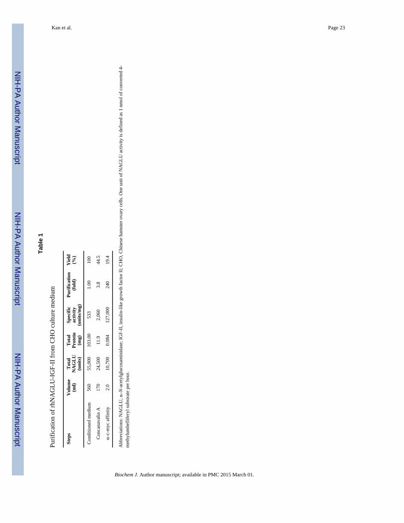
Purified rhNAGLU-IGF-II had a specific activity of 127,000 units/mg with 19.4% final
recovery after the purification process (Table 1). The secreted full-length fusion protein was
engineered to be 832 amino acids in length, with a predicted molecular weight of 91.6 kDa
and theoretical pI of 5.96. However, the enzyme migrated at an apparent molecular weight
of 105 kDa on SDS-PAGE (Fig 1A), likely due to N-glycosylation at one or more of the six
potential N-glycosylation sites present on the NAGLU protein [32]. Western blotting with
anti-NAGLU, anti-c-myc, and anti-IGF-II antibodies verified the presence of all
recombinantly engineered peptide domains in the final, full-length purified product (Fig 1B).
To verify the glycosylation pattern of rhNAGLU-IGF-II, we treated it with the enzymes
PNGase F or Endo Hf to remove attached carbohydrate molecules (Fig 1C). PNGase F
hydrolyzes almost all types of N-linked oligosaccharides, whereas Endo Hf cleaves high-
Kan et al. Page 7
Biochem J. Author manuscript; available in PMC 2015 March 01.
NIH
-PA
Author M
anuscriptN
IH-P
A A
uthor Manuscript
NIH
-PA
Author M
anuscript
mannose N-linked oligosaccharides within the chitobiose core. The rhNAGLU-IGF-II was
resistant to digestion with Endo Hf but sensitive to PNGase F, suggesting that the protein
was N-glycosylated but lacked high-mannose residues.
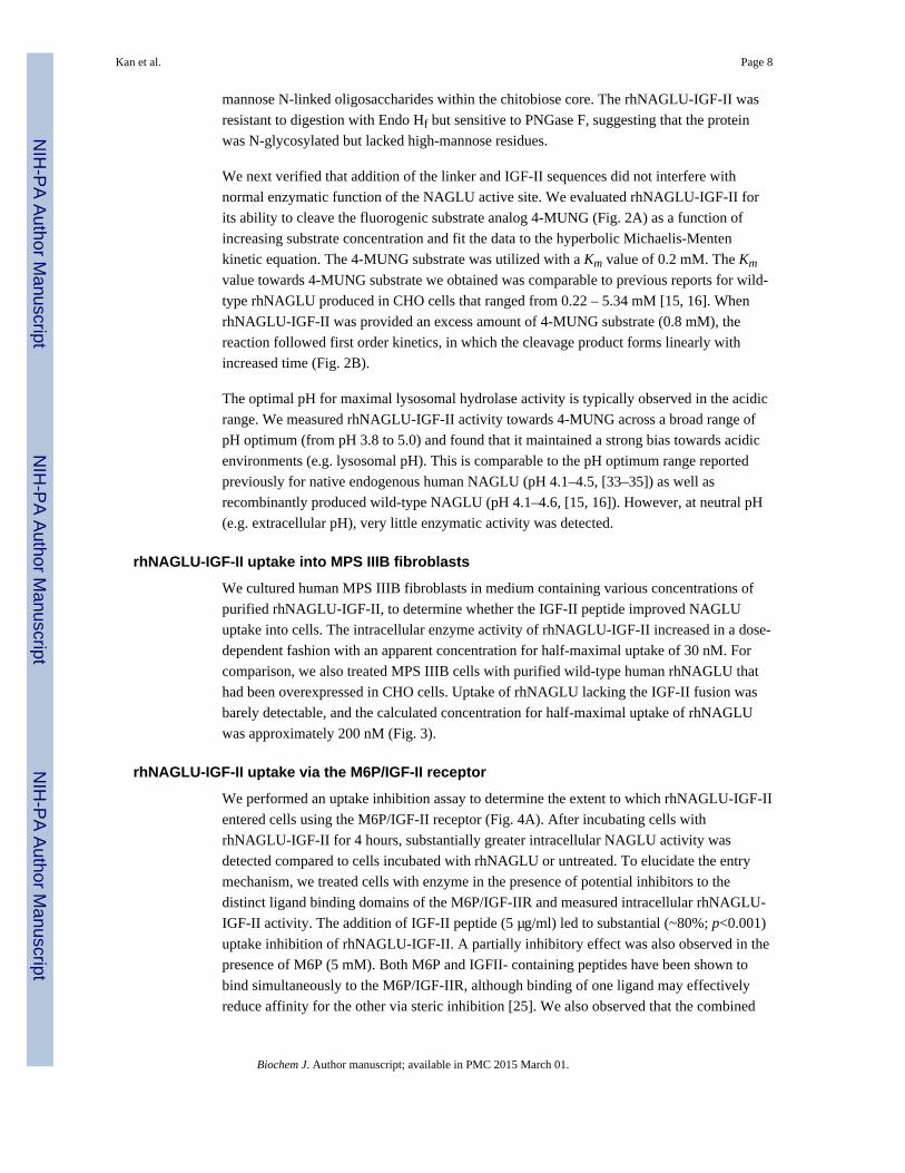
We next verified that addition of the linker and IGF-II sequences did not interfere with
normal enzymatic function of the NAGLU active site. We evaluated rhNAGLU-IGF-II for
its ability to cleave the fluorogenic substrate analog 4-MUNG (Fig. 2A) as a function of
increasing substrate concentration and fit the data to the hyperbolic Michaelis-Menten
kinetic equation. The 4-MUNG substrate was utilized with a Km value of 0.2 mM. The Km
value towards 4-MUNG substrate we obtained was comparable to previous reports for wild-
type rhNAGLU produced in CHO cells that ranged from 0.22 – 5.34 mM [15, 16]. When
rhNAGLU-IGF-II was provided an excess amount of 4-MUNG substrate (0.8 mM), the
reaction followed first order kinetics, in which the cleavage product forms linearly with
increased time (Fig. 2B).
The optimal pH for maximal lysosomal hydrolase activity is typically observed in the acidic
range. We measured rhNAGLU-IGF-II activity towards 4-MUNG across a broad range of
pH optimum (from pH 3.8 to 5.0) and found that it maintained a strong bias towards acidic
environments (e.g. lysosomal pH). This is comparable to the pH optimum range reported
previously for native endogenous human NAGLU (pH 4.1–4.5, [33–35]) as well as
recombinantly produced wild-type NAGLU (pH 4.1–4.6, [15, 16]). However, at neutral pH
(e.g. extracellular pH), very little enzymatic activity was detected.
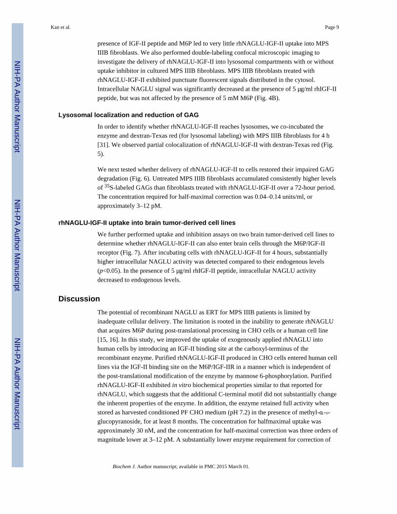
rhNAGLU-IGF-II uptake into MPS IIIB fibroblasts
We cultured human MPS IIIB fibroblasts in medium containing various concentrations of
purified rhNAGLU-IGF-II, to determine whether the IGF-II peptide improved NAGLU
uptake into cells. The intracellular enzyme activity of rhNAGLU-IGF-II increased in a dose-
dependent fashion with an apparent concentration for half-maximal uptake of 30 nM. For
comparison, we also treated MPS IIIB cells with purified wild-type human rhNAGLU that
had been overexpressed in CHO cells. Uptake of rhNAGLU lacking the IGF-II fusion was
barely detectable, and the calculated concentration for half-maximal uptake of rhNAGLU
was approximately 200 nM (Fig. 3).
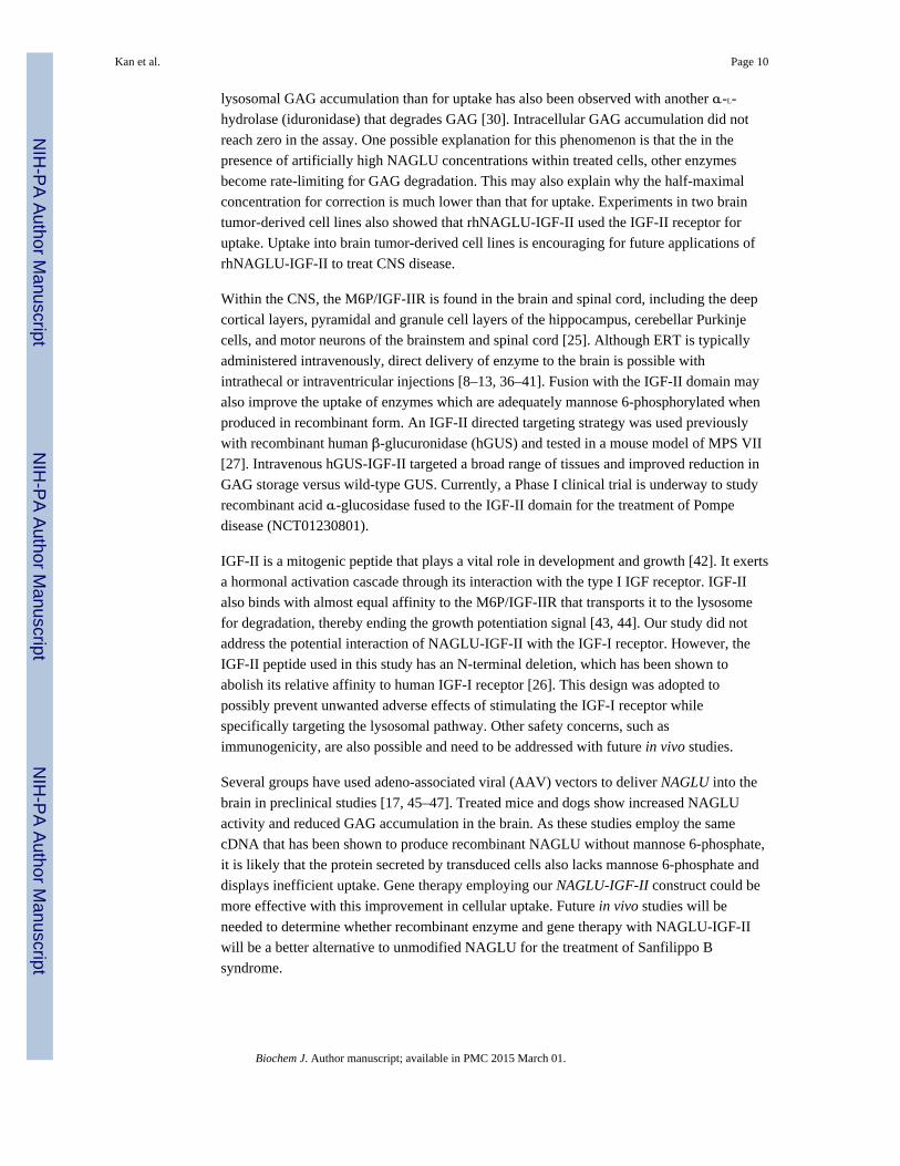
rhNAGLU-IGF-II uptake via the M6P/IGF-II receptor
We performed an uptake inhibition assay to determine the extent to which rhNAGLU-IGF-II
entered cells using the M6P/IGF-II receptor (Fig. 4A). After incubating cells with
rhNAGLU-IGF-II for 4 hours, substantially greater intracellular NAGLU activity was
detected compared to cells incubated with rhNAGLU or untreated. To elucidate the entry
mechanism, we treated cells with enzyme in the presence of potential inhibitors to the
distinct ligand binding domains of the M6P/IGF-IIR and measured intracellular rhNAGLU-
IGF-II activity. The addition of IGF-II peptide (5 µg/ml) led to substantial (~80%; p<0.001)
uptake inhibition of rhNAGLU-IGF-II. A partially inhibitory effect was also observed in the
presence of M6P (5 mM). Both M6P and IGFII- containing peptides have been shown to
bind simultaneously to the M6P/IGF-IIR, although binding of one ligand may effectively
reduce affinity for the other via steric inhibition [25]. We also observed that the combined
Kan et al. Page 8
Biochem J. Author manuscript; available in PMC 2015 March 01.
NIH
-PA
Author M
anuscriptN
IH-P
A A
uthor Manuscript
NIH
-PA
Author M
anuscript
presence of IGF-II peptide and M6P led to very little rhNAGLU-IGF-II uptake into MPS
IIIB fibroblasts. We also performed double-labeling confocal microscopic imaging to
investigate the delivery of rhNAGLU-IGF-II into lysosomal compartments with or without
uptake inhibitor in cultured MPS IIIB fibroblasts. MPS IIIB fibroblasts treated with
rhNAGLU-IGF-II exhibited punctuate fluorescent signals distributed in the cytosol.
Intracellular NAGLU signal was significantly decreased at the presence of 5 µg/ml rhIGF-II
peptide, but was not affected by the presence of 5 mM M6P (Fig. 4B).
Lysosomal localization and reduction of GAG
In order to identify whether rhNAGLU-IGF-II reaches lysosomes, we co-incubated the
enzyme and dextran-Texas red (for lysosomal labeling) with MPS IIIB fibroblasts for 4 h
[31]. We observed partial colocalization of rhNAGLU-IGF-II with dextran-Texas red (Fig.
5).
We next tested whether delivery of rhNAGLU-IGF-II to cells restored their impaired GAG
degradation (Fig. 6). Untreated MPS IIIB fibroblasts accumulated consistently higher levels
of 35S-labeled GAGs than fibroblasts treated with rhNAGLU-IGF-II over a 72-hour period.
The concentration required for half-maximal correction was 0.04–0.14 units/ml, or
approximately 3–12 pM.
rhNAGLU-IGF-II uptake into brain tumor-derived cell lines
We further performed uptake and inhibition assays on two brain tumor-derived cell lines to
determine whether rhNAGLU-IGF-II can also enter brain cells through the M6P/IGF-II
receptor (Fig. 7). After incubating cells with rhNAGLU-IGF-II for 4 hours, substantially
higher intracellular NAGLU activity was detected compared to their endogenous levels
(p<0.05). In the presence of 5 µg/ml rhIGF-II peptide, intracellular NAGLU activity
decreased to endogenous levels.
Discussion
The potential of recombinant NAGLU as ERT for MPS IIIB patients is limited by
inadequate cellular delivery. The limitation is rooted in the inability to generate rhNAGLU
that acquires M6P during post-translational processing in CHO cells or a human cell line
[15, 16]. In this study, we improved the uptake of exogenously applied rhNAGLU into
human cells by introducing an IGF-II binding site at the carboxyl-terminus of the
recombinant enzyme. Purified rhNAGLU-IGF-II produced in CHO cells entered human cell
lines via the IGF-II binding site on the M6P/IGF-IIR in a manner which is independent of
the post-translational modification of the enzyme by mannose 6-phosphorylation. Purified
rhNAGLU-IGF-II exhibited in vitro biochemical properties similar to that reported for
rhNAGLU, which suggests that the additional C-terminal motif did not substantially change
the inherent properties of the enzyme. In addition, the enzyme retained full activity when
stored as harvested conditioned PF CHO medium (pH 7.2) in the presence of methyl-α-D-
glucopyranoside, for at least 8 months. The concentration for halfmaximal uptake was
approximately 30 nM, and the concentration for half-maximal correction was three orders of
magnitude lower at 3–12 pM. A substantially lower enzyme requirement for correction of
Kan et al. Page 9
Biochem J. Author manuscript; available in PMC 2015 March 01.
NIH
-PA
Author M
anuscriptN
IH-P
A A
uthor Manuscript
NIH
-PA
Author M
anuscript
lysosomal GAG accumulation than for uptake has also been observed with another α-L-
hydrolase (iduronidase) that degrades GAG [30]. Intracellular GAG accumulation did not
reach zero in the assay. One possible explanation for this phenomenon is that the in the
presence of artificially high NAGLU concentrations within treated cells, other enzymes
become rate-limiting for GAG degradation. This may also explain why the half-maximal
concentration for correction is much lower than that for uptake. Experiments in two brain
tumor-derived cell lines also showed that rhNAGLU-IGF-II used the IGF-II receptor for
uptake. Uptake into brain tumor-derived cell lines is encouraging for future applications of
rhNAGLU-IGF-II to treat CNS disease.
Within the CNS, the M6P/IGF-IIR is found in the brain and spinal cord, including the deep
cortical layers, pyramidal and granule cell layers of the hippocampus, cerebellar Purkinje
cells, and motor neurons of the brainstem and spinal cord [25]. Although ERT is typically
administered intravenously, direct delivery of enzyme to the brain is possible with
intrathecal or intraventricular injections [8–13, 36–41]. Fusion with the IGF-II domain may
also improve the uptake of enzymes which are adequately mannose 6-phosphorylated when
produced in recombinant form. An IGF-II directed targeting strategy was used previously
with recombinant human β-glucuronidase (hGUS) and tested in a mouse model of MPS VII
[27]. Intravenous hGUS-IGF-II targeted a broad range of tissues and improved reduction in
GAG storage versus wild-type GUS. Currently, a Phase I clinical trial is underway to study
recombinant acid α-glucosidase fused to the IGF-II domain for the treatment of Pompe
disease (NCT01230801).
IGF-II is a mitogenic peptide that plays a vital role in development and growth [42]. It exerts
a hormonal activation cascade through its interaction with the type I IGF receptor. IGF-II
also binds with almost equal affinity to the M6P/IGF-IIR that transports it to the lysosome
for degradation, thereby ending the growth potentiation signal [43, 44]. Our study did not
address the potential interaction of NAGLU-IGF-II with the IGF-I receptor. However, the
IGF-II peptide used in this study has an N-terminal deletion, which has been shown to
abolish its relative affinity to human IGF-I receptor [26]. This design was adopted to
possibly prevent unwanted adverse effects of stimulating the IGF-I receptor while
specifically targeting the lysosomal pathway. Other safety concerns, such as
immunogenicity, are also possible and need to be addressed with future in vivo studies.
Several groups have used adeno-associated viral (AAV) vectors to deliver NAGLU into the
brain in preclinical studies [17, 45–47]. Treated mice and dogs show increased NAGLU
activity and reduced GAG accumulation in the brain. As these studies employ the same
cDNA that has been shown to produce recombinant NAGLU without mannose 6-phosphate,
it is likely that the protein secreted by transduced cells also lacks mannose 6-phosphate and
displays inefficient uptake. Gene therapy employing our NAGLU-IGF-II construct could be
more effective with this improvement in cellular uptake. Future in vivo studies will be
needed to determine whether recombinant enzyme and gene therapy with NAGLU-IGF-II
will be a better alternative to unmodified NAGLU for the treatment of Sanfilippo B
syndrome.
Kan et al. Page 10
Biochem J. Author manuscript; available in PMC 2015 March 01.
NIH
-PA
Author M
anuscriptN
IH-P
A A
uthor Manuscript
NIH
-PA
Author M
anuscript
Acknowledgments
We thank Dr. E. F. Neufeld (University of California, Los Angeles, CA) for providing human NAGLU cDNA,rabbit polyclonal anti-hNAGLU antibody, and thoughtful reading of the manuscript, and Dr. Jon LeBowitz(BioMarin, Novato, CA) for helpful discussions regarding the characterization of rhNAGLU-IGF-II and Dr. J.Lasky (Los Angeles Biomedical Research Institute at Harbor-UCLA, Torrance, CA) for providing DaoY and Escell lines. We also thank Megan Craig and Jon Scott for careful review of the manuscript.
Funding
Funding was provided by grants from the Lauren’s Hope Foundation and the Canadian MPS Society (to P.I.D. andB.L.T.), and in part by National Institute of Health [grant number R21 NS078314-01A1 (to P.I.D.); T32GM8243-27 (to S-h.K.)]. Confocal imaging was supported by the National Center for Advancing TranslationalSciences through UCLA CTSI [grant number UL1TR000124 (to P.I.D)]. The content is solely the responsibility ofthe authors and does not necessarily represent the official views of the NIH.
Abbreviations
GAG glycosaminoglycans
ERT enzyme replacement therapy
MPS mucopolysaccharidosis
M6P/IGF-II mannose 6-phosphate/insulin-like growth factor-II
CHO Chinese hamster ovary
References
1. Valstar MJ, Ruijter GJ, can Diggelen OP, Poorhuis BJ, Wilburg FA. Sanfilippo syndrome: a minireview. J Inherit. Metab. Dis. 2008; 31(2):240–252. [PubMed: 18392742]
2. Kakkis ED, Muenzer J, Tiller GE, Waber L, Belmont J, Passage M, Izykowski B, Phillips J,Doroshow R, Walot I, et al. Enzyme-replacement therapy in mucopolysaccharisosis I. N. Engl. J.Med. 2001; 344:182–188. [PubMed: 11172140]
3. Muenzer J, Wraith JE, Beck M, Giugliani R, Harmatz P, Eng CM, Vellodi A, Martin R, RamaswamiU, Gucsavas-Calikoglu M, et al. A phase II/III clinical study of enzyme replacement therapy withidursulfase in mucopolysaccharidosis II (Hunter syndrome). Genet. Med. 2006; 8:465–473.[PubMed: 16912578]
4. Harmatz P, Giugliani R, Schwartz I, Guffon N, Teles EL, Miranda MCS, Wraith JE, Beck M, ArashL, Scarpa M, et al. Enzyme replacement therapy for mucopolysaccharidosis VI: A phase 3,randomized, double-blind, placebo-controlled, multinational study of recombinant human N-acetylgalactosamine 4-sulfatase (recombinant human arylsulfatase B or rhASB) and follow-on,open-label extension study. J. Pediatr. 2006; 148:533–539. [PubMed: 16647419]
5. Eng CM, Guffon N, Wilcox WR, Germain DP, Lee P, Waldek S, Caplan L, Linthorst GE, DesnickRJ. the International Fabry Disease Study Group. Safety and efficacy of recombinant human α-galactosidase A replacement therapy in Fabry’s disease. N. Engl. J. Med. 2001; 345:9–16.[PubMed: 11439963]
6. Kishnani PS, Corzo D, Nicolino M, Byrne B, Mandel H, Hwu WL, Leslie N, Levine J, Spencer C,McDonald M, et al. Recombinant human acid α-glucosidase: Major clinical benefits in infantile-onset Pompe disease. Neurology. 2007; 68:99–109. [PubMed: 17151339]
7. Barton NW, Brady RO, Dambrosia JM, Di Bisceglie AM, Doppelt SH, Hill SC, Mankin HJ, MurrayGJ, Parker RI, Argoff CE, et al. Replacement therapy for inherited enzyme deficiency: macrophage-targeted glucocerebrosidase for Gaucher’s disease. N. Engl. J. Med. 1991; 324:1464–1470.[PubMed: 2023606]
Kan et al. Page 11
Biochem J. Author manuscript; available in PMC 2015 March 01.
NIH
-PA
Author M
anuscriptN
IH-P
A A
uthor Manuscript
NIH
-PA
Author M
anuscript
8. Dickson P, McEntee M, Vogler C, Le S, Levy B, Peinovich M, Hanson S, Passage M, Kakkis E.Intrathecal enzyme replacement therapy: successful treatment of brain disease via the cerebrospinalfluid. Mol. Genet. Metab. 2007; 91:61–68. [PubMed: 17321776]
9. Lee WC, Tsoi YK, Troendle FJ, DeLucia MW, Ahmed Z, Dicky CA, Dickson DW, Eckman CB.Single-dose intracerebroventricular administration of galactocerebrosidase improves survival in amouse model of globoid cell leukodystrophy. FASEB J. 2007; 21:2520–2527. [PubMed: 17403939]
10. Hemsley KM, King B, Hopwood JJ. Injection of recombinant human sulfamidase into the CSF viathe cerebellomedullary cistern in MPS IIIA mice. Mol. Genet. Metab. 2007; 90:313–328.[PubMed: 17166757]
11. Chang M, Cooper JD, Sleat DE, Cheng SH, Dodge JC, Passini MA, Lobel P, Davidson BL.Intraventricular enzyme replacement improves disease phenotypes in a mouse model of lateinfantile neuronal ceroid lipofuscinosis. Mol. Ther. 2008; 16:649–656. [PubMed: 18362923]
12. Dodge JC, Clarke J, Treleaven CM, Taksir TV, Griffiths DA, Yang W, Fidler JA, Passini MA,Karey KP, Schuchman EH, et al. Intracerebroventricular infusion of acid sphingomyelinasecorrects CNS manifestations in a mouse model of Niemann-Pick A disease. Exp. Neurol. 2009;215:349–357. [PubMed: 19059399]
13. Kondagari GS, King BM, Thomson PC, Williamson P, Clements PR, Fuller M, Hemsley KM,Hopwood JJ, Taylor RM. Treatment of canine fucosidosis by intracisternal enzyme infusion. Exp.Neurol. 2011; 230:218–226. [PubMed: 21575633]
14. Yu WH, Zhao KW, Ryazantsev S, Rozengurt N, Neufeld EF. Shortterm enzyme replacement in themurine model of Sanfilippo syndrome type B. Mol. Genet. Metab. 2000; 71:573–580. [PubMed:11136549]
15. Weber B, Hopwood JJ, Yogalingam G. Expression and characterization of human recombinant and[alpha]-N-actylglucosaminidase. Prot. Expr. Purif. 2001; 21:251–259.
16. Zhao KW, Neufeld EF. Purification and characterization of recombinant human [alpha]-N-acetylglucosaminidase secreted by Chinese Hamster Ovary cells. Prot. Expr. Purif. 2000; 19:202–211.
17. Fu H, Samulski RJ, McCown TJ, Picornell YJ, Fletcher D, Muenzer J. Neurological correction oflysosomal storage in a mucopolysaccharidosis IIIB mouse model by adeno-associated virus-mediated gene delivery. Mol. Ther. 2002; 5:42–49. [PubMed: 11786044]
18. Cressant A, Desmaris N, Verot L, Brejot T, Froissart R, Vanier MT, Maire I, Heard JM. Improvedbehavior and neuropathology in the mouse model of Sanfilippo type IIIB disease after adeno-associated virus-mediated gene transfer in the striatum. J. Neurosci. 2004; 24:10229–10239.[PubMed: 15537895]
19. Zheng Y, Ryazantsev S, Ohmi K, Zhao HZ, Rozengurt N, Kohn DB, Neufeld EF. Retrovirallytransduced bone marrow has a therapeutic effect on brain in the mouse model ofmucopolysaccharidosis IIIB. Mol. Genet. Metab. 2004; 82:286–295. [PubMed: 15308126]
20. Fu H, Kang L, Jennings JS, Moy SS, Perez A, DiRosario J, McCarty DM, Muenzer J. Significantlyincreased lifespan and improved behavioral performances by rAAV gene delivery in adultmucopolysaccharidosis IIIB mice. Gene Ther. 2007; 14:1065–1077. [PubMed: 17460717]
21. McCarty DM, DiRosario J, Gulaid K, Muenzer J, Fu H. Mannitol-facilitated CNS entry of rAAV2vector significantly delayed the neurological disease progression in MPS IIIB mice. Gene Ther.2009; 16:1340–1352. [PubMed: 19587708]
22. Fu H, DiRosario J, Kang L, Muenzer J, McCarty DM. Restoration of central nervous system alpha-N-acetylglucosaminidase activity and therapeutic benefits in mucopolysaccharidosis IIIB mice bya single intracisternal recombinant adeno-associated viral type 2 vector delivery. J. Gene Med.2010; 12:624–633. [PubMed: 20603889]
23. Braulke T, Bonifacino JS. Sorting of lysosomal proteins. Biochim. Biophys. Acta (BBA) - Mol.Cell Res. 2009; 1793:605–614.
24. Hille-Rehfeld A. Mannose 6-phosphate receptors in sorting and transport of lysosomal enzymes.Biochim. Biophys. Acta (BBA) - Rev. Biomembr. 1995; 1241:177–194.
25. Hawkes C, Kar S. The insulin-like growth factor-II/mannose-6-phosphate receptor: structure,distribution and function in the central nervous system. Brain Res. Rev. 2004; 44:117–140.[PubMed: 15003389]
Kan et al. Page 12
Biochem J. Author manuscript; available in PMC 2015 March 01.
NIH
-PA
Author M
anuscriptN
IH-P
A A
uthor Manuscript
NIH
-PA
Author M
anuscript
26. LeBowitz JH, Grubb JH, Maga JA, Schmiel DH, Vogler C, Sly WS. Glycosylation-independenttargeting enhances enzyme delivery to lysosomes and decreases storage in mucopolysaccharidosistype VII mice. Proc. Natl. Acad. Sci. USA. 2004; 101:3083–3088. [PubMed: 14976248]
27. Grubb JH, Vogler C, Sly WS. New strategies for enzyme replacement therapy for lysosomalstorage diseases. Rejuv. Res. 2010; 13:229–236.
28. Chow P, Weissmann B. 4-Methylumbelliferyl 2-acetamido-2-deoxy-α-Dglucopyranoside, afluorogenic substrate for N-acetyl-α-d-glucosaminidase. Carbohydr. Res. 1981; 96:87–93.[PubMed: 7296566]
29. Barton RW, Neufeld EF. The Hurler Corrective Factor. J. Biol. Chem. 1971; 246:7773–7779.[PubMed: 4257494]
30. Kakkis ED, Matynia A, Jonas AJ, Neufeld EF. Overexpression of the human lysosomal enzyme α-l-iduronidase in Chinese hamster ovary cells. Prot. Expr. Purif. 1994; 5:225–232.
31. Chen CB, Dellamaggiore KR, Ouellette CP, Sedano CD, Lizadjohry M, Chernis GA, Gonzales M,Baltasar FE, Fan AL, Myerowitz R, Neufeld EF. Aptamer-based endocytosis of a lysosomalenzyme. Proc. Natl. Acad. Sci. USA. 2008; 105:15908–15913. [PubMed: 18838694]
32. Zhao HG, Li HH, Bach G, Schmidtchen A, Neufeld EF. The molecular basis of Sanfilipposyndrome type B. Proc. Natl. Acad. Sci. 1996; 93:6101–6105. [PubMed: 8650226]
33. von Figura KF, Logering MF, Kresse H. Serum alpha-N-acetylglucosaminidase: determination,characterization, and corrective activity in Sanfilippo B fibroblasts. Z. Klin. Chem. Klin. Biochem.1975; 13:285–289. [PubMed: 242129]
34. Rohrborn WF, von FK. Human placenta alpha-N-acetylglucosaminidase: purification,characterization and demonstration of multiple recognition forms. Hoppe Seylers Z. Physiol.Chem. 1978; 359:1353–1362. [PubMed: 102578]
35. Sasaki T, Sukegawa K, Masue M, Fukuda S, Tomatsu S, Orii T. Purification and partialcharacterization of α-N-acetylglucosaminidase from human liver. J. Biochem. 1991; 110:842–846.[PubMed: 1783617]
36. DeChiara TM, Efstratiadis A, Robertsen EJ. A growth-deficiency phenotype in heterozygous micecarrying an insulin-like growth factor II gene disrupted by targeting. Nature. 1990; 345:78–80.[PubMed: 2330056]
37. Hashimoto R, Fujiwara H, Higashihashi N, Enjoh-Kimura T, Terasawa H, Fujita-Yamaguchi Y,Inagaki F, Perdue JF, Sakano KI. N-terminal deletion mutants of insulin-like growth factor-II(IGF-II) show thr and leu important for binding to insulin and IGF-I receptors and leu critical forall IGF-II functions. J. Biol. Chem. 1995; 270:18013–18018. [PubMed: 7629109]
38. Nakae J, Kido Y, Accili D. Distinct and overlapping functions of insulin and IGF-I receptors.Endo. Rev. 2001; 22:818–835.
39. Sands MS, Vogler CA, Ohlemiller KK, Roberts MS, Grubb JH, Levy B, Sly WS. Biodistribution,kinetics, and efficacy of highly phosphorylated and nonphosphorylated β-glucuronidase in themurine model of mucopolysaccharidosis VII. J. Biol. Chem. 2001; 276:43160–43165. [PubMed:11562370]
40. Dickson PI, Naylor D, Mlikotic A, Victoroff A, Chen A, Passage M, Le S. MPS I IntrathecalResearch Collaborative. Intrathecal recombinant human alpha-Liduronidase alleviates spinal cordcompression symptoms and is well-tolerated in attenuated MPS I patients. Abstract, Mol. Genet.Metab. 2007; 93:247.
41. Garcia, AR.; Pan, J.; Stronge, A.; Tonini, M.; Alessandrini, M.; Neal, C.; Lieb, J.; Lu, Y.; Wiles,M. Intrathecal delivery of iduronate 2-sulfatase to the CNS of cynomolgous monkeys. Abstract,American Society of Human Genetics 57th Annual Meeting; San Diego, California. 2007.
42. Muñoz-Rojas MV, Costa R, Canani SF, Jardim L, Vedolin L, Kakkis E, Dickson P, Vieira T, JohnAB, Raymundo M, Giugliani R. Intrathecal enzyme replacement therapy in a patient withmucopolysaccharidosis type I and symptomatic spinal cord compression. Am. J. Med. Genet. A.2008; 146A:2538–2544. [PubMed: 18792977]
43. Hemsley KM, Luck AJ, Crawley AC, Hassiotis S, Beard H, King B, Rozek TRT, Fuller M,Hopwood JJ. Examination of intravenous and intra-CSF protein delivery for treatment ofneurological disease. Eur. J. Neurosci. 2009; 29:1197–1214. [PubMed: 19302155]
Kan et al. Page 13
Biochem J. Author manuscript; available in PMC 2015 March 01.
NIH
-PA
Author M
anuscriptN
IH-P
A A
uthor Manuscript
NIH
-PA
Author M
anuscript
44. Hemsley KM, Hopwood JJ. Delivery of recombinant proteins via the cerebrospinal fluid as atherapy option for neurodegenerative lysosomal storage diseases. Int. J. Clin. Pharmacol. Ther.2009; 47:S118–S123. [PubMed: 20040322]
45. Heldermon CD, Ohlemiller KK, Herzog ED, Vogler C, Qin E, Wozniak DF, Tan Y, Orrock JL,Sands MS. Therapeutic efficacy of bone marrow transplant, intracranial AAV-mediated genetherapy, or both in the mouse model of MPS IIIB. Mol. Ther. 2010; 18:873–880. [PubMed:20179679]
46. Ellinwood NM, Ausseil J, Desmaris N, Bigou S, Liu S, Jens JK, Snella EM, Mohammed EE,Thomson CB, Raoul S, et al. Safe, efficient, and reproducible gene therapy of the brain in the dogmodels of Sanfilippo and Hurler syndromes. Mol. Ther. 2011; 19:251–259. [PubMed: 21139569]
47. Fu H, DiRosario J, Killedar S, Zaraspe K, McCarty DM. Correction of neurological disease ofmucopolysaccharidosis IIIB in adult mice by rAAV9 trans-bloodbrain barrier gene delivery. Mol.Ther. 2011; 19:1025–1033. [PubMed: 21386820]
Kan et al. Page 14
Biochem J. Author manuscript; available in PMC 2015 March 01.
NIH
-PA
Author M
anuscriptN
IH-P
A A
uthor Manuscript
NIH
-PA
Author M
anuscript
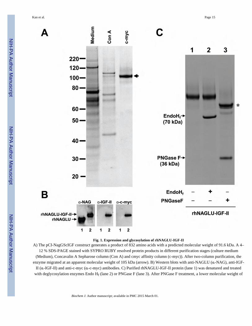
Fig. 1. Expression and glycosylation of rhNAGLU-IGF-IIA) The pCI-NagGScIGF construct generates a product of 832 amino acids with a predicted molecular weight of 91.6 kDa. A 4–
12 % SDS-PAGE stained with SYPRO RUBY resolved protein products in different purification stages (culture medium
(Medium), Concavalin A Sepharose column (Con A) and cmyc affinity column (c-myc)). After two-column purification, the
enzyme migrated at an apparent molecular weight of 105 kDa (arrow). B) Western blots with anti-NAGLU (α-NAG), anti-IGF-
II (α-IGF-II) and anti-c-myc (α-c-myc) antibodies. C) Purified rhNAGLU-IGF-II protein (lane 1) was denatured and treated
with deglycosylation enzymes Endo Hf (lane 2) or PNGase F (lane 3). After PNGase F treatment, a lower molecular weight of
Kan et al. Page 15
Biochem J. Author manuscript; available in PMC 2015 March 01.
NIH
-PA
Author M
anuscriptN
IH-P
A A
uthor Manuscript
NIH
-PA
Author M
anuscript
rhNAGLU-IGF-II of ~92 kDa is seen (asterisk). The enzymes Endo Hf and PNGase F appear in the treated lanes 2 and 3 as
bands at 70 kDa and 36 kDa, respectively.
Kan et al. Page 16
Biochem J. Author manuscript; available in PMC 2015 March 01.
NIH
-PA
Author M
anuscriptN
IH-P
A A
uthor Manuscript
NIH
-PA
Author M
anuscript
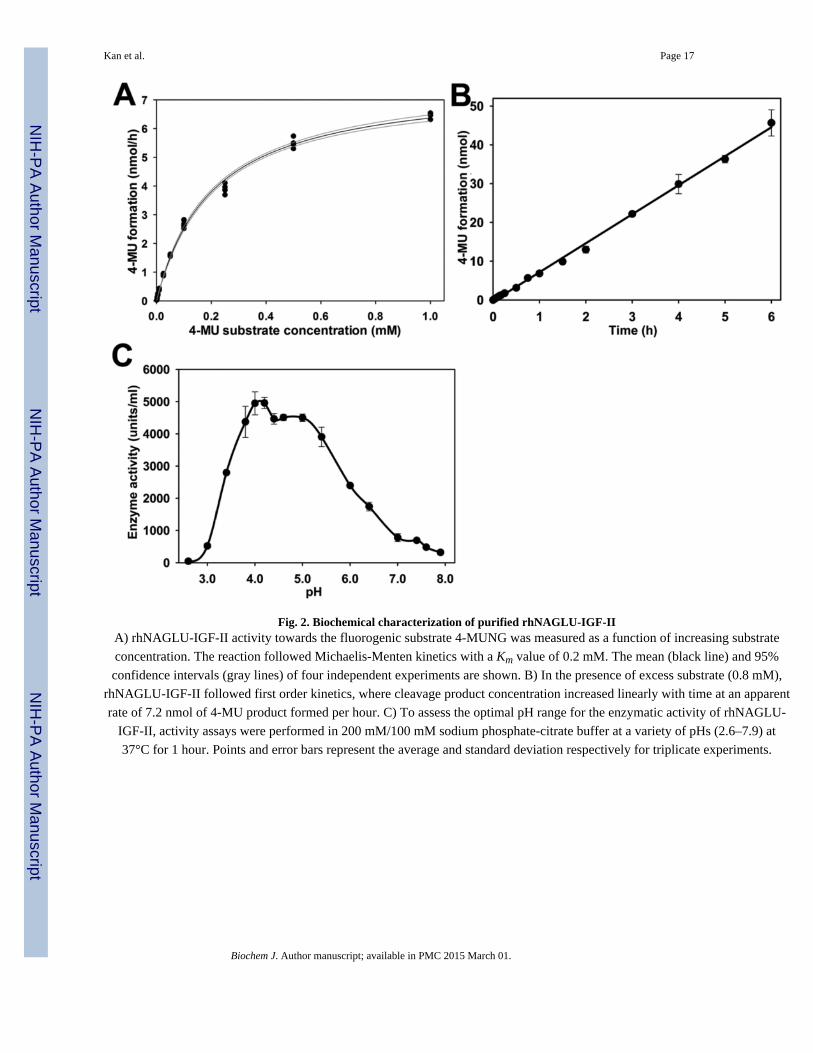
Fig. 2. Biochemical characterization of purified rhNAGLU-IGF-IIA) rhNAGLU-IGF-II activity towards the fluorogenic substrate 4-MUNG was measured as a function of increasing substrate
concentration. The reaction followed Michaelis-Menten kinetics with a Km value of 0.2 mM. The mean (black line) and 95%
confidence intervals (gray lines) of four independent experiments are shown. B) In the presence of excess substrate (0.8 mM),
rhNAGLU-IGF-II followed first order kinetics, where cleavage product concentration increased linearly with time at an apparent
rate of 7.2 nmol of 4-MU product formed per hour. C) To assess the optimal pH range for the enzymatic activity of rhNAGLU-
IGF-II, activity assays were performed in 200 mM/100 mM sodium phosphate-citrate buffer at a variety of pHs (2.6–7.9) at
37°C for 1 hour. Points and error bars represent the average and standard deviation respectively for triplicate experiments.
Kan et al. Page 17
Biochem J. Author manuscript; available in PMC 2015 March 01.
NIH
-PA
Author M
anuscriptN
IH-P
A A
uthor Manuscript
NIH
-PA
Author M
anuscript
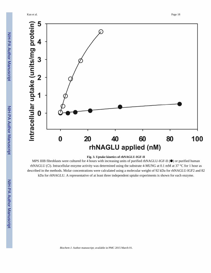
Fig. 3. Uptake kinetics of rhNAGLU-IGF-IIMPS IIIB fibroblasts were cultured for 4 hours with increasing units of purified rhNAGLU-IGF-II (●) or purified human
rhNAGLU (○). Intracellular enzyme activity was determined using the substrate 4-MUNG at 0.1 mM at 37 °C for 1 hour as
described in the methods. Molar concentrations were calculated using a molecular weight of 92 kDa for rhNAGLU-IGF2 and 82
kDa for rhNAGLU. A representative of at least three independent uptake experiments is shown for each enzyme.
Kan et al. Page 18
Biochem J. Author manuscript; available in PMC 2015 March 01.
NIH
-PA
Author M
anuscriptN
IH-P
A A
uthor Manuscript
NIH
-PA
Author M
anuscript
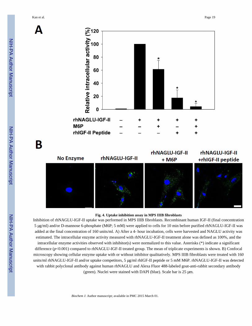
Fig. 4. Uptake inhibition assay in MPS IIIB fibroblastsInhibition of rhNAGLU-IGF-II uptake was performed in MPS IIIB fibroblasts. Recombinant human IGF-II (final concentration
5 µg/ml) and/or D-mannose 6-phosphate (M6P; 5 mM) were applied to cells for 10 min before purified rhNAGLU-IGF-II was
added at the final concentration of 160 units/ml. A) After a 4- hour incubation, cells were harvested and NAGLU activity was
estimated. The intracellular enzyme activity measured with rhNAGLU-IGF-II treatment alone was defined as 100%, and the
intracellular enzyme activities observed with inhibitor(s) were normalized to this value. Asterisks (*) indicate a significant
difference (p<0.001) compared to rhNAGLU-IGF-II treated group. The mean of triplicate experiments is shown. B) Confocal
microscopy showing cellular enzyme uptake with or without inhibitor qualitatively. MPS IIIB fibroblasts were treated with 160
units/ml rhNAGLU-IGF-II and/or uptake competitors, 5 µg/ml rhIGF-II peptide or 5 mM M6P. rhNAGLU-IGF-II was detected
with rabbit polyclonal antibody against human rhNAGLU and Alexa Fluor 488-labeled goat-anti-rabbit secondary antibody
(green). Nuclei were stained with DAPI (blue). Scale bar is 25 µm.
Kan et al. Page 19
Biochem J. Author manuscript; available in PMC 2015 March 01.
NIH
-PA
Author M
anuscriptN
IH-P
A A
uthor Manuscript
NIH
-PA
Author M
anuscript
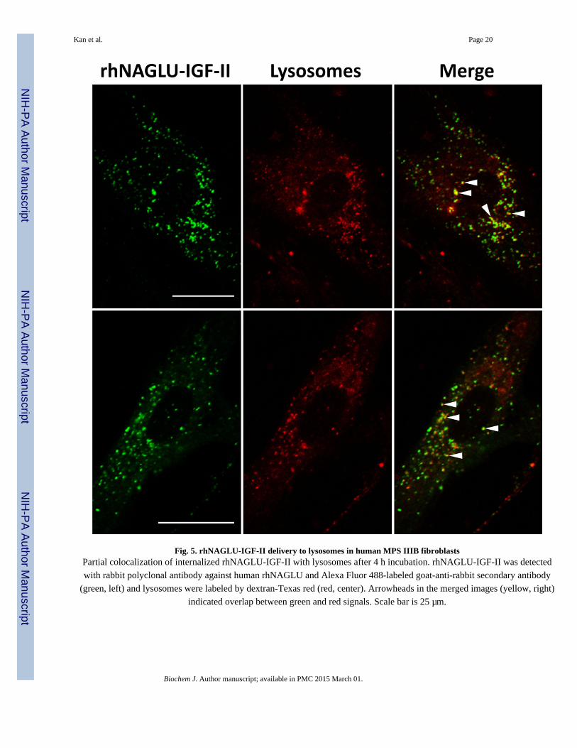
Fig. 5. rhNAGLU-IGF-II delivery to lysosomes in human MPS IIIB fibroblastsPartial colocalization of internalized rhNAGLU-IGF-II with lysosomes after 4 h incubation. rhNAGLU-IGF-II was detected
with rabbit polyclonal antibody against human rhNAGLU and Alexa Fluor 488-labeled goat-anti-rabbit secondary antibody
(green, left) and lysosomes were labeled by dextran-Texas red (red, center). Arrowheads in the merged images (yellow, right)
indicated overlap between green and red signals. Scale bar is 25 µm.
Kan et al. Page 20
Biochem J. Author manuscript; available in PMC 2015 March 01.
NIH
-PA
Author M
anuscriptN
IH-P
A A
uthor Manuscript
NIH
-PA
Author M
anuscript
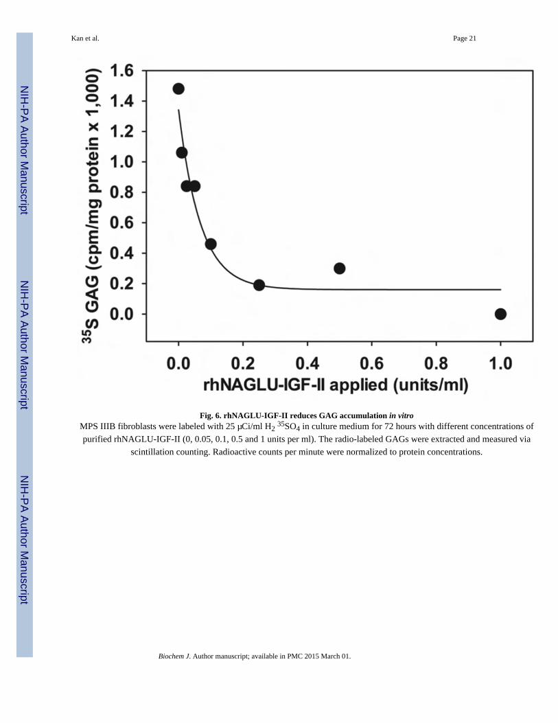
Fig. 6. rhNAGLU-IGF-II reduces GAG accumulation in vitroMPS IIIB fibroblasts were labeled with 25 µCi/ml H2 35SO4 in culture medium for 72 hours with different concentrations of
purified rhNAGLU-IGF-II (0, 0.05, 0.1, 0.5 and 1 units per ml). The radio-labeled GAGs were extracted and measured via
scintillation counting. Radioactive counts per minute were normalized to protein concentrations.
Kan et al. Page 21
Biochem J. Author manuscript; available in PMC 2015 March 01.
NIH
-PA
Author M
anuscriptN
IH-P
A A
uthor Manuscript
NIH
-PA
Author M
anuscript
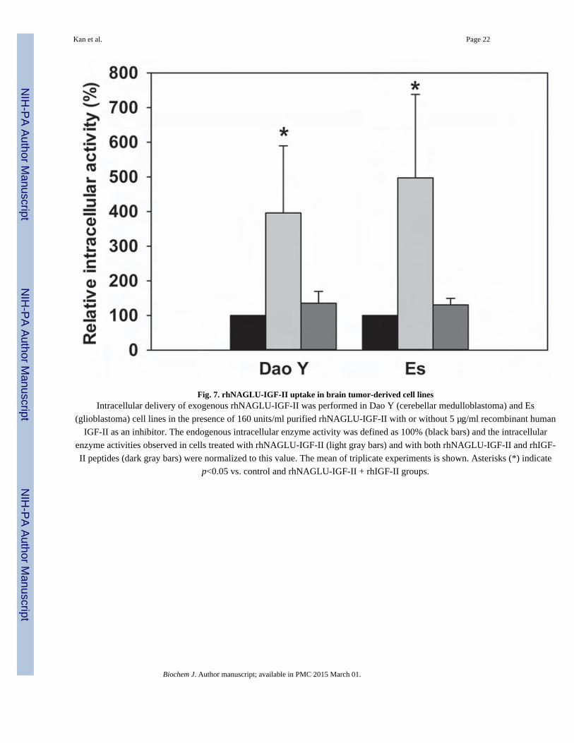
Fig. 7. rhNAGLU-IGF-II uptake in brain tumor-derived cell linesIntracellular delivery of exogenous rhNAGLU-IGF-II was performed in Dao Y (cerebellar medulloblastoma) and Es
(glioblastoma) cell lines in the presence of 160 units/ml purified rhNAGLU-IGF-II with or without 5 µg/ml recombinant human
IGF-II as an inhibitor. The endogenous intracellular enzyme activity was defined as 100% (black bars) and the intracellular
enzyme activities observed in cells treated with rhNAGLU-IGF-II (light gray bars) and with both rhNAGLU-IGF-II and rhIGF-
II peptides (dark gray bars) were normalized to this value. The mean of triplicate experiments is shown. Asterisks (*) indicate
p<0.05 vs. control and rhNAGLU-IGF-II + rhIGF-II groups.
Kan et al. Page 22
Biochem J. Author manuscript; available in PMC 2015 March 01.
NIH
-PA
Author M
anuscriptN
IH-P
A A
uthor Manuscript
NIH
-PA
Author M
anuscript
NIH
-PA
Author M
anuscriptN
IH-P
A A
uthor Manuscript
NIH
-PA
Author M
anuscript
Kan et al. Page 23
Tab
le 1
Puri
fica
tion
of r
hNA
GL
U-I
GF-
II f
rom
CH
O c
ultu
re m
ediu
m
Step
sV
olum
e(m
l)T
otal
NA
GL
U(u
nits
)
Tot
alP
rote
in(m
g)
Spec
ific
acti
vity
(uni
ts/m
g)
Pur
ific
atio
n(f
old)
Yie
ld(%
)
Con
ditio
ned
med
ium
560
55,0
0010
3.00
533
1.00
100
Con
cana
valin
A17
024
,500
11.9
2,06
03.
844
.5
α-c
-myc
aff
inity
2.0
10,7
000.
084
127,
000
240
19.4
Abb
revi
atio
ns: N
AG
LU
, α-N
-ace
tylg
luco
sam
inid
ase;
IG
F-II
, ins
ulin
-lik
e gr
owth
fac
tor
II; C
HO
, Chi
nese
ham
ster
ova
ry c
ells
. One
uni
t of
NA
GL
U a
ctiv
ity is
def
ined
as
1 nm
ol o
f co
nver
ted
4-m
ethy
lum
belli
fery
l sub
stra
te p
er h
our.
Biochem J. Author manuscript; available in PMC 2015 March 01.