Direct Evidence from Single-Cell Analysis that Human -Defensins
9
JOURNAL OF VIROLOGY, Apr. 2010, p. 4041–4049 Vol. 84, No. 8 0022-538X/10/$12.00 doi:10.1128/JVI.02471-09 Copyright © 2010, American Society for Microbiology. All Rights Reserved. Direct Evidence from Single-Cell Analysis that Human -Defensins Block Adenovirus Uncoating To Neutralize Infection Emily K. Nguyen, Glen R. Nemerow,* and Jason G. Smith† Department of Immunology and Microbial Science, The Scripps Research Institute, 10550 N. Torrey Pines Road, La Jolla, California 92037 Received 23 November 2009/Accepted 28 January 2010 Human -defensins are evolutionarily conserved effectors of the innate immune response with broadly acting antibacterial activity. Their role in antiviral immunity is less well understood. We previously showed that these antimicrobial peptides are potent inhibitors of human adenovirus infection. Based on biochemical studies and indirect evidence from confocal microscopy, we proposed that defensins bind to and stabilize the virus capsid and neutralize infection by preventing the release of the endosomalytic protein VI. To determine whether defensin action also restricts exposure of the viral genome, we developed a system to evaluate adenovirus uncoating during cell entry by monitoring the exposure of BrdU-labeled viral genomes. This assay allowed us to determine the kinetics of uncoating of virus particles in single cells. Using this assay, we now provide direct evidence that human -defensins block adenovirus infection by preventing uncoating during cell entry. The protein coat of the adenovirus (AdV) particle protects the virus genome from the extracellular environment; however, this protective coat must be shed during AdV entry to initiate infection in a process termed “uncoating.” The molecular basis for AdV uncoating is poorly understood. Current models of AdV entry, based primarily on studies of species B and C human AdVs (HAdVs), postulate that the virus capsid uncoats in discrete steps (10). After binding to high-affinity attachment receptors (e.g., CAR and CD46) and to integrin coreceptors, the virus particle is internalized by clathrin-mediated endocy- tosis (1, 15). As a consequence of receptor engagement and possibly of other cellular triggers, the pentons at the 12 icosa- hedral vertices, each comprised of a noncovalently coupled complex of penton base and fiber, are shed at or near the virus surface (10, 19). This process may involve either the sequential dissociation of fiber and then penton base or the dissociation of the penton complex as a unit. Subsequent uncoating, including the release of the internal capsid proteins IIIa, VI, and VIII and some of the major capsid protein (hexon), occurs in the endosome and has been proposed to be triggered in part by acidification during endosomal maturation (10). The released viral proteins, likely through the endosomalytic activity of pro- tein VI (32), mediate endosome disruption and allow the par- tially uncoated capsid to enter the cytoplasm. The released particle traffics in the cytoplasm along microtubules to the microtubule-organizing center (MTOC) and then to the nu- clear pore complex, where additional uncoating events occur to allow translocation of the viral DNA into the nucleus (15). The term uncoating, defined conceptually here as the re- moval of the protein capsid shell from the viral genome to the extent that is required to permit viral gene expression and replication, has also been used extensively in the literature as both a functional and a phenotypic descriptor. Uncoating has been measured as an increase in the accessibility of the viral DNA to DNase or DNA-sensitive dyes, either upon recovery from infected cells or in vitro (14, 16, 21, 22, 29, 30, 32). Alternatively, changes in the density of viral protein/DNA complexes or the association of capsid proteins with, or disso- ciation from, the nucleic acid-containing core have been de- termined (14, 16, 22, 26, 27, 29, 32). In some cases, these biochemical measurements have been correlated, directly or indirectly, with morphological changes observed by electron microscopy or immunofluorescence (6, 22, 24, 29); however, the structural basis for the various assays and phenotypes of uncoating are frequently unknown. In particular, existing as- says that measure DNA accessibility cannot be used to distin- guish complete or partial DNA dissociation from the capsid from alterations in the integrity of the capsid that nonetheless maintain the genome within a protein shell. Based on electron microscopy studies and the efficiency of gene delivery by AdV, it has been postulated that infectious particles retain sufficient integrity to sequester the viral genome prior to its docking at the nuclear pore complex; however, the contribution of DNA- capsid dissociation to nonproductive infectious pathways has not been assessed. Components of the adaptive immune system (e.g., neutral- izing antibodies) have been shown to block HAdV infection by impacting one or more steps in this entry pathway (26, 33). We have recently shown that effectors of the innate immune re- sponse, members of a family of human antimicrobial peptides known as -defensins, inhibit HAdV infection at low micro- molar concentrations (27). We provided evidence that these naturally occurring antiviral compounds block infection by binding to HAdV outside the cell and preventing escape of the internalized virus/defensin complex from the endosome. This conclusion was based on the failure of HAdV to mediate the translocation of macromolecules, such as the ribotoxin -sar- cin, into the cytoplasm in the presence of inhibitory concen- trations of defensin. Consistent with this finding, defensins * Corresponding author. Mailing address: 10550 N. Torrey Pines Rd., IMM19, La Jolla, CA 92037. Phone: (858) 784-8072. Fax: (858) 784-8472. E-mail: [email protected]. † Present address: Department of Microbiology, University of Washington School of Medicine, Box 357242, 1705 NE Pacific Street, Seattle, WA 98195-7242. Published ahead of print on 3 February 2010. 4041 Downloaded from https://journals.asm.org/journal/jvi on 06 January 2022 by 125.136.110.145.
Transcript of Direct Evidence from Single-Cell Analysis that Human -Defensins
JOURNAL OF VIROLOGY, Apr. 2010, p. 4041–4049 Vol. 84, No. 80022-538X/10/$12.00 doi:10.1128/JVI.02471-09Copyright © 2010, American Society for Microbiology. All Rights Reserved.
Direct Evidence from Single-Cell Analysis that Human �-DefensinsBlock Adenovirus Uncoating To Neutralize Infection�
Emily K. Nguyen, Glen R. Nemerow,* and Jason G. Smith†Department of Immunology and Microbial Science, The Scripps Research Institute,
10550 N. Torrey Pines Road, La Jolla, California 92037
Received 23 November 2009/Accepted 28 January 2010
Human �-defensins are evolutionarily conserved effectors of the innate immune response with broadly actingantibacterial activity. Their role in antiviral immunity is less well understood. We previously showed that theseantimicrobial peptides are potent inhibitors of human adenovirus infection. Based on biochemical studies andindirect evidence from confocal microscopy, we proposed that defensins bind to and stabilize the virus capsidand neutralize infection by preventing the release of the endosomalytic protein VI. To determine whetherdefensin action also restricts exposure of the viral genome, we developed a system to evaluate adenovirusuncoating during cell entry by monitoring the exposure of BrdU-labeled viral genomes. This assay allowed usto determine the kinetics of uncoating of virus particles in single cells. Using this assay, we now provide directevidence that human �-defensins block adenovirus infection by preventing uncoating during cell entry.
The protein coat of the adenovirus (AdV) particle protectsthe virus genome from the extracellular environment; however,this protective coat must be shed during AdV entry to initiateinfection in a process termed “uncoating.” The molecular basisfor AdV uncoating is poorly understood. Current models ofAdV entry, based primarily on studies of species B and Chuman AdVs (HAdVs), postulate that the virus capsid uncoatsin discrete steps (10). After binding to high-affinity attachmentreceptors (e.g., CAR and CD46) and to integrin coreceptors,the virus particle is internalized by clathrin-mediated endocy-tosis (1, 15). As a consequence of receptor engagement andpossibly of other cellular triggers, the pentons at the 12 icosa-hedral vertices, each comprised of a noncovalently coupledcomplex of penton base and fiber, are shed at or near the virussurface (10, 19). This process may involve either the sequentialdissociation of fiber and then penton base or the dissociation ofthe penton complex as a unit. Subsequent uncoating, includingthe release of the internal capsid proteins IIIa, VI, and VIIIand some of the major capsid protein (hexon), occurs in theendosome and has been proposed to be triggered in part byacidification during endosomal maturation (10). The releasedviral proteins, likely through the endosomalytic activity of pro-tein VI (32), mediate endosome disruption and allow the par-tially uncoated capsid to enter the cytoplasm. The releasedparticle traffics in the cytoplasm along microtubules to themicrotubule-organizing center (MTOC) and then to the nu-clear pore complex, where additional uncoating events occur toallow translocation of the viral DNA into the nucleus (15).
The term uncoating, defined conceptually here as the re-moval of the protein capsid shell from the viral genome to theextent that is required to permit viral gene expression and
replication, has also been used extensively in the literature asboth a functional and a phenotypic descriptor. Uncoating hasbeen measured as an increase in the accessibility of the viralDNA to DNase or DNA-sensitive dyes, either upon recoveryfrom infected cells or in vitro (14, 16, 21, 22, 29, 30, 32).Alternatively, changes in the density of viral protein/DNAcomplexes or the association of capsid proteins with, or disso-ciation from, the nucleic acid-containing core have been de-termined (14, 16, 22, 26, 27, 29, 32). In some cases, thesebiochemical measurements have been correlated, directly orindirectly, with morphological changes observed by electronmicroscopy or immunofluorescence (6, 22, 24, 29); however,the structural basis for the various assays and phenotypes ofuncoating are frequently unknown. In particular, existing as-says that measure DNA accessibility cannot be used to distin-guish complete or partial DNA dissociation from the capsidfrom alterations in the integrity of the capsid that nonethelessmaintain the genome within a protein shell. Based on electronmicroscopy studies and the efficiency of gene delivery by AdV,it has been postulated that infectious particles retain sufficientintegrity to sequester the viral genome prior to its docking atthe nuclear pore complex; however, the contribution of DNA-capsid dissociation to nonproductive infectious pathways hasnot been assessed.
Components of the adaptive immune system (e.g., neutral-izing antibodies) have been shown to block HAdV infection byimpacting one or more steps in this entry pathway (26, 33). Wehave recently shown that effectors of the innate immune re-sponse, members of a family of human antimicrobial peptidesknown as �-defensins, inhibit HAdV infection at low micro-molar concentrations (27). We provided evidence that thesenaturally occurring antiviral compounds block infection bybinding to HAdV outside the cell and preventing escape of theinternalized virus/defensin complex from the endosome. Thisconclusion was based on the failure of HAdV to mediate thetranslocation of macromolecules, such as the ribotoxin �-sar-cin, into the cytoplasm in the presence of inhibitory concen-trations of defensin. Consistent with this finding, defensins
* Corresponding author. Mailing address: 10550 N. Torrey PinesRd., IMM19, La Jolla, CA 92037. Phone: (858) 784-8072. Fax: (858)784-8472. E-mail: [email protected].
† Present address: Department of Microbiology, University ofWashington School of Medicine, Box 357242, 1705 NE Pacific Street,Seattle, WA 98195-7242.
� Published ahead of print on 3 February 2010.
4041
Dow
nloa
ded
from
http
s://j
ourn
als.
asm
.org
/jour
nal/j
vi o
n 06
Jan
uary
202
2 by
125
.136
.110
.145
.
restricted release of the putative endosomalytic protein VI.Moreover, at late time points postinfection virus particles werestrongly colocalized with lysosomes rather than the nucleus.Based on in vitro biochemical studies, we postulated that al-tered virus trafficking is due to a failure of the virus to uncoatupon defensin binding to the capsid. To test this hypothesis, wedeveloped a system to detect HAdV uncoating in single cells byimmunofluorescence based on the prior incorporation of aunique epitope (the thymidine analog 5-bromo-2-deoxyuridine[BrdU]) into the viral genome. This approach allows us tomonitor the kinetics of uncoating of HAdV particles at lowmultiplicities of infection (MOIs) without a requirement forradiolabeling of the viral proteins or nucleic acids. This studyprovides strong evidence to support a model in which �-de-fensins prevent HAdV infection by blocking virus uncoatingand genome exposure during cell entry.
MATERIALS AND METHODS
Cells and viruses. Tissue culture reagents were obtained from Invitrogen(Carlsbad, CA). Human A549 cells were from ATCC and were propagated inDulbecco’s modified Eagle’s medium (DMEM) supplemented with 10% fetalbovine serum (Omega Scientific, Tarzana, CA), 10 mM HEPES, 4 mM L-glutamine, 100 units/ml penicillin, 100 �g/ml streptomycin, and 0.1 mMnonessential amino acids (complete DMEM). Stable 293 cells overexpressingthe human �5 integrin subunit (293�5) were created by transfecting 293 cells(ATCC) with a cytomegalovirus (CMV) promoter-driven expression plasmidcontaining the human �5 gene and a neomycin resistance gene (pCDNA3/�5;a gift from David Cheresh, University of California, San Diego, CA). Trans-fected cells were selected for G418 resistance (700 �g/ml) and sorted for highlevels of integrin expression, using an antibody against human �5 integrin(Millipore, Billerica, MA).
HAdV-2p was obtained from ATCC. The temperature-sensitive mutant ofHAdV type 2 (HAdV-2), HAdV-2ts1, was obtained from Joseph Weber (Uni-versity of Sherbrooke, Quebec, Canada). The replication-defective HAdV-5 vec-tor used in these studies has an E1/E3 deletion and contains a CMV promoter-driven enhanced green fluorescent protein (eGFP) reporter gene cassette.
HAdV-5P137L was created by replacing the codon CCC (proline) with CTC(leucine) by recombineering (31) in a bacterial artificial chromosome (BAC)construct (pAd5-GFPn1; a gift from Zsolt Ruzsics, Max von Pettenkofer Insti-tute, Munich, Germany) (H. Wodrich, D. Henaff, B. Jammart, C. Segura-Mo-rales, S. Seelmeier, O. Coux, Z. Ruzsics, C. Wiethoff and E. J. Kremer, submittedfor publication) containing the entire genome of a HAdV-5 vector with an E1/E3deletion (derived from AdEasy; Qbiogene, Carlsbad, CA) expressing eGFP-n1(derived from pEGFP-N1; Clontech, Mountain View, CA). The fidelity of themutated construct (pAd5GFPn1-P137L) was verified by sequencing the recom-bineered region and by restriction digestion. 293�5 cells were transfected withthe large PacI restriction fragment of pAd5GFPn1-P137L, using Lipofectamine2000 (Invitrogen). Transfected cells were maintained at 33°C until visible, eGFP-positive plaques formed. The lysate from these transfected cells was seriallypassaged on 293�5 cells maintained at 33°C until enough virus was produced forpurification by CsCl density gradient ultracentrifugation as described below. Theidentity of the final virus stock was confirmed by restriction digestion, and a PCRproduct from this stock was sequenced to verify the presence of the engineeredmutation.
Virus production and BrdU labeling. HAdV-2 and the wild-type HAdV-5vector were propagated by infecting 293�5 cells with 300 particles/cell. When acomplete cytopathic effect (CPE) was observed (approximately 48 to 72 h postin-fection), cells were harvested, concentrated by low-speed centrifugation, dis-rupted by three cycles of freezing and thawing, and centrifuged for 10 min at3,200 � g to remove cell debris. The cleared lysate was layered onto a continuous15% to 40% CsCl gradient and centrifuged for 2 to 3 h at 111,000 � g (average),using an SW 41 Ti rotor (Beckman Coulter, Inc., Fullerton, CA). The maturevirus band was collected and purified in a second CsCl density gradient. Themature virus band was collected, dialyzed against three changes of A195 buffer(8), flash frozen in liquid nitrogen, and stored at �80°C. The processes used forthe propagation and purification of HAdV-2ts1 and HAdV-5P137L were iden-tical except that after 2 h of incubation to allow virus internalization, infectedcells were shifted to the nonpermissive temperature of 39.5°C and incubated
until a complete CPE was observed. To BrdU label the viral genome, theinoculum was removed 2 h postinfection and replaced with complete DMEMcontaining 4 �g/ml BrdU (Sigma-Aldrich, St. Louis, MO).
Dot blot for BrdU incorporation. To assay virus for BrdU incorporation,approximately 5 � 1010 particles of HAdV-2 or HAdV-2ts1 were incubated inphosphate-buffered saline (PBS) at the indicated temperatures (Fig. 1) for 10min or boiled for 5 min, cooled to room temperature (RT), and transferred to anitrocellulose membrane, using a Bio-Dot apparatus (Bio-Rad, Hercules, CA).Each sample well was treated for 10 min at RT with 500 units/ml DNase in 150mM NaCl, 5 mM MgCl2, and 10 �M HCl (DNase buffer) and then washed withPBS. The blot was then removed from the Bio-Dot apparatus, blocked with 5%milk in PBS–0.1% Tween-20, and probed for BrdU with a monoclonal antibody(MAb) (BD Biosciences, San Jose, CA). The membrane was then stripped andreprobed with anti-HAdV-2 serum (ATCC).
Confocal microscopy. BrdU-labeled or unlabeled HAdV-5 or HAdV-5P137Lparticles (3 � 109 particles/sample) in ice-cold complete DMEM (50 �l) wereadded to glass coverslips sparsely plated with A549 cells. In some experiments,cells were washed twice with serum-free DMEM (SFM) prior to being incubatedwith virus, and virus was added in SFM. Equivalent results were obtained incomplete medium and SFM. Samples were incubated for 45 min at 4°C and thenshifted to 37°C and incubated for the periods of time for each figure. In someexperiments, cells were washed twice in ice-cold medium prior to being incu-bated at 37°C. Samples were then fixed in 4% paraformaldehyde (PFA) in PBSfor 15 min at RT, quenched and permeabilized with 20 mM glycine–0.5% TritonX-100 for 15 min at RT, treated with 500 units/ml DNase in DNase buffer for 10min at RT, and probed sequentially with an anti-BrdU MAb (BD Biosciences,San Jose, CA), an Alexa Fluor 647-conjugated anti-mouse secondary antibody(Invitrogen), an anti-lamin B1 rabbit polyclonal antibody (PAb) (Abcam, Cam-bridge, MA), an Alexa Fluor 488-conjugated anti-rabbit secondary antibody(Invitrogen), and a Cy3-conjugated anti-HAdV-5 hexon MAb (9C12; hybridomaavailable at the Developmental Studies Hybridoma Bank at the University ofIowa, Iowa City) (26). Samples were mounted for microscopy, using ProLonggold antifade reagent (Invitrogen), and z-series images were acquired with aZeiss LSM 710 laser scanning confocal microscope (Carl Zeiss, Inc., Thornwood,NY). To maintain consistency, all samples from a single experiment were stainedin parallel, and images were acquired with the same microscope using the samesettings.
To measure the effect of the CRM1 inhibitor leptomycin B (LMB; Sigma-Aldrich), cells were incubated with 20 nM LMB for 30 min at 37°C prior to theaddition of virus in medium containing 20 nM LMB. To measure the effect ofHD5, A549 cells on coverslips were washed twice with SFM, and BrdU-labeledHAdV-5 in SFM was added. Samples were incubated for 45 min at 4°C to allowvirus binding, washed twice with ice-cold SFM, and then incubated in SFM withor without 10 �M HD5 (Peptides International, Inc., Louisville, KY) for 45 minat 4°C. Samples were then immediately fixed in 4% PFA or incubated for 45 minat 37°C prior to being fixed in 4% PFA.
Image analysis. A z-profile from the confocal images was created with Zensoftware (Carl Zeiss, Inc.). The signal threshold of the z-profile for each channelwas determined by comparison with that of stained, uninfected control cells,using ImageJ software (W. S. Rasband, National Institutes of Health, Bethesda,MD [http://rsb.info.nih.gov/ij/]). The same threshold values were used for allimages in a given experiment. One exception was the experiments used to test theeffect of HD5. HD5 binding results in a lower-intensity signal for the anti-hexonantibody, perhaps due to competition for similar binding sites on the virus capsid(27); therefore, the hexon thresholds for the HD5-negative and -positive sampleswere set independently such that the mean particle size in the hexon channel attime zero, when virus is mainly monodispersed on the cell surface, was equivalentfor the two conditions. Cell borders were delineated from bright-field images.For each cell, the total pixel area for each channel and the total area of colo-calized pixels between each pair of channels (e.g., red/blue) above the thresholdvalues were determined by using ImageJ software. The percent colocalizedparticles was determined by dividing the area of the colocalized pixels betweentwo channels by the total pixel area for either of those channels. This value is notweighted for signal intensity. The percent uncoated particles was calculated bydividing the BrdU (blue) pixel area by the sum of the hexon (red) and BrdU pixelareas minus the area of red/blue, colocalized pixels to avoid counting the area ofred/blue, colocalized pixels twice. To normalize the uncoating values betweentime course experiments, the average percent uncoating at the zero time pointwas taken as the minimum value, and the average percent uncoating at 60 minwas taken as the maximum value. For the figures, all pixels above the thresholdvalues for the red and blue channels were set to white, and all pixels below thethreshold values were set to black.
4042 NGUYEN ET AL. J. VIROL.
Dow
nloa
ded
from
http
s://j
ourn
als.
asm
.org
/jour
nal/j
vi o
n 06
Jan
uary
202
2 by
125
.136
.110
.145
.
RESULTS
Genomic labeling of AdV particles. We sought to incorpo-rate a unique epitope into the AdV genome as a means tomonitor uncoating of AdV particles during entry. The thymi-dine analog 5-bromo-2-deoxyuridine (BrdU) seemed a likelycandidate to measure exposure of the viral genome, based onthe availability of specific antibodies to detect BrdU and onreports from early literature that AdVs grown in the presenceof BrdU retain the ability to package their genomes (13).Based on preliminary studies, we chose 4 �g/ml BrdU as thelabeling condition for all future experiments. This concentra-tion provided a balance between adequate particle productionand signal intensity.
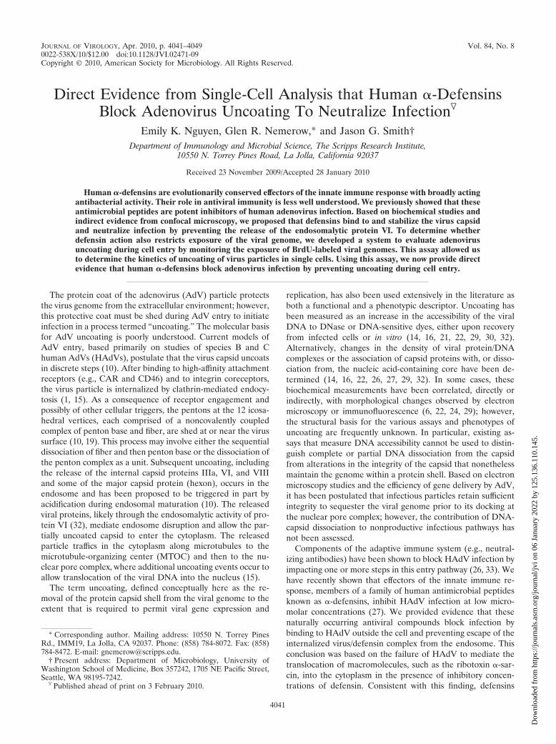
To confirm that the virus genome was labeled, we assayedvirus particles by immunoblotting for BrdU. Previous studiesshowed that HAdV-5 is stable to thermal denaturation up to43°C (26, 27). We exposed BrdU-labeled and unlabeled virusesto increasing temperatures, immobilized the viruses on nitro-cellulose using a dot blot apparatus, and probed the membranefor BrdU or for viral capsid proteins (Fig. 1). No signal wasobserved for the unlabeled virus when probed with the BrdUantibody or with BrdU-labeled virus incubated at 36°C. Incontrast, a positive signal was observed for the BrdU-labeledvirus exposed to temperatures at or above 43°C. Capsid pro-teins were detected in both samples at all temperatures, con-firming the presence of viruses. These results demonstrate thatvirus particles produced in cells exposed to BrdU package agenome incorporating the label. Moreover, they confirm thatthe BrdU epitope is masked in intact virions and can be ex-posed only upon capsid disruption.
Detection of virus uncoating in single cells. Having estab-lished conditions to produce labeled virus particles, we nextsought to ensure that the immunofluorescence signal fromBrdU was specific to uncoated virus in infected cells and thatthe labeling procedure did not alter the entry phenotype of thevirus. One tool to aid these studies was a well-characterizedHAdV-2 temperature sensitive mutant (HAdV-2ts1) that en-codes a mutation in the viral protease gene (P137L) thatgreatly reduces the infectivity of virus grown at the nonpermis-sive temperature due to a failure to uncoat and escape theendosome (12, 21, 25). HAdV-2ts1 was produced under thesame labeling conditions (4 �g/ml BrdU) as wild-type HAdV-2and was also found to contain labeled genomic DNA. Consis-tent with findings from previous studies (27), the HAdV-2ts1particles were very stable to thermal denaturation; however,the BrdU label could be detected upon boiling (Fig. 1). Inorder to use this mutant virus in immunofluorescence studies,
we required a virus with the same phenotype as HAdV-2ts1 butwith a HAdV-5 capsid to enable detection by a HAdV-5-specific antihexon MAb. To this end, we engineered the pro-tease mutation P137L, which alone is responsible for theHAdV-2ts1 phenotype (12), into a HAdV-5-based vector ex-pressing GFP. The high level of sequence conservation be-tween HAdV-2 and HAdV-5 protease (99.5% identical aminoacids) suggested that the phenotype of HAdV-5 bearing thismutation would be identical to that of HAdV-2ts1. Indeed, theHAdV-5 vector bearing this mutation (HAdV5-P137L), whenpropagated at the nonpermissive temperature, contained theprecursor forms of proteins VI, VII, and VIII, a hallmark ofthe HAdV-2ts1 phenotype (data not shown).
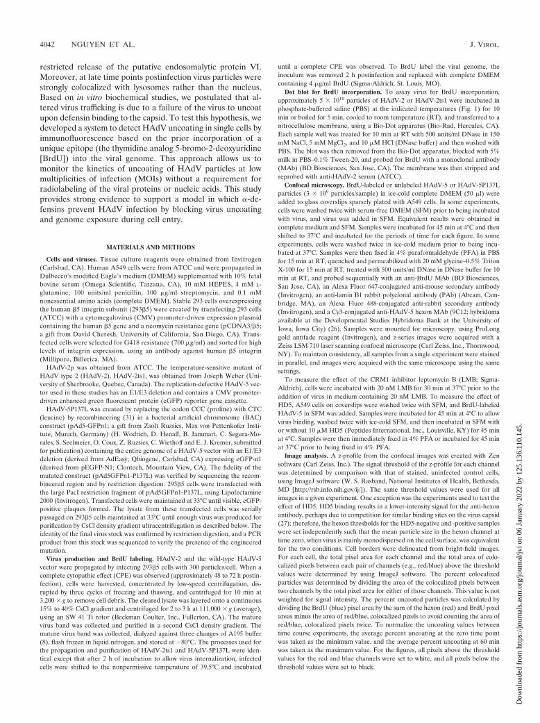
To demonstrate that exposure of the BrdU epitope in in-fected cells is dependent upon virus uncoating, we comparedthe BrdU signals from cells infected with BrdU-labeled andunlabeled wild-type HAdV-5 vectors and BrdU-labeledHAdV5-P137L at 45 min postinfection (Fig. 2). These HAdV-5-based vectors had similar growth characteristics and werelabeled under the same conditions as HAdV-2 and HAdV-2ts1. As expected, no BrdU signal above background was de-tected in cells infected with unlabeled virions. In contrast, alarge number of discrete BrdU-positive puncta (Fig. 2, blueareas) were observed in cells infected with BrdU-labeledHAdV-5. The numbers of these puncta were dramatically re-duced in samples infected with BrdU-labeled HAdV5-P137L.We observed equivalent levels of bright puncta upon stainingfor hexon (Fig. 2, red areas) in all samples. The hexon signalfor HAdV-5 was strongly colocalized with the nucleus (Fig. 2,green areas), while most of the hexon signal for HAdV-P137Lremained cytoplasmic, confirming the defective entry pheno-type of this virus. These results are consistent with a specificsignal from BrdU-labeled viruses that uncoat during cell entryand provide an assay to monitor the effect of antiviral agentson this process.
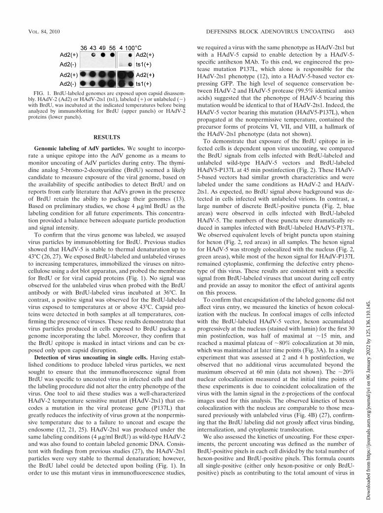
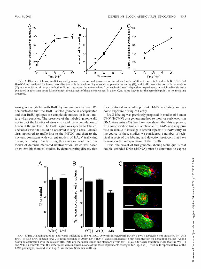
To confirm that encapsidation of the labeled genome did notaffect virus entry, we measured the kinetics of hexon colocal-ization with the nucleus. In confocal images of cells infectedwith the BrdU-labeled HAdV-5 vector, hexon accumulatedprogressively at the nucleus (stained with lamin) for the first 30min postinfection, was half of maximal at �15 min, andreached a maximal plateau of �80% colocalization at 30 min,which was maintained at later time points (Fig. 3A). In a singleexperiment that was assessed at 2 and 4 h postinfection, weobserved that no additional virus accumulated beyond themaximum observed at 60 min (data not shown). The �20%nuclear colocalization measured at the initial time points ofthese experiments is due to coincident colocalization of thevirus with the lamin signal in the z-projections of the confocalimages used for this analysis. The observed kinetics of hexoncolocalization with the nucleus are comparable to those mea-sured previously with unlabeled virus (Fig. 4B) (27), confirm-ing that the BrdU labeling did not grossly affect virus binding,internalization, and cytoplasmic translocation.
We also assessed the kinetics of uncoating. For these exper-iments, the percent uncoating was defined as the number ofBrdU-positive pixels in each cell divided by the total number ofhexon-positive and BrdU-positive pixels. This formula countsall single-positive (either only hexon-positive or only BrdU-positive) pixels as contributing to the total amount of virus in
FIG. 1. BrdU-labeled genomes are exposed upon capsid disassem-bly. HAdV-2 (Ad2) or HAdV-2ts1 (ts1), labeled (�) or unlabeled (�)with BrdU, was incubated at the indicated temperatures before beinganalyzed by immunoblotting for BrdU (upper panels) or HAdV-2proteins (lower panels).
VOL. 84, 2010 DEFENSINS BLOCK ADENOVIRUS UNCOATING 4043
Dow
nloa
ded
from
http
s://j
ourn
als.
asm
.org
/jour
nal/j
vi o
n 06
Jan
uary
202
2 by
125
.136
.110
.145
.
the cell, and pixels that were positive for both hexon and BrdUwere counted only once. Using these criteria, we observed thatthe rate of uncoating was comparable between experiments(Fig. 3B), increasing linearly up to 60 min postinfection with ahalf-life (t1/2) between 30 and 45 min postinfection. However,because of variations in the microscope settings and stainingintensities between experiments, the raw values calculated us-
ing this formula varied considerably between experiments(data not shown). Unlike that of hexon, BrdU colocalizationwith the nucleus was less rapid and more variable betweenexperiments (Fig. 3C). Maximal colocalization ranged from 50to 100% at 60 min postinfection, and the t1/2 was approxi-mately 30 min. These data demonstrate that genome exposurebegins almost immediately postinfection, likely coincident withexposure to cellular triggers (e.g., acidification) and endosomeescape, and that exposed genomes traffic to the nucleus.
The fidelity of cytoplasmic trafficking of BrdU-positive viri-ons was further assessed in experiments using the CRM1 in-hibitor leptomycin B (LMB). In enucleated cells or in cellsexposed to CRM1 inhibitors, HAdV has been shown to accu-mulate at the microtubule-organizing center (MTOC) (2, 28).Based on these observations, it has been proposed that HAdVis actively rescued from the MTOC in a CRM1-dependentfashion prior to docking at the nuclear pore complex, althoughthe molecular basis for this process is unclear. At 45 minpostinfection, inhibitory concentrations of LMB reduced thenuclear localization of hexon without affecting the extent ofuncoating (Fig. 4A and B). The modest quantitative effect ofLMB on nuclear colocalization is primarily due to the fact thatin the z-projections of confocal images used for these analyses,the virus that has accumulated at the MTOC often eitherpartially or completely overlaps the nucleus (Fig. 4C, yellowareas); nonetheless, the dramatic effect of LMB on the subcel-lular distribution of HAdV is readily apparent (compare Fig.4C with Fig. 2). Thus, intracellular trafficking of HAdV to theMTOC is not disrupted by BrdU labeling.
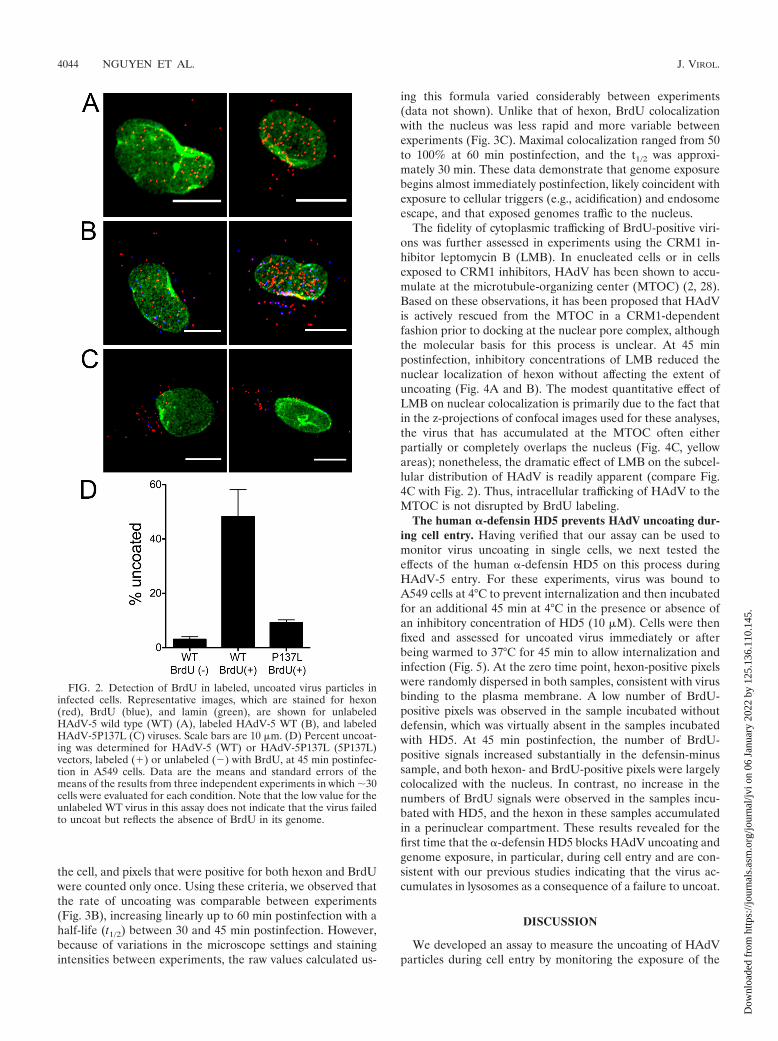
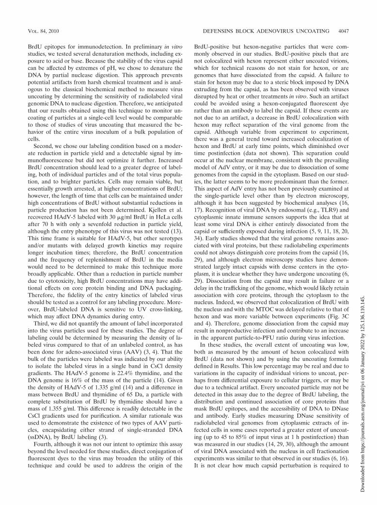
The human �-defensin HD5 prevents HAdV uncoating dur-ing cell entry. Having verified that our assay can be used tomonitor virus uncoating in single cells, we next tested theeffects of the human �-defensin HD5 on this process duringHAdV-5 entry. For these experiments, virus was bound toA549 cells at 4°C to prevent internalization and then incubatedfor an additional 45 min at 4°C in the presence or absence ofan inhibitory concentration of HD5 (10 �M). Cells were thenfixed and assessed for uncoated virus immediately or afterbeing warmed to 37°C for 45 min to allow internalization andinfection (Fig. 5). At the zero time point, hexon-positive pixelswere randomly dispersed in both samples, consistent with virusbinding to the plasma membrane. A low number of BrdU-positive pixels was observed in the sample incubated withoutdefensin, which was virtually absent in the samples incubatedwith HD5. At 45 min postinfection, the number of BrdU-positive signals increased substantially in the defensin-minussample, and both hexon- and BrdU-positive pixels were largelycolocalized with the nucleus. In contrast, no increase in thenumbers of BrdU signals were observed in the samples incu-bated with HD5, and the hexon in these samples accumulatedin a perinuclear compartment. These results revealed for thefirst time that the �-defensin HD5 blocks HAdV uncoating andgenome exposure, in particular, during cell entry and are con-sistent with our previous studies indicating that the virus ac-cumulates in lysosomes as a consequence of a failure to uncoat.
DISCUSSION
We developed an assay to measure the uncoating of HAdVparticles during cell entry by monitoring the exposure of the
FIG. 2. Detection of BrdU in labeled, uncoated virus particles ininfected cells. Representative images, which are stained for hexon(red), BrdU (blue), and lamin (green), are shown for unlabeledHAdV-5 wild type (WT) (A), labeled HAdV-5 WT (B), and labeledHAdV-5P137L (C) viruses. Scale bars are 10 �m. (D) Percent uncoat-ing was determined for HAdV-5 (WT) or HAdV-5P137L (5P137L)vectors, labeled (�) or unlabeled (�) with BrdU, at 45 min postinfec-tion in A549 cells. Data are the means and standard errors of themeans of the results from three independent experiments in which �30cells were evaluated for each condition. Note that the low value for theunlabeled WT virus in this assay does not indicate that the virus failedto uncoat but reflects the absence of BrdU in its genome.
4044 NGUYEN ET AL. J. VIROL.
Dow
nloa
ded
from
http
s://j
ourn
als.
asm
.org
/jour
nal/j
vi o
n 06
Jan
uary
202
2 by
125
.136
.110
.145
.
virus genome labeled with BrdU by immunofluorescence. Wedemonstrated that the BrdU-labeled genome is encapsidatedand that BrdU epitopes are completely masked in intact, ma-ture virus particles. The presence of the labeled genome didnot impact the kinetics of virus entry and the accumulation ofhexon at the nucleus. The BrdU signal was specific to labeled,uncoated virus that could be observed in single cells. Labeledvirus appeared to traffic first to the MTOC and then to thenucleus, consistent with current models of HAdV traffickingduring cell entry. Finally, using this assay we confirmed ourmodel of defensin-mediated neutralization, which was basedon in vitro biochemical studies, by demonstrating directly that
these antiviral molecules prevent HAdV uncoating and ge-nome exposure during cell entry.
BrdU labeling was previously proposed in studies of humanCMV (HCMV) as a general method to monitor early events inDNA virus entry (23). We have now shown that this approach,with some modifications, is applicable to HAdV and may pro-vide an avenue to investigate several aspects of HAdV entry. Inthe course of these studies, we considered a number of tech-nical aspects of the labeling and detection protocols that havebearing on the interpretation of the results.
First, one caveat of this genome-labeling technique is thatdouble-stranded DNA (dsDNA) must be denatured to expose
FIG. 3. Kinetics of hexon trafficking and genome exposure and translocation in infected cells. A549 cells were infected with BrdU-labeledHAdV-5 and analyzed for hexon colocalization with the nucleus (A), normalized percent uncoating (B), and BrdU colocalization with the nucleus(C) at the indicated times postinfection. Points represent the mean values from each of three independent experiments in which �30 cells wereevaluated at each time point. Lines connect the averages of these mean values. In panel C, no value is given for the zero time point, as no uncoatingoccurred.
FIG. 4. BrdU labeling does not alter virus trafficking to the MTOC. A549 cells infected with HAdV-5 (WT), labeled (�) or unlabeled (�) withBrdU, or with BrdU-labeled HAdV-5 in the presence of 20 nM LMB (LMB) were evaluated at 45 min postinfection for percent uncoating (A) andhexon colocalization with the nucleus (B). Data are the mean values and standard errors for �30 cells for each condition. Note that the WT(�)and WT(�) controls from this experiment were included as one of the three experiments averaged for Fig. 2. (C) Three cells representative of theLMB phenotype, colored as in Fig. 2, are shown. Scale bar is 10 �m.
VOL. 84, 2010 DEFENSINS BLOCK ADENOVIRUS UNCOATING 4045
Dow
nloa
ded
from
http
s://j
ourn
als.
asm
.org
/jour
nal/j
vi o
n 06
Jan
uary
202
2 by
125
.136
.110
.145
.
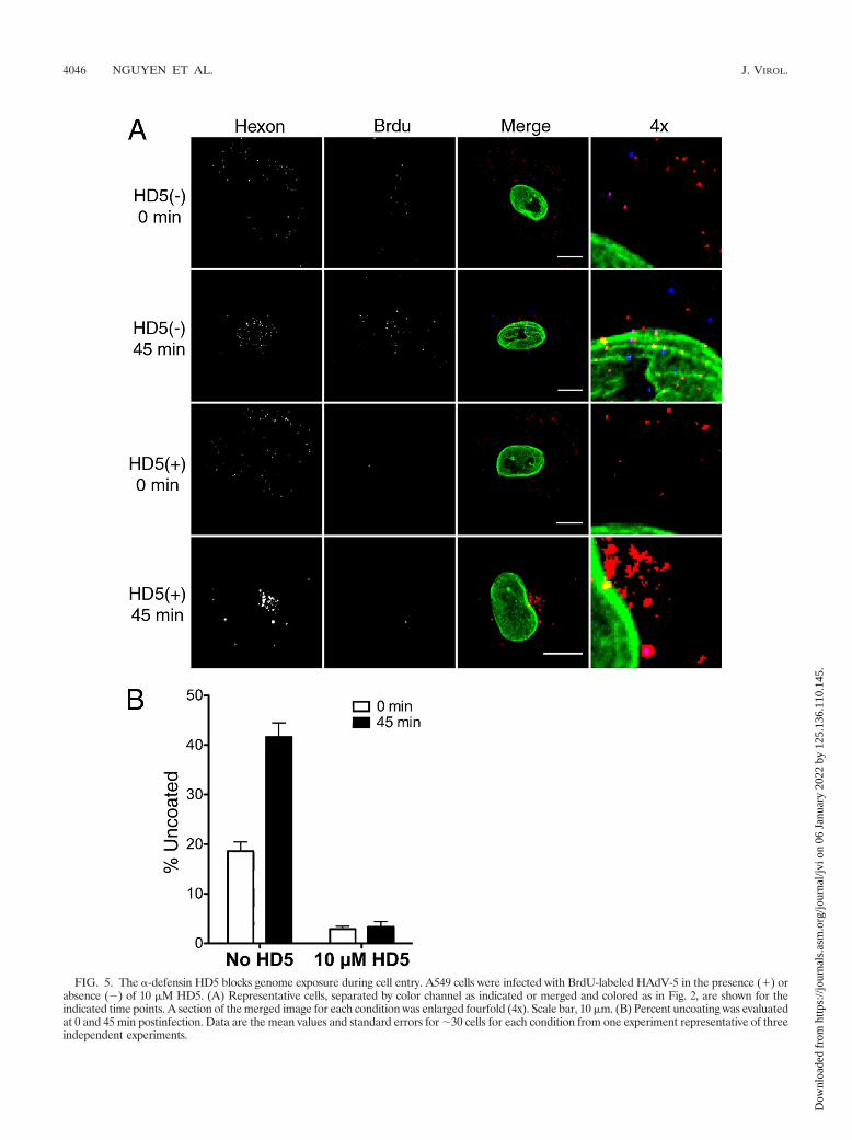
FIG. 5. The �-defensin HD5 blocks genome exposure during cell entry. A549 cells were infected with BrdU-labeled HAdV-5 in the presence (�) orabsence (�) of 10 �M HD5. (A) Representative cells, separated by color channel as indicated or merged and colored as in Fig. 2, are shown for theindicated time points. A section of the merged image for each condition was enlarged fourfold (4x). Scale bar, 10 �m. (B) Percent uncoating was evaluatedat 0 and 45 min postinfection. Data are the mean values and standard errors for �30 cells for each condition from one experiment representative of threeindependent experiments.
4046 NGUYEN ET AL. J. VIROL.
Dow
nloa
ded
from
http
s://j
ourn
als.
asm
.org
/jour
nal/j
vi o
n 06
Jan
uary
202
2 by
125
.136
.110
.145
.
BrdU epitopes for immunodetection. In preliminary in vitrostudies, we tested several denaturation methods, including ex-posure to acid or base. Because the stability of the virus capsidcan be affected by extremes of pH, we chose to denature theDNA by partial nuclease digestion. This approach preventspotential artifacts from harsh chemical treatment and is anal-ogous to the classical biochemical method to measure virusuncoating by determining the sensitivity of radiolabeled viralgenomic DNA to nuclease digestion. Therefore, we anticipatedthat our results obtained using this technique to monitor un-coating of particles at a single-cell level would be comparableto those of studies of virus uncoating that measured the be-havior of the entire virus inoculum of a bulk population ofcells.
Second, we chose our labeling condition based on a moder-ate reduction in particle yield and a detectable signal by im-munofluorescence but did not optimize it further. IncreasedBrdU concentration should lead to a greater degree of label-ing, both of individual particles and of the total virus popula-tion, and to brighter particles. Cells may remain viable, butessentially growth arrested, at higher concentrations of BrdU;however, the length of time that cells can be maintained underhigh concentrations of BrdU without substantial reductions inparticle production has not been determined. Kjellen et al.recovered HAdV-5 labeled with 30 �g/ml BrdU in HeLa cellsafter 70 h with only a sevenfold reduction in particle yield,although the entry phenotype of this virus was not tested (13).This time frame is suitable for HAdV-5, but other serotypesand/or mutants with delayed growth kinetics may requirelonger incubation times; therefore, the BrdU concentrationand the frequency of replenishment of BrdU in the mediawould need to be determined to make this technique morebroadly applicable. Other than a reduction in particle numberdue to cytotoxicity, high BrdU concentrations may have addi-tional effects on core protein binding and DNA packaging.Therefore, the fidelity of the entry kinetics of labeled virusshould be tested as a control for any labeling procedure. More-over, BrdU-labeled DNA is sensitive to UV cross-linking,which may affect DNA dynamics during entry.
Third, we did not quantify the amount of label incorporatedinto the virus particles used for these studies. The degree oflabeling could be determined by measuring the density of la-beled virus compared to that of an unlabeled control, as hasbeen done for adeno-associated virus (AAV) (3, 4). That thebulk of the particles were labeled was indicated by our abilityto isolate the labeled virus in a single band in CsCl densitygradients. The HAdV-5 genome is 22.4% thymidine, and theDNA genome is 16% of the mass of the particle (14). Giventhe density of HAdV-5 of 1.335 g/ml (14) and a difference inmass between BrdU and thymidine of 65 Da, a particle withcomplete substitution of BrdU by thymidine should have amass of 1.355 g/ml. This difference is readily detectable in theCsCl gradients used for purification. A similar rationale wasused to demonstrate the existence of two types of AAV parti-cles, encapsidating either strand of single-stranded DNA(ssDNA), by BrdU labeling (3).
Fourth, although it was not our intent to optimize this assaybeyond the level needed for these studies, direct conjugation offluorescent dyes to the virus may broaden the utility of thistechnique and could be used to address the origin of the
BrdU-positive but hexon-negative particles that were com-monly observed in our studies. BrdU-positive pixels that arenot colocalized with hexon represent either uncoated virions,which for technical reasons do not stain for hexon, or aregenomes that have dissociated from the capsid. A failure tostain for hexon may be due to a steric block imposed by DNAextruding from the capsid, as has been observed with virusesdisrupted by heat or other treatments in vitro. Such an artifactcould be avoided using a hexon-conjugated fluorescent dyerather than an antibody to label the capsid. If these events arenot due to an artifact, a decrease in BrdU colocalization withhexon may reflect separation of the viral genome from thecapsid. Although variable from experiment to experiment,there was a general trend toward increased colocalization ofhexon and BrdU at early time points, which diminished overtime postinfection (data not shown). This separation couldoccur at the nuclear membrane, consistent with the prevailingmodel of AdV entry, or it may be due to dissociation of somegenomes from the capsid in the cytoplasm. Based on our stud-ies, the latter seems to be more predominant than the former.This aspect of AdV entry has not been previously examined atthe single-particle level other than by electron microscopy,although it has been suggested by biochemical analyses (16,17). Recognition of viral DNA by endosomal (e.g., TLR9) andcytoplasmic innate immune sensors supports the idea that atleast some viral DNA is either entirely dissociated from thecapsid or sufficiently exposed during infection (5, 9, 11, 18, 20,34). Early studies showed that the viral genome remains asso-ciated with viral proteins, but these radiolabeling experimentscould not always distinguish core proteins from the capsid (16,29), and although electron microscopy studies have demon-strated largely intact capsids with dense centers in the cyto-plasm, it is unclear whether they have undergone uncoating (6,29). Dissociation from the capsid may result in failure or adelay in the trafficking of the genome, which would likely retainassociation with core proteins, through the cytoplasm to thenucleus. Indeed, we observed that colocalization of BrdU withthe nucleus and with the MTOC was delayed relative to that ofhexon and was more variable between experiments (Fig. 3Cand 4). Therefore, genome dissociation from the capsid mayresult in nonproductive infection and contribute to an increasein the apparent particle-to-PFU ratio during virus infection.
In these studies, the overall extent of uncoating was low,both as measured by the amount of hexon colocalized withBrdU (data not shown) and by using the uncoating formuladefined in Results. This low percentage may be real and due tovariations in the capacity of individual virions to uncoat, per-haps from differential exposure to cellular triggers, or may bedue to a technical artifact. Every uncoated particle may not bedetected in this assay due to the degree of BrdU labeling, thedistribution and continued association of core proteins thatmask BrdU epitopes, and the accessibility of DNA to DNaseand antibody. Early studies measuring DNase sensitivity ofradiolabeled viral genomes from cytoplasmic extracts of in-fected cells in some cases reported a greater extent of uncoat-ing (up to 45 to 85% of input virus at 1 h postinfection) thanwas measured in our studies (14, 29, 30), although the amountof viral DNA associated with the nucleus in cell fractionationexperiments was similar to that observed in our studies (6, 16).It is not clear how much capsid perturbation is required to
VOL. 84, 2010 DEFENSINS BLOCK ADENOVIRUS UNCOATING 4047
Dow
nloa
ded
from
http
s://j
ourn
als.
asm
.org
/jour
nal/j
vi o
n 06
Jan
uary
202
2 by
125
.136
.110
.145
.
generate nuclease sensitivity and to be measured as uncoatedfor either assay. A hole created by removal of just the fiber isunlikely to be sufficient to allow DNA out of or enzymes andantibodies into the interior of the capsid. Therefore, it is likelythat at least one or more of the vertices (penton base and fiber)is removed. Indeed, genome sensitivity to nuclease has beenshown to correlate with vertex removal (22, 24, 29). BrdU maybecome detectable in this assay, therefore, only upon extensivecapsid disruption or when the viral genome has separated orbecome partially externalized from the capsid. Less-drasticuncoating events, although sufficient to release the viral mem-brane lytic proteins and mediate endosomalysis, may not bedetected.
We also observed that uncoating continues to increase oncemaximal hexon colocalization with the nucleus has beenreached. Uncoating may be asynchronous, and the �20% ofvirus that does not reach the nucleus may contribute to thiseffect. Alternatively, this effect may be due to additional un-coating events that occur at the nuclear membrane. Althoughbeyond the scope of our current studies, this technique mayallow for a more-careful analysis of the subcellular localizationof uncoating as well as the fate of the viral DNA in the nucleusat late times postinfection.
We developed this uncoating assay in order to more fullyexplore the mechanism of defensin-mediated neutralization ofAdV infection. Our previous confocal studies provided indirectevidence that AdV fails to uncoat in the presence of inhibitoryconcentrations of defensin, because we observed that most ofthe virus became trapped in the lysosome rather than reachingthe nucleus (27). This effect was also measured as a reductionin the capacity of AdV to mediate sarcin translocation into thecytoplasm in the presence of defensin. From these studies, weproposed that a failure to mediate endosomalysis was due to afailure to uncoat. Biochemical studies showing that defensinbinding increased the thermostability of AdV also supportedthis model. Nonetheless, these studies provided no direct evi-dence that AdV fails to uncoat during entry. An alternativemodel is that defensin could block the membrane lytic activityof proteins (e.g., VI) released from the uncoated virus ratherthan uncoating per se. The marked reduction in BrdU signals incells infected with the virus in the presence of defensin, how-ever, provides strong evidence that these molecules directlyblock uncoating during cell entry. Although we cannot formallyexclude the possibility that the reduction in BrdU signals is dueto HD5 binding to and shielding the DNA directly rather thanpreventing uncoating, we feel that this is unlikely, given thenumber of defensin molecules that would be needed to uni-formly bind the nucleic acid for this to occur and our previouslypublished studies demonstrating that endosome lysis is alsoimpaired. In our current studies, virus was prebound to cellsand monodispersed prior to exposure to defensin. These con-ditions mimic those used in our previous studies to measurethe time course of defensin-mediated inhibition of infectionand provide further evidence that AdV is neutralized by de-fensin due to a discrete block of uncoating rather than a more-general effect such as virus aggregation, which has been pro-posed as the mechanism of defensin-mediated neutralizationof polyomavirus (7). In addition to a blockage of BrdU expo-sure at the 45-min time point postinfection, we also observed areduction in the number of BrdU signals at the zero time point.
Up to 20% uncoating at time zero was also observed in earlystudies, as measured by DNase sensitivity (14). In both treatedand untreated samples, virus was first incubated with cells for45 min at 4°C to allow binding and to synchronize the infection.Therefore, a certain amount of uncoating must occur duringthe subsequent 45-min incubation at 4°C, and defensin stabi-lization of the capsid also prevents this from occurring. Takentogether, these observations provide evidence to support theconcept that defensins neutralize AdV infection by stabilizingthe capsid and preventing uncoating. Although not amenableto live-cell imaging, the assay described here may be used toevaluate the effects of other molecules, including antibodies,clotting factors, complement components, and small-moleculeinhibitors, or mutations in viral proteins, such as the mem-brane lytic protein VI, on AdV uncoating. In addition, thisassay may provide insight into the subcellular localization ofuncoating in relation to innate immune sensors of viral DNAduring infection.
ACKNOWLEDGMENTS
We thank Milena Iacobelli-Martinez for the creation of the 293�5cells and Crystal Moyer for helpful discussions.
This work was supported by NIH grants HL054352 and EY11431 toG.R.N. E.K.N. was supported by the Immunology and Microbial Sci-ences Summer Program at TSRI. The authors declare no competingfinancial interests.
This is Scripps Research Institute manuscript number 20478.
REFERENCES
1. Arnberg, N. 2009. Adenovirus receptors: implications for tropism, treatmentand targeting. Rev. Med. Virol. 19:165–178.
2. Bailey, C. J., R. G. Crystal, and P. L. Leopold. 2003. Association of adeno-virus with the microtubule organizing center. J. Virol. 77:13275–13287.
3. Berns, K. I., and S. Adler. 1972. Separation of two types of adeno-associatedvirus particles containing complementary polynucleotide chains. J. Virol.9:394–396.
4. Berns, K. I., and J. A. Rose. 1970. Evidence for a single-stranded adenovirus-associated virus genome: isolation and separation of complementary singlestrands. J. Virol. 5:693–699.
5. Cerullo, V., M. P. Seiler, V. Mane, N. Brunetti-Pierri, C. Clarke, T. K.Bertin, J. R. Rodgers, and B. Lee. 2007. Toll-like receptor 9 triggers aninnate immune response to helper-dependent adenoviral vectors. Mol. Ther.15:378–385.
6. Chardonnet, Y., and S. Dales. 1970. Early events in the interaction ofadenoviruses with HeLa cells. I. Penetration of type 5 and intracellularrelease of the DNA genome. Virology 40:462–477.
7. Dugan, A. S., M. S. Maginnis, J. A. Jordan, M. L. Gasparovic, K. Manley, R.Page, G. Williams, E. Porter, B. A. O’Hara, and W. J. Atwood. 2008. Humanalpha-defensins inhibit BK virus infection by aggregating virions and block-ing binding to host cells. J. Biol. Chem. 283:31125–31132.
8. Evans, R. K., D. K. Nawrocki, L. A. Isopi, D. M. Williams, D. R. Casimiro,S. Chin, M. Chen, D. M. Zhu, J. W. Shiver, and D. B. Volkin. 2004. Devel-opment of stable liquid formulations for adenovirus-based vaccines.J. Pharm. Sci. 93:2458–2475.
9. Fejer, G., L. Drechsel, J. Liese, U. Schleicher, Z. Ruzsics, N. Imelli, U. F.Greber, S. Keck, B. Hildenbrand, A. Krug, C. Bogdan, and M. A. Freuden-berg. 2008. Key role of splenic myeloid DCs in the IFN-alphabeta responseto adenoviruses in vivo. PLoS Pathog. 4:e1000208.
10. Greber, U. F., M. Willetts, P. Webster, and A. Helenius. 1993. Stepwisedismantling of adenovirus 2 during entry into cells. Cell 75:477–486.
11. Iacobelli-Martinez, M., and G. R. Nemerow. 2007. Preferential activation ofToll-like receptor nine by CD46-utilizing adenoviruses. J. Virol. 81:1305–1312.
12. Imelli, N., Z. Ruzsics, D. Puntener, M. Gastaldelli, and U. F. Greber. 2009.Genetic reconstitution of the human adenovirus type 2 temperature-sensitive1 mutant defective in endosomal escape. Virol. J. 6:174.
13. Kjellen, L., H. G. Pereira, R. C. Valentine, and J. A. Armstrong. 1963. Ananalysis of adenovirus particles and soluble antigens produced in the pres-ence of 5-bromodeoxyuridine. Nature 199:1210–1211.
14. Lawrence, W. C., and H. S. Ginsberg. 1967. Intracellular uncoating of type 5adenovirus deoxyribonucleic acid. J. Virol. 1:851–867.
15. Leopold, P. L., and R. G. Crystal. 2007. Intracellular trafficking of adenovi-rus: many means to many ends. Adv. Drug Deliv. Rev. 59:810–821.
4048 NGUYEN ET AL. J. VIROL.
Dow
nloa
ded
from
http
s://j
ourn
als.
asm
.org
/jour
nal/j
vi o
n 06
Jan
uary
202
2 by
125
.136
.110
.145
.
16. Lonberg-Holm, K., and L. Philipson. 1969. Early events of virus-cell inter-action in an adenovirus system. J. Virol. 4:323–338.
17. Mirza, M. A., and J. Weber. 1979. Uncoating of adenovirus type 2. J. Virol.30:462–471.
18. Muruve, D. A., V. Petrilli, A. K. Zaiss, L. R. White, S. A. Clark, P. J. Ross,R. J. Parks, and J. Tschopp. 2008. The inflammasome recognizes cytosolicmicrobial and host DNA and triggers an innate immune response. Nature452:103–107.
19. Nakano, M. Y., K. Boucke, M. Suomalainen, R. P. Stidwill, and U. F. Greber.2000. The first step of adenovirus type 2 disassembly occurs at the cellsurface, independently of endocytosis and escape to the cytosol. J. Virol.74:7085–7095.
20. Nociari, M., O. Ocheretina, J. W. Schoggins, and E. Falck-Pedersen. 2007.Sensing infection by adenovirus: Toll-like receptor-independent viral DNArecognition signals activation of the interferon regulatory factor 3 masterregulator. J. Virol. 81:4145–4157.
21. Perez-Berna, A. J., R. Marabini, S. H. Scheres, R. Menendez-Conejero, I. P.Dmitriev, D. T. Curiel, W. F. Mangel, S. J. Flint, and C. San Martin. 2009.Structure and uncoating of immature adenovirus. J. Mol. Biol. 392:547–557.
22. Prage, L., U. Pettersson, S. Hoglund, K. Lonberg-Holm, and L. Philipson.1970. Structural proteins of adenoviruses. IV. Sequential degradation of theadenovirus type 2 virion. Virology 42:341–358.
23. Rosenke, K., and E. A. Fortunato. 2004. Bromodeoxyuridine-labeled viralparticles as a tool for visualization of the immediate-early events of humancytomegalovirus infection. J. Virol. 78:7818–7822.
24. Russell, W. C., R. C. Valentine, and H. G. Pereira. 1967. The effect of heaton the anatomy of the adenovirus. J. Gen. Virol. 1:509–522.
25. Silvestry, M., S. Lindert, J. G. Smith, O. Maier, C. M. Wiethoff, G. R.Nemerow, and P. L. Stewart. 2009. Cryo-electron microscopy structure ofadenovirus type 2 temperature-sensitive mutant 1 reveals insight into the cellentry defect. J. Virol. 83:7375–7383.
26. Smith, J. G., A. Cassany, L. Gerace, R. Ralston, and G. R. Nemerow. 2008.Neutralizing antibody blocks adenovirus infection by arresting microtubule-dependent cytoplasmic transport. J. Virol. 82:6492–6500.
27. Smith, J. G., and G. R. Nemerow. 2008. Mechanism of adenovirus neutral-ization by human alpha-defensins. Cell Host Microbe 3:11–19.
28. Strunze, S., L. C. Trotman, K. Boucke, and U. F. Greber. 2005. Nucleartargeting of adenovirus type 2 requires CRM1-mediated nuclear export.Mol. Biol. Cell 16:2999–3009.
29. Sussenbach, J. S. 1967. Early events in the infection process of adenovirustype 5 in HeLa cells. Virology 33:567–574.
30. Svensson, U., and R. Persson. 1984. Entry of adenovirus 2 into HeLa cells.J. Virol. 51:687–694.
31. Warming, S., N. Costantino, D. L. Court, N. A. Jenkins, and N. G. Copeland.2005. Simple and highly efficient BAC recombineering using galK selection.Nucleic Acids Res. 33:e36.
32. Wiethoff, C. M., H. Wodrich, L. Gerace, and G. R. Nemerow. 2005. Adeno-virus protein VI mediates membrane disruption following capsid disassem-bly. J. Virol. 79:1992–2000.
33. Wohlfart, C. 1988. Neutralization of adenoviruses: kinetics, stoichiometry,and mechanisms. J. Virol. 62:2321–2328.
34. Zhu, J., X. Huang, and Y. Yang. 2007. Innate immune response to adenoviralvectors is mediated by both Toll-like receptor-dependent and -independentpathways. J. Virol. 81:3170–3180.
VOL. 84, 2010 DEFENSINS BLOCK ADENOVIRUS UNCOATING 4049
Dow
nloa
ded
from
http
s://j
ourn
als.
asm
.org
/jour
nal/j
vi o
n 06
Jan
uary
202
2 by
125
.136
.110
.145
.