Supplemental Information Direct transcriptional...
23
Supplemental Information Direct transcriptional regulation of Nrp2 by COUP-TFII modulates multiple steps in murine lymphatic vessel development Fu-Jung Lin, Xinpu Chen, Jun Qin, Young-Kwon Hong, Ming-Jer Tsai, and Sophia Y. Tsai Supplemental Experimental Procedures Mouse Strain and Histology The SM22αCre mouse strain has been described previously (1). COUP-TFII flox/flox mice were crossed with SM22αCre; COUP-TFII flox/+ to generate SM22αCre; COUP- TFII flox/flox embryos. The mouse strain was maintained in a mixed genetic background (129/Sv x C57BL/6) and received standard rodent chow. Histological Analysis and Immunohistochemistry Mouse tissues were fixed with 4% paraformaldehyde (PFA), dehydrated, and embedded in paraffin. For histological analysis, sections (7 μm) were stained with hematoxylin and eosin (H&E). For immunohistochemistry, deparaffinized sections after antigen retrieval were blocked with 5% donkey serum and a biotin-blocking system (DakoCytomation). The following primary antibodies were used for immunostaining of mouse tissues: monoclonal mouse anti-human COUP-TFII (Perseus Proteomics), polyclonal goat anti- mouse EphB4, polyclonal goat anti-mouse VEGFR3, polyclonal goat anti-rat Neuropilin- 1, polyclonal goat anti-rat Neuropilin-2, polyclonal goat anti-mouse Endoglin (R&D Systems), monoclonal anti-rabbit VEGFR2 (Cell Signaling Technology), rabbit anti- 1
Transcript of Supplemental Information Direct transcriptional...
Supplemental Information
Direct transcriptional regulation of Nrp2 by COUP-TFII modulates multiple steps
in murine lymphatic vessel development
Fu-Jung Lin, Xinpu Chen, Jun Qin, Young-Kwon Hong, Ming-Jer Tsai, and Sophia Y.
Tsai
Supplemental Experimental Procedures
Mouse Strain and Histology
The SM22αCre mouse strain has been described previously (1). COUP-TFIIflox/flox mice
were crossed with SM22αCre; COUP-TFIIflox/+ to generate SM22αCre; COUP-
TFIIflox/flox embryos. The mouse strain was maintained in a mixed genetic background
(129/Sv x C57BL/6) and received standard rodent chow.
Histological Analysis and Immunohistochemistry
Mouse tissues were fixed with 4% paraformaldehyde (PFA), dehydrated, and embedded
in paraffin. For histological analysis, sections (7 μm) were stained with hematoxylin and
eosin (H&E). For immunohistochemistry, deparaffinized sections after antigen retrieval
were blocked with 5% donkey serum and a biotin-blocking system (DakoCytomation).
The following primary antibodies were used for immunostaining of mouse tissues:
monoclonal mouse anti-human COUP-TFII (Perseus Proteomics), polyclonal goat anti-
mouse EphB4, polyclonal goat anti-mouse VEGFR3, polyclonal goat anti-rat Neuropilin-
1, polyclonal goat anti-rat Neuropilin-2, polyclonal goat anti-mouse Endoglin (R&D
Systems), monoclonal anti-rabbit VEGFR2 (Cell Signaling Technology), rabbit anti-
1
Prox1, rabbit anti-mouse LYVE1 (AngioBio), hamster anti-mouse Podoplanin (clone
8.1.1; Hybridoma Bank, University of Iowa), rabbit anti-laminin (DakoCytomation),
rabbit anti-collagen IV (Abcam), mouse anti-human Ki67 (BD Biosciences), and
monoclonal rat anti-mouse CD31 (PECAM) (clone MEC 13.3; BD Biosciences).
Sections were washed with PBST buffer and incubated with biotinylated secondary
antibodies (Jackson ImmunoResearch). Signal detection was carried out with the Avidin-
Biotin Complex kit (Vector Laboratories) or Tyramide Signal Amplification system
(TSA, Invitrogen). Peroxidase activity was visualized with 3,3’-diaminobenzidine (DAB,
Vector Laboratories). Nuclear staining was carried out with 4,6-diamidino-2-
phenylindole (DAPI; Sigma), and sections were mounted with Vectashield mounting
medium (Vector Laboratories) prior to imaging. Images were captured with a Zeiss
Axiophoto fluorescence microscope.
For whole-mount immunofluorescence staining, embryonic back skins or dorsal segments
of adult mouse ears were dissected and fixed with 4% PFA overnight at 4ºC. Primary
antibodies in a blocking solution (2% nonfat skim milk in phosphate buffered saline
(PBS) with 0.5% Triton X-100) were added and incubated overnight at 4ºC. Tissues were
washed for 5 h and incubated with biotinylated secondary antibodies (Jackson
ImmunoResearch) for 1 h at room temperature. Signals were amplified with a TSA kit
(Invitrogen). Specimens were imaged and analyzed using the Zeiss Axioplan 2 imaging
microscope with MetaMorph software.
2
Western Blot Analysis
24 hours after transfection, LECs were lysed with 1X RIPA buffer containing 1X TBS,
1% Nonidet P-40, 0.5% sodium deoxycholate, 0.1% SDS, a protease inhibitor cocktail
(Roche Applied Science), and a phosphatase inhibitor cocktail (Sigma) for 30 minutes on
ice. After centrifugation for 15 minutes, the supernatant was collected, and protein
content of the samples was analyzed according to the Bradford method. Proteins were
loaded onto SDS-polyacrylamide gels and blotted onto PVDF membranes (Bio-Rad
Laboratories). Western blots were performed using antibodies directed against COUP-
TFII (Perseus Proteomics), Neuropilin-2, and HRP-conjugated β-actin (Santa Cruz
Biotechnology). Enhanced chemiluminescence was performed according to the
manufacturer’s instructions (Amershan Biosciences, UK).
FACS Analysis
Mouse lung EC isolation was carried out as described (2). Briefly, mice were
anesthetized with avertin, perfused with PBS, intra-tracheally injected with 1 mL dispase
(BD Biosciences) and 1mL 1% agarose. Lungs were isolated, minced, and treated with 2
mg/mL collagenase/dispase (Roche) and 10 ug/mL DNase (Sigma) in PBS at 37ºC for 45
minutes to produce a single-cell suspension. The tissue was then filtered through 100 μm
and 40 μm cell strainers (BD Biosciences), and centrifuged at 2000 rpm, 5 minutes.
Subsequently, cells were resuspended and labeled with a biotinylated anti-CD31 antibody
(BD Biosciences) and anti-bitotin microbeads (Miltenyi Biotec). ECs were first separated
from other components by using AutoMACS (Miltenyi Biotec), and then Fluorescence
Activated Cell Sorting (FACS) was performed.
3
For EC isolation from mouse embryos, E14.5 mouse embryos were dissected and the
embryonic liver was removed microscopically. The embryos were then minced into
pieces and digested with 2 mg/mL collagenase/dispase (Roche) and 25 ug/mL DNase
(Sigma) in PBS at 37ºC for 45 minutes. Embryonic ECs were isolated by the method
described above.
In Vitro Proliferation Assay
Human primary LECs were transfected with scrambled or COUP-TFII siRNA for 48
hours. SiRNA-treated LECs were re-plated and incubated at 2.4 x10⁴ cells per well in 48-
well plates coated with fibronectin (10 ng/mL), deprived of FBS for 12 hours, followed
by stimulation with or without 60 ng/mL of VEGF-C in a cultured medium for 48 hours
or 72 hours. Then cells were trypsinized at the indicated time points and cell numbers
were counted using a hemocytometer.
Construction of Luciferase Plasmid and Luciferase Assay
The mouse Nrp2 promoter fragment encompassing nucleotides -2885 to +920 was
generated by PCR with the forward primer 5’-CCAAGAGCTCGCTGAATCCAGCTCC
ACAAACTCC-3’ (SacI site underlined) and the reverse primer 5’-AAAACTCGAGTTT
TGACAGAGAGGCTCT CTCCGG-3’ (XhoI site underlined), using the Expand High
Fidelity system (Roche), according to the manufacturer’s instructions. BAC clone RP24-
163014 (CHORI) was used as a template and the PCR product was digested with SacI
and XhoI and ligated into the SacI/XhoI sites of the pGL2 basic luciferase reporter vector
(Promega). The construct was verified by sequencing. HEK293T cells (2 x 10⁵ per well)
4
were cotransfected with 150 ng of pGL2-Nrp2 or pGL2 basic vectors in the presence of
an empty control vector or a COUP-TFII expressing vector using Lipofectamine 2000
(Invitrogen). Cells were cotransfected with 100 ng of β-gal plasmid to account for
variation in transfection efficiencies. Luciferase activity was measured using the
luciferase assay system following the manufacturer’s protocol (Promega). Luciferase
activity was normalized to β-gal activity.
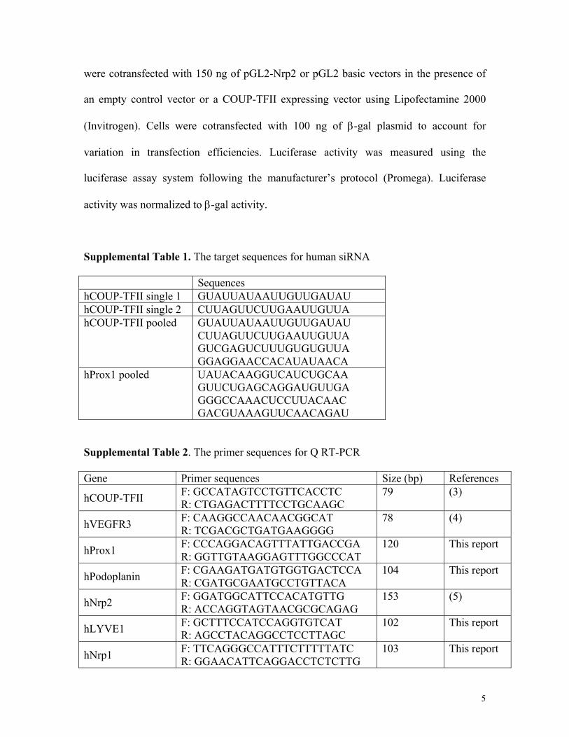
Supplemental Table 1. The target sequences for human siRNA
Sequences hCOUP-TFII single 1 GUAUUAUAAUUGUUGAUAU hCOUP-TFII single 2 CUUAGUUCUUGAAUUGUUA hCOUP-TFII pooled GUAUUAUAAUUGUUGAUAU
CUUAGUUCUUGAAUUGUUA GUCGAGUCUUUGUGUGUUA GGAGGAACCACAUAUAACA
hProx1 pooled UAUACAAGGUCAUCUGCAA GUUCUGAGCAGGAUGUUGA GGGCCAAACUCCUUACAAC GACGUAAAGUUCAACAGAU
Supplemental Table 2. The primer sequences for Q RT-PCR
Gene Primer sequences Size (bp) References
hCOUP-TFII F: GCCATAGTCCTGTTCACCTC R: CTGAGACTTTTCCTGCAAGC
79 (3)
hVEGFR3 F: CAAGGCCAACAACGGCAT R: TCGACGCTGATGAAGGGG
78 (4)
hProx1 F: CCCAGGACAGTTTATTGACCGA R: GGTTGTAAGGAGTTTGGCCCAT
120 This report
hPodoplanin F: CGAAGATGATGTGGTGACTCCA R: CGATGCGAATGCCTGTTACA
104 This report
hNrp2 F: GGATGGCATTCCACATGTTG R: ACCAGGTAGTAACGCGCAGAG
153 (5)
hLYVE1 F: GCTTTCCATCCAGGTGTCAT R: AGCCTACAGGCCTCCTTAGC
102 This report
hNrp1 F: TTCAGGGCCATTTCTTTTTATC R: GGAACATTCAGGACCTCTCTTG
103 This report
5
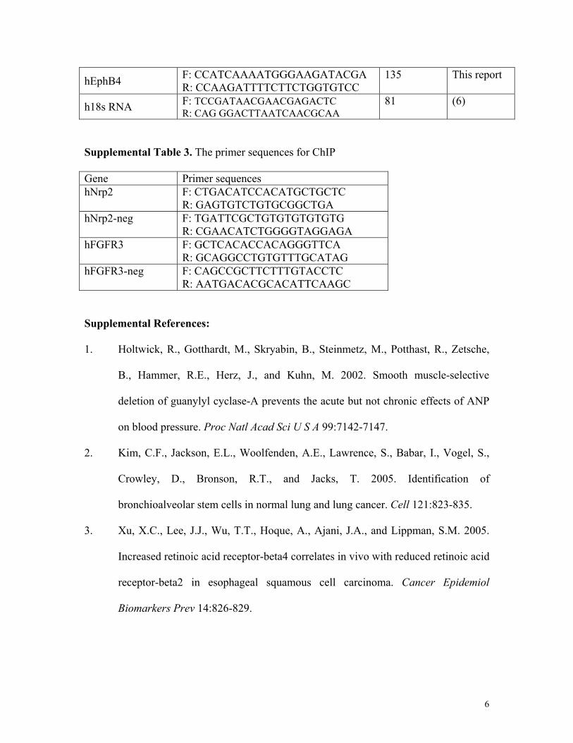
hEphB4 F: CCATCAAAATGGGAAGATACGA R: CCAAGATTTTCTTCTGGTGTCC
135 This report
h18s RNA F: TCCGATAACGAACGAGACTC R: CAG GGACTTAATCAACGCAA
81 (6)
Supplemental Table 3. The primer sequences for ChIP
Gene Primer sequences hNrp2 F: CTGACATCCACATGCTGCTC
R: GAGTGTCTGTGCGGCTGA hNrp2-neg F: TGATTCGCTGTGTGTGTGTG
R: CGAACATCTGGGGTAGGAGA hFGFR3 F: GCTCACACCACAGGGTTCA
R: GCAGGCCTGTGTTTGCATAG hFGFR3-neg F: CAGCCGCTTCTTTGTACCTC
R: AATGACACGCACATTCAAGC Supplemental References:
1. Holtwick, R., Gotthardt, M., Skryabin, B., Steinmetz, M., Potthast, R., Zetsche,
B., Hammer, R.E., Herz, J., and Kuhn, M. 2002. Smooth muscle-selective
deletion of guanylyl cyclase-A prevents the acute but not chronic effects of ANP
on blood pressure. Proc Natl Acad Sci U S A 99:7142-7147.
2. Kim, C.F., Jackson, E.L., Woolfenden, A.E., Lawrence, S., Babar, I., Vogel, S.,
Crowley, D., Bronson, R.T., and Jacks, T. 2005. Identification of
bronchioalveolar stem cells in normal lung and lung cancer. Cell 121:823-835.
3. Xu, X.C., Lee, J.J., Wu, T.T., Hoque, A., Ajani, J.A., and Lippman, S.M. 2005.
Increased retinoic acid receptor-beta4 correlates in vivo with reduced retinoic acid
receptor-beta2 in esophageal squamous cell carcinoma. Cancer Epidemiol
Biomarkers Prev 14:826-829.
6
4. Li, J., Wang, E., Rinaldo, F., and Datta, K. 2005. Upregulation of VEGF-C by
androgen depletion: the involvement of IGF-IR-FOXO pathway. Oncogene
24:5510-5520.
5. Curreli, S., Arany, Z., Gerardy-Schahn, R., Mann, D., and Stamatos, N.M. 2007.
Polysialylated neuropilin-2 is expressed on the surface of human dendritic cells
and modulates dendritic cell-T lymphocyte interactions. J Biol Chem 282:30346-
30356.
6. Harmancey, R., Senard, J.M., Pathak, A., Desmoulin, F., Claparols, C., Rouet, P.,
and Smih, F. 2005. The vasoactive peptide adrenomedullin is secreted by
adipocytes and inhibits lipolysis through NO-mediated beta-adrenergic agonist
oxidation. Faseb J 19:1045-1047.
7. Shin, J.W., Min, M., Larrieu-Lahargue, F., Canron, X., Kunstfeld, R., Nguyen, L.,
Henderson, J.E., Bikfalvi, A., Detmar, M., and Hong, Y.K. 2006. Prox1 promotes
lineage-specific expression of fibroblast growth factor (FGF) receptor-3 in
lymphatic endothelium: a role for FGF signaling in lymphangiogenesis. Mol Biol
Cell 17:576-584.
7
COUP-TFII β-gal DAPI
CO
UP-
TFII
F/F
100 μm
A
A
CV
A
CV
A
CV
CR
E-ER
T2;
CO
UP-
TFII
F/F
COUP-TFII β-gal DAPI
A
CV
A
CV
A
CV
A
CVDA DA
A
CVDA
COUP-TFII β-gal DAPI
A
CV
A
CV
A
CV
A
CVDA
Tam E10.5 (7 hours after Tam)
E11.5 (Tam E10.5)
E12.5 (Tam E10.5)
DA
DA
CO
UP-
TFII
F/F
CR
E-ER
T2;
CO
UP-
TFII
F/F
CO
UP-
TFII
F/F
CR
E-ER
T2;
CO
UP-
TFII
F/F
B
C
100 μm
100 μm
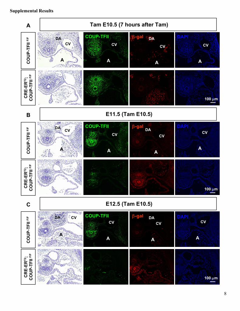
Supplemental Results
8
Supplementary Figure 1. Kinetics of CRE-ERT2
mediated COUP-TFII
inactivation.A single dosage of 3 mg Tam was delivered by intraperitoneal
injection to pregnant dams at E10.5, and embryos were harvested at 7 hours (A), 1 day (B), and 2 days (C)
after Tam treatment. H&E staining and immunofluorescence
for COUP-TFII (green) and β-gal (red) were observed in transverse sections of control COUP-TFII F/F
and CRE-ERT2; COUP-TFII F/F
embryos. Nuclei were counterstained with DAPI. Injection of Tam activates CRE-ERT2, triggering COUP-TFII
excision by recombination. Upon Cre-mediated recombination, the Lac-Z
reporter inserted into the 5’
untranslated
region of COUP-TFII
locus is activated. (A) Embryos exposed to Tam for 7 hours showed few or no β-gal+
cells. (B) One day after Tam administration COUP-TFII
deletion and corresponding β-gal activation were observed in around 80% of cells (C) COUP-TFII
deletion is almost complete 2 days post injection. A, atrium; CV, cardinal vein; DA, dorsal aorta. Scale bar, 100 μm.
9
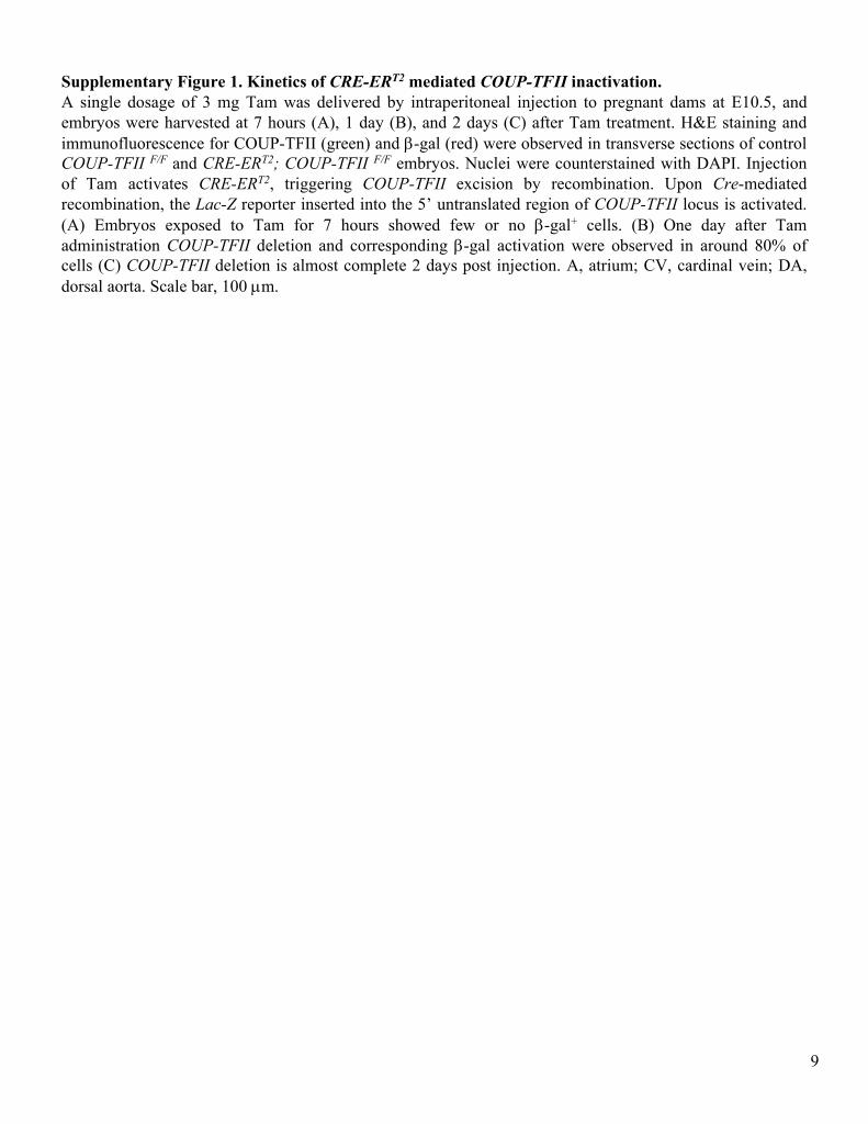
Supplemental Figure 2. Efficient inactivation of COUP-TFII
in embryonic ECs.(A) ECs
were isolated from E14.5 COUP-TFII F/F
(FF) controls and CRE-ERT2; COUP-TFII F/F
(CFF) embryos (administration of Tam at E11.5). Whole embryos were mechanically disrupted and enzymatically digested to create a single-cell suspension. Cells were labeled with PECAM antibodies. The endothelial content was enriched from less than 1% to more than 80% of the total tissue mass using magnetic cell sorting (MACS) separation. The selected fraction of cells was further analyzed by FACS. Gray histograms represent the isotype
control. PECAM-gated ECs
were sorted. (B) Genomic DNAs
were isolated from sorted cells for PCR genotyping and allele excision analysis. β-actin
was used as a loading control. (C) COUP-TFII
expression in sorted ECs
from
COUP-TFII F/F
and CRE-ERT2; COUP-TFII F/F
embryos were analyzed by semi-quantitative RT-PCR. 18S rRNA
was used as a loading control.
COUP-TFII
18S rRNA
FF CFF
FF CFF
floxed
excised
0 10 2 10 3 10 4 10 50
20
40
60
80
100
0 10 2 10 3 10 4 10 50
20
40
60
80
100
Even
ts
FL2-H (PECAM)
COUP-TFII F/F CRE-ERT2; COUP-TFII F/F
A
B C
β-actin
86.9 % 83.2 %
10
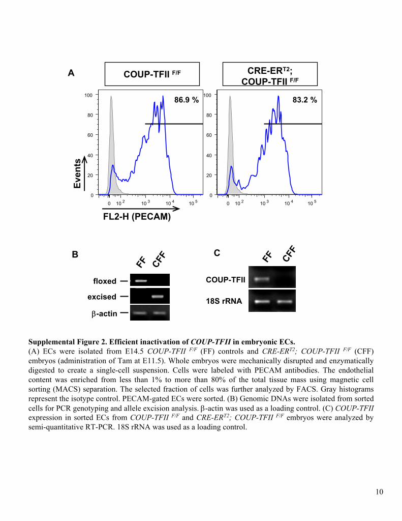
Supplementary Figure 3. Developmental defects observed in inducible COUP-TFII
knockout mutants are due to ablation of COUP-TFII.CRE-ERT2; COUP-TFII F/F
males were crossed to COUP-TFII F/+
females, and pregnant dams were injected with Tam at E13.5. Embryonic hearts were isolated at E16.5 and analyzed by whole mount VEGFR3 staining. (A-D) Gross appearance of CRE-ERT2; COUP-TFII F/+
and CRE-ERT2
controls are indistinguishable from wild-type or COUP-TFII
F/+
controls. (E-H) Whole mount VEGFR3 staining of E16.5 mouse hearts revealed similar cardiac lymphatic patterning in CRE-ERT2; COUP-TFII F/+, CRE-ERT2, wild-
type and COUP-TFII
F/+
embryos. This demonstrates that developmental defects observed in inducible COUP-TFII
knockout mutants (CRE-ERT2; COUP-TFII F/F) are due to COUP-TFII
ablation and not Cre
toxicity.
E16.
5 (T
am E
13.5
)
CRE-ERT2; COUP-TFII F/+ COUP-TFII F/+CRE-ERT2 Wild-type
VEGFR3 VEGFR3 VEGFR3 VEGFR3
A B C D
E F G H
11
100 μm
DA
CV
DA
CVDACV
DA
CV
DACV
DACV
E11.
5 (T
am E
9.5)
COUP-TFII
Prox1
Prox1
VEGFR3
Prox1
Nrp2
DA
CVDA
CV
Prox1
DACV
PECAM
DAPI
COUP-TFII F/F CRE-ERT2; COUP-TFII F/F
A B
C D
E F
G H
(46S) (47S)
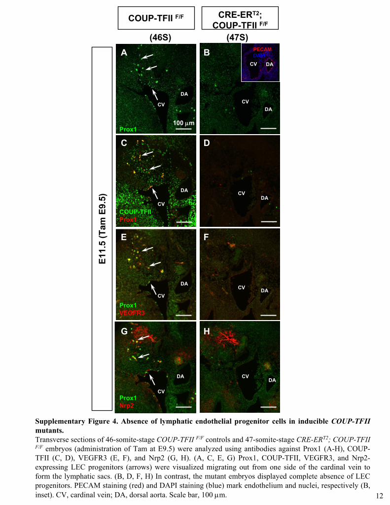
Supplementary Figure 4. Absence of lymphatic endothelial progenitor cells in inducible COUP-TFII
mutants. Transverse sections of 46-somite-stage COUP-TFII F/F
controls and 47-somite-stage CRE-ERT2; COUP-TFII F/F
embryos (administration of Tam at E9.5) were analyzed using antibodies against Prox1 (A-H), COUP-
TFII (C, D), VEGFR3 (E, F), and Nrp2 (G, H). (A, C, E, G) Prox1,
COUP-TFII, VEGFR3, and Nrp2-
expressing LEC progenitors (arrows) were visualized migrating out from one side of the cardinal vein to form the lymphatic sacs. (B, D, F, H) In contrast, the mutant embryos displayed complete absence of LEC progenitors. PECAM staining (red) and DAPI staining (blue) mark endothelium and nuclei, respectively (B, inset). CV, cardinal vein; DA, dorsal aorta. Scale bar, 100 μm. 12
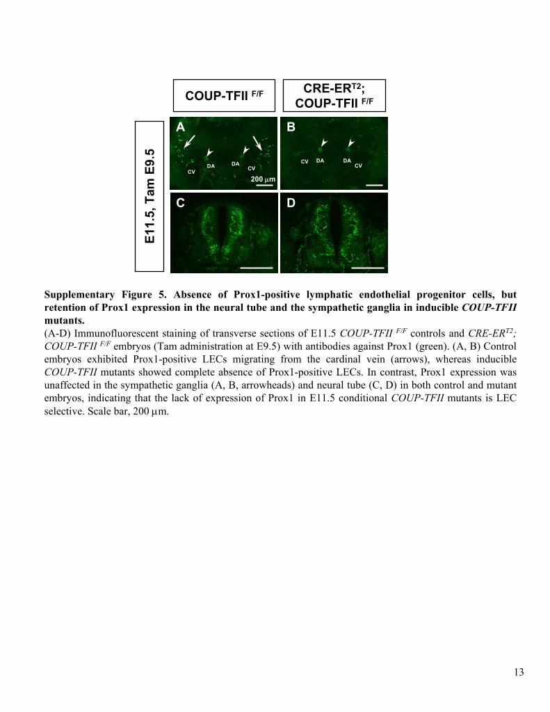
Supplementary Figure 5. Absence of Prox1-positive lymphatic endothelial progenitor cells, but
retention of Prox1 expression in the neural tube and the sympathetic ganglia in inducible COUP-TFII
mutants.(A-D) Immunofluorescent
staining of transverse sections of E11.5 COUP-TFII F/F
controls and CRE-ERT2; COUP-TFII F/F
embryos (Tam administration at E9.5) with antibodies against Prox1 (green). (A, B) Control embryos exhibited Prox1-positive LECs
migrating from the cardinal vein (arrows), whereas inducible COUP-TFII
mutants showed complete absence of Prox1-positive LECs. In contrast, Prox1 expression was unaffected in the sympathetic ganglia (A, B, arrowheads) and neural tube (C, D) in both control and mutant embryos, indicating that the lack of expression of Prox1 in E11.5 conditional COUP-TFII
mutants is LEC selective. Scale bar, 200 μm.
A B
COUP-TFII F/F CRE-ERT2; COUP-TFII F/F
E11.
5, T
am E
9.5
200 μm
C DD
CVDA
CV DACV
DA CVDA
13
VEGFR3
DAPI
C D
JLSJV
COUP-TFII F/F CRE-ERT2; COUP-TFII F/F
JLS
JV
VEGFR2
E F
JLSJV
JLS
JV
Nrp2
DAPI
G H
JLSJV
JLS
JV
LYVE1
DAPI
I J
JLSJV
JLS
JV
E14.
5, T
am E
11.5
A B
100 μm
JLSJV
JLS
JV
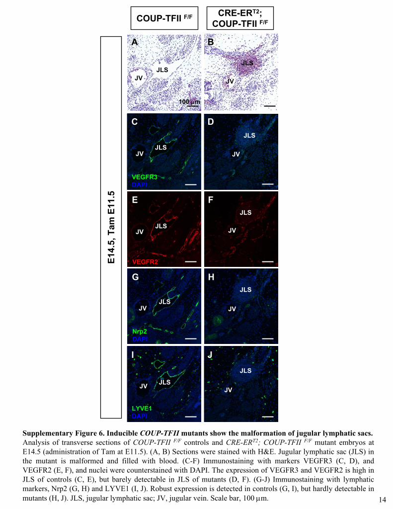
Supplementary Figure 6. Inducible COUP-TFII
mutants show the malformation of jugular lymphatic sacs. Analysis of transverse sections of COUP-TFII F/F
controls and CRE-ERT2; COUP-TFII F/F
mutant embryos at E14.5 (administration of Tam at E11.5). (A, B) Sections were stained with H&E. Jugular lymphatic sac (JLS) in the mutant is malformed and filled with blood. (C-F) Immunostaining
with markers VEGFR3 (C, D), and VEGFR2 (E, F), and nuclei were counterstained with DAPI. The expression of VEGFR3 and VEGFR2 is high in JLS of controls (C, E), but barely detectable in JLS of mutants (D, F). (G-J) Immunostaining
with lymphatic markers, Nrp2 (G, H) and LYVE1 (I, J). Robust expression is detected in controls (G, I), but hardly detectable in mutants (H, J). JLS, jugular lymphatic sac; JV, jugular vein. Scale bar, 100 μm. 14
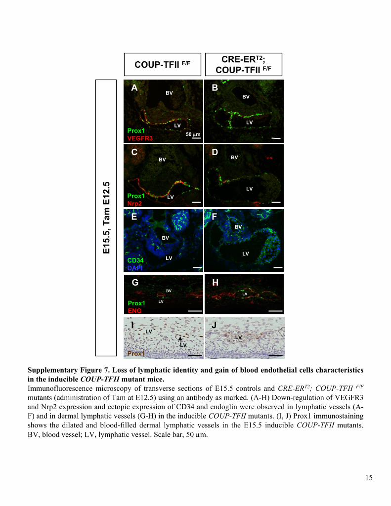
Supplementary Figure 7. Loss of lymphatic identity and gain of blood endothelial cells characteristics in the inducible COUP-TFII
mutant mice. Immunofluorescence
microscopy of transverse sections of E15.5 controls and CRE-ERT2; COUP-TFII F/F
mutants (administration of Tam at E12.5) using an antibody as marked. (A-H) Down-regulation of VEGFR3 and Nrp2 expression and ectopic expression of CD34 and endoglin
were observed in lymphatic vessels (A-
F) and in dermal lymphatic vessels (G-H) in the inducible COUP-TFII
mutants. (I, J) Prox1 immunostaining
shows the dilated and blood-filled dermal lymphatic vessels in the E15.5 inducible COUP-TFII
mutants. BV, blood vessel; LV, lymphatic vessel. Scale bar, 50 μm.
Prox1
Nrp2
Prox1
ENG
LVBV
LV
COUP-TFII F/F CRE-ERT2; COUP-TFII F/F
E15.
5, T
am E
12.5
BV
LV
CD34
DAPI
BV
LV
Prox1
VEGFR3
BV
LV
E F
C D
A B
G H
50 μm
BV
LV
BV
LV
BV
LV
LVLV
I J
Prox1LV
15
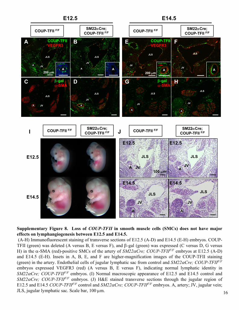
Supplementary Figure 8. Loss of COUP-TFII
in smooth muscle cells (SMCs) does not have major effects on lymphangiogenesis
between E12.5 and E14.5.(A-H) Immunofluorescent
staining of transverse sections of E12.5 (A-D) and E14.5 (E-H) embryos. COUP-
TFII (green) was deleted (A versus B, E versus F), and β-gal (green) was expressed (C versus D, G versus H) in the α-SMA (red)-positive SMCs
of the artery of SM22αCre; COUP-TFIIF/F
embryos at E12.5 (A-D) and E14.5 (E-H). Insets in A, B, E, and F are higher-magnification images of the COUP-TFII staining (green) in the artery. Endothelial cells of jugular lymphatic sac from control and SM22αCre; COUP-TFIIF/F
embryos expressed VEGFR3 (red) (A versus B, E versus F), indicating normal lymphatic identity in
SM22αCre; COUP-TFIIF/F
embryos. (I) Normal macroscopic appearance of E12.5 and E14.5 control and SM22αCre; COUP-TFIIF/F
embryos. (J) H&E stained transverse sections through the jugular region of E12.5 and E14.5 COUP-TFIIF/F
control and SM22αCre; COUP-TFIIF/F
embryos. A, artery; JV, jugular vein; JLS, jugular lymphatic sac. Scale bar, 100 μm.
COUP-TFII F/F SM22αCre; COUP-TFII F/F
JV
JLS
JV
JLS
AA
JV
JLS
JV
JLS
COUP-TFII
VEGFR3
COUP-TFII
α-SMA
100 μm
200 μm
I J COUP-TFII F/F SM22αCre; COUP-TFII F/F
JV
JLS
JV
JLS
JV
JLS
JV
JLS
A A
A A
E12.5 E12.5
E14.5 E14.5
E14.5
E12.5
A
COUP-TFII F/F SM22αCre; COUP-TFII F/F COUP-TFII F/F SM22αCre;
COUP-TFII F/F
COUP-TFII
VEGFR3
β-gal
α-SMA
200 μm
β-gal
α-SMA
JV
JLS
A
JV
JLS
A JV
A
JLS
JV
A
JLS
A A
B
C D
E F
G H
E12.5 E14.5
A A
COUP-TFII
DAPI
16
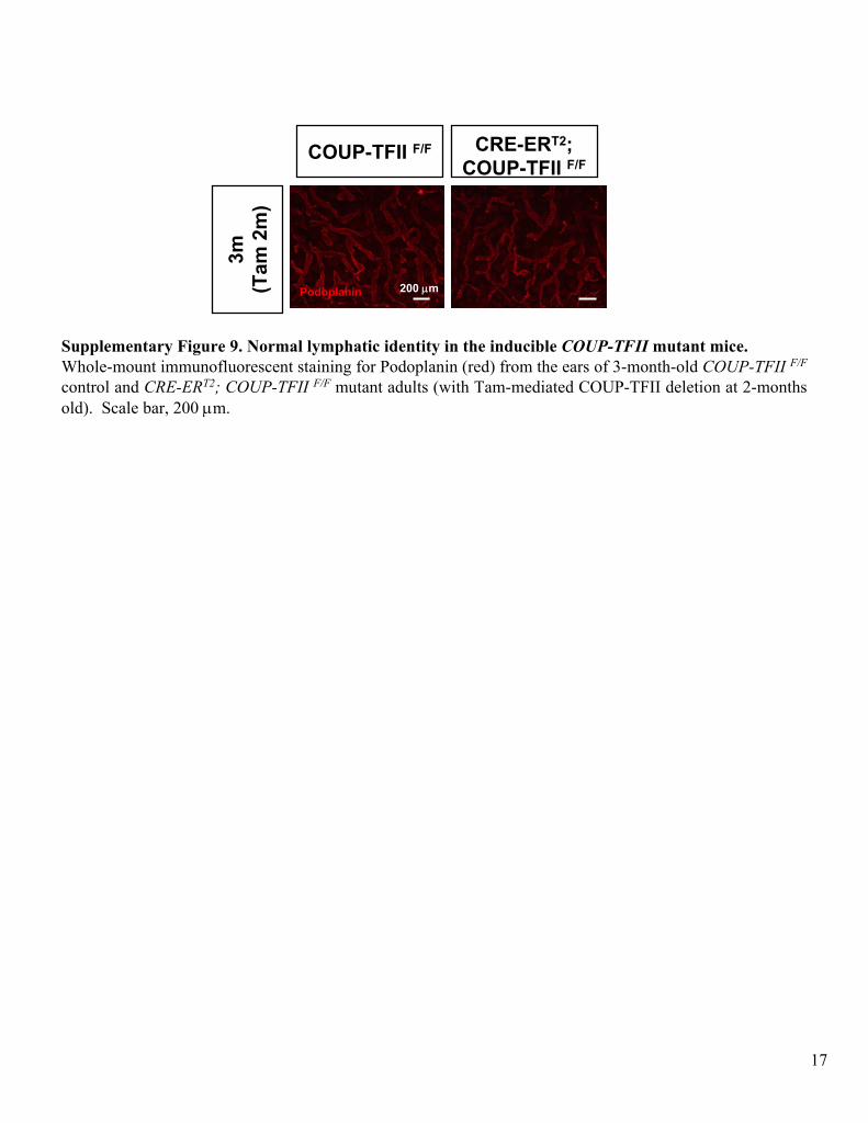
Supplementary Figure 9. Normal lymphatic identity in the inducible COUP-TFII
mutant mice. Whole-mount immunofluorescent
staining for Podoplanin
(red) from the ears of 3-month-old COUP-TFII F/F
control and CRE-ERT2; COUP-TFII F/F
mutant adults (with Tam-mediated COUP-TFII deletion at 2-months old). Scale bar, 200 μm.
COUP-TFII F/F CRE-ERT2; COUP-TFII F/F
3m
(Tam
2m
)Podoplanin 200 μm
17
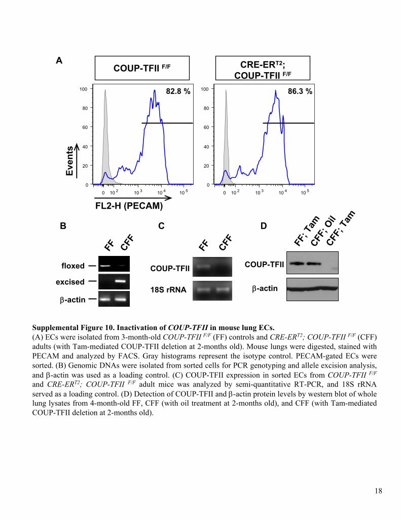
Supplemental Figure 10. Inactivation of COUP-TFII
in mouse lung ECs.(A) ECs
were isolated from 3-month-old COUP-TFII F/F
(FF) controls and CRE-ERT2; COUP-TFII F/F
(CFF) adults (with Tam-mediated COUP-TFII deletion at 2-months old). Mouse lungs were digested, stained with PECAM and analyzed by FACS. Gray histograms represent the isotype
control. PECAM-gated ECs
were sorted. (B) Genomic DNAs
were isolated from sorted cells for PCR genotyping and allele excision analysis, and β-actin
was used as a loading control. (C) COUP-TFII expression in sorted ECs
from
COUP-TFII F/F
and CRE-ERT2; COUP-TFII F/F
adult mice was analyzed by semi-quantitative RT-PCR, and 18S rRNA
served as a loading control. (D) Detection of COUP-TFII and β-actin
protein levels by western blot of whole lung lysates from 4-month-old FF, CFF (with oil treatment at 2-months old), and CFF (with Tam-mediated COUP-TFII deletion at 2-months old).
A
B C
0 10 2 10 3 10 4 10 50
20
40
60
80
100
0 10 2 10 3 10 4 10 50
20
40
60
80
100
Even
ts
FL2-H (PECAM)
COUP-TFII F/F CRE-ERT2; COUP-TFII F/F
COUP-TFII
18S rRNA
FF CFFFF CFF
floxed
excised
COUP-TFII
β-actinFF
; Tam
CFF; O
ilCFF
; TamD
82.8 % 86.3 %
β-actin
18
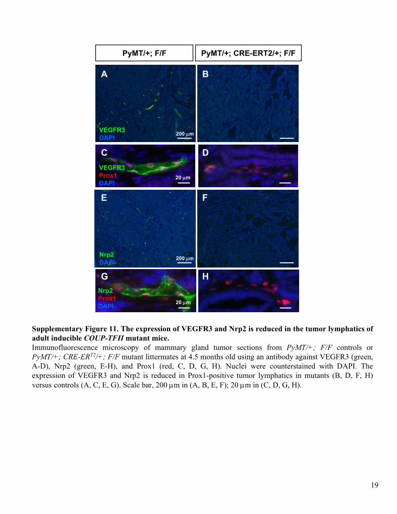
Supplementary Figure 11. The expression of VEGFR3 and Nrp2 is reduced in the tumor lymphatics
of adult inducible COUP-TFII
mutant mice.Immunofluorescence
microscopy of mammary gland tumor sections from PyMT/+; F/F controls or PyMT/+; CRE-ERT2/+; F/F
mutant littermates at 4.5 months old using an antibody against VEGFR3 (green, A-D), Nrp2 (green, E-H), and Prox1 (red, C, D, G, H). Nuclei were counterstained with
DAPI. The
expression of VEGFR3 and Nrp2 is reduced in Prox1-positive tumor lymphatics
in mutants (B, D, F, H) versus controls (A, C, E, G). Scale bar, 200 μm in (A, B, E, F); 20 μm in (C, D, G, H).
VEGFR3
DAPI
Nrp2
DAPI
Nrp2
Prox1
DAPI
VEGFR3
Prox1
DAPI
200 μm
20 μm
20 μm
A B
C D
E F
G H
PyMT/+; F/F PyMT/+; CRE-ERT2/+; F/F
200 μm
19
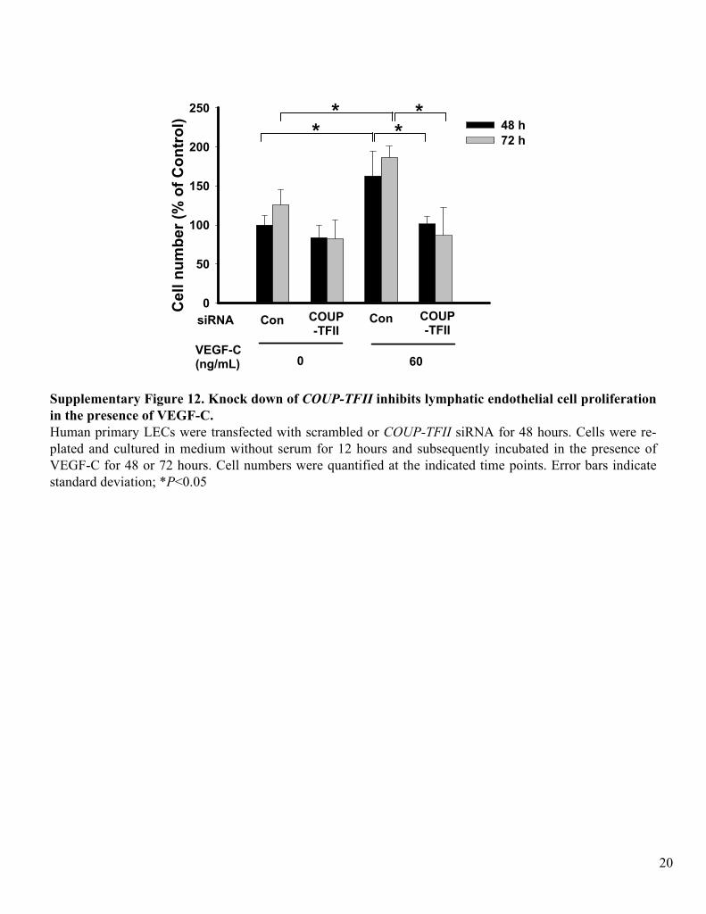
Supplementary Figure 12. Knock down of COUP-TFII
inhibits lymphatic endothelial cell proliferation in the presence of VEGF-C.Human primary LECs
were transfected
with scrambled or COUP-TFII
siRNA
for 48 hours. Cells were re-
plated and cultured in medium without serum for 12 hours and subsequently incubated in the presence of VEGF-C for 48 or 72 hours. Cell numbers were quantified at the indicated time points. Error bars indicate standard deviation; *P<0.05
Cel
l num
ber (
% o
f Con
trol
)
0
50
100
150
200
25048 h72 h
siRNA Con COUP-TFII
Con COUP-TFII
VEGF-C (ng/mL) 0 60
* ** *
20
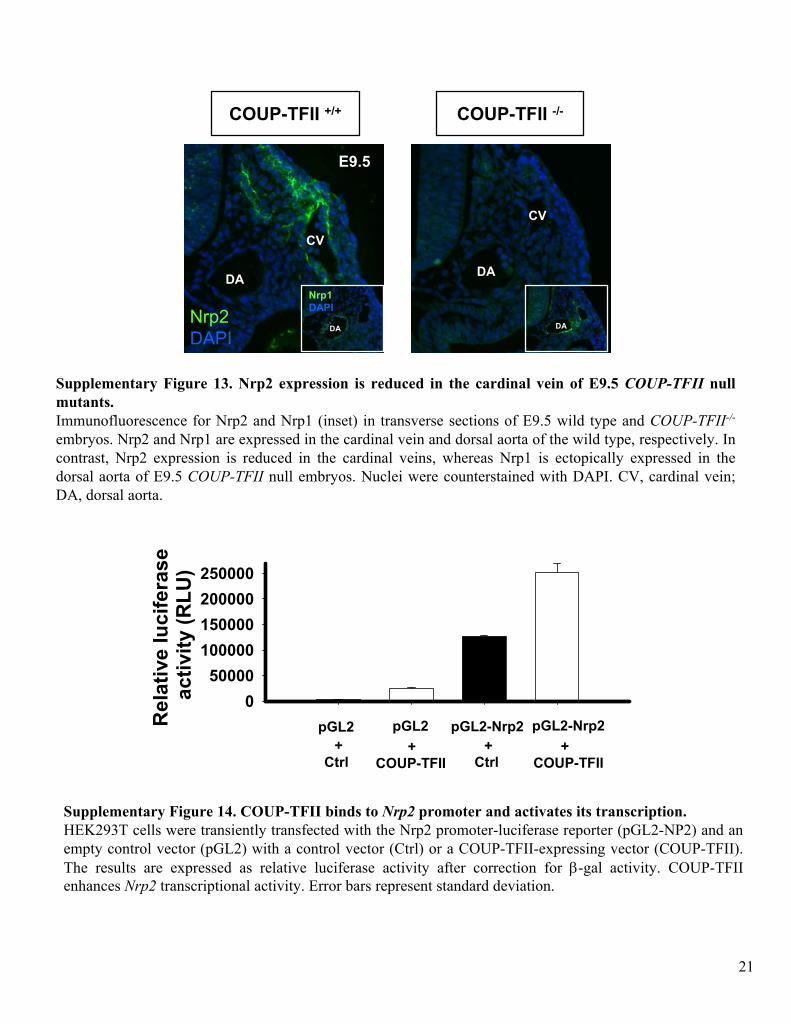
Supplementary Figure 13. Nrp2 expression is reduced in the cardinal vein of E9.5 COUP-TFII
null mutants. Immunofluorescence
for Nrp2 and Nrp1 (inset) in transverse sections of E9.5 wild type and COUP-TFII-/-
embryos. Nrp2 and Nrp1 are expressed in the cardinal vein and dorsal aorta of the wild type, respectively. In contrast, Nrp2 expression is reduced in the cardinal veins, whereas Nrp1 is ectopically expressed in the dorsal aorta of E9.5 COUP-TFII
null embryos. Nuclei were counterstained with DAPI. CV, cardinal vein; DA, dorsal aorta.
DA
CV
DA
CV
Nrp2
DAPI
E9.5
Nrp1
DAPI
DA DA
COUP-TFII +/+ COUP-TFII -/-
21
Supplementary Figure 14. COUP-TFII binds to Nrp2
promoter and activates its transcription. HEK293T cells were transiently transfected
with the Nrp2 promoter-luciferase
reporter (pGL2-NP2) and an empty control vector (pGL2) with a control vector (Ctrl) or a COUP-TFII-expressing vector (COUP-TFII). The results are expressed as relative luciferase
activity after correction for β-gal activity. COUP-TFII enhances Nrp2
transcriptional activity. Error bars represent standard deviation.
Rel
ativ
e lu
cife
rase
act
ivity
(RLU
)
050000
100000150000200000250000
pGL2 pGL2+
Ctrl+
COUP-TFII
pGL2-Nrp2 pGL2-Nrp2+
Ctrl+
COUP-TFII
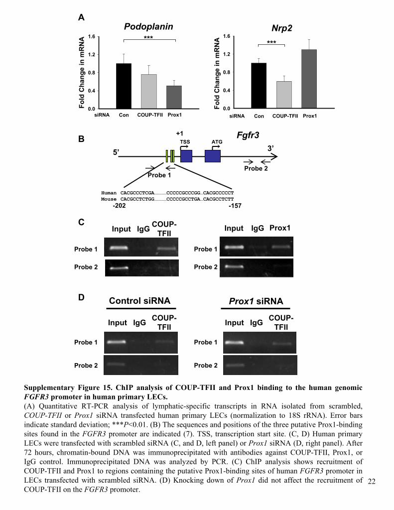
Supplementary Figure 15. ChIP
analysis of COUP-TFII and Prox1 binding to the human genomic FGFR3
promoter in human primary LECs.(A) Quantitative RT-PCR analysis of lymphatic-specific transcripts in RNA isolated from scrambled, COUP-TFII
or Prox1
siRNA
transfected
human primary LECs
(normalization to 18S rRNA). Error bars indicate standard deviation; ***P<0.01. (B) The sequences and positions of the three putative Prox1-binding sites found in the FGFR3
promoter are indicated (7). TSS, transcription start site. (C, D)
Human primary LECs
were transfected
with scrambled siRNA
(C, and D, left panel) or Prox1
siRNA
(D, right panel). After 72 hours, chromatin-bound DNA was immunoprecipitated
with antibodies against COUP-TFII, Prox1, or IgG
control. Immunoprecipitated
DNA was analyzed by PCR. (C) ChIP
analysis shows recruitment of COUP-TFII and Prox1 to regions containing the putative Prox1-binding sites of human FGFR3
promoter in LECs
transfected
with scrambled siRNA. (D) Knocking down of Prox1
did not affect the recruitment of COUP-TFII on the FGFR3
promoter.
Input IgG COUP-
TFII Input IgG Prox1
+1
CACGCCCTCGA…………CCCCCGCCCGG…CACGCCCCCT
CACGCCTCTGG…………CCCCCGCCTGA…CACGCCTCTTHuman
Mouse
5’ 3’Fgfr3
Probe 1Probe 2
Probe 1
Probe 2
Probe 1
Probe 2
TSS ATGB
C
-202 -157Fo
ld C
hang
e in
mR
NA
0.0
0.4
0.8
1.2
1.6Nrp2
***
siRNA Con COUP-TFII Prox1
Fold
Cha
nge
in m
RN
A
0.0
0.4
0.8
1.2
1.6
siRNA Con COUP-TFII Prox1
Podoplanin***
A
D
Input IgG COUP-
TFII Input IgG COUP-
TFII
Control siRNA Prox1 siRNA
Probe 1
Probe 2
Probe 1
Probe 2
22
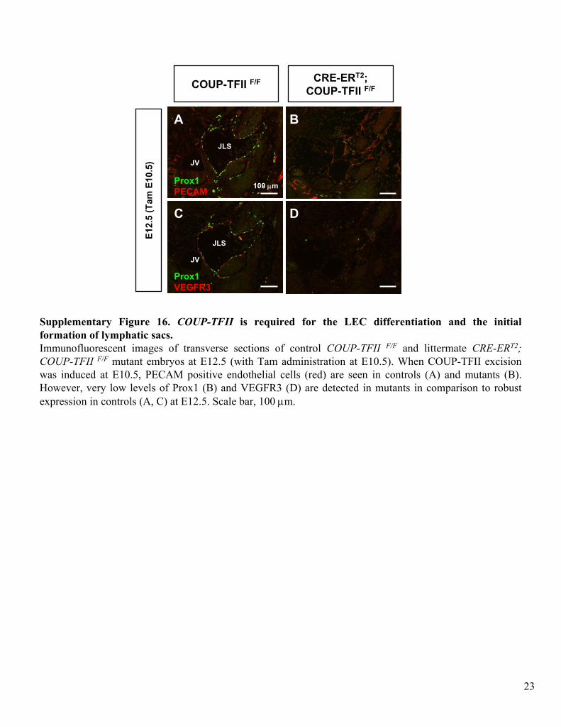
Supplementary Figure 16. COUP-TFII
is required for the LEC differentiation and the initial formation of lymphatic sacs.Immunofluorescent
images of transverse sections of control COUP-TFII F/F
and littermate CRE-ERT2; COUP-TFII F/F
mutant embryos at E12.5 (with Tam administration at E10.5). When COUP-TFII excision was induced at E10.5, PECAM positive endothelial cells (red) are
seen in controls (A) and mutants (B). However, very low levels of Prox1 (B) and VEGFR3 (D) are detected in mutants in comparison to robust expression in controls (A, C) at E12.5. Scale bar, 100 μm.
Prox1
PECAM
Prox1
VEGFR3
E12.
5 (T
am E
10.5
)
COUP-TFII F/F CRE-ERT2; COUP-TFII F/F
100 μm
JLS
JLS
A B
C D
JV
JV
23