Temperature-Dependent Raman Spectroscopic Evidence … · Temperature-Dependent Raman Spectroscopic...
8
Temperature-Dependent Raman Spectroscopic Evidence of and Molecular Mechanism for Irreversible Isomerization of β‑Endosulfan to α‑Endosulfan Walter F. Schmidt, Cathleen J. Hapeman,* Laura L. McConnell, Swati Mookherji, Clifford P. Rice, Julie K. Nguyen, Jianwei Qin, Hoyoung Lee, Kuanglin Chao, and Moon S. Kim Agricultural Research Service, United States Department of Agriculture, 10300 Baltimore Avenue, Beltsville, Maryland 20705-2325, United States ABSTRACT: Endosulfan (6,7,8, 9,10,10-hexachloro-1,5,5a,6,9,9a-hexahydro-6,9-methano-2,4,3-benzodioxathiepine-3-oxide) is a broad-spectrum, organochlorine insecticide used on numerous crops since the 1950s. It is has been identified as a persistent organic pollutant (POP) due to its persistence, bioaccumulation, long-range transport, and adverse effects to human health and aquatic ecosystems; it will be phased out in the United States in 2016. Endosulfan consists of two diastereomers, α and β; α- endosulfan exists as two asymmetrical, twist-chair enantiomers which interchange, while β-endosulfan has a symmetrical-chair conformation. β-Endosulfan has been shown to isomerize to α-endosulfan. Here we document the previously proposed isomerization mechanism using temperature-dependent Raman (TDR) spectroscopy. The bending frequencies in the fingerprint region were assigned to specific bonds. Changes in the signal intensity as a function of temperature were used to identify detailed ring movements and thus conversion of β to α. These movements cannot occur simultaneously nor symmetrically, precluding conversion of α-endosulfan to β-endosulfan. KEYWORDS: endosulfan (6,7,8,9,10,10-hexachloro-1,5,5a,6,9,9a-hexahydro-6,9-methano-2,4,3-benzodioxathiepine-3-oxide), 1,3-dioxepane, temperature-dependent Raman (TDR) spectroscopy, 2D-TDR spectra, Raman ring-bending frequency, temperature-dependent conformational changes ■ INTRODUCTION Endosulfan (6,7,8, 9,10,10-hexachloro-1,5,5a,6,9,9a-hexahydro- 6,9-methano-2,4,3-benzodioxathiepine-3-oxide) is a broad- spectrum organochlorine insecticide that has been used on cereals, fruits, vegetables, and cotton since the 1950s. 1,2 It exists as two diasteromers, α-endosulfan and β-endosulfan, and is typically applied to crops as a 70:30 isomeric ratio of α:β. In 2010, the United States Environmental Protection Agency initiated a Memorandum of Agreement to phase out all uses of endosulfan completely by July 2016 because of the unaccept- able risks to farm workers and wildlife and because it can persist in the environment. 3,4 The United Nations Environmental Programme (UNEP), Parties to the Stockholm Convention on Persistent Organic Pollutants (POPs), agreed to list endosulfan as a POP in 2011. 5 Numerous reviews have documented studies concerning the persistence, bioaccumulation, long-range transport, and adverse effects to aquatic ecosystems and to human health posed by endosulfan [e.g., refs 6-8]. Despite the virtual global ban on endosulfan, the environmental fate of these two compounds will remain a global issue long after their use has ceased. The chemical structure of both endosulfan isomers has been examined and disputed extensively since it was first patented in 1958. 9-12 More recent research confirmed that β-endosulfan is symmetrical as previously suggested but that α-endosulfan is actually composed of two asymmetrical enantiomers (Figure 1). 13 Additional research concerning the Henry’ s Law Constants of endosulfan led to the discovery that β-endosulfan readily converts to α-endosulfan in the aqueous phase. Received: September 30, 2013 Revised: January 31, 2014 Accepted: February 3, 2014 Published: February 3, 2014 Figure 1. Structure of both enantiomers of α-endosulfan and β- endosulfan. For brevity, atom numbers are assigned only to the seven- membered dioxathiepine-3-oxide system, beginning from oxygen site. The β-endosulfan molecule is symmetrical; α-endosulfan is asym- metrical. Article pubs.acs.org/JAFC This article not subject to U.S. Copyright. Published 2014 by the American Chemical Society 2023 dx.doi.org/10.1021/jf404404w | J. Agric. Food Chem. 2014, 62, 2023-2030
Transcript of Temperature-Dependent Raman Spectroscopic Evidence … · Temperature-Dependent Raman Spectroscopic...
Temperature-Dependent Raman Spectroscopic Evidence of andMolecular Mechanism for Irreversible Isomerization of β‑Endosulfanto α‑EndosulfanWalter F. Schmidt, Cathleen J. Hapeman,* Laura L. McConnell, Swati Mookherji, Clifford P. Rice,Julie K. Nguyen, Jianwei Qin, Hoyoung Lee, Kuanglin Chao, and Moon S. Kim
Agricultural Research Service, United States Department of Agriculture, 10300 Baltimore Avenue, Beltsville, Maryland 20705-2325,United States
ABSTRACT: Endosulfan (6,7,8, 9,10,10-hexachloro-1,5,5a,6,9,9a-hexahydro-6,9-methano-2,4,3-benzodioxathiepine-3-oxide) isa broad-spectrum, organochlorine insecticide used on numerous crops since the 1950s. It is has been identified as a persistentorganic pollutant (POP) due to its persistence, bioaccumulation, long-range transport, and adverse effects to human health andaquatic ecosystems; it will be phased out in the United States in 2016. Endosulfan consists of two diastereomers, α and β; α-endosulfan exists as two asymmetrical, twist-chair enantiomers which interchange, while β-endosulfan has a symmetrical-chairconformation. β-Endosulfan has been shown to isomerize to α-endosulfan. Here we document the previously proposedisomerization mechanism using temperature-dependent Raman (TDR) spectroscopy. The bending frequencies in the fingerprintregion were assigned to specific bonds. Changes in the signal intensity as a function of temperature were used to identify detailedring movements and thus conversion of β to α. These movements cannot occur simultaneously nor symmetrically, precludingconversion of α-endosulfan to β-endosulfan.
KEYWORDS: endosulfan (6,7,8,9,10,10-hexachloro-1,5,5a,6,9,9a-hexahydro-6,9-methano-2,4,3-benzodioxathiepine-3-oxide),1,3-dioxepane, temperature-dependent Raman (TDR) spectroscopy, 2D-TDR spectra, Raman ring-bending frequency,temperature-dependent conformational changes
■ INTRODUCTION
Endosulfan (6,7,8, 9,10,10-hexachloro-1,5,5a,6,9,9a-hexahydro-6,9-methano-2,4,3-benzodioxathiepine-3-oxide) is a broad-spectrum organochlorine insecticide that has been used oncereals, fruits, vegetables, and cotton since the 1950s.1,2 It existsas two diasteromers, α-endosulfan and β-endosulfan, and istypically applied to crops as a 70:30 isomeric ratio of α:β. In2010, the United States Environmental Protection Agencyinitiated a Memorandum of Agreement to phase out all uses ofendosulfan completely by July 2016 because of the unaccept-able risks to farm workers and wildlife and because it can persistin the environment.3,4 The United Nations EnvironmentalProgramme (UNEP), Parties to the Stockholm Convention onPersistent Organic Pollutants (POPs), agreed to list endosulfanas a POP in 2011.5 Numerous reviews have documentedstudies concerning the persistence, bioaccumulation, long-rangetransport, and adverse effects to aquatic ecosystems and tohuman health posed by endosulfan [e.g., refs 6−8]. Despite thevirtual global ban on endosulfan, the environmental fate ofthese two compounds will remain a global issue long after theiruse has ceased.The chemical structure of both endosulfan isomers has been
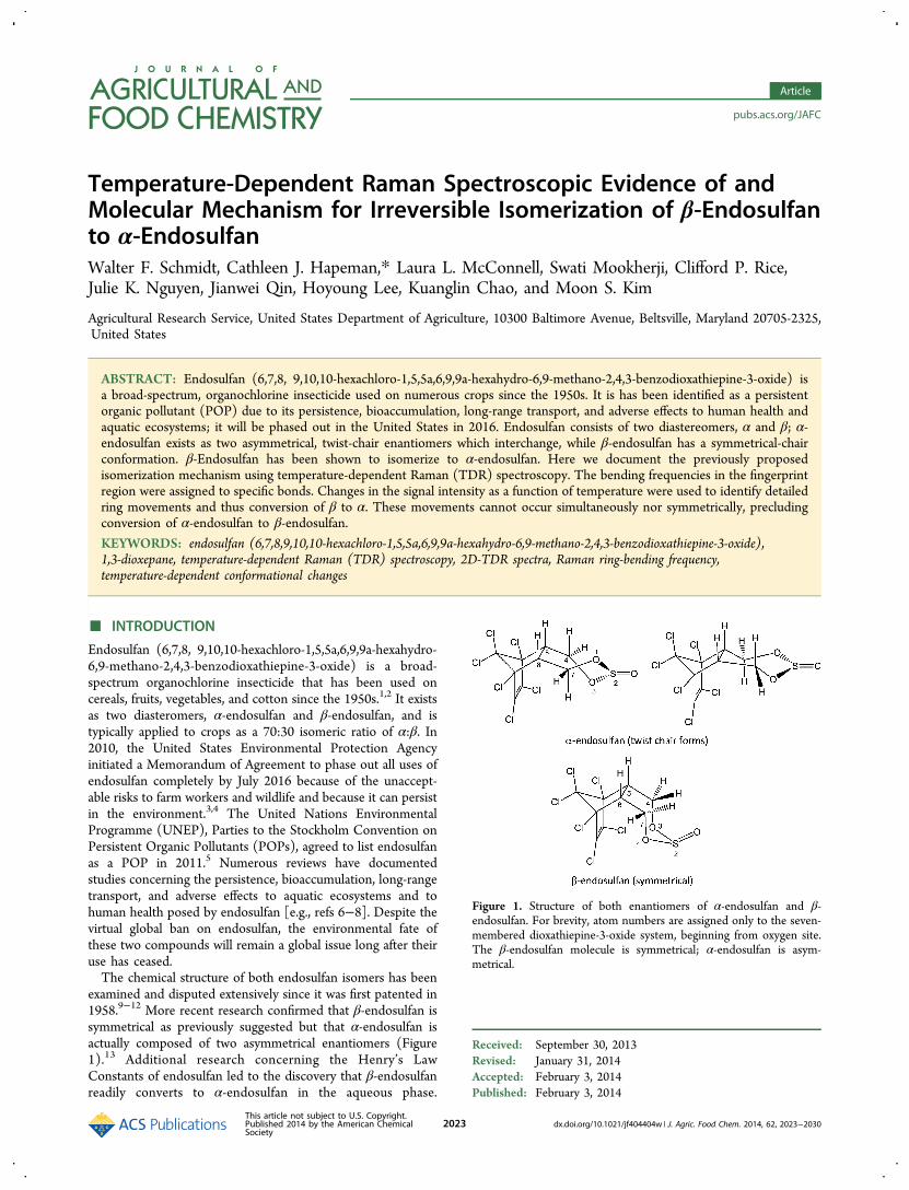
examined and disputed extensively since it was first patented in1958.9−12 More recent research confirmed that β-endosulfan issymmetrical as previously suggested but that α-endosulfan isactually composed of two asymmetrical enantiomers (Figure1).13 Additional research concerning the Henry’s LawConstants of endosulfan led to the discovery that β-endosulfanreadily converts to α-endosulfan in the aqueous phase.
Received: September 30, 2013Revised: January 31, 2014Accepted: February 3, 2014Published: February 3, 2014
Figure 1. Structure of both enantiomers of α-endosulfan and β-endosulfan. For brevity, atom numbers are assigned only to the seven-membered dioxathiepine-3-oxide system, beginning from oxygen site.The β-endosulfan molecule is symmetrical; α-endosulfan is asym-metrical.
Article
pubs.acs.org/JAFC
This article not subject to U.S. Copyright.Published 2014 by the American ChemicalSociety
2023 dx.doi.org/10.1021/jf404404w | J. Agric. Food Chem. 2014, 62, 2023−2030
Experiments were conducted using a wetted-wall column toexamine the partitioning of the nearly pure endosulfan isomersfrom water into air. When starting with β-endosulfan, α-endosulfan was virtually the only isomer observed in airphase.14,15 In regional and long-range transport studies ofendosulfan isomers in air, β-endosulfan was infrequentlyobserved relative to α-endosulfan, so much so that it wasdisregarded in the data analyses.16,17 The isomerization of β-endosulfan to α-endosulfan has been suggested as a possiblereason for the observed overabundance of α-endosulfan in theatmosphere.6,18 Finally, this isomerization has also beenobserved directly on surfaces.19
A mechanism for β-endosulfan conversion to α-endosulfanand its irreversibility have been proposed and has beensupported with data from differential scanning calorimetry(DSC) and NMR experiments and from computationalchemistry calculations.18 DCS scans of pure α-endosulfan,pure β-endosulfan, and mixtures were acquired; temperaturesranged from 25 to 280 °C. Scans of mixtures showed a largephase transition not observed in either of the pure isomerscans, suggesting a new transition which we assigned to theisomerization process. Furthermore, analysis of the remainingmaterial after the scans revealed a eutectic mixture of anapproximately 60:40 mixture of the α:β isomers, againdemonstrating conversion. No degradation other than isomericconversion was observed. Using computational chemistry, amechanism was proposed whereby movement of the SOmoiety followed pseudorotation of the seven-membered ring ofβ-endosulfan resulted in formation of α-endosulfan. Further-more, we concluded that the increase in entropy caused by theasymmetry in α-endosulfan precluded the reverse process of α-endosulfan conversion to β-endosulfan.The three mixtures of endosulfan (nearly pure α-endosulfan,
nearly pure β-endosulfan, and mixtures) have identical chemicalcompositions, i.e., they differ only in spatial arrangement of theatoms, yet the phase transitions observed using DSC weremarkedly different in all three mixtures.18,20 This indicates thatthe thermal energy is absorbed by different sets of atoms. Inaddition, DSC does not identify which part of the molecule orbond(s) absorb the thermal energy. Raman spectroscopyprovides spectral evidence (frequency signals) identifying thesets of atoms or bonds which are affected by energy absorption.Using the technique of temperature-dependent Raman (TDR)spectroscopy recently developed in our laboratory, molecularsites which respond most strongly to the thermal temperaturegradient can be identified. Frequencies which covary withintensity and/or wavenumber shift starting at a specifictemperature can indicate the individual molecular site ormolecular movement that triggers a reversible or irreversibletransition event. From these data, the sequence of molecularmovements absorbing a threshold amount of thermal energycan be organized to explain the mechanism of a transitionevent. The purpose of this article is to provide such evidencethat, in concert with previous work, demonstrates the molecularlevel pathway through which phase transition events occur,particularly, the irreversible process of β-endosulfan convertingto α-endosulfan.
■ MATERIALS AND METHODSMaterials. Certified analytical grade Thiodan (α:β-endosulfan
60:40), β-endosulfan (nearly 100%), and α-endosulfan (99.8% α; 0.2%β) were each obtained commercially from ChemServices (WestChester, PA) and used as is.
Temperature Dependent Raman Measurement System. ARaman spectroscopy system was assembled to collect temperaturedependent Raman (TDR) spectra from the samples. A 16-bit CCDcamera with 1024 × 256 pixels (Newton DU920N-BR-DD, AndorTechnology, South Windsor, CT) was used to acquire Ramanscattering signals. A Raman spectrometer (Raman Explorer 785,Headwall Photonics, Fitchburg, MA) was mounted to the camera. Thespectrometer accepts light through an input slit (5 mm long × 100 μmwide) and detects a Raman shift range of 102.2−2538.1 cm−1 with aspectral resolution of 3.7 cm−1. A 785 nm laser module(I0785MM0350MF-NL, Innovative Photonic Solutions, MonmouthJunction, NJ) served as the excitation source. A fiber optic Ramanprobe (RPB, InPhotonics, Norwood, MA) was used to focus the laserand acquire the Raman signals. A bifurcated fiber bundle was used todeliver the laser light to the probe and transfer the collected Ramansignals to the spectrometer.
A hot plate with a ceramic top surface (Isotemp, Fisher Scientific,Hampton, NH) was used to heat the samples. The powder sampleswere put in a copper sample holder. Two thermocouple probes (typeK) were attached to two sides of the sample area, and they wereconnected to a dual-input thermometer (EasyView EA15, ExtechInstruments, Nashua, NH) to measure the temperature of the samples.Average values of the two probes were used as the sample temperature,and the data were transferred to a computer via a RS-232 port. Acopper heat sink was placed between the ceramic heat surface and thesample holder to slow down the speed of the temperature increase. Arelatively high temperature resolution was consequently achieved forthe temperature-dependent Raman data. The Raman probe, the hotplate, and the sample materials were placed in a closed black box toavoid the influence of ambient light. System software was developedusing LabVIEW (National Instruments, Austin, TX) to fulfill functionssuch as camera control, data acquisition, temperature measurement,and synchronization.21 Each experiment was conducted in triplicate.Over 200 spectra of 1600 data points were collected for eachexperiment, resulting in a data set of approximately 106 data points.
■ RESULTS AND DISCUSSION
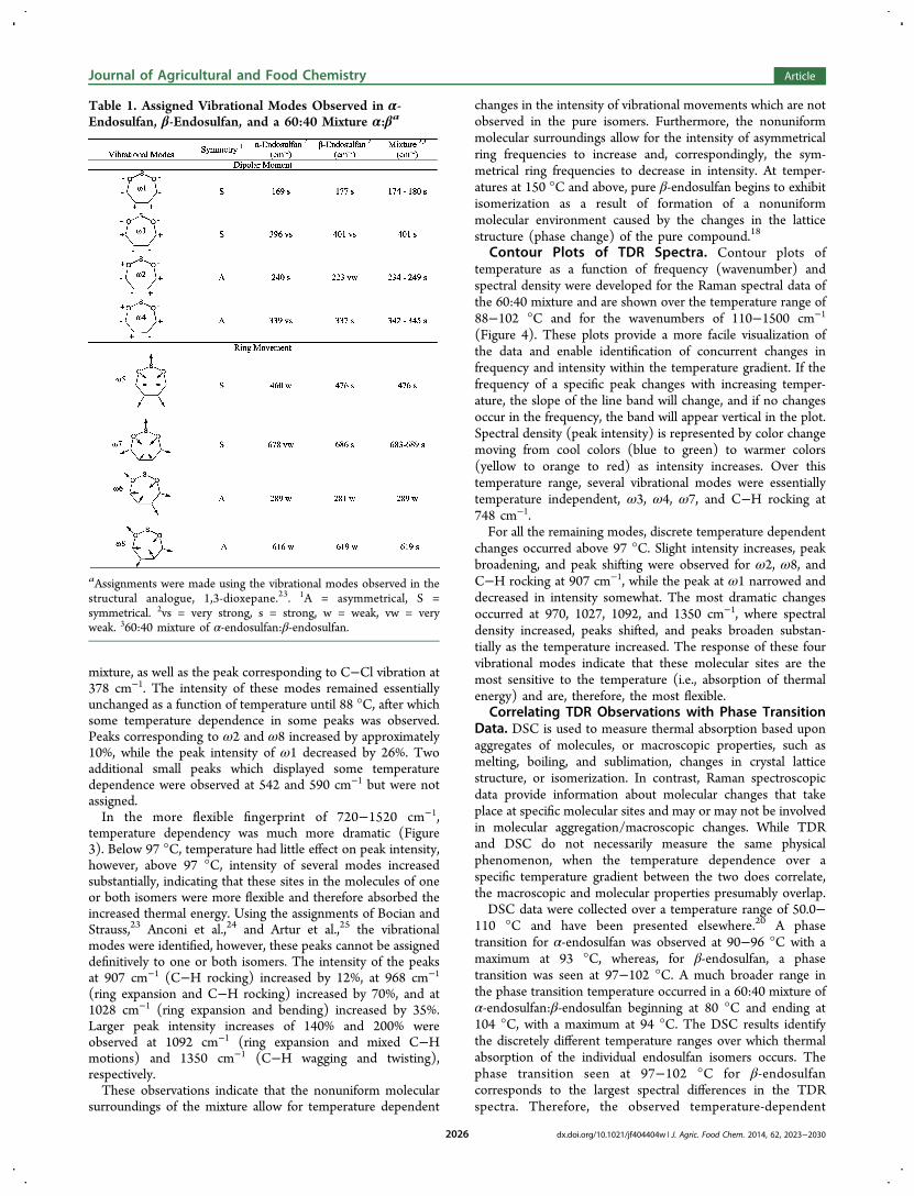
Raman Spectra Mode Assignments for Each Isomer.Endosulfan consists of two structural parts, a hexachlorinatednorboryl moiety and a seven-membered, dioxathiepine-3-oxidering system. The norbene portion is rigid, which is reflected inthe absence of C−Cl Raman vibrational modes except for onesignal at 374 cm−1.22 Vibrational frequencies characteristic ofseven-membered ring structures, including cycloheptane and1,3-dioxepane, have been identified.21 The seven atoms of thesesimpler ring systems gave rise to eight ring-bending frequencies,four symmetrical and four asymmetrical. The seven-memberedring in the endosulfan isomers is a structural analogue of 1,3-dioxepane and, correspondingly, the same eight frequenciesapply. For brevity, the same numbering of ring atoms in 1,3-dioxepane are used here: the ring oxygen atoms are atoms 1and 3, sulfur is atom 2, the methylene carbons are C4 and C7,and the methine carbons are C5 and C6 (Figure 1).The difference in the relative orientation of the axial
hydrogens on the methylene carbons, C4 and C7, is responsiblefor the three discrete forms of endosulfan. In β-endosulfan, onehydrogen atom on each of the methylene carbons is axial abovethe seven-membered ring system, affording a symmetricalmolecule in which CHCH2O on the right half of the ring is inthe same conformation as the left half. This contrasts with thetwo α-endosulfans, where only one hydrogen atom is axialabove the ring, not both, i.e., the axial hydrogen on the othermethylene carbon is axial below the ring system. The right halfof the ring system is not identical to the left half, giving rise totwo asymmetrical enantiomers. As molecules which are direct
Journal of Agricultural and Food Chemistry Article
dx.doi.org/10.1021/jf404404w | J. Agric. Food Chem. 2014, 62, 2023−20302024
mirror images of each other, the two α-endosulfans will haveidentical Raman spectra.Raman spectra were acquired at 1 °C intervals from 50 to
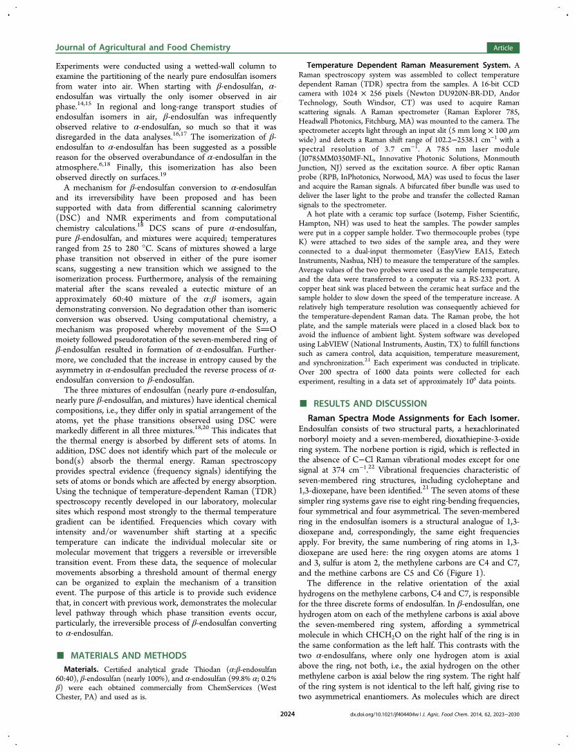
102 °C for nearly pure α-endosulfan and for essentially pure β-endosulfan; four major differences in the 110−810 cm−1 regionwere observed (Figure 2). Two major peaks were found in theα-endosulfan spectra at 229 cm−1 (ω2) and 748 cm−1 but werevery weak in the spectra of β-endosulfan, whereas peaks seen inthe β-endosulfan spectra at 460 cm−1 (ω5) and 686 cm−1 (ω7)were weak in the α-endosulfan spectra. Differences were alsoobserved in the Raman fingerprint region, 820−1520 cm−1.None of the peaks in the spectra of each nearly pure isomershowed a major change in intensity as a function oftemperature.From the work of Bocian and Strauss,23 eight modes in the
ring frequency region, 110−700 cm−1, were assignedcorresponding to the four symmetrical vibrational modes(ω1, ω3, ω5, and ω7) and to four asymmetrical vibrationalmodes (ω2, ω4, ω6, and ω8) (Table 1). Ring frequenciesinvolve only the atoms in the ring, not the hydrogens bound tothe carbons in the ring or the oxygen not part of the ring. Fourof these vibrational modes represent changes in dipolarmoment within the ring, that is, ω1, ω2, ω3, and ω4correspond to changes in electron density among the ringatoms. Modes ω1 and ω3 represent symmetrical dipolar
changes with respect to O−S−O, whereas modes ω2 and ω4are asymmetrical. The asymmetrical dipolar moment ω2 isextremely weak in β-endosulfan. The other four vibrationalmodes correspond to ring-bending, ω5, ω6, ω7, and ω8. Modesω5 and ω7 are symmetrical movements of the ring, andbecause α-endosulfan is asymmetrical, these modes areextremely weak. Asymmetrical ring-bending is depictedmodes ω6 and ω8 and are weak peaks in the symmetrical β-endosulfan.The modes observed in the fingerprint region of 720−1520
cm−1 correspond to the molecular vibrations of C−H. Thesemovements include asymmetrical and symmetrical stretching,rocking, twisting, scissoring, and wagging. A peak at 748 cm−1
was observed in only α-endosulfan and corresponds to anasymmetrical C−H rocking movement. The remaining modesobserved in the spectra of the pure isomers were difficult toassign unambiguously to specific atom vibrations.
Raman Spectra Mode Assignments for a 60:40Mixture of α-Endosulfan:β-Endosulfan. Under similargradient conditions for the individual isomers, Raman spectrawere acquired at 1 °C intervals from 50 to 102 °C for a 60:40mixture of α-endosulfan:β-endosulfan. The same eight ringvibrational modes, ω1−ω8, were observed in the 110−710cm−1 range (Table 1; Figure 3). The peak which was only seenin the α-endosulfan spectra, 748 cm−1, was also present in the
Figure 2. Raman spectra from 110 to 820 cm−1 and 820 to 1520 cm−1 acquired at 1 °C intervals from 50 to 102 °C for nearly pure α-endosulfan andfor essentially pure β-endosulfan. Vibrational modes, ω1 through ω8 (Table 1), and C−H rocking, in the 110−820 cm−1 range are indentified.Relative intensities of each vibrational mode in the individual isomers were essentially temperature independent.
Journal of Agricultural and Food Chemistry Article
dx.doi.org/10.1021/jf404404w | J. Agric. Food Chem. 2014, 62, 2023−20302025
mixture, as well as the peak corresponding to C−Cl vibration at378 cm−1. The intensity of these modes remained essentiallyunchanged as a function of temperature until 88 °C, after whichsome temperature dependence in some peaks was observed.Peaks corresponding to ω2 and ω8 increased by approximately10%, while the peak intensity of ω1 decreased by 26%. Twoadditional small peaks which displayed some temperaturedependence were observed at 542 and 590 cm−1 but were notassigned.In the more flexible fingerprint of 720−1520 cm−1,
temperature dependency was much more dramatic (Figure3). Below 97 °C, temperature had little effect on peak intensity,however, above 97 °C, intensity of several modes increasedsubstantially, indicating that these sites in the molecules of oneor both isomers were more flexible and therefore absorbed theincreased thermal energy. Using the assignments of Bocian andStrauss,23 Anconi et al.,24 and Artur et al.,25 the vibrationalmodes were identified, however, these peaks cannot be assigneddefinitively to one or both isomers. The intensity of the peaksat 907 cm−1 (C−H rocking) increased by 12%, at 968 cm−1
(ring expansion and C−H rocking) increased by 70%, and at1028 cm−1 (ring expansion and bending) increased by 35%.Larger peak intensity increases of 140% and 200% wereobserved at 1092 cm−1 (ring expansion and mixed C−Hmotions) and 1350 cm−1 (C−H wagging and twisting),respectively.These observations indicate that the nonuniform molecular
surroundings of the mixture allow for temperature dependent
changes in the intensity of vibrational movements which are notobserved in the pure isomers. Furthermore, the nonuniformmolecular surroundings allow for the intensity of asymmetricalring frequencies to increase and, correspondingly, the sym-metrical ring frequencies to decrease in intensity. At temper-atures at 150 °C and above, pure β-endosulfan begins to exhibitisomerization as a result of formation of a nonuniformmolecular environment caused by the changes in the latticestructure (phase change) of the pure compound.18
Contour Plots of TDR Spectra. Contour plots oftemperature as a function of frequency (wavenumber) andspectral density were developed for the Raman spectral data ofthe 60:40 mixture and are shown over the temperature range of88−102 °C and for the wavenumbers of 110−1500 cm−1
(Figure 4). These plots provide a more facile visualization ofthe data and enable identification of concurrent changes infrequency and intensity within the temperature gradient. If thefrequency of a specific peak changes with increasing temper-ature, the slope of the line band will change, and if no changesoccur in the frequency, the band will appear vertical in the plot.Spectral density (peak intensity) is represented by color changemoving from cool colors (blue to green) to warmer colors(yellow to orange to red) as intensity increases. Over thistemperature range, several vibrational modes were essentiallytemperature independent, ω3, ω4, ω7, and C−H rocking at748 cm−1.For all the remaining modes, discrete temperature dependent
changes occurred above 97 °C. Slight intensity increases, peakbroadening, and peak shifting were observed for ω2, ω8, andC−H rocking at 907 cm−1, while the peak at ω1 narrowed anddecreased in intensity somewhat. The most dramatic changesoccurred at 970, 1027, 1092, and 1350 cm−1, where spectraldensity increased, peaks shifted, and peaks broaden substan-tially as the temperature increased. The response of these fourvibrational modes indicate that these molecular sites are themost sensitive to the temperature (i.e., absorption of thermalenergy) and are, therefore, the most flexible.
Correlating TDR Observations with Phase TransitionData. DSC is used to measure thermal absorption based uponaggregates of molecules, or macroscopic properties, such asmelting, boiling, and sublimation, changes in crystal latticestructure, or isomerization. In contrast, Raman spectroscopicdata provide information about molecular changes that takeplace at specific molecular sites and may or may not be involvedin molecular aggregation/macroscopic changes. While TDRand DSC do not necessarily measure the same physicalphenomenon, when the temperature dependence over aspecific temperature gradient between the two does correlate,the macroscopic and molecular properties presumably overlap.DSC data were collected over a temperature range of 50.0−
110 °C and have been presented elsewhere.20 A phasetransition for α-endosulfan was observed at 90−96 °C with amaximum at 93 °C, whereas, for β-endosulfan, a phasetransition was seen at 97−102 °C. A much broader range inthe phase transition temperature occurred in a 60:40 mixture ofα-endosulfan:β-endosulfan beginning at 80 °C and ending at104 °C, with a maximum at 94 °C. The DSC results identifythe discretely different temperature ranges over which thermalabsorption of the individual endosulfan isomers occurs. Thephase transition seen at 97−102 °C for β-endosulfancorresponds to the largest spectral differences in the TDRspectra. Therefore, the observed temperature-dependent
Table 1. Assigned Vibrational Modes Observed in α-Endosulfan, β-Endosulfan, and a 60:40 Mixture α:βa
aAssignments were made using the vibrational modes observed in thestructural analogue, 1,3-dioxepane.23. 1A = asymmetrical, S =symmetrical. 2vs = very strong, s = strong, w = weak, vw = veryweak. 360:40 mixture of α-endosulfan:β-endosulfan.
Journal of Agricultural and Food Chemistry Article
dx.doi.org/10.1021/jf404404w | J. Agric. Food Chem. 2014, 62, 2023−20302026
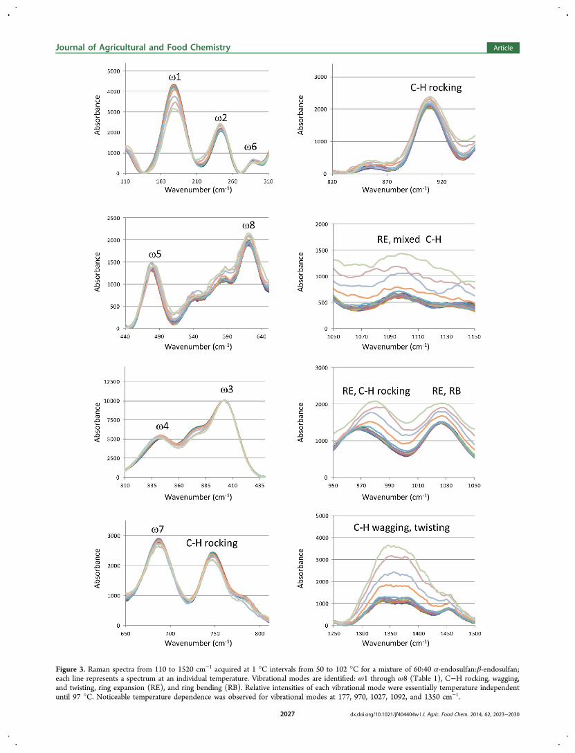
Figure 3. Raman spectra from 110 to 1520 cm−1 acquired at 1 °C intervals from 50 to 102 °C for a mixture of 60:40 α-endosulfan:β-endosulfan;each line represents a spectrum at an individual temperature. Vibrational modes are identified: ω1 through ω8 (Table 1), C−H rocking, wagging,and twisting, ring expansion (RE), and ring bending (RB). Relative intensities of each vibrational mode were essentially temperature independentuntil 97 °C. Noticeable temperature dependence was observed for vibrational modes at 177, 970, 1027, 1092, and 1350 cm−1.
Journal of Agricultural and Food Chemistry Article
dx.doi.org/10.1021/jf404404w | J. Agric. Food Chem. 2014, 62, 2023−20302027
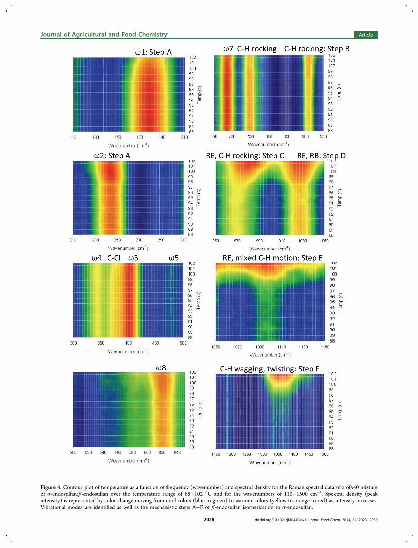
Figure 4. Contour plot of temperature as a function of frequency (wavenumber) and spectral density for the Raman spectral data of a 60:40 mixtureof α-endosulfan:β-endosulfan over the temperature range of 88−102 °C and for the wavenumbers of 110−1500 cm−1. Spectral density (peakintensity) is represented by color change moving from cool colors (blue to green) to warmer colors (yellow to orange to red) as intensity increases.Vibrational modes are identified as well as the mechanistic steps A−F of β-endosulfan isomerization to α-endosulfan.
Journal of Agricultural and Food Chemistry Article
dx.doi.org/10.1021/jf404404w | J. Agric. Food Chem. 2014, 62, 2023−20302028
Raman spectral changes are due to changes in β-endosulfan notα-endosulfan.Mechanism for Isomerization of β-Endosulfan to α-
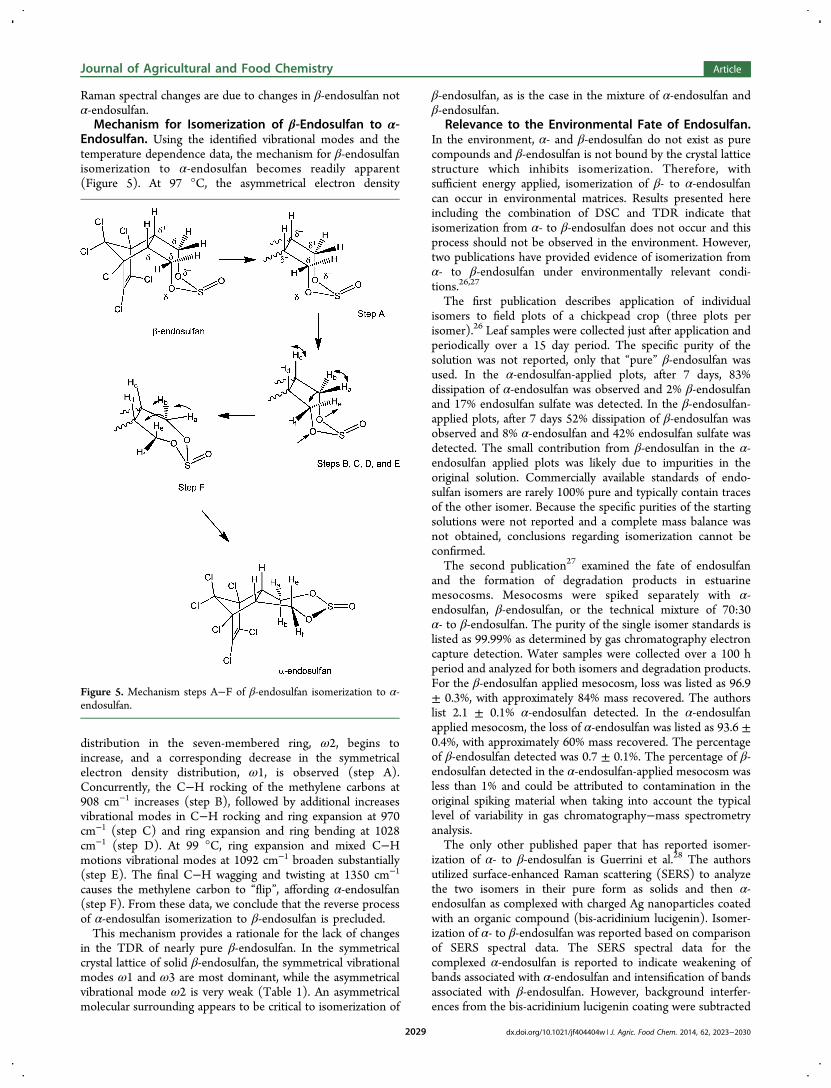
Endosulfan. Using the identified vibrational modes and thetemperature dependence data, the mechanism for β-endosulfanisomerization to α-endosulfan becomes readily apparent(Figure 5). At 97 °C, the asymmetrical electron density
distribution in the seven-membered ring, ω2, begins toincrease, and a corresponding decrease in the symmetricalelectron density distribution, ω1, is observed (step A).Concurrently, the C−H rocking of the methylene carbons at908 cm−1 increases (step B), followed by additional increasesvibrational modes in C−H rocking and ring expansion at 970cm−1 (step C) and ring expansion and ring bending at 1028cm−1 (step D). At 99 °C, ring expansion and mixed C−Hmotions vibrational modes at 1092 cm−1 broaden substantially(step E). The final C−H wagging and twisting at 1350 cm−1
causes the methylene carbon to “flip”, affording α-endosulfan(step F). From these data, we conclude that the reverse processof α-endosulfan isomerization to β-endosulfan is precluded.This mechanism provides a rationale for the lack of changes
in the TDR of nearly pure β-endosulfan. In the symmetricalcrystal lattice of solid β-endosulfan, the symmetrical vibrationalmodes ω1 and ω3 are most dominant, while the asymmetricalvibrational mode ω2 is very weak (Table 1). An asymmetricalmolecular surrounding appears to be critical to isomerization of
β-endosulfan, as is the case in the mixture of α-endosulfan andβ-endosulfan.
Relevance to the Environmental Fate of Endosulfan.In the environment, α- and β-endosulfan do not exist as purecompounds and β-endosulfan is not bound by the crystal latticestructure which inhibits isomerization. Therefore, withsufficient energy applied, isomerization of β- to α-endosulfancan occur in environmental matrices. Results presented hereincluding the combination of DSC and TDR indicate thatisomerization from α- to β-endosulfan does not occur and thisprocess should not be observed in the environment. However,two publications have provided evidence of isomerization fromα- to β-endosulfan under environmentally relevant condi-tions.26,27
The first publication describes application of individualisomers to field plots of a chickpead crop (three plots perisomer).26 Leaf samples were collected just after application andperiodically over a 15 day period. The specific purity of thesolution was not reported, only that “pure” β-endosulfan wasused. In the α-endosulfan-applied plots, after 7 days, 83%dissipation of α-endosulfan was observed and 2% β-endosulfanand 17% endosulfan sulfate was detected. In the β-endosulfan-applied plots, after 7 days 52% dissipation of β-endosulfan wasobserved and 8% α-endosulfan and 42% endosulfan sulfate wasdetected. The small contribution from β-endosulfan in the α-endosulfan applied plots was likely due to impurities in theoriginal solution. Commercially available standards of endo-sulfan isomers are rarely 100% pure and typically contain tracesof the other isomer. Because the specific purities of the startingsolutions were not reported and a complete mass balance wasnot obtained, conclusions regarding isomerization cannot beconfirmed.The second publication27 examined the fate of endosulfan
and the formation of degradation products in estuarinemesocosms. Mesocosms were spiked separately with α-endosulfan, β-endosulfan, or the technical mixture of 70:30α- to β-endosulfan. The purity of the single isomer standards islisted as 99.99% as determined by gas chromatography electroncapture detection. Water samples were collected over a 100 hperiod and analyzed for both isomers and degradation products.For the β-endosulfan applied mesocosm, loss was listed as 96.9± 0.3%, with approximately 84% mass recovered. The authorslist 2.1 ± 0.1% α-endosulfan detected. In the α-endosulfanapplied mesocosm, the loss of α-endosulfan was listed as 93.6 ±0.4%, with approximately 60% mass recovered. The percentageof β-endosulfan detected was 0.7 ± 0.1%. The percentage of β-endosulfan detected in the α-endosulfan-applied mesocosm wasless than 1% and could be attributed to contamination in theoriginal spiking material when taking into account the typicallevel of variability in gas chromatography−mass spectrometryanalysis.The only other published paper that has reported isomer-
ization of α- to β-endosulfan is Guerrini et al.28 The authorsutilized surface-enhanced Raman scattering (SERS) to analyzethe two isomers in their pure form as solids and then α-endosulfan as complexed with charged Ag nanoparticles coatedwith an organic compound (bis-acridinium lucigenin). Isomer-ization of α- to β-endosulfan was reported based on comparisonof SERS spectral data. The SERS spectral data for thecomplexed α-endosulfan is reported to indicate weakening ofbands associated with α-endosulfan and intensification of bandsassociated with β-endosulfan. However, background interfer-ences from the bis-acridinium lucigenin coating were subtracted
Figure 5. Mechanism steps A−F of β-endosulfan isomerization to α-endosulfan.
Journal of Agricultural and Food Chemistry Article
dx.doi.org/10.1021/jf404404w | J. Agric. Food Chem. 2014, 62, 2023−20302029
from the spectra of the nanoparticle complexes, leading to anegative peak in a critical region of the spectra. No otherconfirmatory data concerning presence of β-endosulfan werediscussed. Therefore, α- to β-endosulfan isomerization cannotbe conclusively identified.
■ AUTHOR INFORMATIONCorresponding Author*E-mail: [email protected] work was supported financially by U.S. Department ofAgriculture, Agricultural Research Service intramural projects:1265-42000-018-00 and 1265-12610-001-00.NotesMention of specific products is for identification and does notimply endorsement by the U.S. Department of Agriculture tothe exclusion of other suitable products or suppliers.The authors declare no competing financial interest.
■ ABBREVIATIONS USEDDSC, differential scanning calorimetry; POP, persistent organicpollutants; RE, ring expansion; RB, ring bending; TDR,temperature-dependent Raman; UNEP, United NationsEnvironmental Programme
■ REFERENCES(1) Maier-Bode, H. Properties, effect, residues and analytics of theinsecticide endosulfan. Residue Rev. 1968, 22, 1−44.(2) EPA Action to Terminate Endosulfan; United States EnvironmentalProtection Agency: Washington, DC, June 2010; http://www.epa.gov/pesticides/reregistration/endosulfan/endosulfan-cancl-fs.html#decision (Accessed September 20, 2013).(3) Lubick, N. Endosulfan’s exit: U.S. EPA pesticide review leads to aban. Science 2010, 328, 1466.(4) Endosulfan Phase-Out; United States Environmental ProtectionAgency: Washington, DC, November, 2010; http://www.epa.gov/pesticides/reregistration/endosulfan/endosulfan-agreement.html (Ac-cessed September 20, 2013).(5) United Nations Targets Widely-Used Pesticide Endosulfan for PhaseOut; United Nations Environment Programme: Geneva, May 3, 2011;http://chm.pops.int/TheConvention/Media/PressReleases/COP5Geneva,3May2011Endosulfanphaseout/tabid/2216/Default.aspx (Accessed September 20, 2013).(6) Weber, J.; Halsall, C. J.; Muir, D.; Teixeira, C.; Small, J.; Solomon,K.; Hermanson, M.; Hung, H.; Bidleman, T. Endosulfan, a globalpesticide: a review of its fate in the environment and occurrence in theArctic. Sci. Total Environ. 2010, 408, 2966−2984.(7) Becker, L.; Scheringer, M.; Schenker, U.; Hungerbuhler, K.Assessment of the environmental persistence and long-range transportof endosulfan. Environ. Pollut. 2011, 159, 1737−1743.(8) Janssen, M. P. M. Endosulfan. A Closer Look at the Argumentsagainst a Worldwide Phase Out; RIVM Letter Report 601356002/2011;National Institute for Public Health and the Environment: Bilthoven,Netherlands, 2011; 92 pp, http://www.rivm.nl/bibliotheek/rapporten/601356002.pdf (Accessed September 20, 2013).(9) Riemschneider, R.; Hilscher, J. C. Thiodan und analogeVerbindungen IV: Zur Kenntnis der Thiodan-Isomerie. Z. Naturforsch.,B: Chem. Sci. 1960, 15, 809−810.(10) Riemschneider, R.; Wuscherpfennig, V. Uber den raumlichenBau der Thiodan-Isomeren. Naturwissenschaften 1961, 48 (5), 130−131.(11) Riemschneider, R.; Wucherpfennig, V. Thiodan und analogeVerbindungen, 7. Konstellationen und Dipolmomente isomererThiodane. Z. Naturforsch., B: Chem. Sci. 1962, 17, 723.(12) Forman, S. E.; Durbetaki, A. J.; Cohen, M. V.; Olofson, R. A.Conformational equilibria in cyclic sulfites and sulfates. The
configurations and conformations of the two isomeric thiodans. J.Org. Chem. 1965, 30, 169−175.(13) Schmidt, W. F.; Hapeman, C. J.; Fettinger, J. C.; Rice, C. P.;Bilboulian, S. Structure and asymmetry in the isomeric conversion ofβ- to α-endosulfan. J. Agric. Food Chem. 1997, 45, 1023−1026.(14) Rice, C. P.; Chernyak, S. M.; Hapeman, C. J.; Bilboulian, S. Air−water distribution of the endosulfan isomers. J. Environ. Qual. 1997,26, 1101−1106.(15) Rice, C. P.; Chernyak, S. M.; McConnell, L. L. Henry’s lawconstants for pesticides measured as a function of temperature andsalinity. J. Agric. Food Chem. 1997, 45, 2291−2298.(16) Simonich, S. L.; Hites, R. A. Global distribution of persistentorganochlorine compounds. Science 1995, 269, 1851−1854.(17) Burgoyne, T. W.; Hites, R. A. Effects of temperature and winddirection on the atmospheric concentrations of α-endosulfan. Environ.Sci. Technol. 1993, 27, 910−914.(18) Schmidt, W. F.; Bilboulian, S.; Rice, C. P.; McConnell, L. L.;Fettinger, J. C.; Hapeman, C. J. Thermodynamic, spectroscopic, andcomputational evidence for the irreversible conversion of β- to α-endosulfan. J. Agric. Food Chem. 2001, 49, 5372−5376.(19) Walse, S. S.; Shimizu, K. D.; Ferry, J. L. Surface-catalyzedtransformations of aqueous endosulfan. Environ. Sci. Technol. 2002, 36,4846−4853.(20) Schmidt, W. F., Nguyen, J. K., Qin, J., Kim, M. S., Mookherji, S.,McConnell, L. L., Hapeman, C. J. DSC and Raman spectra of α- andβ-endosulfan plus 60/40 mixture. In SPIE Defense, Security, andSensing; International Society for Optics and Photonics: Bellingham,WA, May 2013; pp 87210R-1−87210R-7.(21) Qin, J.; Chao, K.; Kim, M. S. Raman chemical imaging systemfor food safety and quality inspection. Trans. ASABE 2010, 53, 1873−1882.(22) Moore, W. H.; Ching, J. H. C.; Warrier, A. V. R.; Krimm, S.Assignment of torsion and low frequency bending vibrations ofsecondary chlorides. Spectrochim. Acta, Part A 1973, 29, 1847−1858.(23) Bocian, D. F.; Strauss, H. L. Vibrational spectra, conformations,and potential functions of cycloheptane and related oxepanes. J. Am.Chem. Soc. 1977, 99, 2866−2878.(24) Anconi, C. P. A.; Nascimento, C. S., Jr.; Dos Santos, H. F.;DeAlmeida, W. B. A highly correlated ab initio investigation of thetemperature dependent conformational analysis of cycloheptane.Chem. Phys. Lett. 2006, 418, 459−466.(25) Artur, C.; Le Ru, R. C.; Etchegoin, P. G. Temperaturedependence of the homogeneous broadening of resonant Raman peaksmeasured by single-molecule surface-enhanced Raman spectroscopy. J.Phys. Chem. Lett. 2011, 2, 3002−3005.(26) Gopal, M.; Mukherjee, I. Determination of residues ofendosulfan and endosulfan sulfate on eggplant, mustard and chickpea.Pestic. Sci. 1994, 37, 67−72.(27) Walse, S. S.; Scott, G. I.; Ferry, J. L. Stereoselective degradationof aqueous endosulfan in modular estuarine mesocosms: formation ofendosulfan γ-hydroxycarboxylate. J. Environ. Monit. 2003, 5, 373−379.(28) Guerrini, L.; Aliaga, A. E.; Carcamo, J.; Gomez-Jeria, J. S.;Sanchez-Cortes, S.; Campo-Vallette, M. M.; Garcia-Ramos, J. V.Functionalization of Ag nano-particles with the bis-acridiniumlucigenin as a chemical assembler in the detection of persistentorganic pollutants by surface-enhanced Raman scattering. Anal. Chim.Acta 2008, 624, 286−293.
Journal of Agricultural and Food Chemistry Article
dx.doi.org/10.1021/jf404404w | J. Agric. Food Chem. 2014, 62, 2023−20302030