Price convergence after the Eastern EU enlargement? Evidence from
EVIDENCE ON THE CARCINOGENICITY OF
3-Monochloropropane-1,2-diol (3-MCPD; α-Chlorohydrin) UPDATE September 2010 Reproductive and Cancer Hazard Assessment Branch Office of Environmental Health Hazard Assessment California Environmental Protection Agency
3-MCPD i September 2010 OEHHA
The Office of Environmental Health Hazard Assessment’s (OEHHA) Reproductive and Cancer Hazard Assessment Branch was responsible for the preparation of this document. Primary Authors Rajpal S. Tomar, Ph.D. Staff Toxicologist Cancer Toxicology and Epidemiology Section Feng C. Tsai, Ph.D. Staff Toxicologist Cancer Toxicology and Epidemiology Section Martha S. Sandy, Ph.D. Chief, Cancer Toxicology and Epidemiology Section Reproductive and Cancer Hazard Assessment Branch Internal OEHHA Reviewers George V. Alexeeff, Ph.D., D.A.B.T. Deputy Director for Scientific Affairs Lauren Zeise, Ph.D. Chief, Reproductive and Cancer Hazard Assessment Branch Robert Howd, Ph.D. Chief, Water Toxicology Section Pesticide and Environmental Toxicology Branch John Faust, Ph.D. Staff Toxicologist Cancer Toxicology and Epidemiology Section Reproductive and Cancer Hazard Assessment Branch
3-MCPD ii September 2010 OEHHA
PREFACE Proposition 651 requires the publication of a list of chemicals “known to the state” to cause cancer or reproductive toxicity. It specifies that “a chemical is known to the state to cause cancer … if in the opinion of the state’s qualified experts the chemical has been clearly shown through scientifically valid testing according to generally accepted principles to cause cancer ....” The “state’s qualified experts” regarding findings of carcinogenicity are the members of the Carcinogen Identification Committee (CIC) of the OEHHA Science Advisory Board2
.
The lead agency for implementing Proposition 65 is the Office of Environmental Health Hazard Assessment (OEHHA) of the California Environmental Protection Agency. After consultation with the CIC, OEHHA selected 3-monochloropropane-1,2-diol (3-MCPD) as a chemical for consideration for listing by the CIC. Upon selection, the public was given the opportunity to submit information relevant to the assessment of the evidence on the carcinogenicity of 3-MCPD. OEHHA reviewed and considered those submissions in preparing this document. OEHHA developed this document to provide the CIC with comprehensive information on 3-MCPD carcinogenicity for use in its deliberations on whether or not the chemical should be listed under Proposition 65. This September 2010 update corrects three non-substantive errors on pages ii, 25, and 28. An additional set of two-year drinking water studies in B6C3F1 mice was published in the peer reviewed scientific literature after the release of the June 2010 document, as Jeong et al. (2010)3
. This article was provided to the CIC and was part of the Committee’s consideration of 3-MCPD.
At their September 21, 2010 meeting, the CIC, by a vote of six in favor and one against, found that 3-MCPD had been “clearly shown through scientifically valid testing according to generally accepted principles to cause cancer.” Accordingly, “3-Monochloropropane (3-MCPD)” was placed on the Proposition 65 list of chemicals known to the state to cause cancer.
1 The Safe Drinking Water and Toxic Enforcement Act of 1986 (California Health and Safety Code 25249.5 et seq.) 2 Title 27 Cal. Code of Regs. §25302 3 Jeong J, Han BS, Cho W-S, Choi M, Ha C-S, Lee B-S, Kim Y-B, Son W-C, Kim C-Y (2010). Carcinogenicity study of 3-monochloropropane-1,2-diol (3-MCPD) administered by drinking water to B6C3F1 mice showed no carcinogenic potential. Arch. Toxicol 84:719-729.
3-MCPD iii September 2010 OEHHA
TABLE OF CONTENTS
1. EXECUTIVE SUMMARY ............................................................................................ 1
2. INTRODUCTION ........................................................................................................ 4
2.1 Identity of 3-Monochloropropane-1,2-diol (3-MCPD; α-Chlorohydrin) .................. 4
2.2 Occurrence and Use ............................................................................................. 4
3. DATA ON CARCINOGENICITY ................................................................................. 6
3.1 Carcinogenicity Studies in Humans ....................................................................... 6
3.2 Carcinogenicity Studies in Animals ....................................................................... 6
3.2.1 Studies in mice ................................................................................................ 7
3.2.2 Studies in rats .................................................................................................. 7
3.3 Other Relevant Data ............................................................................................ 11
3.3.1 Pharmacokinetics and Metabolism ................................................................ 11
3.3.2 Genotoxicity ................................................................................................... 14
3.3.3 In Vitro Transformation Study ........................................................................ 18
3.3.4 Animal Tumor Pathology ............................................................................... 18
3.3.5 Effects on Immunological Systems ................................................................ 19
3.3.6 Structure Activity Comparisons ..................................................................... 20
4. MECHANISMS ......................................................................................................... 26
5. REVIEWS BY OTHER AGENCIES .......................................................................... 27
6. SUMMARY AND CONCLUSIONS ........................................................................... 27
6.1 Summary of Evidence ......................................................................................... 27
6.2 Conclusion........................................................................................................... 29
7. REFERENCES .......................................................................................................... 30
3-MCPD iv September 2010 OEHHA
LIST OF TABLES
Table 1. Incidence of treatment-related lesions in Fischer rats administered 3-MCPD in drinking water for 104 weeks ................................................................................. 8
Table 2. Incidence of treatment-related lesions in Sprague Dawley rats administered 3-MCPD in drinking water for two years ................................................................... 10
Table 3. In Vitro Genotoxicity Studies on 3-MCPD ....................................................... 15
Table 4. In Vivo Genotoxicity Studies on 3-MCPD …… .............................................. 17
Table 5. Structure-Activity Comparisons for 3-MCPD ................................................ 23
LIST OF FIGURES
Figure 1. Chemical structure of 3-MCPD ....................................................................... 4 Figure 2. Metabolic Pathways Proposed for 3-MCPD .................................................. 13
3-MCPD 1 September 2010 OEHHA
1. EXECUTIVE SUMMARY 3-Monochloropropane-1,2-diol (3-MCPD) is used in the manufacture of dye intermediates, as a solvent for cellulose acetate, and to lower the freezing point of dynamite. It is registered at the federal level as a rodenticide under the name α-chlorohydrin; however, there are no pesticide products containing 3-MCPD currently registered in California. 3-MCPD is one of several chloropropanols that can be formed in foods during processing, cooking and storage as a result of chloride ions reacting with glycerol and other lipids present in the food. 3-MCPD has been detected in a variety of foods, including foods containing acid-hydrolyzed vegetable protein (acid-HVP), such as soy and oyster sauces. It has also been found in foods that do not contain acid-HVP, such as malt products; cold-smoked meats, sausage, and fish; cooked meats; anchovies packed in oil; some cheeses; roasted or toasted cereals; breads and biscuits; and instant coffee and roasted coffee beans. 3-MCPD also may be present as an impurity in epichlorohydrin-containing products, such as water flocculants and “wet-strength” resins used in food-contact materials and some paper products. 3-MCPD has been tested in two sets of two-year drinking water studies conducted in male and female Fischer and Sprague-Dawley rats. Treatment-related increases in tumors of the kidney, mammary gland, and testes (Leydig cell) were observed, as follows. Kidney tumors
• 3-MCPD significantly increased the incidence of malignant, and benign and malignant kidney tumors in male Sprague-Dawley rats.
o Tumors appeared early. o Kidney tumors are rare in untreated male Sprague-Dawley rats.
• Combined benign and malignant kidney tumors were significantly increased in treated female Sprague-Dawley rats.
o Kidney tumors are rare in untreated female Sprague-Dawley rats. • Benign kidney tumors were increased in treated male Fischer rats. • Benign kidney tumors were increased in treated female Fischer rats. • 3-MCPD increased renal tubular hyperplasia in both sexes and strains. • 3-MCPD exacerbated chronic progressive nephropathy in both sexes and
strains. Mammary tumors
• 3-MCPD significantly increased the incidence of benign and malignant mammary tumors in male Fischer rats.
• 3-MCPD increased glandular hyperplasia of the mammary gland in male Fischer rats.
• Mammary gland tumors are uncommon in male Fischer rats.
3-MCPD 2 September 2010 OEHHA
Leydig cell tumors
• 3-MCPD significantly increased the incidence of benign and malignant Leydig cell tumors in male Fischer rats.
• 3-MCPD significantly increased the incidence of Leydig cell tumors in male Sprague-Dawley rats.
• Leydig cell tumors are uncommon in Sprague-Dawley rats. No treatment-related tumors were observed in 19-month studies conducted in female Swiss mice exposed to 3-MCPD either via topical application (thrice per week) or by subcutaneous injection (once per week), or in 72-week studies in male and female Charles River (CD-1) rats administered 3-MCPD by gavage (twice per week). 3-MCPD has been found to be genotoxic in a variety of assays involving prokaryotic and eukaryotic cells, and to induce malignant transformation of mouse M2 fibroblasts in culture. There is some suggestion from in vitro and in vivo studies in mice that 3-MCPD may impair certain immune functions, such as NK cell activity which is involved in tumor surveillance, and altered production of cytokines, which is involved in regulation of inflammatory responses. Additional evidence of carcinogenicity comes from eight compounds that are either structurally similar to 3-MCPD, precursors or metabolites of 3-MCPD, or share common metabolites with 3-MCPD. All have positive results in genotoxicity assays performed in vitro, and all but 3-MCPD, 1,3-DCP, and 2,3-dibromo-1-propanol also have some positive genotoxicity results in vivo. All have some positive carcinogenic data in rodent studies, and six are currently listed under Proposition 65 as causing cancer and classified by the International Agency for Research on Cancer as either Group 2A or Group 2B carcinogens (glycidol, epichlorohydrin, 2,3-dibromo-1-propanol, 1,2,3-trichloropropane, 1,2-dibromo-3-chloropropane (DBCP), and tris(2,3-dibromopropyl)phosphate (TDPP)). Many of these compounds induce tumors at multiple sites, and in most cases in more than one sex/species. Many of these compounds also induce tumors at the same sites in rats as 3-MCPD. Specifically, 3-MCPD, 1,3-DCP, 1,2,3-trichloropropene, DBCP, tris(1,3-dichloro-2-propyl)phosphate (TDCPP) and TDPP induce rat kidney tumors; 3-MCPD and TDCPP induce rat Leydig cell tumors; and 3-MCPD and glycidol induce mammary gland tumors in male rats. In summary, the evidence for carcinogenicity of 3-MCPD comes from:
• Multiple studies in rats o Kidney tumors in male Fischer rats o Kidney tumors in male Sprague-Dawley rats o Kidney tumors in female Fischer rats o Kidney tumors in male Sprague-Dawley rats o Leydig cell tumors of the testes in male Fischer rats o Leydig cell tumors of the testes in male Sprague-Dawley rats o Mammary tumors in male Fischer rats
• Positive findings in a variety of in vitro genotoxicity test systems • Malignant transformation of mammalian cells in culture
3-MCPD 3 September 2010 OEHHA
• Metabolism of 3-MCPD to glycidol, a genotoxic carcinogen • Structure-activity considerations with six carcinogens identified by IARC and
listed under Proposition 65.
3-MCPD 4 September 2010 OEHHA
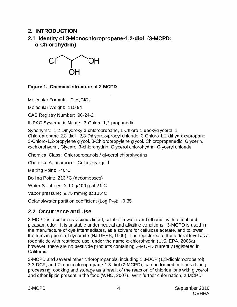
2. INTRODUCTION2.1 Identity of 3-Monochloropropane-1,2-diol (3-MCPD;
α-Chlorohydrin)
Figure 1. Chemical structure of 3-MCPD
Molecular Formula: C3H7ClO2 Molecular Weight: 110.54 CAS Registry Number: 96-24-2 IUPAC Systematic Name: 3-Chloro-1,2-propanediol Synonyms: 1,2-Dihydroxy-3-chloropropane, 1-Chloro-1-deoxyglycerol, 1-Chloropropane-2,3-diol, 2,3-Dihydroxypropyl chloride, 3-Chloro-1,2-dihydroxypropane, 3-Chloro-1,2-propylene glycol, 3-Chloropropylene glycol, Chloropropanediol Glycerin,α-chlorohydrin, Glycerol 3-chlorohydrin, Glycerol chlorohydrin, Glyceryl chloride
Chemical Class: Chloropropanols / glycerol chlorohydrins Chemical Appearance: Colorless liquid Melting Point: -40°C Boiling Point: 213 °C (decomposes) Water Solubility: ≥ 10 g/100 g at 21°C Vapor pressure: 9.75 mmHg at 115°C Octanol/water partition coefficient (Log Pow): -0.85
2.2 Occurrence and Use 3-MCPD is a colorless viscous liquid, soluble in water and ethanol, with a faint andpleasant odor. It is unstable under neutral and alkaline conditions. 3-MCPD is used inthe manufacture of dye intermediates, as a solvent for cellulose acetate, and to lowerthe freezing point of dynamite (NJ DHSS, 1999). It is registered at the federal level as arodenticide with restricted use, under the name α-chlorohydrin (U.S. EPA, 2006a);however, there are no pesticide products containing 3-MCPD currently registered inCalifornia.3-MCPD and several other chloropropanols, including 1,3-DCP (1,3-dichloropropanol),2,3-DCP, and 2-monochloropropane-1,3-diol (2-MCPD), can be formed in foods duringprocessing, cooking and storage as a result of the reaction of chloride ions with glyceroland other lipids present in the food (WHO, 2007). With further chlorination, 2-MCPD
3-MCPD 5 September 2010 OEHHA
and 3-MCPD may form dichloropropanols. 3-MCPD and 1,3-DCP are among the major chloropropanols identified in foods (Baer et al., 2010). The formation of 3-MCPD in food is influenced by many factors, including temperature, pH, moisture content, sugar and lipid content, the type of processing method employed, and storage conditions (Baer et al., 2010). Three mechanisms of 3-MCPD formation have been proposed: i) Acid hydrolysis of glycerol and other lipids in the presence of chloride ions, as happens during the formation of acid-hydrolyzed vegetable protein (acid-HVP). ii) Heat processing of lipids with sodium chloride (present naturally or added), occurring in the absence of acid-HVP. iii) Hydrolysis of 3-MCPD esters by lipases, as can occur in baked bread (Baer et al., 2010). 3-MCPD has been detected in a variety of foods. These include:
• A wide range of prepared and processed foods to which acid-HVP is added, including many soy sauces, oyster sauces, other sauces, instant soups, bouillon cubes, gravy mixes, savory snacks, spreads, stuffings, ready-to-eat meals, instant noodles, frozen dinners, and other frozen prepared foods (ILS, 2005).
• Some processed foods that do not contain acid-HVP, such as malt products; cold-smoked meats, sausage, and fish; cooked meats (salami, bacon, hamburgers); anchovies packed in oil; melted or grilled cheese; processed cheese and cheese alternatives; roasted or toasted cereals, breads, and biscuits; instant coffee and roasted coffee beans (Hamlet et al., 2002; FSANZ, 2003; WHO, 2007; Baer et al., 2010).
A recent study found that pyrolysis of the synthetic sweetener sucralose, a polychlorinated compound, in the presence of glycerol, can also result in the formation of chloropropanols (>75% 3-MCPD, 15%-23% 1,3-DCP, <5% 1,2-DCP) (Rahn and Yaylayan, 2010). This finding suggests that the use of sucralose in baked goods may lead to chloropropanol formation. 3-MCPD esters, such as mono- and diesters of fatty acids with 3-MCPD, have been detected in a variety of foods (e.g., vegetable oils, infant formula, human breast milk), prompting concerns that 3-MCPD may be released directly into the food, or in the body after consumption, as a result of lipase-catalyzed hydrolysis (Zelinkova et al., 2008; Zelinkova et al., 2009; Baer et al., 2010). Regarding the presence of chloropropanols in soy sauces and related foods, if these products are prepared using traditional fermentation processes (i.e., without the addition of acid-HVP), they generally do not contain 3-MCPD or other chloropropanols (Crews et al., 2003; Nyman et al., 2003; WHO, 2007). Efforts to reduce the levels of chloropropanols in soy sauces and other foods include carefully controlling the acid hydrolysis process, following the acid hydrolysis step with an alkaline hydrolysis step, and reducing the concentrations of fats and oils in the starting materials (Nyman et al., 2003; Crews et al., 2003; Macarthur et al., 2000). Baer
3-MCPD 6 September 2010 OEHHA
et al. (2010) identified other control strategies for limiting the amount of 3-MCPD in food, including:
• Raising the pH of high moisture content food • Lowering the maximum processing temperature and salt content of the food • Avoiding low water/high temperature treatments • Limiting the amount of glycerol produced in the food during preparation and
storage • Avoiding the use of partial glycerides as additives • Using spice extracts instead of native spices • Reducing microbial load via thermal treatment • Confirming the purity of food additives • Inactivating lipases and esterases • Screening food contact material for 3-MCPD precursors
3-MCPD and other chloropropanols can be formed from the degradation of epichlorohydrin in aqueous media because of epichlorohydrin’s slow hydrolysis in the presence of chloride ions (ILS, 2005). 3-MCPD and other chloropropanols also may be present as impurities in epichlorohydrin compounds and products derived from them, such as epoxy resins and dimethylamine-epichlorohydrin copolymers (DECs) (ILS, 2005). DECs are used in flocculants and coagulants for water purification, and 3-MCPD has been detected in finished drinking water as a result of such use (EC, 2004). Epichlorohydrin copolymers containing low concentrations of 3-MCPD and 1,3-DCP are also used as “wet-strength” resins for products such as tea bags, coffee filters, sausage casings, absorbents packaged with meats, and paper towels and tissues (Tritscher, 2004; ILS, 2005). DECs are also used as decolorizing agents and flocculants in the clarification of refinery sugar liquors and juices, and in the production of high-fructose corn syrup (ILS, 2005).
3. DATA ON CARCINOGENICITY
3.1 Carcinogenicity Studies in Humans No carcinogenicity studies in humans were found in the published literature.
3.2 Carcinogenicity Studies in Animals A number of long-term animal carcinogenicity studies have been reported in the literature. In two studies, female Swiss mice were exposed to 3-MCPD either via topical application or by subcutaneous injection. Two sets of two-year drinking water studies were conducted in Fischer and Sprague-Dawley rats of both sexes. Oral gavage studies in male and female Sprague-Dawley rats have also been conducted. This section reports on the designs and results of the 3-MCPD carcinogenicity studies.
3-MCPD 7 September 2010 OEHHA
3.2.1 Studies in mice 19-month dermal study in CHR/Ha Swiss mice (Van Duuren et al., 1974)
3-MCPD was administered to 50 female CHR/Ha Swiss mice by topical application at a dose of 2 milligrams (mg) three times per week. 3-MCPD was dissolved in 0.1 milliliters (mL) of acetone. The duration of treatment and the study was 580 days. A control group of 50 female CHR/Ha Swiss mice was given vehicle alone. No treatment-related neoplastic findings were reported. 19-month subcutaneous injection study in CHR/Ha Swiss mice (Van Duuren et al., 1974)
3-MCPD was administered to 50 female CHR/Ha Swiss mice by weekly subcutaneous injections at a dose of 1 mg/week for 580 days. The chemical was dissolved in 0.5 mL of tricaprylin. A control group of 50 female CHR/Ha Swiss mice received weekly injections of the vehicle alone. A local sarcoma at the site of injection was observed in one treated and one control animal.
3.2.2 Studies in rats Two-year drinking water studies in Fischer 344 rats (Sunahara et al., 1993, as reported in WHO, 2002)
3-MCPD (purity 98%) was administered in drinking water to groups of 50 male and 50 female Fischer 344 rats at concentrations of 0, 20, 100, and 500 milligrams/liter (mg/L) for 104 weeks. The 3-MCPD solution was prepared twice a week and was reported to be stable for more than four days in water. Food and water consumption were routinely recorded. Hematological parameters and blood chemistry values were assessed at weeks 103-105 for all surviving animals. All animals were subjected to complete necropsy and histopathological examination. There was a significant reduction in food and water consumption in male and female rats at the highest dose of 500 mg/L. This was accompanied by a significant reduction in body weight after the first week. At study termination, weights were lower than controls by 33 percent in high-dose males and 35 percent in high-dose females. At termination the body weight was also significantly lower in the mid-dose males and females. No significant dose-related effects were observed on hematological and blood clinical chemistry parameters except for increased blood urea nitrogen and creatinine in the mid- and high-dose groups in both males and females. Significant dose-related histopathological changes were observed in the testis, mammary gland, kidneys and pancreas in male rats, and kidneys in female rats. Among males, there was a significant dose-dependent decrease in Leydig cell hyperplasia with increasing dose (p < 0.01), and a dose-dependent increase with increasing dose in the incidence of Leydig cell adenoma (p < 0.01) and a positive trend for carcinoma (p < 0.001). The incidence of adenoma significantly differed between control and treated animals in the mid- (p < 0.001) and high- (p < 0.05) dose groups. The combined Leydig cell adenoma and carcinoma showed a significant trend and
3-MCPD 8 September 2010 OEHHA
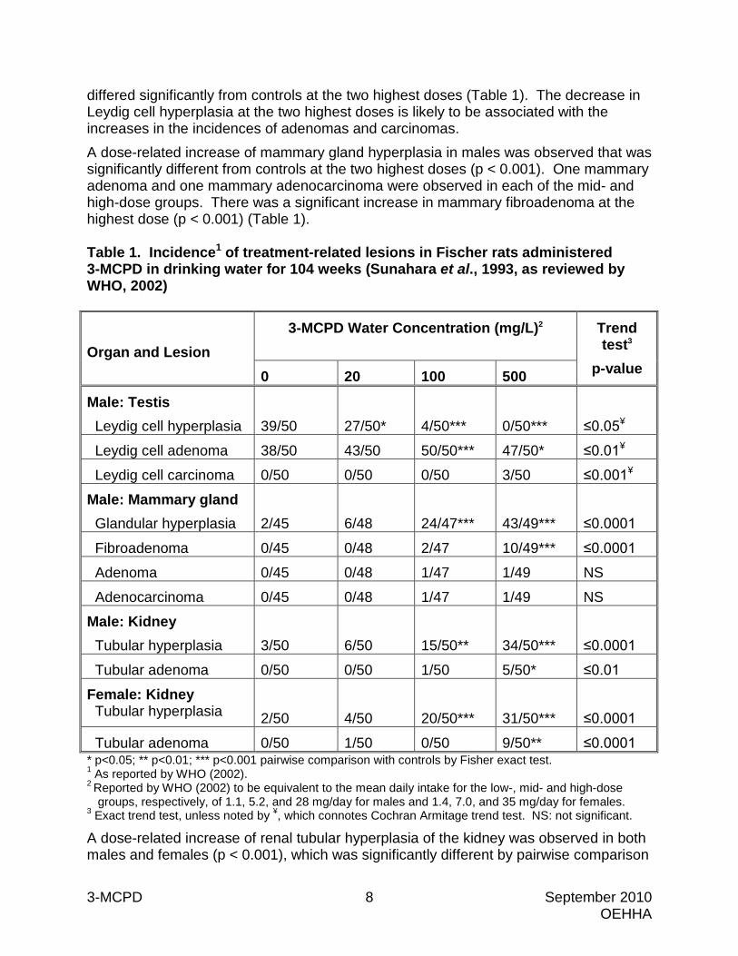
differed significantly from controls at the two highest doses (Table 1). The decrease in Leydig cell hyperplasia at the two highest doses is likely to be associated with the increases in the incidences of adenomas and carcinomas. A dose-related increase of mammary gland hyperplasia in males was observed that was significantly different from controls at the two highest doses (p < 0.001). One mammary adenoma and one mammary adenocarcinoma were observed in each of the mid- and high-dose groups. There was a significant increase in mammary fibroadenoma at the highest dose (p < 0.001) (Table 1). Table 1. Incidence1 of treatment-related lesions in Fischer rats administered 3-MCPD in drinking water for 104 weeks (Sunahara et al., 1993, as reviewed by WHO, 2002)
Organ and Lesion 3-MCPD Water Concentration (mg/L)2
Trend test3
p-value 0 20 100 500
Male: Testis Leydig cell hyperplasia
39/50
27/50*
4/50***
0/50***
≤0.05¥
Leydig cell adenoma 38/50 43/50 50/50*** 47/50* ≤0.01¥
Leydig cell carcinoma 0/50 0/50 0/50 3/50 ≤0.001¥
Male: Mammary gland Glandular hyperplasia
2/45
6/48
24/47***
43/49***
≤0.0001
Fibroadenoma 0/45 0/48 2/47 10/49*** ≤0.0001
Adenoma 0/45 0/48 1/47 1/49 NS
Adenocarcinoma 0/45 0/48 1/47 1/49 NS
Male: Kidney Tubular hyperplasia
3/50
6/50
15/50**
34/50***
≤0.0001
Tubular adenoma 0/50 0/50 1/50 5/50* ≤0.01
Female: Kidney Tubular hyperplasia
2/50
4/50
20/50***
31/50***
≤0.0001
Tubular adenoma 0/50 1/50 0/50 9/50** ≤0.0001 * p<0.05; ** p<0.01; *** p<0.001 pairwise comparison with controls by Fisher exact test. 1 As reported by WHO (2002). 2 Reported by WHO (2002) to be equivalent to the mean daily intake for the low-, mid- and high-dose
groups, respectively, of 1.1, 5.2, and 28 mg/day for males and 1.4, 7.0, and 35 mg/day for females. 3 Exact trend test, unless noted by ¥, which connotes Cochran Armitage trend test. NS: not significant.
A dose-related increase of renal tubular hyperplasia of the kidney was observed in both males and females (p < 0.001), which was significantly different by pairwise comparison

3-MCPD 9 September 2010 OEHHA
at the two highest doses compared to controls (p < 0.001). In females, a significant increase in tubular adenoma (p < 0.01) was also seen in the highest dose groups, while in males there was a positive trend for adenoma (Table 1). Chronic progressive nephropathy was found in all dose groups in both males and females, but was significant only at the two highest doses. A correlation was observed with progressive nephropathy and incidence of renal hyperplasia and adenoma in both sexes. There was a dose-related decrease in pancreatic hyperplasia, adenoma and carcinoma in males. Two-year drinking water studies in Sprague-Dawley rats (Cho et al., 2008)
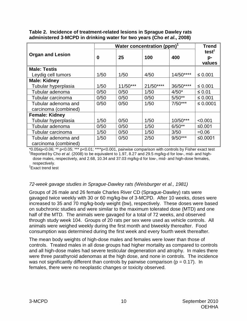
3-MCPD (98% purity) was administered in drinking water to groups of 50 male and 50 female Sprague-Dawley rats at concentrations of 0, 25, 100, or 400 parts per million (ppm) for two years. Body weight, water and food consumption were recorded weekly. Based on water consumption the average daily intakes of 3-MCPD were 0, 1.97, 8.27, and 29.50 milligrams/kilogram per day (mg/kg/day) for males and 0, 2.68, 10.34, and 37.03 mg/kg/day for females. These doses were selected based on 13-week toxicity studies conducted in Sprague-Dawley rats. There were no significant differences in survival at the end of two years among the male or female rats. However, survival was less than 50 percent due to spontaneous pituitary tumors in all male and female groups. There was no difference in food consumption among the groups, though water consumption was significantly lower in high-dose males and females, as compared to controls. The body weights of high-dose males and females were significantly lower than controls throughout the course of the studies. Male and female rats were necropsied at 100 and 104 weeks, respectively, and complete necropsies were performed on all animals. In male rats, there was a significant increase in Leydig cell tumors at the highest dose as compared to controls (p < 0.001) and a significant dose-related trend (p < 0.001) (Table 2). The incidences of combined renal tubular adenoma and carcinoma were significantly increased (p < 0.01) in high-dose males and high-dose females as compared to controls, and these increases occurred with significant dose-related trends (p < 0.0001). In high-dose males renal tubular carcinoma was significantly increased (p < 0.05), and adenoma was marginally increased (0.05 < p < 0.1). In high-dose females renal tubular adenoma was significantly increased (p < 0.05). These increases in kidney tumors were accompanied by highly significant increases in renal tubular hyperplasia in all male treatment groups and at the highest dose in females. The study authors stated that renal tubular adenoma and carcinoma were first observed in males at 78 and 74 weeks, respectively.
3-MCPD 10 September 2010 OEHHA
Table 2. Incidence of treatment-related lesions in Sprague Dawley rats administered 3-MCPD in drinking water for two years (Cho et al., 2008)
Organ and Lesion Water concentration (ppm)1 Trend
test2 p-
values 0 25 100 400
Male: Testis Leydig cell tumors
1/50
1/50
4/50
14/50****
≤ 0.001
Male: Kidney Tubular hyperplasia
1/50
11/50***
21/50****
36/50****
≤ 0.001
Tubular adenoma 0/50 0/50 1/50 4/50* ≤ 0.01 Tubular carcinoma 0/50 0/50 0/50 5/50** ≤ 0.001 Tubular adenoma and carcinoma (combined)
0/50 0/50 1/50 7/50*** ≤ 0.0001
Female: Kidney Tubular hyperplasia
1/50
0/50
1/50
10/50***
<0.001
Tubular adenoma 0/50 0/50 1/50 6/50** ≤0.001 Tubular carcinoma 1/50 0/50 1/50 3/50 =0.06 Tubular adenoma and carcinoma (combined)
1/50 0/50 2/50 9/50*** ≤0.0001
*0.05≤p<0.06; ** p<0.05; *** p<0.01; ****p<0.001, pairwise comparison with controls by Fisher exact test 1Reported by Cho et al. (2008) to be equivalent to 1.97, 8.27 and 29.5 mg/kg-d for low-, mid- and high-
dose males, respectively, and 2.68, 10.34 and 37.03 mg/kg-d for low-, mid- and high-dose females, respectively.
2Exact trend test 72-week gavage studies in Sprague-Dawley rats (Weisburger et al., 1981)
Groups of 26 male and 26 female Charles River CD (Sprague-Dawley) rats were gavaged twice weekly with 30 or 60 mg/kg-bw of 3-MCPD. After 10 weeks, doses were increased to 35 and 70 mg/kg-body weight (bw), respectively. These doses were based on subchronic studies and were similar to the maximum tolerated dose (MTD) and one half of the MTD. The animals were gavaged for a total of 72 weeks, and observed through study week 104. Groups of 20 rats per sex were used as vehicle controls. All animals were weighed weekly during the first month and biweekly thereafter. Food consumption was determined during the first week and every fourth week thereafter. The mean body weights of high-dose males and females were lower than those of controls. Treated males in all dose groups had higher mortality as compared to controls and all high-dose males had severe testicular degeneration and atrophy. In males there were three parathyroid adenomas at the high dose, and none in controls. The incidence was not significantly different than controls by pairwise comparison (p = 0.17). In females, there were no neoplastic changes or toxicity observed.
3-MCPD 11 September 2010 OEHHA
3.3 Other Relevant Data
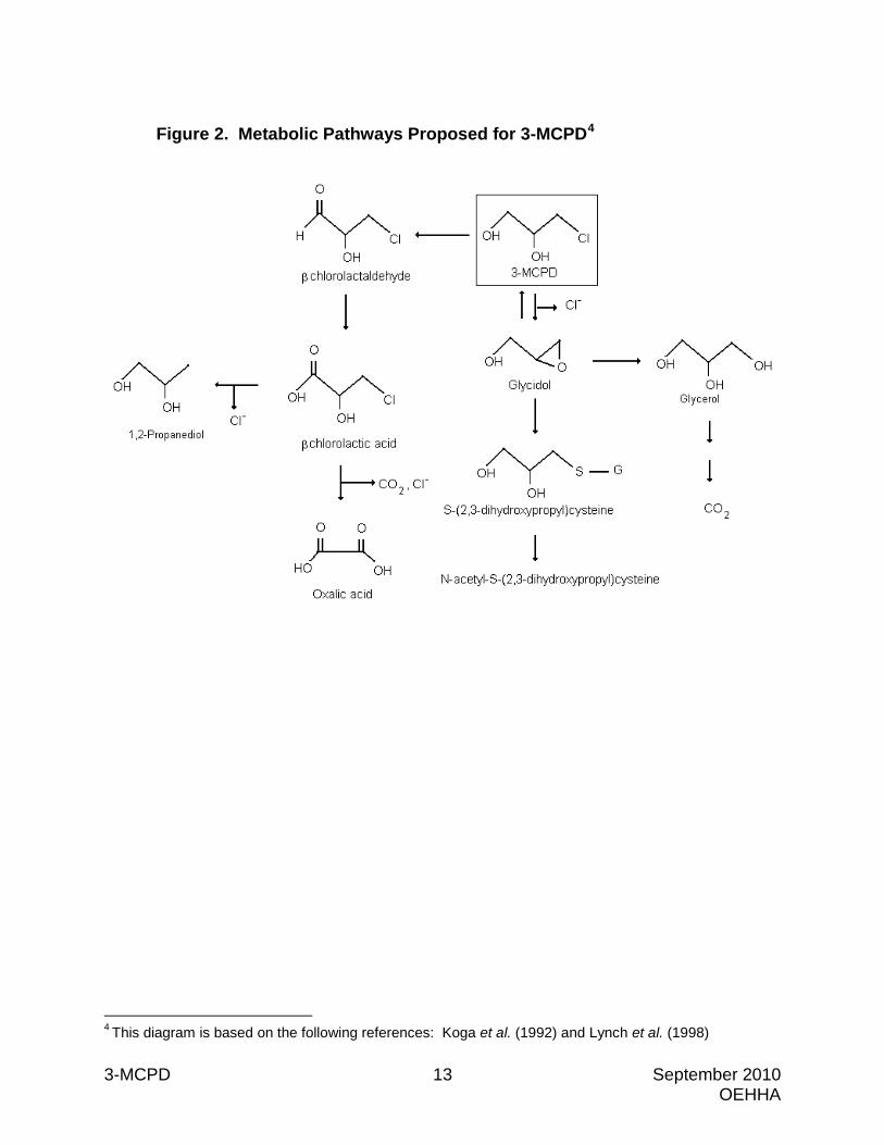
3.3.1 Pharmacokinetics and Metabolism Studies in rats and mice indicate that 3-MCPD is rapidly absorbed and distributed throughout the body (Jones et al., 1978; WHO, 2002). It crosses the blood-brain barrier, the blood-testis barrier, and the placenta (Jones et al., 1978; WHO, 2002; El Ramy et al., 2006). 3-MCPD is metabolized via an epoxidation pathway involving the formation of glycidol, and via an oxidation pathway involving the formation of β-chlorolactic acid and oxalic acid (Jones et al., 1978) (Figure 2). 3-MCPD is excreted in the breath as CO2 and in the urine as the parent compound and metabolites (Jones, 1975; Jones et al., 1969; ibid, 1978; WHO, 2002). Jones et al. (1969) analyzed the urine of Wistar rats administered 50 mg/kg 3-MCPD by oral or intraperitoneal (i.p.) injection. 3-MCPD and two metabolites were detected in the urine: S-(2,3-dihydroxypropyl) cysteine and the corresponding mercapturic acid, N-acetyl-S-(2,3-dihydroxypropyl) cysteine. These cysteine compounds are derived from a glutathione conjugate. Jones (1975) administered [14C]3-MCPD by i.p. injection to male Wister rats and male ICI Swiss mice and measured exhalation of 14CO2 and excretion of radioactivity in the urine. Thirty percent of the administered dose of [14C]3-MCPD was expired as 14CO2 by rats and 23 percent by mice within 24 hours of dosing. The collected urine was chromatographed on paper and thin layer chromatography and identification of chemicals was confirmed by mass spectra. In rats, about 8.5 percent of the administered dose was excreted in the urine as the parent compound, and in mice, about 11 percent was excreted as the parent compound. The same two urinary metabolites reported by Jones et al. (1969), S-(2,3-dihydroxypropyl)cysteine and N-acetyl-S-(2,3-dihydroxypropyl)cysteine, were observed in rats and mice in these studies. Since administration of glycidol also results in these same urinary metabolites, the authors conducted studies to investigate if 3-MCPD could react directly with glutathione or if formation of glycidol was a necessary intermediate step in the formation of 3-MCPD-derived glutathione conjugates. Using in vitro incubations of rat liver supernatant with [36Cl]3-MCPD and glutathione, under pH conditions that precluded non-enzymatic formation of glycidol, less than 5% of free 36Cl- was detected after three hours, suggesting that little hydrolysis or enzyme-catalyzed reactions with glutathione occurred. Similarly, when [14C]3-MCPD was used, only trace amounts of S-(2,3-dihydroxypropyl)glutathione were detected. In contrast, incubation of [14C]glycidol resulted in significant formation of S-(2,3-dihydroxypropyl)glutathione. Thus, Jones (1975) concluded that conjugation of 3-MCPD with glutathione requires formation of glycidol as the reactive intermediate. Jones et al. (1978) studied the tissue distribution, metabolism and excretion of [36Cl]3-MCPD (100 mg/kg by i.p.) in male Sprague-Dawley rats. Radioactivity was rapidly distributed throughout the body, with levels in the cauda epididymis, caput epididymis, testes, brain, liver, pituitary, muscle, and red blood cells ranging from 0.3 to 0.5% of the total radioactive dose/g tissue within 30 minutes of administration. After 24 hours, the levels in these tissues ranged from 0.2 to 0.3% of the total radioactive
3-MCPD 12 September 2010 OEHHA
dose/gram (g) tissue. Radioactivity was not found to concentrate in any specific tissue. The authors discussed this finding in light of the study of Crabo and Appelgren (1972), in which the administration of [14C]3-MCPD resulted in the accumulation of radioactivity in the rat cauda epididymis, together with preliminary studies indicating that administration of [14C]glycidol or [14C]glycerol also results in the accumulation of radioactivity in the rat cauda epididymis. Jones et al. (1978) concluded that the accumulated 14C radioactivity from each of these compounds was a dechlorinated product of the 3-MCPD epoxidation pathway. 3-MCPD was detected in all tissues examined at four hours, but was not detected in any tissues after 12 hours (Jones et al., 1978). 36Cl- was detected at two hours, was present in all tissues up to 48 hours, and in plasma up to 250 hours after [36Cl]3-MCPD administration. β- Chlorolactic acid was also present in the tissues at two hours, and at 14 hours was the dominant radioactive metabolite present in the tissues. A small amount of a minor radiolabeled metabolite, putatively identified as β-chlorolactaldehyde, was detected in the brain within the first four hours. The presence of 36Cl- and β-chlorolactic acid in the tissues after two hours indicates that 3-MCPD is metabolized through two separate pathways, one involving the loss of chlorine and the formation of glycidol, and the other involving oxidation to yield β-chlorolactic acid.
Twenty-two percent of the total administered radioactivity was excreted in the urine within 10 hours of dosing (Jones et al., 1978). Nine percent of the administered dose was excreted as the parent compound (i.e., [36Cl]3-MCPD), which was present in the urine for up to eight hours after dosing. Twenty-three percent of the total urinary radioactivity was β-chlorolactic acid (first seen in the urine five hours after dosing), approximately 0.9 percent was putatively identified as β-chlorolactaldehyde (first seen in the urine three to five hours after dosing), and the remainder was free 36Cl. Jones et al. (1978) concluded that epoxide formation (i.e., glycidol) is the major pathway of 3-MCPD metabolism in the rat, but that at higher doses, 3-MCPD can undergo oxidation.
3-MCPD 13 September 2010 OEHHA
Figure 2. Metabolic Pathways Proposed for 3-MCPD4
4 This diagram is based on the following references: Koga et al. (1992) and Lynch et al. (1998)
3-MCPD 14 September 2010 OEHHA
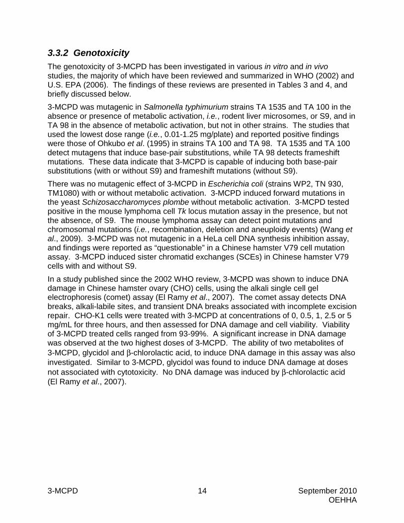
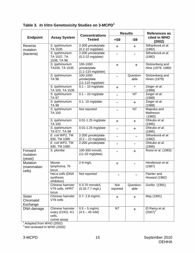
3.3.2 Genotoxicity The genotoxicity of 3-MCPD has been investigated in various in vitro and in vivo studies, the majority of which have been reviewed and summarized in WHO (2002) and U.S. EPA (2006). The findings of these reviews are presented in Tables 3 and 4, and briefly discussed below. 3-MCPD was mutagenic in Salmonella typhimurium strains TA 1535 and TA 100 in the absence or presence of metabolic activation, i.e., rodent liver microsomes, or S9, and in TA 98 in the absence of metabolic activation, but not in other strains. The studies that used the lowest dose range (i.e., 0.01-1.25 mg/plate) and reported positive findings were those of Ohkubo et al. (1995) in strains TA 100 and TA 98. TA 1535 and TA 100 detect mutagens that induce base-pair substitutions, while TA 98 detects frameshift mutations. These data indicate that 3-MCPD is capable of inducing both base-pair substitutions (with or without S9) and frameshift mutations (without S9). There was no mutagenic effect of 3-MCPD in Escherichia coli (strains WP2, TN 930, TM1080) with or without metabolic activation. 3-MCPD induced forward mutations in the yeast Schizosaccharomyces plombe without metabolic activation. 3-MCPD tested positive in the mouse lymphoma cell Tk locus mutation assay in the presence, but not the absence, of S9. The mouse lymphoma assay can detect point mutations and chromosomal mutations (i.e., recombination, deletion and aneuploidy events) (Wang et al., 2009). 3-MCPD was not mutagenic in a HeLa cell DNA synthesis inhibition assay, and findings were reported as “questionable” in a Chinese hamster V79 cell mutation assay. 3-MCPD induced sister chromatid exchanges (SCEs) in Chinese hamster V79 cells with and without S9. In a study published since the 2002 WHO review, 3-MCPD was shown to induce DNA damage in Chinese hamster ovary (CHO) cells, using the alkali single cell gel electrophoresis (comet) assay (El Ramy et al., 2007). The comet assay detects DNA breaks, alkali-labile sites, and transient DNA breaks associated with incomplete excision repair. CHO-K1 cells were treated with 3-MCPD at concentrations of 0, 0.5, 1, 2.5 or 5 mg/mL for three hours, and then assessed for DNA damage and cell viability. Viability of 3-MCPD treated cells ranged from 93-99%. A significant increase in DNA damage was observed at the two highest doses of 3-MCPD. The ability of two metabolites of 3-MCPD, glycidol and β-chlorolactic acid, to induce DNA damage in this assay was also investigated. Similar to 3-MCPD, glycidol was found to induce DNA damage at doses not associated with cytotoxicity. No DNA damage was induced by β-chlorolactic acid (El Ramy et al., 2007).
3-MCPD 15 September 2010 OEHHA
Table 3. In Vitro Genotoxicity Studies on 3-MCPD1
Endpoint Assay System Concentrations Tested
Results References as cited in WHO
(2002) +S9 -S9 Reverse mutation (bacteria)
S. typhimurium TA 1535
2-200 μmole/plate (0.2-22 mg/plate)
+ + Silhankovà et al. (1982)
S. typhimurium TA 1537, TA 1538, TA 98
2-200 μmole/plate (0.2-22 mg/plate)
- - Silhankovà et al. (1982)
S. typhimurium TA100, TA 1535
100-1000 μmole/plate (1.1-110 mg/plate)
+ + Stolzenberg and Hine (1979, 1980)
S. typhimurium TA 98
100-1000 μmole/plate (11-110 mg/plate)
- Question-able
Stolzenberg and Hines (1979)
S. typhimurium TA 100, TA 1535
0.1 – 10 mg/plate + + Zeiger et al. (1988)
S. typhimurium TA 97
0.1 – 10 mg/plate - NT Zeiger et al. (1988)
S. typhimurium TA 98
0.1- 10 mg/plate - + Zeiger et al. (1988)
S. typhimurium TA 100
Not reported - NT Majeska and Matheson (1983)
S. typhimurium TA 100
0.01-1.25 mg/plate + + Ohkubo et al. (1995)
S. typhimurium TA 677, TA 98
0.01-1.25 mg/plate - + Ohkubo et al. (1995)
E. coli WP2, TM 930, TM 1080
2-200 μmole/plate (0.2 – 22 mg/plate)
- - Silhankovà et al. (1982)
E. coli WP2, TM 930, TM 1080
2-200 μmole/plate - - Ohkubo et al. (1995)
Forward mutation (yeast)
S. plombe 100-300 mmol/L (11-33 mg/plate)
- + Rossi et al. (1983)
Mutation (mammalian cells)
Mouse lymphoma, Tk locus
2-9 mg/L + - Henderson et al. (1987)
HeLa cells (DNA synthesis inhibition)
Not reported - - Painter and Howard (1982)
Chinese hamster V79 cells, HPRT locus
0.3-70 mmole/L (0.33-7.7 mg/L)
Not reported
Question-able
Gorlitz (1991)
Sister Chromatid Exchange
Chinese hamster V79 cells
0.7- 2.8 mg/mL
+ + May (1991)
DNA damage Chinese hamster ovary (CHO) -K1 cells, comet assay
0.5 – 5 mg/mL (4.5 – 45 mM)
NT + El Ramy et al. (2007)2
1 Adapted from WHO (2002) 2 Not reviewed in WHO (2002)
3-MCPD 16 September 2010 OEHHA
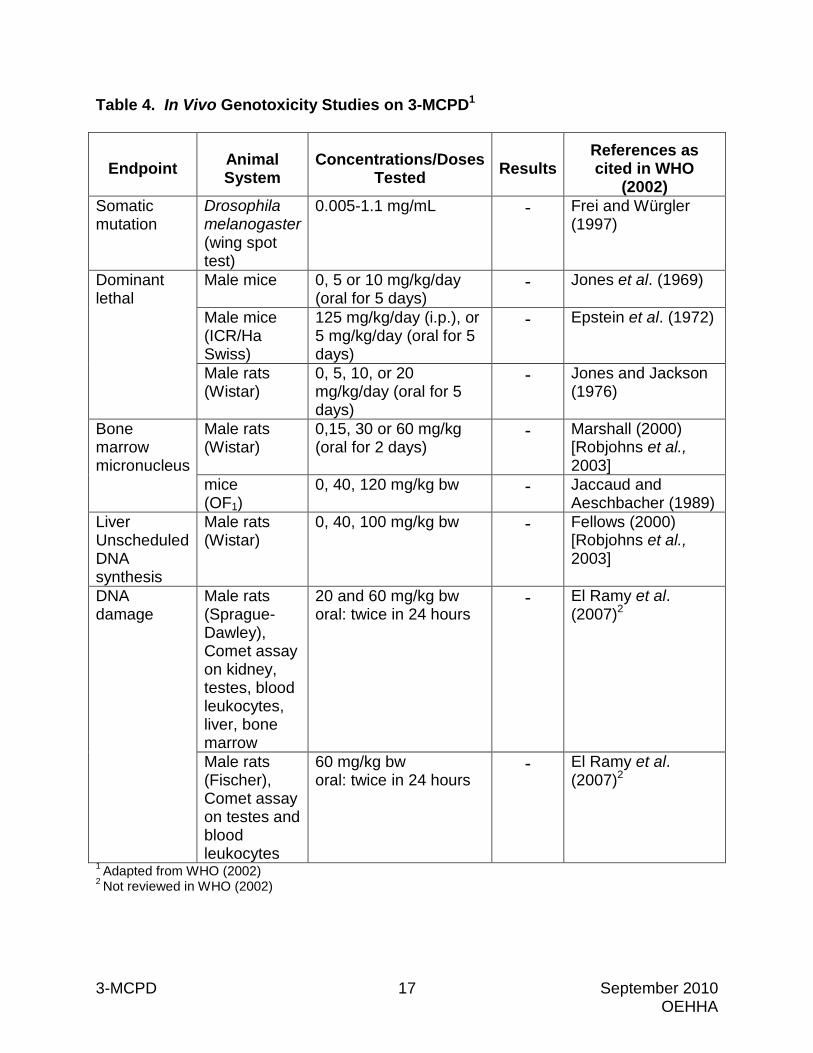
3-MCPD has not been found to be genotoxic in any of the in vivo studies conducted to date (Table 4). As reviewed by WHO (2002), 3-MCPD was negative for the induction of somatic mutations in the Drosophila wing spot test, dominant lethal mutations in mice and rats, micronuclei in the bone marrow of mice and rats, and unscheduled DNA synthesis in the liver of rats. In studies published since the WHO review (2002), 3-MCPD was investigated for the ability to induce DNA damage, as assessed by the comet assay, in certain target and non-target tissues in male Sprague-Dawley rats and in two tissues in male Fischer rats (El Ramy et al., 2007). Sprague-Dawley rats were administered 3-MCPD at doses of 0, 25 or 60 mg/kg-bw by oral gavage at 24 and 3 hours before sacrifice. At sacrifice, kidney and testes (i.e., target tissues), and blood leukocytes, liver, and bone marrow (i.e., non-target tissues) were collected for analysis in the comet assay. 3-MCPD did not induce DNA damage in any of the tissues tested at any dose level. Fischer rats were dosed in an identical manner with 60 mg/kg-bw 3-MCPD. At sacrifice, testes and blood leukocytes were collected and analyzed in the comet assay. No DNA damage was detected in Fischer rat testes or blood leukocytes.
3-MCPD 17 September 2010 OEHHA
Table 4. In Vivo Genotoxicity Studies on 3-MCPD1
Endpoint Animal System
Concentrations/Doses Tested Results
References as cited in WHO
(2002) Somatic mutation
Drosophila melanogaster (wing spot test)
0.005-1.1 mg/mL - Frei and Würgler (1997)
Dominant lethal
Male mice 0, 5 or 10 mg/kg/day (oral for 5 days)
- Jones et al. (1969)
Male mice (ICR/Ha Swiss)
125 mg/kg/day (i.p.), or 5 mg/kg/day (oral for 5 days)
- Epstein et al. (1972)
Male rats (Wistar)
0, 5, 10, or 20 mg/kg/day (oral for 5 days)
- Jones and Jackson (1976)
Bone marrow micronucleus
Male rats (Wistar)
0,15, 30 or 60 mg/kg (oral for 2 days)
- Marshall (2000) [Robjohns et al., 2003]
mice (OF1)
0, 40, 120 mg/kg bw - Jaccaud and Aeschbacher (1989)
Liver Unscheduled DNA synthesis
Male rats (Wistar)
0, 40, 100 mg/kg bw - Fellows (2000) [Robjohns et al., 2003]
DNA damage
Male rats (Sprague-Dawley), Comet assay on kidney, testes, blood leukocytes, liver, bone marrow
20 and 60 mg/kg bw oral: twice in 24 hours
- El Ramy et al. (2007)2
Male rats (Fischer), Comet assay on testes and blood leukocytes
60 mg/kg bw oral: twice in 24 hours
- El Ramy et al. (2007)2
1 Adapted from WHO (2002) 2 Not reviewed in WHO (2002)
3-MCPD 18 September 2010 OEHHA
3.3.3 In Vitro Transformation Study 3-MCPD was assayed for malignant transformation of mouse M2 fibroblasts by Piasecki et al. (1990). This assay is designed to detect a change in the growth pattern of fibroblasts which is indicative of loss of contact inhibition, a phenotype that is characteristic of cancer cells. The number of morphologically transformed foci in the 3-MCPD treated plates was significantly increased (p < 0.001) at 250 and 500 micrograms/milliliter (μg/mL), as compared to the untreated control plates. In addition, a dose-dependent reduction in plating efficiency, indicative of cytotoxicity, was observed.
3.3.4 Animal Tumor Pathology 3-MCPD significantly increased the incidence of kidney tumors in male and female Sprague-Dawley and Fischer rats, mammary tumors in male Fischer rats, and testicular tumors in male Sprague-Dawley and Fischer rats (WHO, 2002; Cho et al., 2008). The kidney tumors observed in treated male and female Sprague-Dawley rats were renal tubular adenomas and carcinomas (Cho et al., 2008). Renal tubular adenomas were observed in treated male and female Fischer rats (WHO, 2002). Adenomas can progress to carcinomas, and the two tumor phenotypes are aggregated when evaluating study results (IARC, 2006; McConnell et al., 1986). In Sprague-Dawley rats, no renal tubular tumors were observed in control males, while one carcinoma was observed in a control female. Cho et al. (2008) stated that renal tubular tumors are rare in untreated Sprague-Dawley rats, and noted the early appearance of renal tubular adenomas (week 78) and carcinomas (week 74) in males treated with 3-MCPD. In Fischer rats, no renal tubular tumors were observed in male or female controls. Dose-related increases in renal tubular hyperplasia were observed in male and female Sprague-Dawley and Fischer rats. 3-MCPD treatment was also associated with exacerbation of chronic progressive nephropathy in these studies. Chronic progressive nephropathy is characterized by basophilic renal tubules, a thickened basement membrane, hyaline cast formation and glomerulosclerosis (Hard and Khan, 2004). The mammary tumors observed in treated male Fischer rats were fibroadenomas, adenomas, and adenocarcinomas (WHO, 2002). Fibroadenomas of the mammary gland are benign epithelial cell tumors comprised of a mixture of glandular tissue, connective tissue and ducts. The proportion of glandular to connective tissue varies widely among fibroadenomas. On occasion, fibroadenomas can give rise to adenocarcinomas (Boorman and Everitt, 2006). Mammary gland adenomas are benign tumors consisting primarily of glandular epithelium, with little connective tissue. Mammary adenomas may progress to adenocarcinomas (Boorman et al., 1990b). Mammary gland fibroadenomas, adenomas and adenocarcinomas are typically aggregated for study evaluation (IARC, 2006; McConnell et al., 1986). The combined incidence of these mammary tumors in male Fischer rats treated with 3-MCPD was not reported (WHO, 2002), so the increase in the combined incidence of these tumors could not be evaluated. Tumors of the mammary gland are generally uncommon in male Fischer rats (Boorman et al., 1990b). No mammary gland tumors were observed in male controls. A dose-related increase in glandular hyperplasia of the mammary gland was also observed in treated male Fischer rats.
3-MCPD 19 September 2010 OEHHA
Testicular interstitial cell tumors, also referred to as Leydig cell tumors, were observed in male Sprague-Dawley and Fischer rats. Leydig cells are located in the interstitium of the testis, between the seminiferous tubules. There is a continuum of Leydig cell proliferative response, ranging from hyperplasia to adenomas and carcinomas (Boorman et al., 1990a). Differential diagnosis is based on size of the lesion. The incidences of Leydig cell adenomas and carcinomas in Fischer rats were reported in WHO (2002), while the report of Cho et al. (2008) did not specify whether the Leydig cell tumors observed in 3-MCPD-treated Sprague-Dawley rats were benign or malignant. Leydig cell adenomas and carcinomas are aggregated for carcinogen identification (IARC, 2006; McConnell et al., 1986). Leydig cell tumors are known to occur with high spontaneous incidence in untreated aged Fischer rats. In the 3-MCPD study in Fischer rats, 76% incidence of Leydig cell adenomas and 0% incidence of carcinomas were observed in controls (WHO, 2002). 3-MCPD treatment increased the incidence of Leydig cell adenomas in the mid- and high-dose groups to 100% and 94%, respectively. In addition, Leydig cell carcinomas were observed in three high-dose animals, but not in any other treated or control group (WHO, 2002). In other rat strains, including Sprague-Dawley rats, the spontaneous incidence of Leydig cell tumors is generally low (~1% incidence). In the 3-MCPD study in Sprague-Dawley rats, Leydig cell tumors occurred in just one out of 50 control animals (2% incidence), while in treated animals the incidence increased to four out of 50 (8%) mid-dose and 14 out of 50 (28%) high-dose animals, in the absence of any increase in Leydig cell hyperplasia (Cho et al., 2008). Thus, the finding of Leydig cell tumors is significant.
3.3.5 Effects on Immunological Systems Lee et al. (2004) studied various parameters of immunological responsiveness in female Balb/c mice using in vivo/in vitro methods. Groups of 6-week old mice (n= 7 or 8) were administered doses of 3-MCPD at 0, 25, 50 or 100 mg/kg/day by oral gavage for 14 days. No effects were observed on spleen lymphocyte proliferative responses to the mitogen concanavalin A (con A), lipopolysaccharides (LPS) or anti-CD3 antibody. There was reduced spleen cellularity in the high-dose group compared to controls. Phenotypic analysis did not detect any differences in the relative proportions of spleen lymphocyte subsets between treated and control mice. Absolute and relative thymus weights were significantly reduced in the high-dose group. A decrease in thymus cellularity accompanied the reduction in thymus weight. Also, antibody production against sheep red blood cells (SRBC) was reduced in high-dose mice and natural killer (NK) cell activity was reduced in mid- and high-dose mice. The basic function of the thymus is to produce various T cell subsets, which regulate all aspects of the adaptive immune response. Antibody production to SRBC requires the cooperation of T cells, B cells, macrophages, and cytokines. The reduced antibody response to SRBC (a T cell-dependent antigen) seen in the high-dose animals suggests that 3-MCPD may reduce the immune system’s ability to combat bacterial infection. This assay does not provide information on which cell type or cytokine is responsible for the reduced response, however. Natural killer cell activity, which does not require prior exposure or antibody formation, results in the lysis of target cells. Reduced NK cell activity will compromise the immune system’s ability to conduct tumor surveillance.
3-MCPD 20 September 2010 OEHHA
Using a similar exposure protocol to their earlier study, Lee et al. (2005) studied the effects of 3-MCPD on several other immunological functions in female Balb/c mice. These include effects on thymus cell subsets, delayed type hypersensitivity, mixed lymphocyte reactivity and lymphocyte proliferation in auricular lymph node cells, and TNF-α production by peritoneal macrophages. The decrease in thymus cellularity induced by 3-MCPD was associated with an increase in apoptosis in the thymus. There was also a significant decrease in CD4+CD8+ T cells and an increase in CD4-CD8+ T cells in the low- and high-dose groups, as measured by flow cytometric analysis. CD4+CD8+ T cells are precursors to CD4+ T helper cells and CD+ T cytotoxic cells in the thymus. No effect was observed on delayed hypersensitivity as measured by ear thickness and cell proliferation. Also, no effects were observed on mixed lymphocyte reaction to allogenic lymphocytes and TNF-α production by peritoneal macrophages. An observed slight dose-dependent decrease in macrophage oxide production suggests that the macrophages may have had a reduced capacity to kill pathogens. This research group also evaluated the effects of 3-MCPD on murine splenocyte proliferative response and cytokine production and on peritoneal macrophage function in vitro (Byun et al., 2006). 3-MCPD significantly decreased the proliferative response to con A and anti-CD3 antibody at ≥ 2 mM, and to LPS at ≥ 0.4 mM. 3-MCPD significantly decreased spleen cell production of IFN-γ (interferon-γ) and IL-10 (interleukin 10) at ≥ 1 mM and IL-4 at ≥ 4 mM, and peritoneal macrophage production of TNFα (tumor necrosis factor α) at ≥ 0.5 mM and nitric oxide at ≥ 4 mM. 3-MCPD was not cytotoxic to spleen cells at concentrations up to 6.0 mM or to peritoneal macrophages at concentrations up to 10 mM. In summary, the in vivo mouse studies suggest that 3-MCPD may reduce antibody production involved in combating bacterial infection and may reduce NK cell activity involved in cell lysis and tumor surveillance. The in vitro studies suggest that 3-MCPD impairs the ability of T and B cells to proliferate (a prerequisite in the generation of the adaptive immune response) and to produce various cytokines such as IL-10 and IL-4, which are involved in immunoregulation, inflammation, and antibody production.
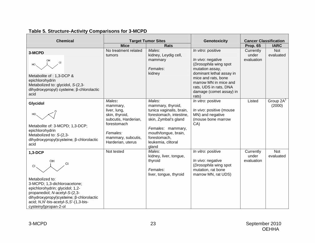
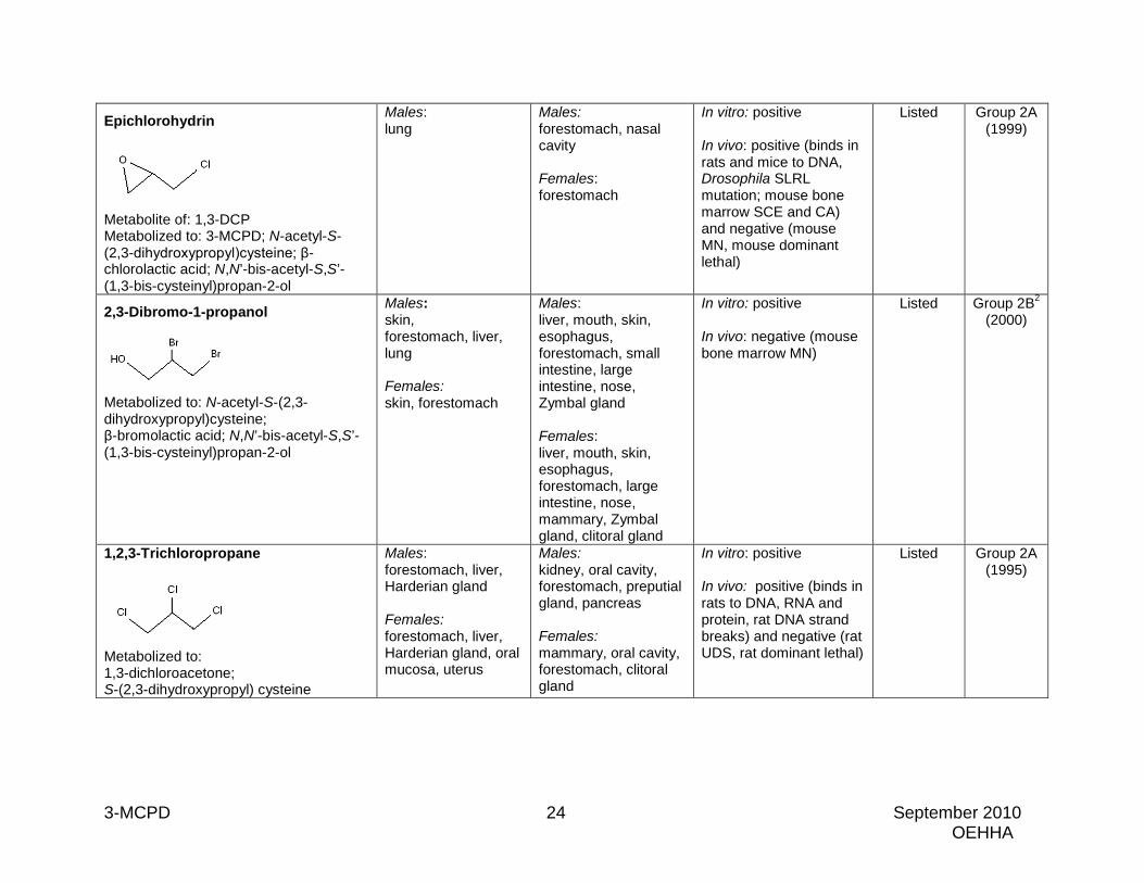
3.3.6 Structure Activity Comparisons 3-MCPD is a chlorinated three-carbon alcohol. Information on cancer bioassay findings in mice and rats, in vitro and in vivo genotoxicity findings, and cancer classification (Proposition 65 and IARC) for 3-MCPD and eight structurally-related compounds are presented in Table 5. Glycidol is a three-carbon epoxide compound that is a metabolite of 3-MCPD, epichlorohydrin, and 1,3-DCP. Glycidol is carcinogenic in both sexes of rats and mice, inducing tumors at multiple sites, including the mammary gland, and is genotoxic in vitro and in vivo (IARC, 2000). 1,3-DCP is a chlorinated three-carbon alcohol that is metabolized to 3-MCPD via epichlorohydrin. 1,3-DCP induced tumors in male and female rats (kidney, liver, tongue, thyroid) and is genotoxic in in vitro, but not in vivo assays (OEHHA, 2010).
3-MCPD 21 September 2010 OEHHA
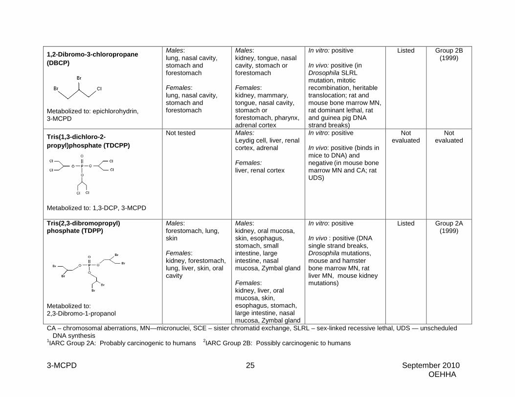
Epichlorohydrin is a chlorinated three-carbon epoxide compound that is metabolized to 3-MCPD. Epichlorohydrin is carcinogenic in male and female rats (forestomach, nasal cavity) and male mice (lung) and is genotoxic in vitro without metabolic activation and in several in vivo assays (ILS, 2005; IARC, 1999). 2,3-Dibromo-1-propanol is a brominated three-carbon alcohol that is likely to be metabolized to epibromohydrin, with subsequent metabolism via similar pathways as 3-MCPD. This is supported by the fact that 2,3-dibromo-1-propanol is metabolized to the same mercapturic acid metabolite as is 3-MCPD, and to β-bromolactic acid, instead of β-chlorolactic acid. 2,3-Dibromo-1-propanol is carcinogenic in both sexes of rats and mice and is genotoxic in vitro, but not in in vivo assays (IARC, 2000). 1,2,3-Trichloropropane is a chlorinated three-carbon compound that has been shown to be metabolized to the same mercapturic acid precursor as 3-MCPD, S-(2,3-dihydroxypropyl)cysteine. 1,2,3-Trichloropropane is carcinogenic in both sexes of rats and mice and is genotoxic in vitro and in vivo (IARC, 1995). 1,2-Dibromo-3-chloropropane (DBCP) is a chlorinated and brominated three-carbon compound that is metabolized to 3-MCPD via epichlorohydrin. It is carcinogenic in both sexes of rats and mice and is genotoxic in vitro and in vivo (IARC, 1999). Tris(1,3-dichloro-2-propyl)phosphate (TDCPP) is a chlorinated phosphate triester that is metabolized to 3-MCPD via 1,3-DCP. TDCPP induced tumors in male and female rats (liver, kidney, Leydig cell, adrenal) and is genotoxic in in vitro, and in some in vivo assays (Babich, 2006). Tris(2,3-dibromopropyl)phosphate (TDPP) is a brominated phosphate triester that is metabolized to 2,3-dibromo-1-propanol. TDPP is carcinogenic in both sexes of rats and mice and is genotoxic in vitro and in vivo (IARC, 1999). In summary, the compounds discussed here are structurally similar to 3-MCPD, are metabolized to 3-MCPD or a structurally similar compound, or share common metabolites with 3-MCPD. All have some positive carcinogenicity data in rodent studies, and six are currently listed under Proposition 65 as causing cancer and classified by IARC as either Group 2A or Group 2B carcinogens (glycidol, epichlorohydrin, 2,3-dibromo-1-propanol, 1,2,3-trichloropropane, DBCP, and TDPP). Most of the compounds included in Table 5 induce tumors at multiple sites, and in most cases in more than one sex/species. Certain tumor sites such as kidney, mammary gland, liver, tongue, and mouth or oral cavity are common target sites shared by many of these compounds. All of the compounds have positive results in genotoxicity assays performed in vitro. All but 3-MCPD, 1,3-DCP, and 2,3-dibromo-1-propanol also have some positive genotoxicity results in vivo. Some of the chemicals tested negative in some of the same in vivo assay systems in which 3-MCPD tested negative:
• 3-MCPD and 1,3-DCP were negative in the Drosophila wing spot mutation assay.
• 3-MCPD and epichlorohydrin were negative in the dominant lethal assay in mice. • 3-MCPD and 1,2,3-trichloropropane were negative in the dominant lethal assay
in rats.
3-MCPD 22 September 2010 OEHHA
• 3-MCPD, epichlorohydrin, 2,3-dibromo-1-propanol, and TDCPP were negative in the mouse bone marrow MN assay.
• 3-MCPD and 1,3-DCP were negative in the rat bone marrow MN assay. • 3-MCPD, 1,3-DCP, 1,2,3-trichloropropane, and TDCPP were negative in the rat
liver UDS assay.
3-MCPD 23 September 2010 OEHHA
Table 5. Structure-Activity Comparisons for 3-MCPD
Chemical
Target Tumor Sites
Genotoxicity
Cancer Classification Mice Rats Prop. 65 IARC
3-MCPD
Metabolite of : 1,3-DCP & epichlorohydrin Metabolized to: glycidol, S-(2,3-dihydroxypropyl) cysteine; β-chlorolactic acid
No treatment related tumors
Males: kidney, Leydig cell, mammary Females: kidney
In vitro: positive In vivo: negative (Drosophila wing spot mutation assay, dominant lethal assay in mice and rats, bone marrow MN in mice and rats, UDS in rats, DNA damage (comet assay) in rats)
Currently under
evaluation
Not evaluated
Glycidol
Metabolite of: 3-MCPD; 1,3-DCP; epichlorohydrin Metabolized to: S-(2,3-dihydroxypropyl)cysteine; β-chlorolactic acid
Males: mammary, liver, lung, skin, thyroid, subcutis, Harderian, forestomach Females: mammary, subcutis, Harderian, uterus
Males: mammary, thyroid, tunica vaginalis, brain, forestomach, intestine, skin, Zymbal’s gland Females: mammary, mouth/tongue, brain, forestomach, leukemia, clitoral gland
In vitro: positive In vivo: positive (mouse MN) and negative (mouse bone marrow CA)
Listed
Group 2A1
(2000)
1,3-DCP
Metabolized to: 3-MCPD; 1,3-dichloroacetone; epichlorohydrin; glycidol; 1,2-propanediol; N-acetyl-S-(2,3-dihydroxypropyl)cysteine; β-chlorolactic acid; N,N’-bis-acetyl-S,S’-(1,3-bis-cysteinyl)propan-2-ol
Not tested Males: kidney, liver, tongue, thyroid Females: liver, tongue, thyroid
In vitro: positive In vivo: negative (Drosophila wing spot mutation, rat bone marrow MN, rat UDS)
Currently under
evaluation
Not evaluated
3-MCPD 24 September 2010 OEHHA
Epichlorohydrin
Metabolite of: 1,3-DCP Metabolized to: 3-MCPD; N-acetyl-S-(2,3-dihydroxypropyl)cysteine; β-chlorolactic acid; N,N’-bis-acetyl-S,S’-(1,3-bis-cysteinyl)propan-2-ol
Males: lung
Males: forestomach, nasal cavity Females: forestomach
In vitro: positive In vivo: positive (binds in rats and mice to DNA, Drosophila SLRL mutation; mouse bone marrow SCE and CA) and negative (mouse MN, mouse dominant lethal)
Listed
Group 2A (1999)
2,3-Dibromo-1-propanol
Metabolized to: N-acetyl-S-(2,3-dihydroxypropyl)cysteine; β-bromolactic acid; N,N’-bis-acetyl-S,S’-(1,3-bis-cysteinyl)propan-2-ol
Males: skin, forestomach, liver, lung Females: skin, forestomach
Males: liver, mouth, skin, esophagus, forestomach, small intestine, large intestine, nose, Zymbal gland Females: liver, mouth, skin, esophagus, forestomach, large intestine, nose, mammary, Zymbal gland, clitoral gland
In vitro: positive In vivo: negative (mouse bone marrow MN)
Listed Group 2B2 (2000)
1,2,3-Trichloropropane
Metabolized to: 1,3-dichloroacetone; S-(2,3-dihydroxypropyl) cysteine
Males: forestomach, liver, Harderian gland Females: forestomach, liver, Harderian gland, oral mucosa, uterus
Males: kidney, oral cavity, forestomach, preputial gland, pancreas Females: mammary, oral cavity, forestomach, clitoral gland
In vitro: positive In vivo: positive (binds in rats to DNA, RNA and protein, rat DNA strand breaks) and negative (rat UDS, rat dominant lethal)
Listed
Group 2A (1995)
3-MCPD 25 September 2010 OEHHA
1,2-Dibromo-3-chloropropane (DBCP)
Metabolized to: epichlorohydrin, 3-MCPD
Males: lung, nasal cavity, stomach and forestomach Females: lung, nasal cavity, stomach and forestomach
Males: kidney, tongue, nasal cavity, stomach or forestomach Females: kidney, mammary, tongue, nasal cavity, stomach or forestomach, pharynx, adrenal cortex
In vitro: positive In vivo: positive (in Drosophila SLRL mutation, mitotic recombination, heritable translocation; rat and mouse bone marrow MN, rat dominant lethal, rat and guinea pig DNA strand breaks)
Listed
Group 2B (1999)
Tris(1,3-dichloro-2-propyl)phosphate (TDCPP)
Metabolized to: 1,3-DCP, 3-MCPD
Not tested Males: Leydig cell, liver, renal cortex, adrenal Females: liver, renal cortex
In vitro: positive In vivo: positive (binds in mice to DNA) and negative (in mouse bone marrow MN and CA; rat UDS)
Not evaluated
Not evaluated
Tris(2,3-dibromopropyl) phosphate (TDPP)
Metabolized to: 2,3-Dibromo-1-propanol
Males: forestomach, lung, skin Females: kidney, forestomach, lung, liver, skin, oral cavity
Males: kidney, oral mucosa, skin, esophagus, stomach, small intestine, large intestine, nasal mucosa, Zymbal gland Females: kidney, liver, oral mucosa, skin, esophagus, stomach, large intestine, nasal mucosa, Zymbal gland
In vitro: positive In vivo : positive (DNA single strand breaks, Drosophila mutations, mouse and hamster bone marrow MN, rat liver MN, mouse kidney mutations)
Listed Group 2A (1999)
CA – chromosomal aberrations, MN—micronuclei, SCE – sister chromatid exchange, SLRL – sex-linked recessive lethal, UDS — unscheduled DNA synthesis
1IARC Group 2A: Probably carcinogenic to humans 2IARC Group 2B: Possibly carcinogenic to humans
3-MCPD 26 September 2010 OEHHA
4. MECHANISMS 3-MCPD was found to induce kidney tumors in male and female rats of two strains, Leydig cell tumors of the testes in male rats of two strains, and mammary tumors in male rats of one strain. 3-MCPD was also shown to induce the malignant transformation of mouse cells in vitro. While the mechanisms by which 3-MCPD induces tumors are not known, there is evidence to suggest that 3-MCPD may act by a genotoxic mechanism. 3-MCPD has been shown to induce mutations in bacteria and yeast, and mutations, SCEs, and DNA damage in cultured mammalian cells (see Section 3.3.2). 3-MCPD is metabolized to glycidol, a reactive epoxide and genotoxic carcinogen. Like 3-MCPD, glycidol induces DNA damage in CHO cells, and mammary tumors in male rats, suggesting that the carcinogenicity of 3-MCPD may be due to the formation of glycidol, at least. It is unclear if other observed effects of 3-MCPD may be involved in its carcinogenic action. For example, 3-MCPD induces infertility in male rats by inhibiting enzymes involved in glycolysis in epithelial cells of the testis and caput epididymis, and in spermatozoa (WHO, 2002). One of the enzymes 3-MCPD inhibits is glyceraldehyde-3-phosphate-dehydrogenase (GAPDH) (Jones, 1983). GAPDH has multiple cellular functions, in addition to its role in glycolysis. These include involvement in the control of DNA repair, cell death, cell cycle progression, and the regulation of mRNA stability (Colell et al., 2009). Studies have implicated GAPDH as both a promoter and an inhibitor of carcinogenesis (Colell et al., 2009). Based on the current limited understanding of the role GAPDH plays in carcinogenesis, it is unclear whether the ability of 3-MCPD to inhibit GAPDH is involved in the chemical’s carcinogenic action. Kidney toxicity was observed in 3-MCPD-treated male and female rats, manifested as an exacerbation of the chronic progressive nephropathy commonly observed in aging rats. 3-MCPD’s toxic effects on the kidney may be related to inhibition of glycolysis, or to the formation of the metabolite oxalic acid, which is known to induce kidney damage. The extent to which toxicity may contribute to the development of kidney tumors induced by 3-MCPD is not known. 3-MCPD also has been shown to suppress certain immune functions, such as NK cell activity which is involved in tumor surveillance, and altered production of cytokines which is involved in regulation of inflammatory responses and other functions. The implications of these immune effects for the carcinogenicity of 3-MCPD are not known. There is little information to support a role for hormonal effects in 3-MCPD carcinogenesis. One study in male Wistar rats given a single injection of 3-MCPD reported that serum follicle-stimulating hormone (FSH) levels increased on day one and remained elevated for up to 91 days; serum prolactin levels increased by day two and slowly returned to normal by day 28; serum luteinizing hormone (LH) levels increased on day seven, and then returned to normal by day 42 (Morris and Jackson, 1978). In this same study, no change in pituitary levels of FSH or prolactin was observed, while LH levels were steadily increased from day 21 to day 56. The implications for carcinogenesis of these hormone level changes, in response to a single dose of
3-MCPD 27 September 2010 OEHHA
3-MCPD, are unclear. In an in vitro study of isolated rat interstitial (Leydig) cells, 3-MCPD decreased cellular testosterone production. A reduction in oxygen consumption by these cells was also observed, suggesting that reduced testosterone production may have resulted from the inhibition of glycolysis (Paz et al., 1985).
5. REVIEWS BY OTHER AGENCIES Data relating to the carcinogenicity of 3-MCPD have been reviewed by the World Health Organization (WHO, 2002; 2007). Based on the data reviewed, the WHO concluded that 3-MCPD should be considered a non-genotoxic carcinogen (Tritscher, 2004). The U.S. Environmental Protection Agency (U.S. EPA) conducted a human health risk assessment of 3-MCPD for proposed use as a rodenticide in 2006, but the Agency has not classified the compound as to its carcinogenic potential (U.S. EPA, 2006b). Since the WHO and U.S. EPA reviews occurred, additional positive long-term cancer studies of 3-MCPD in Sprague-Dawley rats have been published (Cho et al., 2008). 3-MCPD has not been classified as to its potential carcinogenicity by the U.S. EPA, the U.S. Food and Drug Administration, the National Toxicology Program, the National Institute for Occupational Safety and Health, or the International Agency for Research on Cancer.
6. SUMMARY AND CONCLUSIONS
6.1 Summary of Evidence 3-MCPD has been tested in multiple rodent carcinogenicity studies employing various routes of exposure. No treatment-related tumors were observed in 19-month studies conducted in female Swiss mice exposed to 3-MCPD either via topical application (thrice per week) or by subcutaneous injection (once per week), or in 72-week studies in male and female Charles River (CD-1) rats administered 3-MCPD by gavage (twice per week). However, treatment-related increases in tumors were observed in two sets of two-year studies conducted in male and female Fischer and Sprague-Dawley rats, involving daily exposure to 3-MCPD via drinking water. 3-MCPD significantly increased the incidence of kidney tumors in male and female Sprague-Dawley and Fischer rats, mammary tumors in male Fischer rats, and Leydig cell tumors in male Sprague-Dawley and Fischer rats. Salient features regarding the treatment-related tumors seen in these studies include: Kidney tumors
• 3-MCPD significantly increased the incidence of malignant, and benign and malignant kidney tumors in male Sprague-Dawley rats.
o Tumors appeared early. o Kidney tumors are rare in untreated male Sprague-Dawley rats.
• Combined benign and malignant kidney tumors were significantly increased in treated female Sprague-Dawley rats.
o Kidney tumors are rare in untreated female Sprague-Dawley rats. • Benign kidney tumors increased in treated male Fischer rats. • Benign kidney tumors increased in treated female Fischer rats.
3-MCPD 28 September 2010 OEHHA
• 3-MCPD increased renal tubular hyperplasia in both sexes and strains. • 3-MCPD exacerbated chronic progressive nephropathy in both sexes and
strains. Mammary tumors
• 3-MCPD significantly increased the incidence of benign and malignant mammary tumors in male Fischer rats.
• 3-MCPD increased glandular hyperplasia of the mammary gland in male Fischer rats.
• Mammary gland tumors are uncommon in male Fischer rats. Leydig cell tumors
• 3-MCPD significantly increased the incidence of benign and malignant Leydig cell tumors in male Fischer rats.
• 3-MCPD significantly increased the incidence of Leydig cell tumors in male Sprague-Dawley rats.
• Leydig cell tumors are uncommon in Sprague-Dawley rats.
Evidence indicating that 3-MCPD is genotoxic comes from a number of in vitro test systems:
• 3-MCPD induced mutations in Salmonella typhimurium, in the presence or absence of exogenous metabolic activation, inducing both base-pair substitution and frameshift mutations.
• 3-MCPD induced mutations in the yeast S. plombe. • 3-MCPD induced mutations in mouse lymphoma cells in the presence of
exogenous metabolic activation. • 3-MCPD induced SCEs in Chinese hamster V79 cells. • 3-MCPD induced DNA damage in CHO cells.
3-MCPD has not been shown to be genotoxic in several in vivo test systems. 3-MCPD induced malignant transformation of mammalian cells in culture. There is some suggestion from in vitro and in vivo studies in mice that 3-MCPD may impair some aspects of immune function. Structure-activity considerations include:
• 3-MCPD is structurally related to six carcinogens identified by IARC and listed under Proposition 65:
o Glycidol o Epichlorohydrin o 2,3-Dibromo-1-propanol o 1,2,3-Trichloropropane o DBCP o TDPP
• 3-MCPD is metabolized to a genotoxic carcinogen: o Glycidol
3-MCPD 29 September 2010 OEHHA
• Four structurally-related chemicals that are metabolized to 3-MCPD and cause cancer in rodents are:
o Epichlorohydrin o DBCP o 1,3-DCP o TDCPP
• Three structurally-related chemicals that are metabolized in a similar manner as 3-MCPD, share the same mercapturic acid metabolite as 3-MCPD, and cause cancer in rodents are:
o 1,2,3-Trichloropropane o 2,3-Dibromo-1-propanol o TDPP
• Many of these structurally-related chemicals induce tumors at the same sites in rats as 3-MCPD:
o Kidney tumors 1,3-DCP 1,2,3-Trichloropropene DBCP TDCPP TDPP
o Leydig cell tumors: TDCPP
o Mammary gland tumors in male rats: Glycidol
6.2 Conclusion The evidence for carcinogenicity of 3-MCPD comes from:
• Multiple studies in rats o Kidney tumors in male Fischer rats o Kidney tumors in male Sprague-Dawley rats o Kidney tumors in female Fischer rats o Kidney tumors in male Sprague-Dawley rats o Leydig cell tumors of the testes in male Fischer rats o Leydig cell tumors of the testes in male Sprague-Dawley rats o Mammary tumors in male Fischer rats
• Positive findings in a variety of in vitro genotoxicity test systems • Malignant transformation of mammalian cells in culture • Metabolism of 3-MCPD to glycidol, a genotoxic carcinogen • Structure-activity considerations with six carcinogens identified by IARC and
listed under Proposition 65.
3-MCPD 30 September 2010 OEHHA
7. REFERENCES Baer I, de la Calle B, Taylor P (2010). 3-MCPD in food other than soy sauce or hydrolysed vegetable protein (HVP). Anal Bioanal Chem 396:443-456. Boorman GA Chapin RE and Mitsumori (1990a). Testis and epididymis. In Pathology of the Fischer Rat (Boorman GA, Eustis SL, Elwel MR, Montgomery CA Jr, Mackenzie WF, eds) Academic Press, Inc. pp. 405-417. Boorman GA, Wilson JT, Van Zwienten MJ, Eutis SL (1990b). Mammary gland. In Pathology of the Fischer Rat (Boorman GA, Eustis SL, Elwel MR, Montgomery CA Jr, Mackenzie WF, eds) Academic Press, Inc. pp. 295-312. Boorman GA, Everitt JI (2006). Neoplastic disease. In “The Laboratory Rat” (MA Suckow, SH Weisbroth and CL Franklin, eds), Elsevier Academic Press, Inc. pp. 479-511. Byun JA, Ryu MH, Lee JK (2006). The immunomodulatory effects of 3-monochloro-1,2-propanediol on murine splenocyte and peritoneal macrophage function in vitro. Toxicology In Vitro 20:272-278. Cho WS, Han BS, Nam KT, Park K, Choi M, Kim SH, Jeong J, Jang DD (2008). Carcinogenicity study of 3-monochloropropane-1,2-diol in Sprague-Dawley rats. Food Chem Toxicol 46:3172-3177.
Crabo B, Appelgren LE (1972). Distribution of [14C]α-chlorohydrin in mice and rats. J Reprod Fert 30:161-163. Crews C, Hasnip S, Chapman S, Hough P, Potter N, Todd J, Brereton P, Matthews W (2003). Survey of chloropropanols in soy sauces and related products purchased in the UK in 2000 and 2002. Food Add Contam 20:916-922. Colell A, Green DR, Ricci JE (2009). Novel role of GAPDH in cell death and carcinogenesis. Cell Death Differentiation 16:1573-1581. El Ramy RM, Elhkim O, Poul M, Forest MG, Leduque, P, Maguere (2006). Lack of effect on rat testicular organogenesis after in utero exposure to 3-monochloropropane-1,2-diol (3-MCPD). Repro Toxicol 22(3):485-492. El Ramy R, Elhkim MO, Lezmi S, Poul JM (2007). Evaluation of the genotoxic potential of 3-monochloropropane-1,2-diol (3-MCPD) and its metabolites, glycidol and beta-chlorolactic acid, using the single cell gel/comet assay. Food Chem Toxicol 45:41-48. European Commission (EC, 2004). Collection and collation of data on levels of 3-monochloropropanediol (3-MCPD) and related substances in food (scientific co-operation report). EC Directorate-General of Health and Consumer Protection, Brussels. Food Standards Australia New Zealand (FSANZ, 2003). Chloropropanols in food: An analysis of the public health risk. Technical report series No. 15. Available at www.foodstandards.gov.au.
3-MCPD 31 September 2010 OEHHA
Hamlet CG, Sadd PA, Crews C, Velisek J, Baxter DE (2002). Occurrence of 3-chloro-propane-1,2-diol (3-MCPD) and related compounds in foods: a review. Food Add Contam 19:619-631. Hard GC, Khan KN (2004). A contemporary overview of chronic progressive nephropathy in the laboratory rat, and its significance for human risk assessment. Toxicol Pathol 32:171-80. Integrated Laboratory Systems, Incorporated (ILS, 2005). 1,3-Dichloro-2-propanol [CAS No. 96-23-1]. Review of toxicological literature. Prepared by Integrated Laboratory Systems, Inc., Research Triangle Park, North Carolina, for the National Toxicology Program, National Institute of Environmental Health Sciences. International Agency for Research on Cancer (IARC, 1995). IARC Monographs on the Evaluation of Carcinogenic Risks to Humans. Volume 63. IARC, Lyon, France. International Agency for Research on Cancer (IARC, 1999). IARC Monographs on the Evaluation of Carcinogenic Risks to Humans. Volume 71. Re-evaluation of Some Organic Chemicals, Hydrazine and Hydrogen Peroxide (Parts Two and Three). IARC, Lyon, France. International Agency for Research on Cancer (IARC, 2000). IARC Monographs on the Evaluation of Carcinogenic Risks to Humans. Volume 77. IARC, Lyon, France. International Agency for Research on Cancer (IARC, 2006). IARC Monographs on the Evaluation of Carcinogenic Risks to Humans. Preamble. IARC, Lyon, France, 2006. Available at http://monographs.iarc.fr/ENG/Preamble/index.php. Jones AR, Davies P, Edwards K, Jackson H (1969). Antifertility effects and metabolism of α and epi-chlorohydrins in the rat. Nature 224:83. Jones AR (1975). The metabolism of 3-chloro, 3-bromo- and 3-iodopropan-1,2-diol in rats and mice. Xenobiotica 5:155-165. Jones AR, Milton DH, Murcott C (1978). The oxidative metabolism of alpha-chlorohydrin in the male rat and the formation of spermatocoeles. Xenobiotica 8:573-582. Kalla NR, Bansal MP (1977). In vivo & in vitro alkylation of testicular cysteine by alpha-chlorohydrin. Indian J Exp Biol 15:232-233. Koga M, Inoue N, Imazu K, Yamada N, Shinoki Y (1992). Identification and quantitative analysis of urinary metabolites of dichloropropanols in rats. J Univ Occup Environ Health Jpn 14:13-22. Lee JK, Byun JA, Park SH, Choi HJ, Kim HS, Oh HY (2005). Evaluation of the potential immunotoxicity of 3-monochloro-1,2-propanediol in Balb/c mice - II. Effect on thymic subset, delayed-type hypersensitivity, mixed-lymphocyte reaction, and peritoneal macrophage activity. Toxicology 211:187-196.
3-MCPD 32 September 2010 OEHHA
Lee JK, Byun JA, Park SH, Kim HS, Park JH, Eom JH, Oh HY (2004). Evaluation of the potential immunotoxicity of 3-monochloro-1,2-propanediol in Balb/c mice - I. Effect on antibody forming cell, mitogen-stimulated lymphocyte proliferation, splenic subset, and natural killer cell activity. Toxicology 204:1-11. Lynch BS, Bryant DW, Hook GJ, Nestmann ER, Munro IC (1998). Carcinogenicity of monochloro-1,2-propanediol (alpha-chlorohydrin, 3-MCPD). Inter J Toxicol 17:47-76. Macarthur R, Crews C, Davies A, Brereton P, Hough P, Harvey D (2000). 3-Monochloropropane-1,2-diol (3-MCPD) in soy sauces and similar products available from retail outlets in the UK. Food Add Contam 17:903-906. McConnell EE, Solleveld HA, Swenberg JA, Boorman GA (1986). Guidelines for combining neoplasms for evaluation of rodent carcinogenesis studies. JNCI 76:283-289. Morris ID, Jackson CM (1978). Gonadotrophin response after castration and selective destruction of the testicular interstitium in the normal and aspermatogenic rat. Acta Endocrinol 88:38-47. New Jersey Department of Health and Senior Services (NJ DHSS, 1999). Glycerol-alpha-monochlorohydrin. Hazardous Substance Fact Sheet. Available at http://nj.gov/health/eoh/rtkweb/documents/fs/2453.pdf. Nyman PJ, Diachenko GW, Perfetti GA (2003). Determination of 1,3-dichloropropanol in soy and related sauces by using gas chromatography/mass spectrometry. Food Add Contam 20:903-908. Office of Environmental Health Hazard Assessment (OEHHA, 2010). Evidence on the Carcinogenicity of 1,3-Dichloro-2-propanol. OEHHA, Reproductive and Cancer Hazard Assessment Branch, California Environmental Protection Agency, Oakland, June 2010.
Paz G, Carmon A, Homonnai ZT (1985). Effect of α-chlorohydrin on metabolism and testosterone secretion by rat testicular interstitial cells. Int J Androl 8:139-146. Piasecki A, Ruge A, Marquardt H (1990). Malignant transformation of mouse M2-fibroblasts by glycerol chlorohydrines contained in protein hydrolysates and commercial food. Arzneimittel-Forschung/Drug Research 402:1054-1055. Rahn A, Yaylayan VA (2010). Thermal degradation of sucralose and its potential in generating chloropropanols in the presence of glycerol. Food Chem 118:56-61. Sunahara G, Perrin I, Marchessini M (1993). Carcinogenicity study on 3-monochloropropane-1,2-diol (3-MCPD) administered in drinking water to Fischer 344 rats. Unpublished report No. RE-SR93003 submitted to WHO by Nestec, Ltd., Research and Development, Switzerland. Tritscher AM (2004). Human health risk assessment of processing-related compounds in food. Toxicol Lett 149:177-186. U.S. Environmental Protection Agency (U.S. EPA, 2006a). Pesticide fact sheet: alpha-chlorohydrin. Office of Prevention, Pesticides and Toxic Substances, U.S. EPA. Available at http://www.epa.gov/opprd001/factsheets/alpha_chlorohydrin.pdf.
3-MCPD 33 September 2010 OEHHA
U.S. Environmental Protection Agency (U.S. EPA, 2006b). Memorandum on Alpha-chlorohydrin: Human health risk assessment for proposed use as a rodenticide. PC Code: 117101; DP# 326010. Health Effects Division, U.S. EPA, August 17, 2006. Van Duuren BL, Goldschmidt BM, Katz C, Seidman I, Paul JS (1974). Carcinogenic activity of alkalating agents. J Natl Cancer Inst 53(3):695-700. Wang J, Sawyer JR, Chen L, Chen T, Honma M, Mei N, Moore MM (2009). The mouse lymphoma assay detects recombination, deletion, and aneuploidy. Toxicol Sci 109:96-105. Weisburger EK, Ulland BM, Nam J, Gart JJ, Weisburger JH (1981). Carcinogenicity tests of certain environmental and industrial chemicals. J Natl Cancer Inst 67(1):75-88. World Health Organization (WHO, 2002). Safety evaluation of certain food additives and contaminants – 3-Chloro-1,2-propanediol. WHO Food Additives Series, Vol. 48. Available at: www.inchem.org/documents/jecfa/v48je19.htm#10.0. World Health Organization (WHO, 2007). Safety evaluation of certain food additives and contaminants. WHO Food Additives Series, Vol. 58. 3-Chloro-1,2-propanediol (addendum). Joint FAO/WHO Expert Committee on Food Additives, International Programme of Chemical Safety, WHO. Available at http://whqlibdoc.who.int/publications/2007/9789241660587_eng.pdf.
Zelinkova Z, Novotny O, Schurek J, Velisek J, Hajslova J, Dolezal M (2008). Occurrence of 3-MCPD fatty acid esters in human breast milk. Food Add Contam 25:669-676. Zelinkova Z, Dolezal M, Velisek J (2009). Occurrence of 3-chloropropane-1,2-diol fatty acid esters in infant and baby foods. Eur Food Res Technol 228:571-578.


![Enhancement of ceramide formation increases endocytosis of ......Cytokine production differs in both type and magnitude dependent on the type of microbial stimulation [1,2]. The type](https://static.fdocument.org/doc/165x107/5f33e885a4573a2325398318/enhancement-of-ceramide-formation-increases-endocytosis-of-cytokine-production.jpg)