Antimicrobial mechanisms of phagocytes and bacterial evasion strategies · 2009-10-30 ·...
12
Professional phagocytes, such as macrophages, neu- trophils and dendritic cells, are uniquely qualified to engulf large (≥ 0.5 μm) particles, including microorgan- isms. The internalization and subsequent destruction of pathogens are key to the innate immune response, and promote antigen presentation and the develop- ment of adaptive immunity. After engulfment, the microorganisms are trapped, together with extracellu- lar fluid, in a vacuole, or phagosome, derived from the plasma membrane. Because the nascent phagosomal membrane and its contents are innocuous, they must undergo a drastic conversion to acquire the microbi- cidal and degradative features associated with innate immunity. This conversion, known as phagosomal maturation, is accomplished through a strictly cho- reographed sequence of fusion and fission events that involve defined compartments of the endocytic pathway (FIG. 1). Effective phagocytosis therefore requires two components: particle internalization and phagosomal maturation. Although most bacteria are successfully internal- ized and eliminated by phagocytes, several patho- gens have developed survival strategies that interfere with the internalization and/or maturation processes. Prevention and management of the infections caused by such pathogens would obviously benefit from understanding the manner in which they circum- vent and often co-opt the immune response. This, in turn, requires detailed knowledge of the basic mecha- nisms underlying phagocytosis. To this end, we briefly summarize our current knowledge of phagocytosis and describe salient examples of bacterial species that have evolved distinct strategies to evade killing. Phagosome formation The interaction of the microorganism with the phago- cyte can be direct, through recognition of pathogen- associated molecules (such as surface carbohydrates, peptidoglycans or lipoproteins) by pattern recognition receptors, or indirect, through mediation by opsonins. Opsonins are host factors, such as immunoglobulin G (IgG), and components of the complement cascade that attach to the pathogen surface, acquiring a conforma- tion that is recognized by phagocytic receptors, such as Fcγ receptors (FcγRs) and complement receptor 3 (CR3) 1 . The signalling that is triggered by the particle varies depending on the nature of the receptors engaged. The pathway elicited by FcγR is best understood. Exposure to multivalent ligands induces clustering of these receptors in the plane of the membrane, initiating phosphorylation of their cytoplasmic immunoreceptor tyrosine-based activation motifs (ITAMs) by Src-family kinases 2 . ITAM phosphorylation recruits and activates the tyrosine kinase SYK, which in turn phosphorylates various substrates 3 . The events that follow SYK activa- tion and culminate in particle engulfment are not as clearly understood. Remodelling of actin is unambigu- ously required for pseudopod extension and, in the case of FcγR, polymerization is driven by Rac1 and/ or Rac2, and cell division control protein 42 (Cdc42) 4 . Program in Cell Biology, The Hospital for Sick Children, 555 University Avenue, Toronto, Ontario M5G 1X8, Canada. Correspondence to S.G. e-mail: [email protected] *These authors contributed equally to this work. doi:10.1038/nrmicro2128 Endocytic pathway The route followed inside the cell by vesicles derived from the plasma membrane by endocytosis, including their membrane-associated cargo and trapped fluid. Vesicles or vacuoles derived from the plasma membrane undergo fusion and fission events that deliver some of their components to lysosomes for degradation, whereas others are recycled. Antimicrobial mechanisms of phagocytes and bacterial evasion strategies Ronald S. Flannagan*, Gabriela Cosío* and Sergio Grinstein Abstract | Professional phagocytes have a vast and sophisticated arsenal of microbicidal features. They are capable of ingesting and destroying invading organisms, and can present microbial antigens on their surface, eliciting acquired immune responses. To survive this hostile response, certain bacterial species have developed evasive strategies that often involve the secretion of effectors to co-opt the cellular machinery of the host. In this Review, we present an overview of the antimicrobial defences of the host cell, with emphasis on macrophages, for which phagocytosis has been studied most extensively. In addition, using Mycobacterium tuberculosis, Listeria monocytogenes, Legionella pneumophila and Coxiella burnetii as examples, we describe some of the evasive strategies used by bacteria. NATURE REVIEWS | MICROBIOLOGY VOLUME 7 | MAY 2009 | 355 FOCUS ON MICROBIAL HOST CELL SUBVERSION © 2009 Macmillan Publishers Limited. All rights reserved
Transcript of Antimicrobial mechanisms of phagocytes and bacterial evasion strategies · 2009-10-30 ·...
Professional phagocytes, such as macrophages, neu-trophils and dendritic cells, are uniquely qualified to engulf large (≥ 0.5 μm) particles, including microorgan-isms. The internalization and subsequent destruction of pathogens are key to the innate immune response, and promote antigen presentation and the develop-ment of adaptive immunity. After engulfment, the microorganisms are trapped, together with extracellu-lar fluid, in a vacuole, or phagosome, derived from the plasma membrane. Because the nascent phagosomal membrane and its contents are innocuous, they must undergo a drastic conversion to acquire the microbi-cidal and degradative features associated with innate immunity. This conversion, known as phagosomal maturation, is accomplished through a strictly cho-reographed sequence of fusion and fission events that involve defined compartments of the endocytic pathway (FIG. 1). Effective phagocytosis therefore requires two components: particle internalization and phagosomal maturation.
Although most bacteria are successfully internal-ized and eliminated by phagocytes, several patho-gens have developed survival strategies that interfere with the internalization and/or maturation processes. Prevention and management of the infections caused by such pathogens would obviously benefit from understanding the manner in which they circum-vent and often co-opt the immune response. This, in turn, requires detailed knowledge of the basic mecha-nisms underlying phagocytosis. To this end, we briefly
summarize our current knowledge of phagocytosis and describe salient examples of bacterial species that have evolved distinct strategies to evade killing.
Phagosome formationThe interaction of the microorganism with the phago-cyte can be direct, through recognition of pathogen-associated molecules (such as surface carbohydrates, peptidoglycans or lipoproteins) by pattern recognition receptors, or indirect, through mediation by opsonins. Opsonins are host factors, such as immunoglobulin G (IgG), and components of the complement cascade that attach to the pathogen surface, acquiring a conforma-tion that is recognized by phagocytic receptors, such as Fcγ receptors (FcγRs) and complement receptor 3 (CR3)1. The signalling that is triggered by the particle varies depending on the nature of the receptors engaged. The pathway elicited by FcγR is best understood. Exposure to multivalent ligands induces clustering of these receptors in the plane of the membrane, initiating phosphorylation of their cytoplasmic immunoreceptor tyrosine-based activation motifs (ITAMs) by Src-family kinases2. ITAM phosphorylation recruits and activates the tyrosine kinase SYK, which in turn phosphorylates various substrates3. The events that follow SYK activa-tion and culminate in particle engulfment are not as clearly understood. Remodelling of actin is unambigu-ously required for pseudopod extension and, in the case of FcγR, polymerization is driven by Rac1 and/or Rac2, and cell division control protein 42 (Cdc42)4.
Program in Cell Biology, The Hospital for Sick Children, 555 University Avenue, Toronto, Ontario M5G 1X8, Canada. Correspondence to S.G. e-mail: [email protected] *These authors contributed equally to this work.doi:10.1038/nrmicro2128
Endocytic pathwayThe route followed inside the cell by vesicles derived from the plasma membrane by endocytosis, including their membrane-associated cargo and trapped fluid. Vesicles or vacuoles derived from the plasma membrane undergo fusion and fission events that deliver some of their components to lysosomes for degradation, whereas others are recycled.
Antimicrobial mechanisms of phagocytes and bacterial evasion strategiesRonald S. Flannagan*, Gabriela Cosío* and Sergio Grinstein
Abstract | Professional phagocytes have a vast and sophisticated arsenal of microbicidal features. They are capable of ingesting and destroying invading organisms, and can present microbial antigens on their surface, eliciting acquired immune responses. To survive this hostile response, certain bacterial species have developed evasive strategies that often involve the secretion of effectors to co-opt the cellular machinery of the host. In this Review, we present an overview of the antimicrobial defences of the host cell, with emphasis on macrophages, for which phagocytosis has been studied most extensively. In addition, using Mycobacterium tuberculosis, Listeria monocytogenes, Legionella pneumophila and Coxiella burnetii as examples, we describe some of the evasive strategies used by bacteria.
R E V I E W S
nATuRE REvIEwS | Microbiology vOluME 7 | MAY 2009 | 355
f o c u S o n m I c R o b I a l h o S t c E l l S u b V E R S I o n
© 2009 Macmillan Publishers Limited. All rights reserved
CD63
HOPS Rab5
ESCRT Cathepsins
Cathepsins
Rab7
Hydrolases
HydrolasesLAMP1
LAMP2
RILP
MicrotubulesDynein ordynactin
VPS34 PI(3)P
Rab5
RABEX
Multivesicular body Late endosomeEarly endosome Lysosome
EEA1
V-ATPase
a Early
pH 4.5
b Intermediate c Late
PI(3)P
Trans-Golgi networkMHC II
presentationRecycling endosome
BacteriumBacterium
Bacterium
d Phagolysosome
LBPALBPA
LAMP1
LAMP2
7.4
Bacterium
Nature Reviews | Microbiology
PhosphoinositideAn inositol phospholipid that can be phosphorylated separately or at all possible combinations of the D-3, D-4 and D-5 positions of the inositol ring.
SNARE proteinA member of the soluble N-ethylmaleimide sensitive factor attachment protein receptor family that mediates docking and fusion of cellular membranes. Cognate SNARE pairs on the vesicular and target membranes intertwine to form a SNARE pin that brings the membranes into close apposition, driving their fusion.
The identity of the guanine nucleotide exchange fac-tors (GEFs) that are responsible for Rac and Cdc42 activation are the subject of debate5,6. By contrast, it is generally agreed that downstream effectors, such as wiskott–Aldrich syndrome protein7, which in turn interacts with and activates actin-related protein 2/3 (Arp2/3), are actively involved in actin polymerization during FcγR-initiated phagocytosis8. In the case of CR3-mediated phagocytosis, the diaphanous-related formin Dia1 (also known as DIAPH1) is thought to initiate actin filament nucleation and elongation4,9.
Phosphoinositides also provide an important contri-bution to actin remodelling during phagocytosis. Both phosphatidylinositol-4,5-bisphosphate and phosphati-dylinositol-3,4,5-trisphosphate accumulate at sites of particle engagement and are instrumental in timing the onset and termination of actin assembly. whereas phosphatidylinositol-4,5-bisphosphate is essential for the initial polymerization that drives pseudopod formation, its conversion to phosphatidylinositol-3,4,5-trisphosphate seems to be required for pseudo-pod extension and phagosomal closure10, at least in part by recruitment of myosin X11. The metabolism of other phospholipids by phospholipases A and D is also necessary for successful completion of phagocyto-sis12,13, although the precise products and mechanisms involved have not been fully resolved.
During phagocytosis of large or multiple particles, a considerable amount of membrane is internalized, and the cell needs to compensate for the loss of surface
area. Paradoxically, capacitance measurements have shown that the plasmalemmal surface in fact increases during phagocytosis14. This has been attributed to focal exocytosis of endomembranes at sites of phagocyto-sis. Recycling15 and late endosomes16 are thought to be the main contributors, but even lysosomes have been reported to fuse when the demand for membrane is excessive17,18. Rab and ADP-ribosylation factor (Arf) GTPases are thought to be important in directing and tethering the endomembrane organelles to form phagosomes19,20, whereas SNARE proteins (soluble nSF-attachment protein receptor proteins), including vesicle-associated membrane protein 3 (vAMP3)15 and vAMP7 (REF. 16) underpin the fusion reaction.
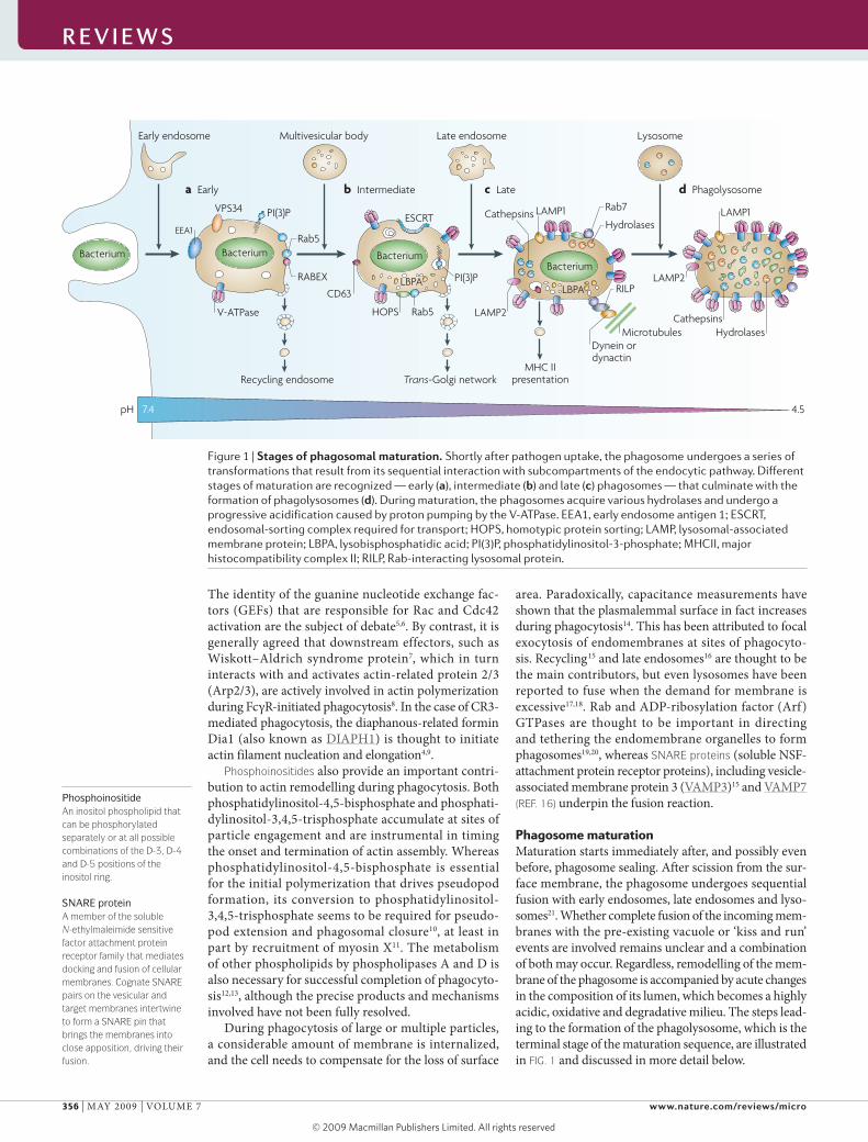
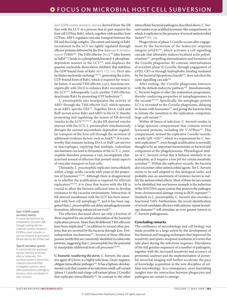
Phagosome maturationMaturation starts immediately after, and possibly even before, phagosome sealing. After scission from the sur-face membrane, the phagosome undergoes sequential fusion with early endosomes, late endosomes and lyso-somes21. whether complete fusion of the incoming mem-branes with the pre-existing vacuole or ‘kiss and run’ events are involved remains unclear and a combination of both may occur. Regardless, remodelling of the mem-brane of the phagosome is accompanied by acute changes in the composition of its lumen, which becomes a highly acidic, oxidative and degradative milieu. The steps lead-ing to the formation of the phagolysosome, which is the terminal stage of the maturation sequence, are illustrated in FIG. 1 and discussed in more detail below.
Figure 1 | Stages of phagosomal maturation. Shortly after pathogen uptake, the phagosome undergoes a series of transformations that result from its sequential interaction with subcompartments of the endocytic pathway. Different stages of maturation are recognized — early (a), intermediate (b) and late (c) phagosomes — that culminate with the formation of phagolysosomes (d). During maturation, the phagosomes acquire various hydrolases and undergo a progressive acidification caused by proton pumping by the V-ATPase. EEA1, early endosome antigen 1; ESCRT, endosomal-sorting complex required for transport; HOPS, homotypic protein sorting; LAMP, lysosomal-associated membrane protein; LBPA, lysobisphosphatidic acid; PI(3)P, phosphatidylinositol-3-phosphate; MHCII, major histocompatibility complex II; RILP, Rab-interacting lysosomal protein.
R E V I E W S
356 | MAY 2009 | vOluME 7 www.nature.com/reviews/micro
R E V I E W S
© 2009 Macmillan Publishers Limited. All rights reserved
Multivesicular body(MVB). A defined stage in the transit between early endosomes and late endosomes or lysosomes. MVBs are characterized by a limiting membrane that encloses internal vesicles rich in lyso-bisphosphatidic acid, CD63 and phosphatidylinositol-3-phosphate. Proteins destined for degradation are sorted to internal vesicles of MVBs.
The early phagosome. newly formed phagosomes rap-idly gain many of the properties of early endosomes. They have a propensity to fuse with sorting and recy-cling endosomes and are refractory to fusion with lyso-somes22,23. Their lumen is mildly acidic (pH 6.1–6.5) and poor in hydrolytic activity24.
The small GTPase Rab5A integrates the targeting, teth-ering and fusion of early endosomes25, and also seems to be involved in the dynamics of early phagosomes, in which it is activated by the GAPvD1 (GTPase-activating protein and vPS9 domain-containing protein 1) exchange factor after the ingestion of apoptotic cells26. Rab5A acts using multiple effectors, including the p150–hvPS34 complex, early endosome antigen 1 (EEA1) and SnARE proteins. The Ser and Thr kinase p150 supports the recruitment of hvPS34, a class III phosphatidylinositol-3-kinase that generates phosphatidylinositol-3-phosphate (PI(3)P) on the early phagosomal membrane27. PI(3)P anchors effec-tor proteins, such as EEA1, to the cytosolic face of the phagosome through FYvE and PX domains28,29. EEA1, which also interacts directly with Rab5 (REF. 30), is thought to act as a bridge that tethers early endosomes to incoming endocytic vesicles31, and probably has an equivalent role in phagosomes. Additionally, EEA1 interacts with syntaxin 13 (REF. 32), a SnARE protein required for membrane fusion, and with an N-ethylmaleimide-sensitive fusion protein that is essential for the disassembly and reuse of SnARE complexes33.
Despite repeated rounds of fusion with endomem-brane vesicles, the surface area of the phagosomal membrane does not increase perceptibly, and contin-ues to envelop the internalized particle tightly. This probably results from the concomitant occurrence of membrane fission events. Similarly to early endosomes, phagosomes are thought to be able to recycle molecules to the plasma membrane by a process involving coat protein I (COPI), and Arf and Rab GTPases34. Rab11A, which was previously known to mediate recycling of endosomes to the plasma membrane, also participates in the retrieval of phagosomal constituents to the plas-malemma35, a process that is regulated by the Rab-coupling protein36,37. In addition, cargo is retrieved to endosomes and the trans-Golgi network by a complex of carrier vesicles, tubules and molecular motors38. The retromer complex of sorting nexin 1 (SnX1), SnX2, vacuolar protein sorting-associated protein 26A (vPS26A), vPS29 and vPS35, which links cargo selec-tion to tubule generation in endosomes, is likely to play a similar part in phagosomes. SnX4 and EH domain-containing protein 1 (EHD1), two other components that are active in retrieval and tubule stabilization in other systems39,40, may also contribute to phagosomal maturation.
In addition to budding outwards for the purpose of retrieval, phagosomes divert membrane-associated cargo that is destined for degradation to intraluminal vesicles. Such vesicles are thought to arise from inwards budding and pinching of the limiting membrane of the phagosome, in a manner akin to the generation of multivesicular bodies (MvBs). This initially occurs at a stage we designated as intermediate in FIG. 1, as it
possesses features that are not present in early phago-somes, but lacks other features that are typical of late phagosomes (discussed below). As in endosomes, phago-somal membrane proteins destined for degradation are ubiquitinated and associate with the endosomal-sorting complex required for transport (ESCRT)41. In MvBs, the final component of the complex, ESCRTIII, forms a lat-tice that in conjunction with the ATPase vPS4A forces the extrusion of vesicles into the organellar lumen42,43. Phosphatidylinositol-(3,5)-bisphosphate synthesized by the FYvE finger-containing phosphoinositide kinase PIP5K3 (PIKfyve kinase) may also be important for vesiculation, as it binds to ESCRTIII44.
The late phagosome. Once the recycling proteins are removed, the phagosome proceeds to the late stage, which is characterized by a more acidic luminal pH (5.5–6.0) brought about by the acquisition of additional proton-pumping v-ATPases21. The late phagosome is also enriched in proteases and lysosomal-associated membrane proteins (lAMPs), which are either imported from the Golgi complex or acquired by fusion with late endosomes. little is known about late phagosome dynam-ics21. The small GTPase Rab7A is a characteristic marker of this organelle, and is known to mediate the traffic between phagosomes and late endosomes or lysosomes45,46. The vpsC–homotypic protein sorting (HOPS) complex, which mediates the transition from Rab5A- to Rab7A-positive endosomes47, probably serves a similar function in phago-some maturation. However, whereas vpsC–HOPS does regulate vesicular traffic and fusion during lysosome biogenesis, it is not needed for Rab7A recruitment48,49. Regardless of how it is acquired, Rab7A recruits several effectors to the vacuolar membrane. One such effector, Rab-interacting lysosomal protein (RIlP), promotes the centripetal movement of late phagosomes and lysosomes by bridging the membrane to the dynein–dynactin motor complex process46,50. Fusion of endosomes and lysosomes is facilitated by bringing the organelles in close apposition so that SnAREs such as vAMP7 and vAMP8 can complete membrane coalescence51,52, and physical proximity is equally likely to favour fusion of phagosomes. Although necessary, Rab7A and RIlP are not the only mediators of late phagosome maturation. Phosphatidylinositol-3-kinase antagonists block phago-some maturation despite the acquisition of Rab7A and RIlP53, implying that a separate, inositide-dependent event is also essential.
Retrieval and disposal of membrane components also occur at this stage. Similarly to late endosomes, late phagosomes contain lysobisphosphatidic acid (lBPA), a unique lipid found in luminal vesicles of MvBs. Programmed cell death 6-interacting protein (PDCD6IP; also known as AlIX), which binds lBPA and can link ESCRTI and ESCRTIII54 in endosomes, is speculated to participate in the inward budding process.
Phagolysosome. The maturation process culminates with the formation of the phagolysosome, the ultimate micro-bicidal organelle. Phagolysosomes are endowed with a complete, sophisticated armamentarium to eliminate and
R E V I E W S
nATuRE REvIEwS | Microbiology vOluME 7 | MAY 2009 | 357
f o c u S o n m I c R o b I a l h o S t c E l l S u b V E R S I o n
© 2009 Macmillan Publishers Limited. All rights reserved
Nature Reviews | Microbiology
Cytoplasm
PhagosomepH ~4.0–5.0
Bacterium
p47
p67
V-ATPase
Antimicrobialpeptides
SOD
iNOS
Citruline Arginine
NO ONOO– NO2•
HOCl
MPO
Defensins
Metal transporters Proteases
+ + + + + +
Stress proteases and proteasome
Lys
Lys + H+
CadAROSIntactDNAandprotein
Oxidizedand damagedDNA andprotein
Cadaverine
iNOS
Citruline Arginine
O2–
p40
Fe2+
Fe2+
H+
H+
NADP+ + O2–
NADPHCytochrome b558
NADPH oxidase
H2O2
2 H2O2
HO•
Lactoferrin
NADPH oxidase
2 H2O + O2
ProteasesAra4N
Catalase
a Host microbicidal factors b Microbial defensive mechanisms
degrade microorganisms (discussed below). They are generated by fusion with lysosomes through a Rab7A-dependent process and are highly acidic (luminal pH values as low as 4.5 have been reported). Insertion of addi-tional v-ATPases and tightening of the H+ ‘leak’ account for the accentuated acidification. Phagolysosomes can be differentiated from late phagosomes by their paucity of lBPA or PI(3)P-enriched internal membranes55,56, by their elevated mature cathepsin content and by their lack of mannose-6-phosphate receptors57.
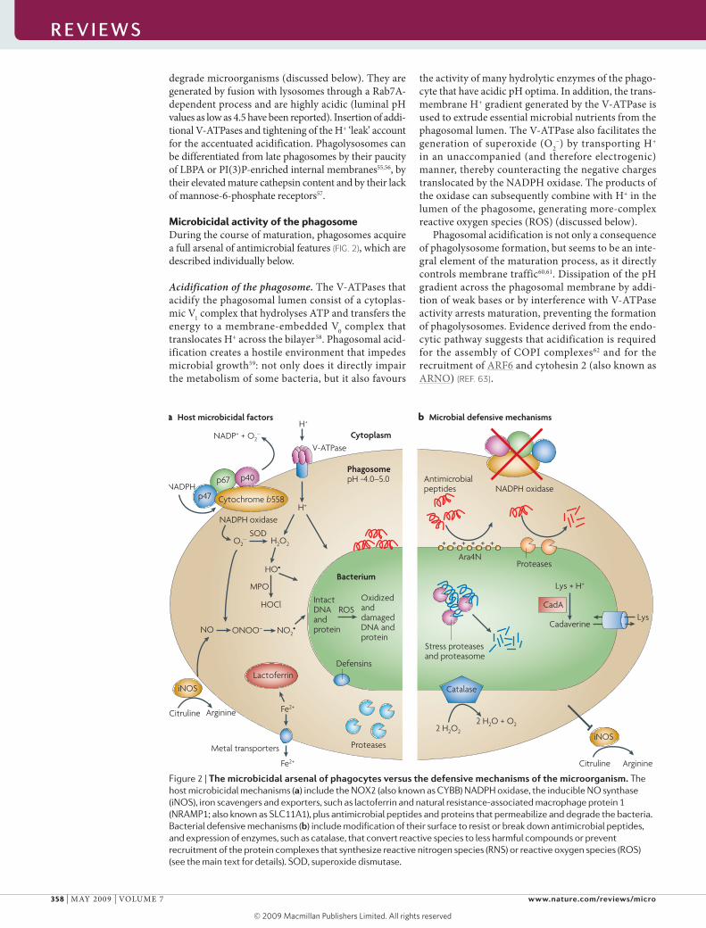
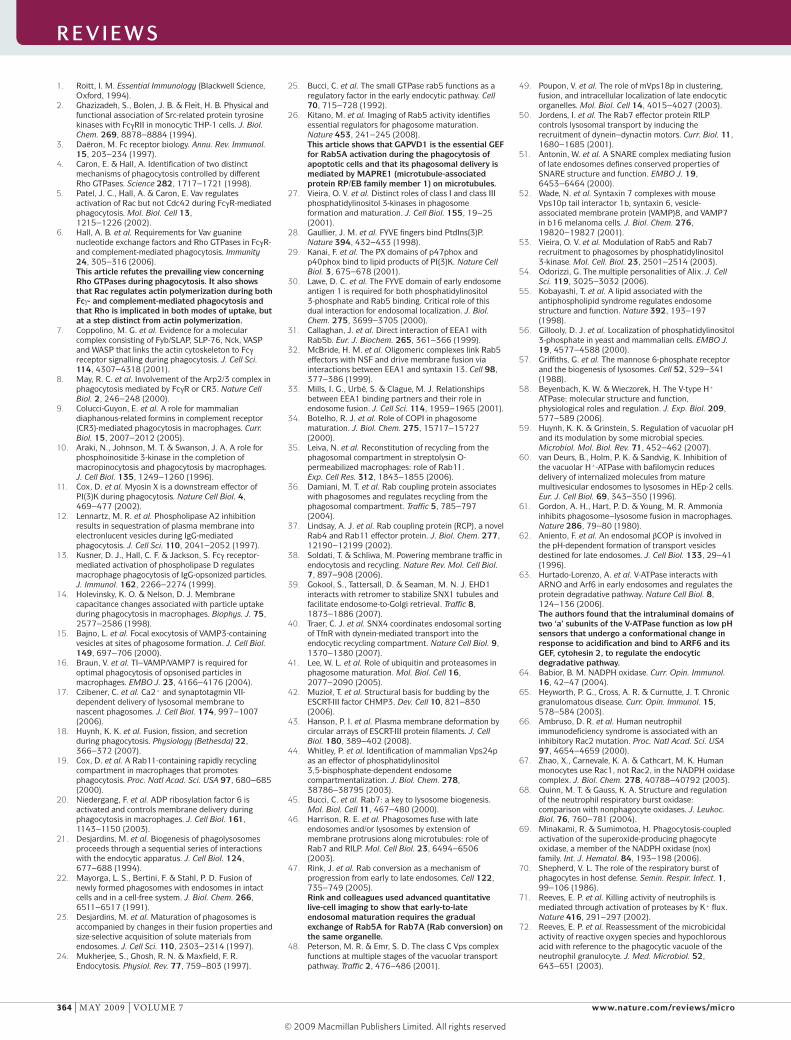
Microbicidal activity of the phagosomeDuring the course of maturation, phagosomes acquire a full arsenal of antimicrobial features (FIG. 2), which are described individually below.
Acidification of the phagosome. The v-ATPases that acidify the phagosomal lumen consist of a cytoplas-mic v1 complex that hydrolyses ATP and transfers the energy to a membrane-embedded v0 complex that translocates H+ across the bilayer58. Phagosomal acid-ification creates a hostile environment that impedes microbial growth59: not only does it directly impair the metabolism of some bacteria, but it also favours
the activity of many hydrolytic enzymes of the phago-cyte that have acidic pH optima. In addition, the trans-membrane H+ gradient generated by the v-ATPase is used to extrude essential microbial nutrients from the phagosomal lumen. The v-ATPase also facilitates the generation of superoxide (O2
–) by transporting H+ in an unaccompanied (and therefore electrogenic) manner, thereby counteracting the negative charges translocated by the nADPH oxidase. The products of the oxidase can subsequently combine with H+ in the lumen of the phagosome, generating more-complex reactive oxygen species (ROS) (discussed below).
Phagosomal acidification is not only a consequence of phagolysosome formation, but seems to be an inte-gral element of the maturation process, as it directly controls membrane traffic60,61. Dissipation of the pH gradient across the phagosomal membrane by addi-tion of weak bases or by interference with v-ATPase activity arrests maturation, preventing the formation of phagolysosomes. Evidence derived from the endo-cytic pathway suggests that acidification is required for the assembly of COPI complexes62 and for the recruitment of ARF6 and cytohesin 2 (also known as ARnO) (REF. 63).
Figure 2 | The microbicidal arsenal of phagocytes versus the defensive mechanisms of the microorganism. The host microbicidal mechanisms (a) include the NOX2 (also known as CYBB) NADPH oxidase, the inducible NO synthase (iNOS), iron scavengers and exporters, such as lactoferrin and natural resistance-associated macrophage protein 1 (NRAMP1; also known as SLC11A1), plus antimicrobial peptides and proteins that permeabilize and degrade the bacteria. Bacterial defensive mechanisms (b) include modification of their surface to resist or break down antimicrobial peptides, and expression of enzymes, such as catalase, that convert reactive species to less harmful compounds or prevent recruitment of the protein complexes that synthesize reactive nitrogen species (RNS) or reactive oxygen species (ROS) (see the main text for details). SOD, superoxide dismutase.
R E V I E W S
358 | MAY 2009 | vOluME 7 www.nature.com/reviews/micro
R E V I E W S
© 2009 Macmillan Publishers Limited. All rights reserved
Azurophil or primary granuleA specialized neutrophil granule, also called a peroxi-dase-positive granule, that resembles lysosomes, in that it contains degradative enzymes, such as β-glucuronidase, cathepsins, elastase, lysozyme and myeloperoxidase, as well as antimicrobial peptides, such as defensins.
Specific or secondary granuleA specialized neutrophil granule, also called a peroxi-dase-negative granule, that exists as a heterogeneous continuum of granules with varying amounts of lactoferrin, collagenase, heparanase, lysozyme and antimicrobial cathelicidins.
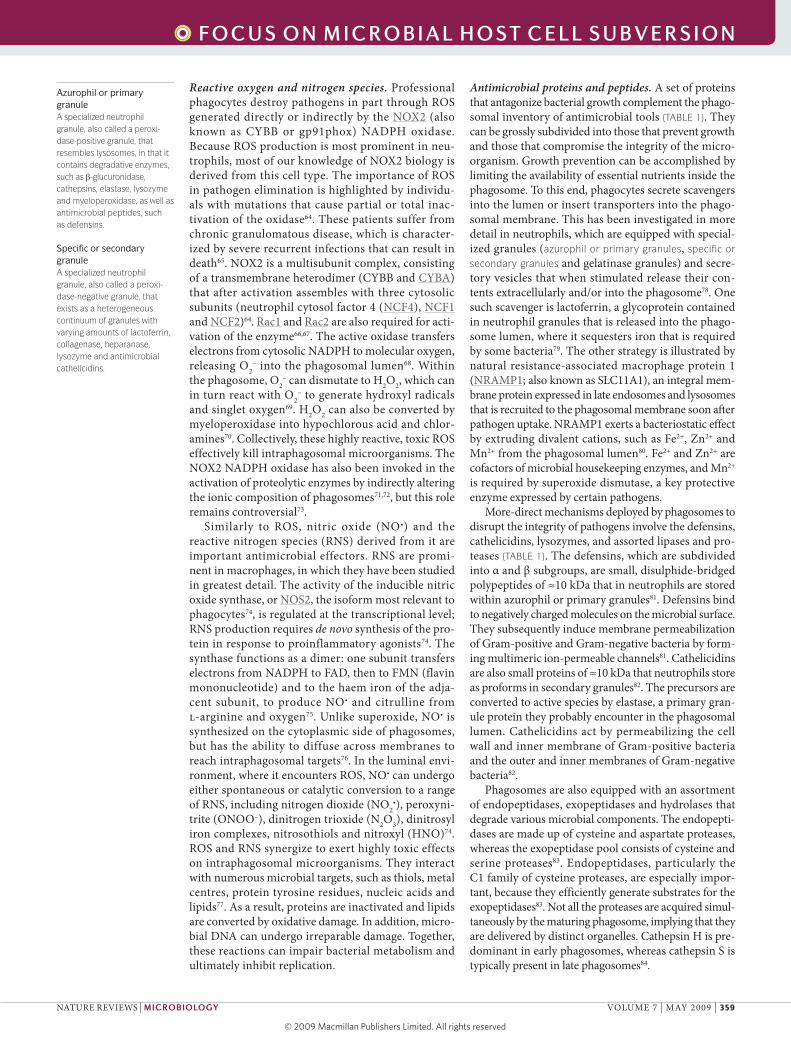
Reactive oxygen and nitrogen species. Professional phagocytes destroy pathogens in part through ROS generated directly or indirectly by the nOX2 (also known as CYBB or gp91phox) nADPH oxidase. Because ROS production is most prominent in neu-trophils, most of our knowledge of nOX2 biology is derived from this cell type. The importance of ROS in pathogen elimination is highlighted by individu-als with mutations that cause partial or total inac-tivation of the oxidase64. These patients suffer from chronic granulomatous disease, which is character-ized by severe recurrent infections that can result in death65. nOX2 is a multisubunit complex, consisting of a transmembrane heterodimer (CYBB and CYBA) that after activation assembles with three cytosolic subunits (neutrophil cytosol factor 4 (nCF4), nCF1 and nCF2)64. Rac1 and Rac2 are also required for acti-vation of the enzyme66,67. The active oxidase transfers electrons from cytosolic nADPH to molecular oxygen, releasing O2
– into the phagosomal lumen68. within the phagosome, O2
– can dismutate to H2O2, which can in turn react with O2
– to generate hydroxyl radicals and singlet oxygen69. H2O2 can also be converted by myeloperoxidase into hypochlorous acid and chlor-amines70. Collectively, these highly reactive, toxic ROS effectively kill intraphagosomal microorganisms. The nOX2 nADPH oxidase has also been invoked in the activation of proteolytic enzymes by indirectly altering the ionic composition of phagosomes71,72, but this role remains controversial73.
Similarly to ROS, nitric oxide (nO•) and the reactive nitrogen species (RnS) derived from it are important antimicrobial effectors. RnS are promi-nent in macrophages, in which they have been studied in greatest detail. The activity of the inducible nitric oxide synthase, or nOS2, the isoform most relevant to phagocytes74, is regulated at the transcriptional level; RnS production requires de novo synthesis of the pro-tein in response to proinflammatory agonists74. The synthase functions as a dimer: one subunit transfers electrons from nADPH to FAD, then to FMn (flavin mononucleotide) and to the haem iron of the adja-cent subunit, to produce nO• and citrulline from l-arginine and oxygen75. unlike superoxide, nO• is synthesized on the cytoplasmic side of phagosomes, but has the ability to diffuse across membranes to reach intraphagosomal targets76. In the luminal envi-ronment, where it encounters ROS, nO• can undergo either spontaneous or catalytic conversion to a range of RnS, including nitrogen dioxide (nO2
•), peroxyni-trite (OnOO–), dinitrogen trioxide (n2O3), dinitrosyl iron complexes, nitrosothiols and nitroxyl (HnO)74. ROS and RnS synergize to exert highly toxic effects on intraphagosomal microorganisms. They interact with numerous microbial targets, such as thiols, metal centres, protein tyrosine residues, nucleic acids and lipids77. As a result, proteins are inactivated and lipids are converted by oxidative damage. In addition, micro-bial DnA can undergo irreparable damage. Together, these reactions can impair bacterial metabolism and ultimately inhibit replication.
Antimicrobial proteins and peptides. A set of proteins that antagonize bacterial growth complement the phago-somal inventory of antimicrobial tools (TABLE 1). They can be grossly subdivided into those that prevent growth and those that compromise the integrity of the micro-organism. Growth prevention can be accomplished by limiting the availability of essential nutrients inside the phagosome. To this end, phagocytes secrete scavengers into the lumen or insert transporters into the phago-somal membrane. This has been investigated in more detail in neutrophils, which are equipped with special-ized granules (azurophil or primary granules, specific or secondary granules and gelatinase granules) and secre-tory vesicles that when stimulated release their con-tents extracellularly and/or into the phagosome78. One such scavenger is lactoferrin, a glycoprotein contained in neutrophil granules that is released into the phago-some lumen, where it sequesters iron that is required by some bacteria79. The other strategy is illustrated by natural resistance-associated macrophage protein 1 (nRAMP1; also known as SlC11A1), an integral mem-brane protein expressed in late endosomes and lysosomes that is recruited to the phagosomal membrane soon after pathogen uptake. nRAMP1 exerts a bacteriostatic effect by extruding divalent cations, such as Fe2+, Zn2+ and Mn2+ from the phagosomal lumen80. Fe2+ and Zn2+ are cofactors of microbial housekeeping enzymes, and Mn2+ is required by superoxide dismutase, a key protective enzyme expressed by certain pathogens.
More-direct mechanisms deployed by phagosomes to disrupt the integrity of pathogens involve the defensins, cathelicidins, lysozymes, and assorted lipases and pro-teases (TABLE 1). The defensins, which are subdivided into α and β subgroups, are small, disulphide-bridged polypeptides of ≈10 kDa that in neutrophils are stored within azurophil or primary granules81. Defensins bind to negatively charged molecules on the microbial surface. They subsequently induce membrane permeabilization of Gram-positive and Gram-negative bacteria by form-ing multimeric ion-permeable channels81. Cathelicidins are also small proteins of ≈10 kDa that neutrophils store as proforms in secondary granules82. The precursors are converted to active species by elastase, a primary gran-ule protein they probably encounter in the phagosomal lumen. Cathelicidins act by permeabilizing the cell wall and inner membrane of Gram-positive bacteria and the outer and inner membranes of Gram-negative bacteria82.
Phagosomes are also equipped with an assortment of endopeptidases, exopeptidases and hydrolases that degrade various microbial components. The endopepti-dases are made up of cysteine and aspartate proteases, whereas the exopeptidase pool consists of cysteine and serine proteases83. Endopeptidases, particularly the C1 family of cysteine proteases, are especially impor-tant, because they efficiently generate substrates for the exopeptidases83. not all the proteases are acquired simul-taneously by the maturing phagosome, implying that they are delivered by distinct organelles. Cathepsin H is pre-dominant in early phagosomes, whereas cathepsin S is typically present in late phagosomes84.
R E V I E W S
nATuRE REvIEwS | Microbiology vOluME 7 | MAY 2009 | 359
f o c u S o n m I c R o b I a l h o S t c E l l S u b V E R S I o n
© 2009 Macmillan Publishers Limited. All rights reserved
Hydrolases that target carbohydrates (for example, α-hexosaminidase, β-glucuronidase and lysozyme) and lipids (for example, phospholipase A2) are also delivered to the phagosomes.
Bacterial resistance to phagocyte killingDespite the presence of these antimicrobial host factors, many pathogens can survive inside the host cell. Such pathogens, which include bacteria, fungi and viruses, have evolved a multitude of strategies to counteract host defences. For simplicity, we confine the remain-der of this Review to bacterial pathogens. Some bac-terial species interfere with the ability of phagocytes to engulf them85,86, either by scavenging, inhibiting or even degrading opsonic antibodies or complement87–89, or by directly impairing the phagocytic machinery of macrophages and neutrophils85,86,90. Other bacteria have become resistant to one or more of the antimicrobial fac-tors of phagocytes (FIG. 2). Some species have developed metabolic pathways to counteract acid accumulation inside phagosomes or have acquired uniquely resistant proteins to withstand the low pH91,92. Yet other bacteria protect themselves by actively degrading93 or shielding themselves94,95 from the antimicrobial peptides and pro-teins produced by phagocytes, or by expressing detoxi-fying enzymes, such as catalase, that neutralize ROS and/or RnS96,97. Alternatively, some bacterial species prevent RnS and ROS formation by impairing recruit-ment of the proteins that mediate their synthesis98,99. Other species have devised means of overcoming the
scarcity of iron by secreting specialized iron-scavenging molecules called siderophores, which sequester and target the cation for bacterial use100, or by expressing iron storage101 or transport proteins102. lastly, many bacteria improve their intraphagosomal survival by mounting a vigorous stress response to dispose of and replace damaged proteins103.
Although most bacteria use one or more of these resistance mechanisms, only a select group of bacteria are ‘professional’ intracellular pathogens. These species survive and replicate inside phagocytes, effectively avoid-ing attack by their antimicrobial factors. To accomplish this feat, such pathogens have evolved multiple strate-gies towards one common goal: to perturb phagosomal maturation. These different strategies are exemplified by the mechanisms used by Mycobacterium tuberculo-sis, Listeria monocytogenes, Legionella pneumophila and Coxiella burnetii. These bacteria parasitize host cells by arresting or reprogramming phagosomal maturation, by escaping maturing phagosomes or by withstanding the microbicidal properties of the phagolysosome.
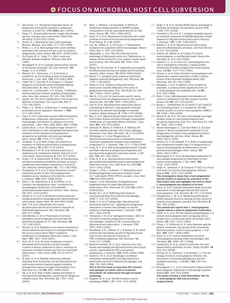
M. tuberculosis: inhibition of phagosomal maturation. The pathogenicity of M. tuberculosis is largely attributed to its ability to survive within macrophages by arresting phagosomal maturation104. This bacterium is exquisitely adapted to life within macrophages and not only arrests phagolysosome formation but can also escape the phagosome105 and modulate other macrophage defences to promote its survival106,107. Phagosomal escape, a previ-ously unappreciated facet of intracellular M. tuberculo-sis, requires the expression of a novel bacterial secretion system, ESX105, which is lacking in avirulent mycobacte-ria (reviewed in REF. 108). Phagocytosis of M. tuberculo-sis by macrophages occurs through the engagement of various receptors, including CR3 (REF. 109). However, unlike other particles that are engulfed by the same receptors, the Mycobacterium-containing phagosome fails to progress and become a phagolysosome and is instead arrested at an early stage110 (FIG. 3a). Arrested M. tuberculosis-containing phagosomes are charac-terized by the presence of Rab5A, but the recruitment of Rab5A effectors, such as EEA1 and hvPS34, is impaired110,111, and as a result, PI(3)P does not accu-mulate. M. tuberculosis uses a range of protein and lipid effectors to alter PI(3)P signalling112,113 (TABLE 2). The mycobacterial phosphoinositide lipoarabinoman-nan112, a component of the cell wall that is shed from live bacteria and becomes distributed throughout the endocytic network114, prevents the increase in cytosolic [Ca2+] that normally accompanies phagocytosis and that is thought to be required to activate hvPS34 through calmodulin112. M. tuberculosis further impairs cytosolic Ca2+ flux by inhibiting sphingosine kinase, which con-verts sphingosine to sphingosine-1-phosphate, which in turn promotes Ca2+ efflux from the endoplasmic reticu-lum (ER)115,116. M. tuberculosis also produces the phos-phatase SapM, which specifically hydrolyses PI(3)P113. This combined strategy effectively depletes PI(3)P from early phagosomes and prevents the transition to the late and phagolysosomal stages.
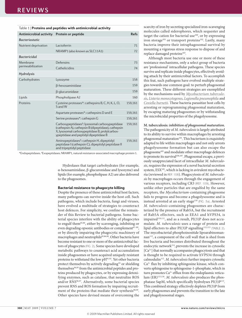
Table 1 | Proteins and peptides with antimicrobial activity
Antimicrobial activity Protein or peptide refs
Bacteriostatic
Nutrient deprivation Lactoferrin 71
NRAMP1 (also known as SLC11A1) 72
Bactericidal
Membrane permeabilization
Defensins 73
Cathelicidins 74
Hydrolysis
Carbohydrates Lysozyme 158
β-hexosaminidase 159
β-glucuronidase 159
Lipids Phospholipase A2 160
Proteins Cysteine proteases*: cathepsins B, C, H, K, L, O, S and W
159,161
Aspartate proteases*: cathepsins D and E 159,161
Serine proteases*: cathepsin G 159,161
Carboxypeptidases‡: lysosomal carboxypeptidase (cathepsin A), cathepsin B (dipeptidase), cathepsin X, lysosomal carboxypeptidase B, prolylcarbox-ypeptidase and peptidyl dipeptidase B
159,161
Aminopeptidases‡: cathepsin H, dipeptidyl peptidase I (cathepsin C), dipeptidyl peptidase II and tripeptidyl peptidase
159,161
*Endopeptidases. ‡Exopeptidases. NRAMP1, natural resistance-associated macrophage protein 1.
R E V I E W S
360 | MAY 2009 | vOluME 7 www.nature.com/reviews/micro
R E V I E W S
© 2009 Macmillan Publishers Limited. All rights reserved
a Mycobacterium tuberculosis d Coxiella burnetiib Legionella pneumophila c Listeria monocytogenes
PIMLAM
?
SapM
PIPIM
hVPS34EEA1
Ca2+
Nucleus Nucleus
ER-derived vesicle
DrrARab1
ARF1
GDI
LidA
RalF
Nucleus
LLO
Ca2+ or H+
PlcA
PlcB
Nucleus
APV LC3
V-ATPase
Delayed
Nature Reviews | Microbiology
Rab5
Early endosome
Trans-Golgi network
ER
Late endosome
Lysosome
ActA
LC3
LC3
Rab7
Activation of macrophages increases their ability to eradicate intracellular M. tuberculosis and other organ-isms117,118. This is highlighted by the observation that interferon-γ (IFnγ)-stimulated macrophages demon-strate enhanced bacterial clearance; in stimulated cells, M. tuberculosis-containing phagosomes are seques-tered by autophagic compartments that ultimately fuse with lysosomes119. This autophagic response can be enhanced by Toll-like receptor ligands120 and the activation of immunity-related p47 guanosine triphos-phatase protein121. Immunity to pathogens such as myco-bacteria is in part attributable to the activation of the inflammasome, a multiprotein complex that facilitates the killing of intra cellular bacteria and is required for interleukin-1β (Il-1β) processing. Il-1β enables mac-rophages to overcome the arrested maturation of the M. tuberculosis-containing phagosome122,123 through an unknown mechanism that may involve restored PI(3)P production and subsequent maturation of the phago-some. Interestingly, the bacteria have also evolved a way to counteract the inflammatory response: M. tubercu-losis secretes ZmpA, a predicted zinc metalloprotease
that inhibits Il-1β processing by the host cells123. Intracellular survival of the bacteria therefore depends on an ongoing, multilevel tug of war between the patho-gen and host macrophage.
L. monocytogenes: a phagosomal escape artist. listeriosis, a potentially fatal disease caused by the Gram-positive bacterium L. monocytogenes124, is frequently contracted through the consumption of contaminated foods. L. monocytogenes is internalized by both non-phagocytic cells and professional phagocytes124, which is crucial for bacterial propagation and dissemination. uptake by epi-thelial cells is mediated by the surface proteins internalin A and internalin B (InlA125 and InlB126), which func-tion as ligands for the adhesion molecule E cadherin127, the hepatocyte growth factor receptor Met128 and the complement receptor, C1qR129. However, phagocytosis of L. monocytogenes by macrophages is mediated by scavenger receptors that recognize lipoteichoic acid, a component of the Gram-positive bacterial cell wall130. In addition, the surface of L. monocytogenes can become decorated with the complement components C1q131 and
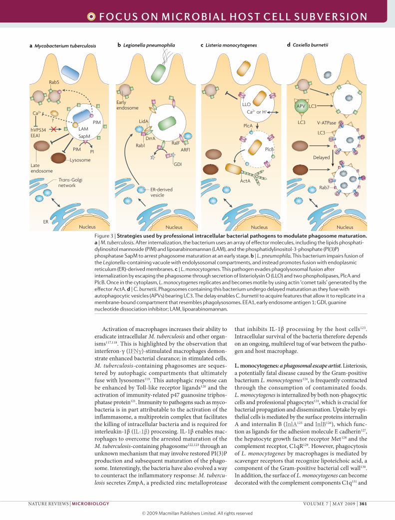
Figure 3 | Strategies used by professional intracellular bacterial pathogens to modulate phagosome maturation. a | M. tuberculosis. After internalization, the bacterium uses an array of effector molecules, including the lipids phosphati-dylinositol mannoside (PIM) and lipoarabinomannan (LAM), and the phosphatidylinositol-3-phosphate (PI(3)P) phosphatase SapM to arrest phagosome maturation at an early stage. b | L. pneumophila. This bacterium impairs fusion of the Legionella-containing vacuole with endolysosomal compartments, and instead promotes fusion with endoplasmic reticulum (ER)-derived membranes. c | L. monocytogenes. This pathogen evades phagolysosomal fusion after internalization by escaping the phagosome through secretion of listeriolysin O (LLO) and two phospholipases, PlcA and PlcB. Once in the cytoplasm, L. monocytogenes replicates and becomes motile by using actin ‘comet tails’ generated by the effector ActA. d | C. burnetii. Phagosomes containing this bacterium undergo delayed maturation as they fuse with autophagocytic vesicles (APVs) bearing LC3. The delay enables C. burnetii to acquire features that allow it to replicate in a membrane-bound compartment that resembles phagolysosomes. EEA1, early endosome antigen 1; GDI, guanine nucleotide dissociation inhibitor; LAM, lipoarabinomannan.
R E V I E W S
nATuRE REvIEwS | Microbiology vOluME 7 | MAY 2009 | 361
f o c u S o n m I c R o b I a l h o S t c E l l S u b V E R S I o n
© 2009 Macmillan Publishers Limited. All rights reserved
AutophagyA complex cellular process by which intracellular components, including entire organelles, are sequestered in double-membrane vesicles or vacuoles called autophagosomes that eventually fuse with lysosomes, bringing about the degradation of their contents.
C3 (REF. 132), which are ligands for macrophage comple-ment receptors. lastly, InlB also functions as a ligand for the C1q receptor129.
L. monocytogenes, a facultative intracellular patho-gen, survives intracellularly by modifying and subse-quently escaping from phagosomes (FIG. 3c). To this end, the bacteria use a sophisticated combination of effec-tors. The cholesterol-dependent cytolysin listeriolysin O (llO) creates pores in the phagosomal membrane as early as 5 minutes after infection133. The effect of llO is restricted to the phagosome, as it needs to be activated by acidification and/or by the host enzyme GIlT (IFnγ-inducible lysosomal thiol reductase) that is found inside the phagosome134. Secretion of llO inhibits the matura-tion of phagosomes135 owing to a loss of luminal H+ and Ca2+, which are thought to be required for fusion with endosomes and/or lysosomes136. Listeria also expresses two membrane-active phospholipase C enzymes, phos-phoinositol-specific phospholipase C (PI-PlC; encoded by plcA) and broad-range phospholipase C (PC-PlC; encoded by plcB). Together with llO, PI-PlC and PC-PlC cause the breakdown of the membrane of the L. monocytogenes-containing phagosome and thereby enable the bacteria to escape and take up residence in the cytosol136,137, where bacterial replication occurs138.
Cytosolic L. monocytogenes replicates efficiently, and has a generation time of ~30 minutes, owing to the expression of genes that enable nutrients to be used directly from the host cell139. In the cytosol, L. mono-cytogenes becomes motile by usurping the host’s actin cytoskeletal machinery. The bacterial surface protein ActA induces the assembly of spectacular actin ‘comet tails’ by recruiting host cell Arp2/3 complexes, G actin and vasodilator-stimulated phosphoprotein (vASP)
family members (reviewed in REF. 140). Although this motility is not required for phagosomal escape, ActA con-tributes substantially to L. monocytogenes dissemination during infection141.
Although L. monocytogenes was previously thought to reside primarily in the cytosol, under some cir-cumstances, it replicates in macrophages, inside large, lAMP1-positive vacuoles called spacious Listeria-containing phagosomes (SlAPs)142. The formation of SlAPs is strictly dependent on low levels of llO produc-tion and the recruitment of the autophagy protein lC3 to the phagosome142. SlAP formation in macrophages allows L. monocytogenes to replicate slowly (generation time >8 hours) without destroying the infected cell142. This newly discovered facet of the L. monocytogenes life cycle could contribute to the development of chronic L. monocytogenes infections.
L. pneumophila: reprogramming the phagosomal mat-uration pathway. L. pneumophila is a Gram-negative bacterium that is found ubiquitously in aquatic environ-ments, growing in biofilms or within freshwater proto-zoa143. In humans, it can survive and replicate within professional phagocytes144 by redirecting the matura-tion of phagosomes to create a unique intracellular niche suited for bacterial replication (FIG. 3b).
After inhalation of L. pneumophila, the major outer membrane protein on the surface of the bacteria effec-tively fixes complement145, thereby promoting phagocyto-sis by macrophages through complement receptors146 and leading to the formation of Legionella-containing vacuoles (lCvs). Internalized L. pneumophila rapidly modulates the maturation of the lCv, avoiding interaction with the default endolysosomal pathway147,148. Shortly afterwards,
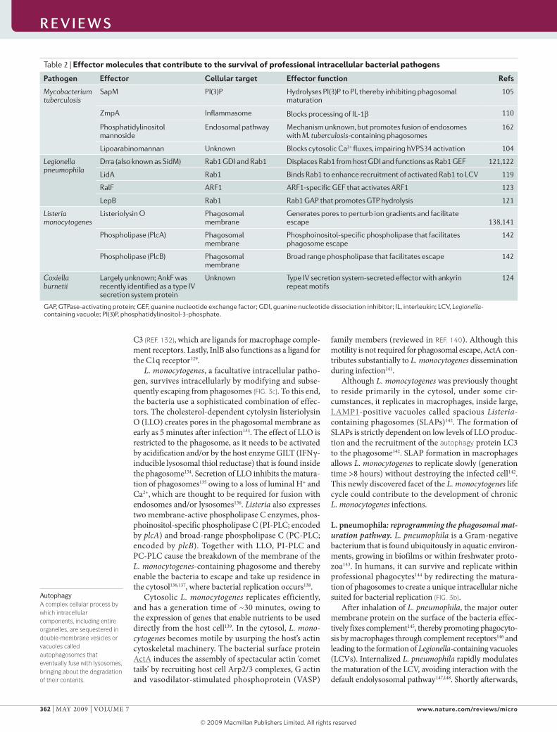
Table 2 | Effector molecules that contribute to the survival of professional intracellular bacterial pathogens
Pathogen Effector cellular target Effector function refs
Mycobacterium tuberculosis
SapM PI(3)P Hydrolyses PI(3)P to PI, thereby inhibiting phagosomal maturation
105
ZmpA Inflammasome Blocks processing of IL-1β 110
Phosphatidylinositol mannoside
Endosomal pathway Mechanism unknown, but promotes fusion of endosomes with M. tuberculosis-containing phagosomes
162
Lipoarabinomannan Unknown Blocks cytosolic Ca2+ fluxes, impairing hVPS34 activation 104
Legionella pneumophila
Drra (also known as SidM) Rab1 GDI and Rab1 Displaces Rab1 from host GDI and functions as Rab1 GEF 121,122
LidA Rab1 Binds Rab1 to enhance recruitment of activated Rab1 to LCV 119
RalF ARF1 ARF1-specific GEF that activates ARF1 123
LepB Rab1 Rab1 GAP that promotes GTP hydrolysis 121
Listeria monocytogenes
Listeriolysin O Phagosomal membrane
Generates pores to perturb ion gradients and facilitate escape
138,141
Phospholipase (PlcA) Phagosomal membrane
Phosphoinositol-specific phospholipase that facilitates phagosome escape
142
Phospholipase (PlcB) Phagosomal membrane
Broad range phospholipase that facilitates escape 142
Coxiella burnetii
Largely unknown; AnkF was recently identified as a type IV secretion system protein
Unknown Type IV secretion system-secreted effector with ankyrin repeat motifs
124
GAP, GTPase-activating protein; GEF, guanine nucleotide exchange factor; GDI, guanine nucleotide dissociation inhibitor; IL, interleukin; LCV, Legionella-containing vacuole; PI(3)P, phosphatidylinositol-3-phosphate.
R E V I E W S
362 | MAY 2009 | vOluME 7 www.nature.com/reviews/micro
R E V I E W S
© 2009 Macmillan Publishers Limited. All rights reserved
SarI–COPII-coated secretory vesicleA vesicle derived from the endoplasmic reticulum (ER) through coating with the coatomer protein complex II (COPII) protein complex, a process initiated at specialized ER exit sites by the GTPase SarI.
Type IV secretion systemA macromolecular apparatus used by bacteria to secrete effector molecules. This secretion system is ancestrally related to bacterial DNA conjugation systems, and is often expressed by pathogenic bacteria, which contributes to their virulence.
SarI–COPII-coated secretory vesicles derived from the ER fuse with the lCv in a process that in part requires the host cell GTPase Rab1, which, together with another host GTPase, ARF1, regulates vesicular transport between the ER and the Golgi complex. The extent and timing of Rab1 recruitment to the lCv are tightly regulated through effector proteins delivered by the Dot–Icm type IV secretion system (T4SS)149. The T4SS effector DrrA150 (also known as SidM151) binds in a phosphatidylinositol-4-phosphate-dependent manner to the lCv152, and displaces the guanine nucleotide dissociation inhibitor that stabilizes the GDP-bound form of Rab1 (REFS 153,154). DrrA also facilitates nucleotide exchange150,151, generating the active, GTP-bound form of Rab1, which is required for vesicu-lar fusion. A second T4SS effector, lidA, functions syn-ergistically with DrrA to enhance Rab1 recruitment to the lCv151. Subsequently lepB, another T4SS effector, deactivates Rab1 by promoting GTP hydrolysis153.
L. pneumophila also manipulates the activity of ARF1 through the T4SS effector RalF, which operates as an ARF1-specific GEF155. Together, DrrA, lidA and RalF recruit active Rab1 and ARF1 to the lCv, thereby promoting and regulating the fusion of ER-derived vesicles to the lCv150,153,155. As the ER-derived vesicles interact with the lCv, L. pneumophila simultaneously disrupts the normal microtubule-dependent organel-lar transport of the host cell through the secretion of additional virulence factors, such as AnkX156. It is note-worthy that mutants lacking DrrA or RalF can survive in macrophages, implying that multiple, redundant mechanisms can lead to formation of the lCv. L. pneu-mophila therefore possesses a vast, incompletely char-acterized arsenal of effectors that perturb many aspects of vesicular transport in host cells.
ultimately, L. pneumophila replicates intracellularly within a large, acidic vacuole with some of the proper-ties of lysosomes157,158. Although there is disagreement as to whether the acidification is required for efficient replication158,159, it is clear that fusion with the ER is crucial to allow the bacteria sufficient time to develop resistance to the vacuolar environment. Interaction of ER-derived membranes with the lCv has been associ-ated with host cell autophagy160, and it has been sug-gested that L. pneumophila can delay autophagolysosome formation, allowing enhanced survival161.
The effectors discussed above are only a fraction of those required for successful culmination of the bacterial replication process. More than 80 different T4SS effectors have been implicated162, in addition to several other pro-teins that are secreted by the bacteria through Icm–Dot-independent mechanisms163. Several of these effectors possess motifs that are commonly identified in eukaryotic proteins, suggesting that L. pneumophila has the potential to manipulate additional host cell processes156,163.
C. burnetii: weathering the storm. C. burnetii, the causa-tive agent of Q fever, is a highly infectious, Gram-negative, obligate intracellular pathogen164. It has a biphasic develop-mental cycle that consists of an infectious small-cell variant (phase 1 Coxiella) and a large-cell variant (phase 2 Coxiella) that replicates intracellularly164. In contrast to the other
intracellular bacterial pathogens described above, C. bur-netii resides in an acidified lysosome-like compartment, in which it replicates in the presence of several antimicrobial factors165 (FIG. 3d).
Phagocytosis of phase I Coxiella occurs after engage-ment by the bacterium of the leukocyte response integrin (αvβ3)166, which activates a cell signalling cascade that ultimately induces localized actin polym-erization167, propelling internalization and formation of the Coxiella phagosome. By contrast, internalization of avirulent phase II Coxiella, through engagement of αvβ3, CR3 or through hydrophobic binding mediated by the bacterial lipopolysaccharide164, does not elicit the same signalling cascade168.
After sealing, the Coxiella phagosome interacts with the default endocytic pathway165. Simultaneously, C. burnetii begins to alter the maturation programme, thereby conferring properties of autophagosomes to the vacuole169,170. Specifically, the autophagic protein lC3 is recruited to the Coxiella phagosome, delaying its fusion with lysosomes171 and giving the bacteria time to initiate the transition to the replication-competent, large-cell variant170.
within 48 hours of infection, C. burnetii resides in a large spacious compartment that contains several lysosomal proteins, including the v-ATPase165. This compartment, termed the replicative Coxiella vacuole, is acidic (pH ≈4.8)172, which is a requirement for C. bur-netii replication165, even though acidification is normally thought to be an important bacteriostatic or bactericidal component of the phagolysosome. Physiological stud-ies of C. burnetii reveal that it behaves similarly to an acidophile, as it requires a low pH for certain metabolic activities173. within the replicative vacuole, the bacteria also encounter other antimicrobial agents. Yet C. burnetii seems to be well adapted to this biological niche, and probably uses an assortment of virulence factors to nul-lify the antimicrobial effects. Most of these factors remain to be identified, but one known example is the induction of the SOS DnA repair system that protects the pathogen from chromosomal damage owing to ROS exposure174. Similarly to L. pneumophila, C. burnetii also encodes a functional T4SS. Furthermore, the recent identification of several candidate effectors with ankyrin-repeat homol-ogy domains156 will stimulate an even greater interest in C. burnetii pathogenesis.
Concluding remarksThe confluence of microbiology and cell biology was made possible to a large extent by the development of biochemical and imaging techniques that improved the sensitivity and spatio-temporal resolution of events that take place during the infectious sequence. Elucidation of the full genome sequences of a number of pathogens, together with the increased sensitivity and accuracy of proteomic analyses and the implementation of power-ful intravital imaging will further accelerate the pace of knowledge acquisition in the new discipline of cel-lular microbiology. As a consequence, more fascinating insights into the interaction between phagocytes and pathogens are certain to emerge.
R E V I E W S
nATuRE REvIEwS | Microbiology vOluME 7 | MAY 2009 | 363
f o c u S o n m I c R o b I a l h o S t c E l l S u b V E R S I o n
© 2009 Macmillan Publishers Limited. All rights reserved
1. Roitt, I. M. Essential Immunology (Blackwell Science, Oxford, 1994).
2. Ghazizadeh, S., Bolen, J. B. & Fleit, H. B. Physical and functional association of Src-related protein tyrosine kinases with FcγRII in monocytic THP-1 cells. J. Biol. Chem. 269, 8878–8884 (1994).
3. Daëron, M. Fc receptor biology. Annu. Rev. Immunol. 15, 203–234 (1997).
4. Caron, E. & Hall, A. Identification of two distinct mechanisms of phagocytosis controlled by different Rho GTPases. Science 282, 1717–1721 (1998).
5. Patel, J. C., Hall, A. & Caron, E. Vav regulates activation of Rac but not Cdc42 during FcγR-mediated phagocytosis. Mol. Biol. Cell 13, 1215–1226 (2002).
6. Hall, A. B. et al. Requirements for Vav guanine nucleotide exchange factors and Rho GTPases in FcγR- and complement-mediated phagocytosis. Immunity 24, 305–316 (2006).This article refutes the prevailing view concerning Rho GTPases during phagocytosis. It also shows that Rac regulates actin polymerization during both Fcγ- and complement-mediated phagocytosis and that Rho is implicated in both modes of uptake, but at a step distinct from actin polymerization.
7. Coppolino, M. G. et al. Evidence for a molecular complex consisting of Fyb/SLAP, SLP-76, Nck, VASP and WASP that links the actin cytoskeleton to Fcγ receptor signalling during phagocytosis. J. Cell Sci. 114, 4307–4318 (2001).
8. May, R. C. et al. Involvement of the Arp2/3 complex in phagocytosis mediated by FcγR or CR3. Nature Cell Biol. 2, 246–248 (2000).
9. Colucci-Guyon, E. et al. A role for mammalian diaphanous-related formins in complement receptor (CR3)-mediated phagocytosis in macrophages. Curr. Biol. 15, 2007–2012 (2005).
10. Araki, N., Johnson, M. T. & Swanson, J. A. A role for phosphoinositide 3-kinase in the completion of macropinocytosis and phagocytosis by macrophages. J. Cell Biol. 135, 1249–1260 (1996).
11. Cox, D. et al. Myosin X is a downstream effector of PI(3)K during phagocytosis. Nature Cell Biol. 4, 469–477 (2002).
12. Lennartz, M. R. et al. Phospholipase A2 inhibition results in sequestration of plasma membrane into electronlucent vesicles during IgG-mediated phagocytosis. J. Cell Sci. 110, 2041–2052 (1997).
13. Kusner, D. J., Hall, C. F. & Jackson, S. Fcγ receptor-mediated activation of phospholipase D regulates macrophage phagocytosis of IgG-opsonized particles. J. Immunol. 162, 2266–2274 (1999).
14. Holevinsky, K. O. & Nelson, D. J. Membrane capacitance changes associated with particle uptake during phagocytosis in macrophages. Biophys. J. 75, 2577–2586 (1998).
15. Bajno, L. et al. Focal exocytosis of VAMP3-containing vesicles at sites of phagosome formation. J. Cell Biol. 149, 697–706 (2000).
16. Braun, V. et al. TI–VAMP/VAMP7 is required for optimal phagocytosis of opsonised particles in macrophages. EMBO J. 23, 4166–4176 (2004).
17. Czibener, C. et al. Ca2+ and synaptotagmin VII-dependent delivery of lysosomal membrane to nascent phagosomes. J. Cell Biol. 174, 997–1007 (2006).
18. Huynh, K. K. et al. Fusion, fission, and secretion during phagocytosis. Physiology (Bethesda) 22, 366–372 (2007).
19. Cox, D. et al. A Rab11-containing rapidly recycling compartment in macrophages that promotes phagocytosis. Proc. Natl Acad. Sci. USA 97, 680–685 (2000).
20. Niedergang, F. et al. ADP ribosylation factor 6 is activated and controls membrane delivery during phagocytosis in macrophages. J. Cell Biol. 161, 1143–1150 (2003).
21. Desjardins, M. et al. Biogenesis of phagolysosomes proceeds through a sequential series of interactions with the endocytic apparatus. J. Cell Biol. 124, 677–688 (1994).
22. Mayorga, L. S., Bertini, F. & Stahl, P. D. Fusion of newly formed phagosomes with endosomes in intact cells and in a cell-free system. J. Biol. Chem. 266, 6511–6517 (1991).
23. Desjardins, M. et al. Maturation of phagosomes is accompanied by changes in their fusion properties and size-selective acquisition of solute materials from endosomes. J. Cell Sci. 110, 2303–2314 (1997).
24. Mukherjee, S., Ghosh, R. N. & Maxfield, F. R. Endocytosis. Physiol. Rev. 77, 759–803 (1997).
25. Bucci, C. et al. The small GTPase rab5 functions as a regulatory factor in the early endocytic pathway. Cell 70, 715–728 (1992).
26. Kitano, M. et al. Imaging of Rab5 activity identifies essential regulators for phagosome maturation. Nature 453, 241–245 (2008).This article shows that GAPVD1 is the essential GEF for Rab5A activation during the phagocytosis of apoptotic cells and that its phagosomal delivery is mediated by MAPRE1 (microtubule-associated protein RP/EB family member 1) on microtubules.
27. Vieira, O. V. et al. Distinct roles of class I and class III phosphatidylinositol 3-kinases in phagosome formation and maturation. J. Cell Biol. 155, 19–25 (2001).
28. Gaullier, J. M. et al. FYVE fingers bind PtdIns(3)P. Nature 394, 432–433 (1998).
29. Kanai, F. et al. The PX domains of p47phox and p40phox bind to lipid products of PI(3)K. Nature Cell Biol. 3, 675–678 (2001).
30. Lawe, D. C. et al. The FYVE domain of early endosome antigen 1 is required for both phosphatidylinositol 3-phosphate and Rab5 binding. Critical role of this dual interaction for endosomal localization. J. Biol. Chem. 275, 3699–3705 (2000).
31. Callaghan, J. et al. Direct interaction of EEA1 with Rab5b. Eur. J. Biochem. 265, 361–366 (1999).
32. McBride, H. M. et al. Oligomeric complexes link Rab5 effectors with NSF and drive membrane fusion via interactions between EEA1 and syntaxin 13. Cell 98, 377–386 (1999).
33. Mills, I. G., Urbé, S. & Clague, M. J. Relationships between EEA1 binding partners and their role in endosome fusion. J. Cell Sci. 114, 1959–1965 (2001).
34. Botelho, R. J. et al. Role of COPI in phagosome maturation. J. Biol. Chem. 275, 15717–15727 (2000).
35. Leiva, N. et al. Reconstitution of recycling from the phagosomal compartment in streptolysin O- permeabilized macrophages: role of Rab11. Exp. Cell Res. 312, 1843–1855 (2006).
36. Damiani, M. T. et al. Rab coupling protein associates with phagosomes and regulates recycling from the phagosomal compartment. Traffic 5, 785–797 (2004).
37. Lindsay, A. J. et al. Rab coupling protein (RCP), a novel Rab4 and Rab11 effector protein. J. Biol. Chem. 277, 12190–12199 (2002).
38. Soldati, T. & Schliwa, M. Powering membrane traffic in endocytosis and recycling. Nature Rev. Mol. Cell Biol. 7, 897–908 (2006).
39. Gokool, S., Tattersall, D. & Seaman, M. N. J. EHD1 interacts with retromer to stabilize SNX1 tubules and facilitate endosome-to-Golgi retrieval. Traffic 8, 1873–1886 (2007).
40. Traer, C. J. et al. SNX4 coordinates endosomal sorting of TfnR with dynein-mediated transport into the endocytic recycling compartment. Nature Cell Biol. 9, 1370–1380 (2007).
41. Lee, W. L. et al. Role of ubiquitin and proteasomes in phagosome maturation. Mol. Biol. Cell 16, 2077–2090 (2005).
42. Muzioł, T. et al. Structural basis for budding by the ESCRT-III factor CHMP3. Dev. Cell 10, 821–830 (2006).
43. Hanson, P. I. et al. Plasma membrane deformation by circular arrays of ESCRT-III protein filaments. J. Cell Biol. 180, 389–402 (2008).
44. Whitley, P. et al. Identification of mammalian Vps24p as an effector of phosphatidylinositol 3,5-bisphosphate-dependent endosome compartmentalization. J. Biol. Chem. 278, 38786–38795 (2003).
45. Bucci, C. et al. Rab7: a key to lysosome biogenesis. Mol. Biol. Cell 11, 467–480 (2000).
46. Harrison, R. E. et al. Phagosomes fuse with late endosomes and/or lysosomes by extension of membrane protrusions along microtubules: role of Rab7 and RILP. Mol. Cell Biol. 23, 6494–6506 (2003).
47. Rink, J. et al. Rab conversion as a mechanism of progression from early to late endosomes. Cell 122, 735–749 (2005).Rink and colleagues used advanced quantitative live-cell imaging to show that early-to-late endosomal maturation requires the gradual exchange of Rab5A for Rab7A (Rab conversion) on the same organelle.
48. Peterson, M. R. & Emr, S. D. The class C Vps complex functions at multiple stages of the vacuolar transport pathway. Traffic 2, 476–486 (2001).
49. Poupon, V. et al. The role of mVps18p in clustering, fusion, and intracellular localization of late endocytic organelles. Mol. Biol. Cell 14, 4015–4027 (2003).
50. Jordens, I. et al. The Rab7 effector protein RILP controls lysosomal transport by inducing the recruitment of dynein–dynactin motors. Curr. Biol. 11, 1680–1685 (2001).
51. Antonin, W. et al. A SNARE complex mediating fusion of late endosomes defines conserved properties of SNARE structure and function. EMBO J. 19, 6453–6464 (2000).
52. Wade, N. et al. Syntaxin 7 complexes with mouse Vps10p tail interactor 1b, syntaxin 6, vesicle-associated membrane protein (VAMP)8, and VAMP7 in b16 melanoma cells. J. Biol. Chem. 276, 19820–19827 (2001).
53. Vieira, O. V. et al. Modulation of Rab5 and Rab7 recruitment to phagosomes by phosphatidylinositol 3-kinase. Mol. Cell. Biol. 23, 2501–2514 (2003).
54. Odorizzi, G. The multiple personalities of Alix. J. Cell Sci. 119, 3025–3032 (2006).
55. Kobayashi, T. et al. A lipid associated with the antiphospholipid syndrome regulates endosome structure and function. Nature 392, 193–197 (1998).
56. Gillooly, D. J. et al. Localization of phosphatidylinositol 3-phosphate in yeast and mammalian cells. EMBO J. 19, 4577–4588 (2000).
57. Griffiths, G. et al. The mannose 6-phosphate receptor and the biogenesis of lysosomes. Cell 52, 329–341 (1988).
58. Beyenbach, K. W. & Wieczorek, H. The V-type H+ ATPase: molecular structure and function, physiological roles and regulation. J. Exp. Biol. 209, 577–589 (2006).
59. Huynh, K. K. & Grinstein, S. Regulation of vacuolar pH and its modulation by some microbial species. Microbiol. Mol. Biol. Rev. 71, 452–462 (2007).
60. van Deurs, B., Holm, P. K. & Sandvig, K. Inhibition of the vacuolar H+-ATPase with bafilomycin reduces delivery of internalized molecules from mature multivesicular endosomes to lysosomes in HEp-2 cells. Eur. J. Cell Biol. 69, 343–350 (1996).
61. Gordon, A. H., Hart, P. D. & Young, M. R. Ammonia inhibits phagosome–lysosome fusion in macrophages. Nature 286, 79–80 (1980).
62. Aniento, F. et al. An endosomal βCOP is involved in the pH-dependent formation of transport vesicles destined for late endosomes. J. Cell Biol. 133, 29–41 (1996).
63. Hurtado-Lorenzo, A. et al. V-ATPase interacts with ARNO and Arf6 in early endosomes and regulates the protein degradative pathway. Nature Cell Biol. 8, 124–136 (2006).The authors found that the intraluminal domains of two ‘a’ subunits of the V-ATPase function as low pH sensors that undergo a conformational change in response to acidification and bind to ARF6 and its GEF, cytohesin 2, to regulate the endocytic degradative pathway.
64. Babior, B. M. NADPH oxidase. Curr. Opin. Immunol. 16, 42–47 (2004).
65. Heyworth, P. G., Cross, A. R. & Curnutte, J. T. Chronic granulomatous disease. Curr. Opin. Immunol. 15, 578–584 (2003).
66. Ambruso, D. R. et al. Human neutrophil immunodeficiency syndrome is associated with an inhibitory Rac2 mutation. Proc. Natl Acad. Sci. USA 97, 4654–4659 (2000).
67. Zhao, X., Carnevale, K. A. & Cathcart, M. K. Human monocytes use Rac1, not Rac2, in the NADPH oxidase complex. J. Biol. Chem. 278, 40788–40792 (2003).
68. Quinn, M. T. & Gauss, K. A. Structure and regulation of the neutrophil respiratory burst oxidase: comparison with nonphagocyte oxidases. J. Leukoc. Biol. 76, 760–781 (2004).
69. Minakami, R. & Sumimotoa, H. Phagocytosis-coupled activation of the superoxide-producing phagocyte oxidase, a member of the NADPH oxidase (nox) family. Int. J. Hematol. 84, 193–198 (2006).
70. Shepherd, V. L. The role of the respiratory burst of phagocytes in host defense. Semin. Respir. Infect. 1, 99–106 (1986).
71. Reeves, E. P. et al. Killing activity of neutrophils is mediated through activation of proteases by K+ flux. Nature 416, 291–297 (2002).
72. Reeves, E. P. et al. Reassessment of the microbicidal activity of reactive oxygen species and hypochlorous acid with reference to the phagocytic vacuole of the neutrophil granulocyte. J. Med. Microbiol. 52, 643–651 (2003).
R E V I E W S
364 | MAY 2009 | vOluME 7 www.nature.com/reviews/micro
R E V I E W S
© 2009 Macmillan Publishers Limited. All rights reserved
73. DeCoursey, T. E. During the respiratory burst, do phagocytes need proton channels or potassium channels, or both? Sci. STKE 2004, pe21 (2004).
74. Fang, F. C. Antimicrobial reactive oxygen and nitrogen species: concepts and controversies. Nature Rev. Microbiol. 2, 820–832 (2004).
75. Stuehr, D. J. Mammalian nitric oxide synthases. Biochim. Biophys. Acta 1411, 217–230 (1999).
76. Webb, J. L. et al. Macrophage nitric oxide synthase associates with cortical actin but is not recruited to phagosomes. Infect. Immun. 69, 6391–6400 (2001).
77. De Groote, M. A., Fang, F. G. Nitric oxide and Infection (Kluwer Academic–Plenum, New York, 1999).
78. Borregaard, N. et al. Granules and secretory vesicles of the human neutrophil. Clin. Exp. Immunol. 101 (Suppl. 1), 6–9 (1995).
79. Masson, P. L., Heremans, J. F. & Schonne, E. Lactoferrin, an iron-binding protein in neutrophilic leukocytes. J. Exp. Med. 130, 643–658 (1969).
80. Cellier, M. F., Courville, P. & Campion, C. Nramp1 phagocyte intracellular metal withdrawal defense. Microbes Infect. 9, 1662–1670 (2007).
81. Lehrer, R. I., Lichtenstein, A. K. & Ganz, T. Defensins: antimicrobial and cytotoxic peptides of mammalian cells. Annu. Rev. Immunol. 11, 105–128 (1993).
82. Zanetti, M. The role of cathelicidins in the innate host defenses of mammals. Curr. Issues Mol. Biol. 7, 179–196 (2005).
83. Pillay, C. S., Elliott, E. & Dennison, C. Endolysosomal proteolysis and its regulation. Biochem. J. 363, 417–429 (2002).
84. Claus, V. et al. Lysosomal enzyme trafficking between phagosomes, endosomes, and lysosomes in J774 macrophages. Enrichment of cathepsin H in early endosomes. J. Biol. Chem. 273, 9842–9851 (1998).
85. Garrity-Ryan, L. et al. The arginine finger domain of ExoT contributes to actin cytoskeleton disruption and inhibition of internalization of Pseudomonas aeruginosa by epithelial cells and macrophages. Infect. Immun. 68, 7100–7113 (2000).
86. Grosdent, N. et al. Role of Yops and adhesins in resistance of Yersinia enterocolitica to phagocytosis. Infect. Immun. 70, 4165–4176 (2002).
87. Rooijakkers, S. H. M. et al. Immune evasion by a staphylococcal complement inhibitor that acts on C3 convertases. Nature Immunol. 6, 920–927 (2005).
88. Hong, Y. Q. & Ghebrehiwet, B. Effect of Pseudomonas aeruginosa elastase and alkaline protease on serum complement and isolated components C1q and C3. Clin. Immunol. Immunopathol. 62, 133–138 (1992).
89. Prasadarao, N. V. et al. A novel interaction of outer membrane protein A with C4b binding protein mediates serum resistance of Escherichia coli K1. J. Immunol. 169, 6352–6360 (2002).
90. Davis, J. M., Rasmussen, S. B. & O’Brien, A. D. Cytotoxic necrotizing factor type 1 production by uropathogenic Escherichia coli modulates polymorphonuclear leukocyte function. Infect. Immun. 73, 5301–5310 (2005).
91. Vandal, O. H. et al. A membrane protein preserves intrabacterial pH in intraphagosomal Mycobacterium tuberculosis. Nature Med. 14, 849–854 (2008).
92. Park, Y. K. et al. Internal pH crisis, lysine decarboxylase and the acid tolerance response of Salmonella typhimurium. Mol. Microbiol. 20, 605–611 (1996).
93. Schmidtchen, A. et al. Proteinases of common pathogenic bacteria degrade and inactivate the antibacterial peptide LL-37. Mol. Microbiol. 46, 157–168 (2002).
94. Peschel, A. et al. Staphylococcus aureus resistance to human defensins and evasion of neutrophil killing via the novel virulence factor MprF is based on modification of membrane lipids with l-lysine. J. Exp. Med. 193, 1067–1076 (2001).
95. Trent, M. S. et al. An inner membrane enzyme in Salmonella and Escherichia coli that transfers 4-amino-4-deoxy-l-arabinose to lipid A: induction on polymyxin-resistant mutants and role of a novel lipid-linked donor. J. Biol. Chem. 276, 43122–43131 (2001).
96. St John, G. et al. Peptide methionine sulfoxide reductase from Escherichia coli and Mycobacterium tuberculosis protects bacteria against oxidative damage from reactive nitrogen intermediates. Proc. Natl Acad. Sci. USA 98, 9901–9906 (2001).
97. Ng, V. H. et al. Role of KatG catalase-peroxidase in mycobacterial pathogenesis: countering the phagocyte oxidative burst. Mol. Microbiol. 52, 1291–1302 (2004).
98. Mott, J., Rikihisa, Y. & Tsunawaki, S. Effects of Anaplasma phagocytophila on NADPH oxidase components in human neutrophils and HL-60 cells. Infect. Immun. 70, 1359–1366 (2002).
99. Davis, A. S. et al. Mechanism of inducible nitric oxide synthase exclusion from mycobacterial phagosomes. PLoS Pathog. 3, e186 (2007).
100. Luo, M., Fadeev, E. A. & Groves, J. T. Mycobactin-mediated iron acquisition within macrophages. Nature Chem. Biol. 1, 149–153 (2005).
101. Velayudhan, J. et al. The role of ferritins in the physiology of Salmonella enterica sv. Typhimurium: a unique role for ferritin B in iron–sulphur cluster repair and virulence. Mol. Microbiol. 63, 1495–1507 (2007).
102. Robey, M. & Cianciotto, N. P. Legionella pneumophila feoAB promotes ferrous iron uptake and intracellular infection. Infect. Immun. 70, 5659–5669 (2002).
103. Raivio, T. L. Envelope stress responses and Gram-negative bacterial pathogenesis. Mol. Microbiol. 56, 1119–1128 (2005).
104. Pethe, K. et al. Isolation of Mycobacterium tuberculosis mutants defective in the arrest of phagosome maturation. Proc. Natl Acad. Sci. USA 101, 13642–13647 (2004).
105. Wel, N. V. D. et al. M. tuberculosis and M. leprae translocate from the phagolysosome to the cytosol in myeloid cells. Cell 129, 1287–1298 (2007).
106. Gan, H. et al. Mycobacterium tuberculosis blocks crosslinking of annexin-1 and apoptotic envelope formation on infected macrophages to maintain virulence. Nature Immunol. 9, 1189–1197 (2008).
107. Rao, V. et al. Mycobacterium tuberculosis controls host innate immune activation through cyclopropane modification of a glycolipid effector molecule. J. Exp. Med. 201, 535–543 (2005).
108. Simeone, R., Bottai, D. & Brosch, R. ESX/type VII secretion systems and their role in host–pathogen interaction. Curr. Opin. Microbiol. 12, 4–10 (2009).
109. Schlesinger, L. S. et al. Phagocytosis of Mycobacterium tuberculosis is mediated by human monocyte complement receptors and complement component C3. J. Immunol. 144, 2771–2780 (1990).
110. Fratti, R. A. et al. Role of phosphatidylinositol 3-kinase and Rab5 effectors in phagosomal biogenesis and mycobacterial phagosome maturation arrest. J. Cell Biol. 154, 631–644 (2001).
111. Fratti, R. A. et al. Mycobacterium tuberculosis glycosylated phosphatidylinositol causes phagosome maturation arrest. Proc. Natl Acad. Sci. USA 100, 5437–5442 (2003).
112. Vergne, I., Chua, J. & Deretic, V. Tuberculosis toxin blocking phagosome maturation inhibits a novel Ca2+/calmodulin–PI3K hVPS34 cascade. J. Exp. Med. 198, 653–659 (2003).
113. Vergne, I. et al. Mechanism of phagolysosome biogenesis block by viable Mycobacterium tuberculosis. Proc. Natl Acad. Sci. USA 102, 4033–4038 (2005).
114. Beatty, W. L. et al. Trafficking and release of mycobacterial lipids from infected macrophages. Traffic 1, 235–247 (2000).
115. Malik, Z. A. et al. Cutting edge: Mycobacterium tuberculosis blocks Ca2+ signaling and phagosome maturation in human macrophages via specific inhibition of sphingosine kinase. J. Immunol. 170, 2811–2815 (2003).
116. Thompson, C. R. et al. Sphingosine kinase 1 (SK1) is recruited to nascent phagosomes in human macrophages: inhibition of SK1 translocation by Mycobacterium tuberculosis. J. Immunol. 174, 3551–3561 (2005).
117. Randhawa, A. K., Ziltener, H. J. & Stokes, R. W. CD43 controls the intracellular growth of Mycobacterium tuberculosis through the induction of TNF-α-mediated apoptosis. Cell. Microbiol. 10, 2105–2117 (2008).
118. Bonecini-Almeida, M. G. et al. Induction of in vitro human macrophage anti-Mycobacterium tuberculosis activity: requirement for IFN-γ and primed lymphocytes. J. Immunol. 160, 4490–4499 (1998).
119. Gutierrez, M. G. et al. Autophagy is a defense mechanism inhibiting BCG and Mycobacterium tuberculosis survival in infected macrophages. Cell 119, 753–766 (2004).This is the first study to show that IFNγ-treated macrophages are better able to eradicate intracellular M. tuberculosis through increased autophagy.
120. Delgado, M. A. et al. Toll-like receptors control autophagy. EMBO J. 27, 1110–1121 (2008).
121. Singh, S. B. et al. Human IRGM induces autophagy to eliminate intracellular mycobacteria. Science 313, 1438–1441 (2006).
122. Fremond, C. M. et al. IL-1 receptor-mediated signal is an essential component of MyD88-dependent innate response to Mycobacterium tuberculosis infection. J. Immunol. 179, 1178–1189 (2007).
123. Master, S. S. et al. Mycobacterium tuberculosis prevents inflammasome activation. Cell Host Microbe 3, 224–232 (2008).
124. Hamon, M., Bierne, H. & Cossart, P. Listeria monocytogenes: a multifaceted model. Nature Rev. Microbiol. 4, 423–434 (2006).
125. Gaillard, J. L. et al. Entry of L. monocytogenes into cells is mediated by internalin, a repeat protein reminiscent of surface antigens from Gram-positive cocci. Cell 65, 1127–1141 (1991).
126. Dramsi, S. et al. Entry of Listeria monocytogenes into hepatocytes requires expression of InIB, a surface protein of the internalin multigene family. Mol. Microbiol. 16, 251–261 (1995).
127. Mengaud, J. et al. E-cadherin is the receptor for internalin, a surface protein required for entry of L. monocytogenes into epithelial cells. Cell 84, 923–932 (1996).
128. Shen, Y. et al. InIB-dependent internalization of Listeria is mediated by the Met receptor tyrosine kinase. Cell 103, 501–510 (2000).
129. Braun, L., Ghebrehiwet, B. & Cossart, P. gC1q-R/p32, a C1q-binding protein, is a receptor for the InlB invasion protein of Listeria monocytogenes. EMBO J. 19, 1458–1466 (2000).
130. Dunne, D. W. et al. The type I macrophage scavenger receptor binds to Gram-positive bacteria and recognizes lipoteichoic acid. Proc. Natl Acad. Sci. USA 91, 1863–1867 (1994).
131. Alvarez-Dominguez, C., Carrasco-Marin, E. & Leyva-Cobian, F. Role of complement component C1q in phagocytosis of Listeria monocytogenes by murine macrophage-like cell lines. Infect. Immun. 61, 3664–3672 (1993).
132. Drevets, D. A. & Campbell, P. A. Roles of complement and complement receptor type 3 in phagocytosis of Listeria monocytogenes by inflammatory mouse peritoneal macrophages. Infect. Immun. 59, 2645–2652 (1991).
133. Beauregard, K. E. et al. pH-dependent perforation of macrophage phagosomes by listeriolysin O from Listeria monocytogenes. J. Exp. Med. 186, 1159–1163 (1997).
134. Singh, R., Jamieson, A. & Cresswell, P. GILT is a critical host factor for Listeria monocytogenes infection. Nature 455, 1244–1247 (2008).This investigation shows that a host phagosome- specific protein is required for activation of the listeriolysin toxin and highlights the level to which L. monocytogenes has adapted to its host.
135. Henry, R. et al. Cytolysin-dependent delay of vacuole maturation in macrophages infected with Listeria monocytogenes. Cell. Microbiol. 8, 107–119 (2006).
136. Shaughnessy, L. M. et al. Membrane perforations inhibit lysosome fusion by altering pH and calcium in Listeria monocytogenes vacuoles. Cell. Microbiol. 8, 781–792 (2006).This manuscript reports how L. monocytogenes rapidly induces a delay in phagosome maturation.
137. Smith, G. A. et al. The two distinct phospholipases C of Listeria monocytogenes have overlapping roles in escape from a vacuole and cell-to-cell spread. Infect. Immun. 63, 4231–4237 (1995).
138. Tilney, L. G. & Portnoy, D. A. Actin filaments and the growth, movement, and spread of the intracellular bacterial parasite, Listeria monocytogenes. J. Cell Biol. 109, 1597–1608 (1989).
139. Eylert, E. et al. Carbon metabolism of Listeria monocytogenes growing inside macrophages. Mol. Microbiol. 69, 1008–1017 (2008).
140. Lambrechts, A. et al. Listeria comet tails: the actin-based motility machinery at work. Trends Cell Biol. 18, 220–227 (2008).
141. Portnoy, D. A., Auerbuch, V. & Glomski, I. J. The cell biology of Listeria monocytogenes infection: the intersection of bacterial pathogenesis and cell-mediated immunity. J. Cell Biol. 158, 409–414 (2002).
142. Birmingham, C. L. et al. Listeriolysin O allows Listeria monocytogenes replication in macrophage vacuoles. Nature 451, 350–354 (2008).This study revealed a novel intracellular fate for L. monocytogenes that was previously unappreciated.
R E V I E W S
nATuRE REvIEwS | Microbiology vOluME 7 | MAY 2009 | 365
f o c u S o n m I c R o b I a l h o S t c E l l S u b V E R S I o n
© 2009 Macmillan Publishers Limited. All rights reserved

143. Borella, P. et al. Water ecology of Legionella and protozoan: environmental and public health perspectives. Biotechnol. Annu. Rev. 11, 355–380 (2005).
144. Brüggemann, H., Cazalet, C. & Buchrieser, C. Adaptation of Legionella pneumophila to the host environment: role of protein secretion, effectors and eukaryotic-like proteins. Curr. Opin. Microbiol. 9, 86–94 (2006).
145. Bellinger-Kawahara, C. & Horwitz, M. A. Complement component C3 fixes selectively to the major outer membrane protein (MOMP) of Legionella pneumophila and mediates phagocytosis of liposome–MOMP complexes by human monocytes. J. Exp. Med. 172, 1201–1210 (1990).
146. Payne, N. R. & Horwitz, M. A. Phagocytosis of Legionella pneumophila is mediated by human monocyte complement receptors. J. Exp. Med. 166, 1377–1389 (1987).
147. Clemens, D. L., Lee, B. Y. & Horwitz, M. A. Deviant expression of Rab5 on phagosomes containing the intracellular pathogens Mycobacterium tuberculosis and Legionella pneumophila is associated with altered phagosomal fate. Infect. Immun. 68, 2671–2684 (2000).
148. Joshi, A. D., Sturgill-Koszycki, S. & Swanson, M. S. Evidence that Dot-dependent and -independent factors isolate the Legionella pneumophila phagosome from the endocytic network in mouse macrophages. Cell. Microbiol. 3, 99–114 (2001).
149. Robinson, C. G. & Roy, C. R. Attachment and fusion of endoplasmic reticulum with vacuoles containing Legionella pneumophila. Cell. Microbiol. 8, 793–805 (2006).
150. Murata, T. et al. The Legionella pneumophila effector protein DrrA is a Rab1 guanine nucleotide-exchange factor. Nature Cell Biol. 8, 971–977 (2006).
151. Machner, M. P. & Isberg, R. R. Targeting of host Rab GTPase function by the intravacuolar pathogen Legionella pneumophila. Dev. Cell 11, 47–56 (2006).
152. Brombacher, E. et al. Rab1 guanine nucleotide exchange factor SidM is a major phosphatidylinositol 4-phosphate-binding effector protein of Legionella pneumophila. J. Biol. Chem. 284, 4846–4856 (2009).
153. Ingmundson, A. et al. Legionella pneumophila proteins that regulate Rab1 membrane cycling. Nature 450, 365–369 (2007).
154. Machner, M. P. & Isberg, R. R. A bifunctional bacterial protein links GDI displacement to Rab1 activation. Science 318, 974–977 (2007).
155. Nagai, H. et al. A bacterial guanine nucleotide exchange factor activates ARF on Legionella phagosomes. Science 295, 679–682 (2002).This is the first study to show that an Icm–Dot-secreted factor from L. pneumophila targets host vesicle trafficking.
156. Pan, X. et al. Ankyrin repeat proteins comprise a diverse family of bacterial type IV effectors. Science 320, 1651–1654 (2008).The authors show that C. burnetii secretes effectors into host cells.
157. Sauer, J. et al. Specificity of Legionella pneumophila and Coxiella burnetii vacuoles and versatility of Legionella pneumophila revealed by coinfection. Infect. Immun. 73, 4494–4504 (2005).
158. Sturgill-Koszycki, S. & Swanson, M. S. Legionella pneumophila replication vacuoles mature into acidic, endocytic organelles. J. Exp. Med. 192, 1261–1272 (2000).
159. Wieland, H., Goetz, F. & Neumeister, B. Phagosomal acidification is not a prerequisite for intracellular multiplication of Legionella pneumophila in human monocytes. J. Infect. Dis. 189, 1610–1614 (2004).
160. Swanson, M. S., Fernandez-Moreira, E. & Fernandez-Moreia, E. A microbial strategy to multiply in macrophages: the pregnant pause. Traffic 3, 170–177 (2002).
161. Amer, A. O. & Swanson, M. S. Autophagy is an immediate macrophage response to Legionella pneumophila. Cell. Microbiol. 7, 765–778 (2005).
162. Isberg, R. R., O’Connor, T. J. & Heidtman, M. The Legionella pneumophila replication vacuole: making a cosy niche inside host cells. Nature Rev. Microbiol. 7, 13–24 (2009).
163. Albert-Weissenberger, C., Cazalet, C. & Buchrieser, C. Legionella pneumophila — a human pathogen that co-evolved with fresh water protozoa. Cell. Mol. Life Sci. 64, 432–448 (2007).
164. Voth, D. E. & Heinzen, R. A. Lounging in a lysosome: the intracellular lifestyle of Coxiella burnetii. Cell. Microbiol. 9, 829–840 (2007).
165. Heinzen, R. A. et al. Differential interaction with endocytic and exocytic pathways distinguish parasitophorous vacuoles of Coxiella burnetii and Chlamydia trachomatis. Infect. Immun. 64, 796–809 (1996).
166. Capo, C. et al. Subversion of monocyte functions by Coxiella burnetii: impairment of the cross-talk between αvβ3 integrin and CR3. J. Immunol. 163, 6078–6085 (1999).
167. Meconi, S. et al. Activation of protein tyrosine kinases by Coxiella burnetii: role in actin cytoskeleton
reorganization and bacterial phagocytosis. Infect. Immun. 69, 2520–2526 (2001).
168. Meconi, S. et al. Coxiella burnetii induces reorganization of the actin cytoskeleton in human monocytes. Infect. Immun. 66, 5527–5533 (1998).
169. Berón, W. et al. Coxiella burnetii localizes in a Rab7-labeled compartment with autophagic characteristics. Infect. Immun. 70, 5816–5821 (2002).
170. Gutierrez, M. G. et al. Autophagy induction favours the generation and maturation of the Coxiella-replicative vacuoles. Cell. Microbiol. 7, 981–993 (2005).
171. Romano, P. S. et al. The autophagic pathway is actively modulated by phase II Coxiella burnetii to efficiently replicate in the host cell. Cell. Microbiol. 9, 891–909 (2007).
172. Maurin, M. et al. Phagolysosomes of Coxiella burnetii-infected cell lines maintain an acidic pH during persistent infection. Infect. Immun. 60, 5013–5016 (1992).
173. Hackstadt, T. & Williams, J. C. Biochemical stratagem for obligate parasitism of eukaryotic cells by Coxiella burnetii. Proc. Natl Acad. Sci. USA 78, 3240–3244 (1981).
174. Mertens, K. et al. Constitutive SOS expression and damage-inducible AddAB-mediated recombinational repair systems for Coxiella burnetii as potential adaptations for survival within macrophages. Mol. Microbiol. 69, 1411–1426 (2008).
Acknowledgements Work in the authors’ laboratory is supported by the Canadian Cystic Fibrosis Foundation, the Heart and Stroke Foundation of Ontario and the Canadian Institutes of Health Research (CIHR). R.S.F. is supported by a CIHR Fellowship and G.C. is supported by the National Council of Science and Technology of Mexico (CONACYT).
DATABASESEntrez Genome Project: http://www.ncbi.nlm.nih.gov/entrez/query.fcgi?db=genomeprjCoxiella burnetii | Legionella pneumophila | Listeria monocytogenes | Mycobacterium tuberculosisuniProtKb: http://www.uniprot.orgActA | ARF6 | ARNO | Cdc42 | CYBA | DIAPH1 | DrrA | EEA1 | EHD1 | GAPVD1| IFNγ | IL-1β | InlA | InlB | LAMP1 | LepB | LidA | NCF1 | NCF2 | NCF4 | NOS2 | NOX2 | NRAMP1 | PIP5K3 | Rac1 | Rac2 | Rab5A | Rab7A | Rab11A | RalF | RILP | SNX1 | SNX2 | SNX4 | SYK | VAMP3 | VAMP7 | VAMP8 | VPS4A | VPS26A | VPS29 | VPS35
All linkS ArE AcTivE in ThE onlinE Pdf
R E V I E W S
366 | MAY 2009 | vOluME 7 www.nature.com/reviews/micro
R E V I E W S
© 2009 Macmillan Publishers Limited. All rights reserved