JPET #143826 CHEMOTHERAPY-INDUCED CXC...
51
JPET #143826 1 CHEMOTHERAPY-INDUCED CXC-CHEMOKINE/CXCR2 SIGNALING IN METASTATIC PROSTATE CANCER CELLS CONFERS RESISTANCE TO OXALIPLATIN THROUGH POTENTIATION OF NF- B TRANSCRIPTION AND EVASION OF APOPTOSIS. Catherine Wilson, Colin Purcell, Angela Seaton, Olabode Oladipo, Pamela J. Maxwell, Joe M. O’Sullivan, Richard H. Wilson, Patrick G. Johnston & David J.J. Waugh Centre for Cancer Research and Cell Biology, Queen’s University Belfast, 97 Lisburn Road, Belfast BT9 7BL, Northern Ireland (CW, CP, AS, OO, PJM, JMO’S, RHW, PGJ, DJJW) JPET Fast Forward. Published on September 9, 2008 as DOI:10.1124/jpet.108.143826 Copyright 2008 by the American Society for Pharmacology and Experimental Therapeutics. This article has not been copyedited and formatted. The final version may differ from this version. JPET Fast Forward. Published on September 9, 2008 as DOI: 10.1124/jpet.108.143826 at ASPET Journals on October 21, 2018 jpet.aspetjournals.org Downloaded from
Transcript of JPET #143826 CHEMOTHERAPY-INDUCED CXC...
JPET #143826
1
CHEMOTHERAPY-INDUCED CXC-CHEMOKINE/CXCR2 SIGNALING IN
METASTATIC PROSTATE CANCER CELLS CONFERS RESISTANCE TO
OXALIPLATIN THROUGH POTENTIATION OF NF-κB TRANSCRIPTION AND
EVASION OF APOPTOSIS.
Catherine Wilson, Colin Purcell, Angela Seaton, Olabode Oladipo, Pamela J. Maxwell, Joe
M. O’Sullivan, Richard H. Wilson, Patrick G. Johnston & David J.J. Waugh
Centre for Cancer Research and Cell Biology, Queen’s University Belfast,
97 Lisburn Road, Belfast BT9 7BL, Northern Ireland (CW, CP, AS, OO, PJM, JMO’S, RHW,
PGJ, DJJW)
JPET Fast Forward. Published on September 9, 2008 as DOI:10.1124/jpet.108.143826
Copyright 2008 by the American Society for Pharmacology and Experimental Therapeutics.
This article has not been copyedited and formatted. The final version may differ from this version.JPET Fast Forward. Published on September 9, 2008 as DOI: 10.1124/jpet.108.143826
at ASPE
T Journals on O
ctober 21, 2018jpet.aspetjournals.org
Dow
nloaded from
JPET #143826
2
a) Running Title: CXC-chemokine signaling attenuates platinum cytotoxicity.
b) Address correspondence to: Dr. David Waugh, Centre for Cancer Research and Cell
Biology, Queens University Belfast, 97 Lisburn Road, Belfast BT9 7BL, Northern Ireland. Tel:
+44-(0)2890-972942; Fax: +44-(0)2890-972776; e-mail: [email protected]
c) Text Pages: 42 (entire manuscript including references)
Number of Tables: 0
Number of Figures: 9
Number of References: 39
Number of Words in Abstract: 211
Number of Words in Introduction: 721
Number of Words in Discussion: 1418
d) Non-Standard Abbreviations:
AIPC, androgen-independent prostate cancer
CaP, prostate cancer
EMSA, electromobility shift assay
L-OHP, oxaliplatin;
e) Chemotherapy, Antibiotics, and Gene Therapy
This article has not been copyedited and formatted. The final version may differ from this version.JPET Fast Forward. Published on September 9, 2008 as DOI: 10.1124/jpet.108.143826
at ASPE
T Journals on O
ctober 21, 2018jpet.aspetjournals.org
Dow
nloaded from
JPET #143826
3
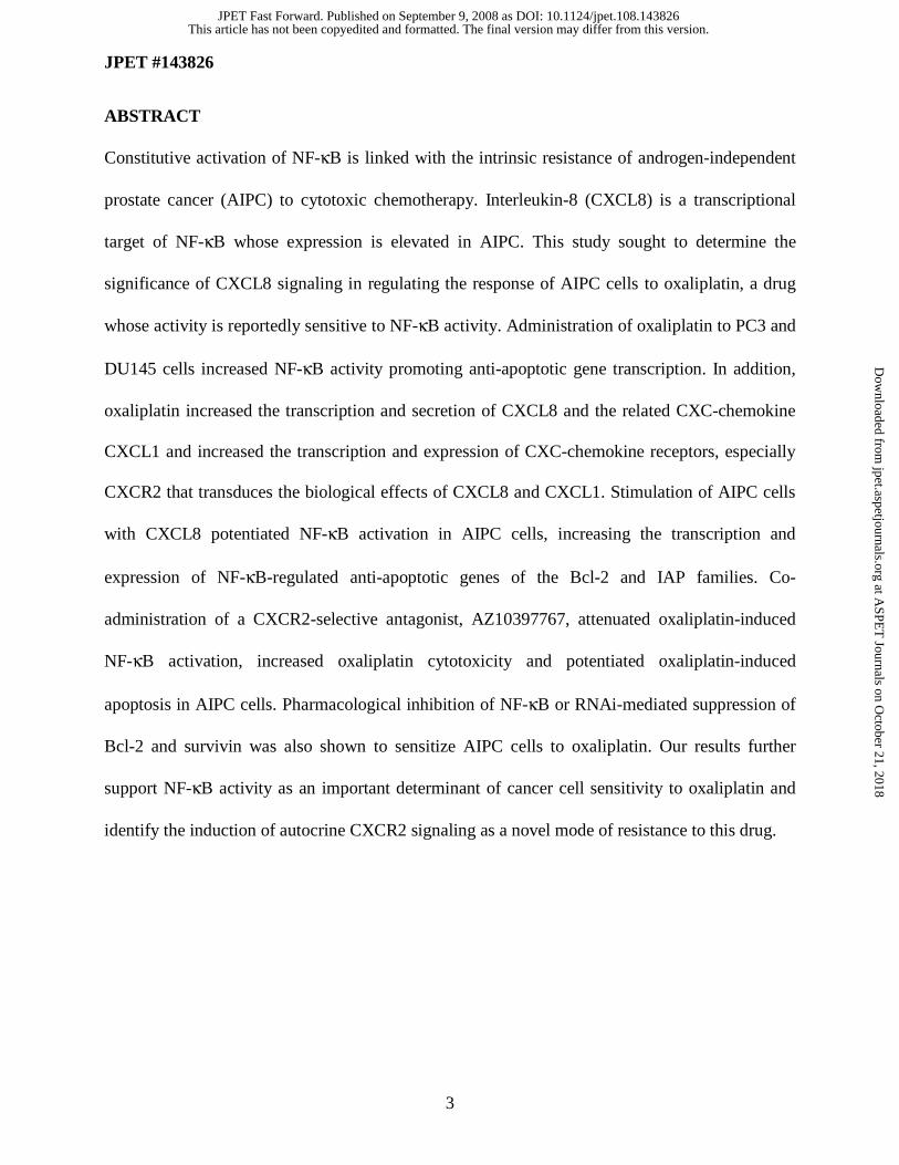
ABSTRACT
Constitutive activation of NF-κB is linked with the intrinsic resistance of androgen-independent
prostate cancer (AIPC) to cytotoxic chemotherapy. Interleukin-8 (CXCL8) is a transcriptional
target of NF-κB whose expression is elevated in AIPC. This study sought to determine the
significance of CXCL8 signaling in regulating the response of AIPC cells to oxaliplatin, a drug
whose activity is reportedly sensitive to NF-κB activity. Administration of oxaliplatin to PC3 and
DU145 cells increased NF-κB activity promoting anti-apoptotic gene transcription. In addition,
oxaliplatin increased the transcription and secretion of CXCL8 and the related CXC-chemokine
CXCL1 and increased the transcription and expression of CXC-chemokine receptors, especially
CXCR2 that transduces the biological effects of CXCL8 and CXCL1. Stimulation of AIPC cells
with CXCL8 potentiated NF-κB activation in AIPC cells, increasing the transcription and
expression of NF-κB-regulated anti-apoptotic genes of the Bcl-2 and IAP families. Co-
administration of a CXCR2-selective antagonist, AZ10397767, attenuated oxaliplatin-induced
NF-κB activation, increased oxaliplatin cytotoxicity and potentiated oxaliplatin-induced
apoptosis in AIPC cells. Pharmacological inhibition of NF-κB or RNAi-mediated suppression of
Bcl-2 and survivin was also shown to sensitize AIPC cells to oxaliplatin. Our results further
support NF-κB activity as an important determinant of cancer cell sensitivity to oxaliplatin and
identify the induction of autocrine CXCR2 signaling as a novel mode of resistance to this drug.
This article has not been copyedited and formatted. The final version may differ from this version.JPET Fast Forward. Published on September 9, 2008 as DOI: 10.1124/jpet.108.143826
at ASPE
T Journals on O
ctober 21, 2018jpet.aspetjournals.org
Dow
nloaded from

JPET #143826
4
INTRODUCTION
Interleukin-8 (CXCL8) is a pro-inflammatory CXC-chemokine whose expression is
primarily regulated by the AP-1 and NF-κB transcription factors (Brat et al., 2005). Over-
expression of this chemokine has been detected in the serum of patients with metastatic prostate
cancer (CaP) (Veltri et al., 1999; McCarron et al., 2002) while colorimetric in situ hybridization
has reported elevated expression of CXCL8 in the tumor cells of androgen-independent CaP
(AIPC) tissue (Uehara et al., 2005). Recently, we and others have demonstrated elevated CXCL8
expression and CXCL8 receptor expression in cancer cells of prostate biopsy tissue (Murphy et
al., 2005; Huang et al., 2005). Using immunohistochemistry, we determined that the intensity of
CXCL8, CXCR1 and CXCR2 staining increased with stage of disease, reaching a maximal level
in AIPC. The concurrent expression of the ligand and its receptors suggests that CaP cells are
subject to a continuous autocrine CXCL8 signaling stimulus in situ. Significantly, increased
expression of CXCL8 has been correlated with the angiogenesis, tumorigenicity and incidence of
lymph node metastasis arising from orthotopic or xenograft implantation of human AIPC cells in
athymic nude mice (Inoue et al., 2000; Kim et al., 2001), suggesting that de-regulation of this
chemokine in patients may have functional significance with regard to disease progression.
Increasingly, monitoring CXCL8 expression in cancer patients has been used as a
prognostic marker in assessing patient response to chemotherapy. In ovarian cancer, elevated
serum CXCL8 levels identify those patients with a high residual tumor burden following
paclitaxel-therapy (Uslu et al., 2005), while high levels of CXCL8 expression in the peritoneal
fluid and serum correlate with a poor initial response to paclitaxel chemotherapy (Penson et al.,
2000). Similarly, decreased CXCL8 serum levels have been described as an indicator of response
to chemotherapy in stage IV melanoma (Brennecke et al., 2005) and non-small cell lung cancer
This article has not been copyedited and formatted. The final version may differ from this version.JPET Fast Forward. Published on September 9, 2008 as DOI: 10.1124/jpet.108.143826
at ASPE
T Journals on O
ctober 21, 2018jpet.aspetjournals.org
Dow
nloaded from
JPET #143826
5
patients (Orditura et al., 2002) while reductions in intra-tumoral CXCL8 expression has been
reported in esophageal adenocarcinoma patients exhibiting a complete pathological response to
chemotherapy (Abdel-Latif et al., 2005). Chemotherapy agents have been shown to directly
regulate CXCL8 transcription in cancer cells. Paclitaxel increases CXCL8 transcription and
secretion in ovarian, breast and lung cancer cell lines (Uslu et al., 2005; Collins et al., 2000).
Similarly, administration of adriamycin and 5-fluoro-2’-deoxyuridine to breast cancer cells
(DeLarco et al., 2001), the addition of 5-FU to oral cancer cells (Tamatani et al., 2004),
doxorubicin addition to small cell lung cancer cells (Shibakura et al., 2003) and dacarbazine
administration to melanoma cells (Lev et al., 2003) all result in increased CXCL8 expression.
However, the significance of this chemokine in modulating the response of cancer cells to
chemotherapy is less well understood. Tumour necrosis factor-related apoptosis inducing ligand
(TRAIL)-mediated increases in CXCL8 expression attenuate cell death as a consequence of
decreased DR4 expression and reduced caspase-8 activation in ovarian carcinoma cells
(Abdollahi et al., 2003). We have also demonstrated that CXCL8 signaling regulates the
transcription of the native caspase-8 inhibitory protein, c-FLIP to attenuate TRAIL-induced
apoptosis in prostate cancer cells (Wilson et al., 2008). Conversely, the increased expression of
CXCL8 was not shown to affect the sensitivity of osteosarcoma cells to paclitaxel (Duan et al.,
2002).
The constitutive activity of NF-κB detected in AIPC cell lines and tissue is proposed to
underpin the poor response of this disease to chemotherapy (Fradet et al., 2004; Sweeney et al.,
2004). Since CXCL8 transcription is regulated by NF-κB (Brat et al., 2005), our study aimed to
determine whether this CXC-chemokine may contribute to the resistance of AIPC cells to
cytotoxic chemotherapeutic agents. We report our findings that the expression of CXCL8, a
This article has not been copyedited and formatted. The final version may differ from this version.JPET Fast Forward. Published on September 9, 2008 as DOI: 10.1124/jpet.108.143826
at ASPE
T Journals on O
ctober 21, 2018jpet.aspetjournals.org
Dow
nloaded from
JPET #143826
6
related CXC-chemokine CXCL1 and their signaling competent receptors CXCR1 and CXCR2
are each increased in AIPC cells in response to administration of the platinum agent, oxaliplatin,
a chemotherapy agent whose activity has been shown to be modulated by NF-κB activity
(Rakitina et al., 2003). In addition, we demonstrate that oxaliplatin-induced CXCR2 signaling
potentiates NF-κB activation and potentiates anti-apoptotic gene expression. Consistent with a
role for CXCL8signaling in promoting cellular resistance to a chemical stress, we further show
that inhibition of CXCR2 signaling and its downstream effectors, NF-κB, Bcl-2 and survivin
increases the sensitivity of AIPC cells to undergo oxaliplatin-induced apoptosis. These findings
support our recent characterization of CXCL8 signaling in mediating the attenuated response and
reduced sensitivity of hypoxic AIPC cells to etoposide (Maxwell et al., 2007).
This article has not been copyedited and formatted. The final version may differ from this version.JPET Fast Forward. Published on September 9, 2008 as DOI: 10.1124/jpet.108.143826
at ASPE
T Journals on O
ctober 21, 2018jpet.aspetjournals.org
Dow
nloaded from
JPET #143826
7
METHODS
Cell Culture. PC3 and DU145 cells were sourced and cultured as previously described
(Maxwell et al., 2007; MacManus et al., 2007).
Chemicals. Chemicals were sourced from Sigma Chemical Company (St. Louis, MO) unless
otherwise stated. BAY 11-7082 and SC-514 were purchased from Calbiochem (La Jolla, CA).
Oxaliplatin (L-OHP) (Woynarowski et al., 2000) was obtained from the Bridgewater
Chemotherapy Suite, Belfast City Hospital. AZ10397767 (AZ767) was kindly provided by Dr.
Simon T. Barry and Dr. David Blakey (AstraZeneca, Alderley Park, Cheshire, UK) (Walters et
al., 2007).
Immunoblotting. Protein was prepared, resolved and blotted as previously described (Murphy
et al, 2005; MacManus et al., 2007). Membranes were probed with monoclonal antibodies to anti-
CXCL8 (1:150 dilution) (Abcam Ltd., Cambridgeshire, UK), anti-CXCR1 (1:500 dilution) or
anti-CXCR2 (1:250 dilution) (Biosource, Camirillo, CA) anti-BCl2 (1:500 dilution), anti-Survivin
(1:1000 dilution), (all from Cell Signaling Technology, Beverly, MA) and Poly(ADP-ribose)
polymerase (1:500 dilution) (PARP; e-biosciences, San Diego, CA) overnight at 4oC. Following
washing in TBS / 0.1% Tween, membranes were incubated with HRP-conjugated secondary
antibodies (Amersham Life Sciences). Specific staining was detected using chemiluminescence
(Supersignal, Pierce, Rockford, IL or ECL plus Amersham, Buckinghamshire, UK). Equal
loading was assessed using a GAPDH mouse monoclonal primary antibody.
This article has not been copyedited and formatted. The final version may differ from this version.JPET Fast Forward. Published on September 9, 2008 as DOI: 10.1124/jpet.108.143826
at ASPE
T Journals on O
ctober 21, 2018jpet.aspetjournals.org
Dow
nloaded from
JPET #143826
8
ELISA. Cells (1x10⁵ cells/well) were incubated overnight at 37oC in a humidified 5% CO2
atmosphere and replenished in serum free RPMI 1640 medium prior to treatment with
oxaliplatin. Cell media was collected at indicated times, processed and subjected to specific
ELISA. CXCL8 levels were measured using the Pelikine CompactTM CXCL8Elisa Kit
(SanquinReagents, Amsterdam, The Netherlands) while CXCL1 levels were determined using
the Quantikine® kit (R&D Systems, Abingdon, UK). The manufacturer’s instructions were
employed in the application of each ELISA kit.
Electromobility Shift Assays. Nuclear extracts (8μg of protein) were incubated with 35,000 cpm
of a 22-base pair oligonucleotide containing the NF-κB consensus sequence, which had been
previously end-labeled with [γ-32P] ATP (10mCi/mmol) by T4 polynucleotide kinase.
Incubations were performed for 30 min at room temperature in the presence of 2μl of poly(dI.dC)
and 100mM Tris-HCl, pH 7.5, containing 10mM EDTA, 50mM DTT, 40% (v/v) glycerol. The
NF-κB complexes were resolved on 5% acrylamide gels and the migration of NF-κB complexes
determined by detection of radioactivity on the gel by autoradiography.
Luciferase Reporter Assays. Cells (1 x10⁵ cells/well) were seeded in 6-well plates and incubated
in RPMI 1640 medium at 37 ˚C for 48 h. Cells were then transfected using GeneJuice (Merck
Chemicals) according to manufacturer’s protocol incorporating 2µg pGL3 basic vector (Promega,
Madison, WI) or 2µg of an NF-κB-LUC-pGL3 plasmid (kindly provided by Dr. James Purcell,
QUB). Cells were also co-transfected with 0.2µg of a Renilla luciferase plasmid as a transfection
control for pGL3 and NF-κB. Transfected cells were incubated for a further 24 h prior to drug
addition. Either oxaliplatin or the CXC-chemokine was added for the desired time and the
This article has not been copyedited and formatted. The final version may differ from this version.JPET Fast Forward. Published on September 9, 2008 as DOI: 10.1124/jpet.108.143826
at ASPE
T Journals on O
ctober 21, 2018jpet.aspetjournals.org
Dow
nloaded from
JPET #143826
9
samples were analyzed by luciferase assay using the Promega Dual Luciferase assay kit
(Promega, Madison, WI) according to manufacturer’s protocol.
Quantitative Real-Time PCR Analysis. RNA was harvested from cultured cells using
RNAStat60TM (Biogenesis Ltd) according to manufacturer’s instructions and cDNA was
synthesised from 2µg total RNA by priming with random hexamers (Invitrogen™ Life
Technologies, Paisley, Scotland) and reverse transcribing with MMLV reverse transcriptase
(Invitrogen™ Life Technologies, Paisley, Scotland), as per the manufacturer’s protocol.
Quantitative real time PCR (qPCR) analysis was performed on the DNA Engine Opticon®2
Continuous Fluorescence Detector (MJ Research, Bio-Rad Laboratories, Inc. Waltham, MA)
with product amplification determined by SYBR Green 1 fluorescence detection (Finnzymes, Oy,
Espo, Finland). cDNA was synthesized as described previously (Maxwell et al., 2007). The
forward and reverse primers used were: CXCL8 Forward: ATg ACT TCC AAg CTg gCC gTg g;
CXCL8 Reverse: CAT AAT TTC TgT gTT ggC gCA gTg Tgg (Invitrogen™ Life Technologies,
Paisley, Scotland); BCl2 Forward: AAA ggA CCT gAT CAT Tgg gg; BCl2 Reverse: CAA CTC
TTT TCC TCC CAC CA; Survivin Forward: TCT gCT TCA Agg AgC Tgg AA; Survivin
Reverse: CgC ACT TTC TTC gCA gTT TC; CXCR1 Forward: TgC ATC AgT gTg gAC CgT
TA; CXCR1 Reverse: TgT CAT TTC CCA ggA CCT CA; CXCR2 Forward: TgC ATC AgT
gTg gAC CgT TA; CXCR2 Reverse: CCg CCA gTT TgC TgT ATT g; CXCL1 Forward CCC
AAg AAC ATC CAA AgT gTC A; CXCL1 Reverse gTg gCT ATg ACT TCg gTT Tg; 18S
Forward CAT TCg TAT TgC gCC gCT A; 18S Reverse CgA Cgg TAT CTg ATC gTC. (MWG
Biotech AG, Ebersberg, Germany). The qPCR reaction consisted of 1µl undiluted cDNA,
0.225μM final concentration of forward and reverse primers and 2 X SYBR Green 1 master-mix
This article has not been copyedited and formatted. The final version may differ from this version.JPET Fast Forward. Published on September 9, 2008 as DOI: 10.1124/jpet.108.143826
at ASPE
T Journals on O
ctober 21, 2018jpet.aspetjournals.org
Dow
nloaded from
JPET #143826
10
(Finnzymes, Oy, Espoo, Finland). Standard cycling procedures were employed with an
annealing temperature of 55ºC for all primer pairs tested. Specific amplicon formation with each
primer pair was confirmed by melt curve analysis. Gene expression was quantified relative to an
18S housekeeping gene.
Cell Count Analysis. Cells (1 x 10⁵ cells/well) were allowed to adhere overnight. Media was
replaced with fresh RPMI 1640 medium and treated with a range of concentrations of oxaliplatin
in the absence or presence of AZ10397767 (20nM) or BAY 11-7082 (1μM). Cells were
incubated at 37oC in a humidified 5% CO2 atmosphere for 72 h, then trypsinized and counted in
triplicate using a Coulter® ZTM Series particle count and size analyzer (Beckman Coulter, Miami,
FL) at threshold values of TU 21 and TL 5. Cell numbers were normalized to control values and
statistical analyses of the data performed using GraphPad PRISM 3.0 software.
siRNA strategy. Cells were seeded at 5 x 10⁵ per P90 plate in Optimem 1 (Invitrogen)-10%
(v/v) FCS medium and allowed to grow to 50% confluency. SiRNA oligonucleotide sequences
were designed as follows – Bcl2 Targeted CAg CUg CAC CUg ACg CCC UUC; Bcl2 Scrambled
CCU UCA gCC gCA UgC UgC CCA; Survivin Targeted AAC gAg CCA gAC UUg gCC CA;
Survivin Scrambled ACU ggC AAU CCA gCA gCC. SiRNA transfections were conducted by
incubating cells in unsupplemented Optimem 1 medium using oligofectamine reagent
(Invitrogen) for 4 h at 37oC, following which cells were replenished in 20% (v/v) FCS-enriched
medium. The concentration of oligonucleotides employed and the period of incubation for each
experiment is described in the relevant textual description of results and the corresponding figure
legend.
This article has not been copyedited and formatted. The final version may differ from this version.JPET Fast Forward. Published on September 9, 2008 as DOI: 10.1124/jpet.108.143826
at ASPE
T Journals on O
ctober 21, 2018jpet.aspetjournals.org
Dow
nloaded from
JPET #143826
11
FACS Analysis. Cells (5 x 10⁴ cells) were seeded in a 6-well tissue culture plate, allowed to
adhere overnight, then replenished in fresh RPMI 1640 medium. Cells were treated with
increasing concentrations of oxaliplatin and incubated for a further 72 h prior to fixation in 100%
ethanol. Cellular DNA profile was evaluated by propidium iodide staining of cells using an
EPICS XL Flow Cytometer (Beckman Coulter). In some experiments, oxaliplatin was
administered in the presence of 20nM AZ10397767 or 1µM BAY 11-7082.
Statistical Analysis. The nature of the interaction between oxaliplatin and AZ10397767 was
determined by calculating the R index (RI), using the methods recently described (Longley et al.,
2004). Differences observed in comparing apoptosis levels observed in control and drug-treated
groups were analyzed for statistical significance using a two-tailed students-t-test comparison
(Prism 3.0 software).
This article has not been copyedited and formatted. The final version may differ from this version.JPET Fast Forward. Published on September 9, 2008 as DOI: 10.1124/jpet.108.143826
at ASPE
T Journals on O
ctober 21, 2018jpet.aspetjournals.org
Dow
nloaded from
JPET #143826
12
RESULTS
Oxaliplatin promotes NF-κB activation in AIPC cells.
Oxaliplatin induces cytotoxicity through formation of adducts with cellular DNA
(Woynarowski et al., 2000). Previously published studies suggest that the sensitivity of cancer
cell lines to oxaliplatin is modulated by the level of constitutive NF-κB activity (Rakitina et al.,
2003). An electromobility shift assay (EMSA) and luciferase reporter assays were each
conducted to determine whether oxaliplatin promoted NF-κB activation in AIPC cells. Initial
studies conducted in the PC3 cell line confirmed that administration of 1 µM oxaliplatin resulted
in a rapid increase in the DNA binding activity of NF-κB (Fig. 1a). Subsequently, luciferase
reporter assays confirmed an increased level of NF-κB transcriptional activity in two AIPC cell
lines, PC3 and DU145 cells (Fig. 1b). Furthermore, using real-time PCR analysis, we observed
increased mRNA transcript levels for two representative NF-κB target genes, Bcl-2 and survivin
following exposure of PC3 and DU145 cells to 1μM oxaliplatin (Fig. 1c, left and right panels,
respectively). In addition, oxaliplatin was also shown to increase Bcl-2 and survivin protein
expression in PC3 (Fig. 1c, left panel) and DU145 cells (Fig. 1c, right panel).
Oxaliplatin potentiates CXCL8 and CXC-chemokine receptor expression in PC3 cells.
Transcription of the CXCL8 gene is regulated by NF-κB. Using a qPCR protocol,
oxaliplatin was shown to induce a rapid and sustained increase in the CXCL8 mRNA transcript
level in PC3 and DU145 cells (Fig. 2a, left panel). Furthermore, oxaliplatin treatment induced the
expression of the related CXC-chemokine, CXCL1 in each of these CaP cell lines (Fig. 2a, right
panel). This induction of CXCL8 and CXCL1 by oxaliplatin was attenuated by addition of the
This article has not been copyedited and formatted. The final version may differ from this version.JPET Fast Forward. Published on September 9, 2008 as DOI: 10.1124/jpet.108.143826
at ASPE
T Journals on O
ctober 21, 2018jpet.aspetjournals.org
Dow
nloaded from
JPET #143826
13
NF-κB inhibitor, BAY-11-7082 (Fig. 2b), suggesting that NF-κB activation is the predominant
factor underpinning oxaliplatin-induced transcriptional regulation of these CXC-chemokines.
Immunoblotting also confirmed a time-dependent increase in the intracellular CXCL8 protein
level in response to 1 μM oxaliplatin (Fig. 2c) while ELISA-based analysis of PC3 or DU145 cell
culture medium revealed an increased level of CXCL8and CXCL1 secretion from each of these
CaP cell lines following exposure to 1 μM oxaliplatin for 3h (Fig. 2d).
Significantly, the administration of oxaliplatin also increased the expression of CXCR1
and CXCR2, the receptors that mediate the biological activity of both CXCL8 and CXCL1. The
increase in the mRNA transcript level detected for CXCR2 was more sustained and greater in
magnitude than that detected for CXCR1 in both the PC3 and DU145 cells in response to 1 μM
oxaliplatin, respectively (Fig. 2e). Immunoblotting also confirmed increased CXCR1 and
CXCR2 receptor expression in the PC3 and DU145 cells following addition of oxaliplatin,
especially that of the CXCR2 receptor (Fig. 2f). Consequently, the potentiation of CXCL8 and
CXCL1 secretion and the increase in CXCR2 expression suggests that exposure to oxaliplatin
increases the magnitude of the autocrine CXC-chemokine signaling stimulus that AIPC cells are
subject to and furthermore, promotes a selective increase in CXCR2-mediated signaling.
CXC-chemokine signaling potentiates NF-κB transcription in AIPC cells.
Further experiments were conducted to characterize the effect of CXCL8 signaling upon
established cell survival pathways, focusing on whether NF-κB was a downstream effector of
CXCL8signaling in these AIPC cells. PC3 cells were stimulated with 3nM recombinant human-
CXCL8 (rCXCL8) and an EMSA used to determine whether this stimulus increased the DNA
binding activity of NF-κB. Increased complexing of NF-κB with DNA was detected within 20
This article has not been copyedited and formatted. The final version may differ from this version.JPET Fast Forward. Published on September 9, 2008 as DOI: 10.1124/jpet.108.143826
at ASPE
T Journals on O
ctober 21, 2018jpet.aspetjournals.org
Dow
nloaded from
JPET #143826
14
min of adding rCXCL8, reaching a peak level of interaction at 40 min (Fig. 3a, left panel).
Analysis over a longer time-course also confirmed that NF-κB complexing with DNA was
detected within 1 h of stimulation with rCXCL8 and identified a secondary increase in the DNA
binding of this transcription factor 4 h post-addition of rCXCL8 (Fig. 3a, right panel). The
secondary increase in NF-κB complexing was lower in intensity than the initial response to
exogenous rCXCL8. We also examined the receptor selectivity underpinning the CXCL8-
induced activation of NF-κB and the CXCL8-induced potentiation of Bcl-2 expression. Blockade
of CXCR2 receptor signaling was effected using a small-molecule CXCR2 receptor antagonist,
AZ10397767, which when administered at a concentration (20nM) selectively antagonizes
CXCR2 activation but has no activity on the CXCR1 receptor in vitro (Walters et al., 2008). In
EMSA analysis, CXCL8-promoted DNA binding of NF-κB was attenuated in the presence of
AZ10397767 (Fig. 3b). Luciferase reporter assays were also conducted to demonstrate that CXC-
chemokine signaling not only potentiated nuclear translocation and DNA-binding of NF-κB but
also induced the transcriptional activity of this factor in AIPC cells. Stimulation with either 3nM
CXCL8 (Fig. 3c, left panel) or 3nM CXCL1 (Fig. 3c, right panel), was observed to increase NF-
κB-promoted luciferase activity in both PC3 and DU145 cells. Consistent with the EMSA
analysis, the CXC-chemokine promoted NF-κB transcriptional activation was attenuated by co-
administration of the CXCR2 antagonist, AZ10397767.
CXCL8 signaling potentiates anti-apoptotic gene transcription in AIPC cells.
qPCR analysis was conducted to determine whether CXCL8 signaling increased the
transcriptional of established, NF-κB-regulated anti-apoptotic genes. Bcl-2 mRNA transcript
levels were shown to increase by a factor in excess of 3-fold and 2.5-fold within 1 h of adding
This article has not been copyedited and formatted. The final version may differ from this version.JPET Fast Forward. Published on September 9, 2008 as DOI: 10.1124/jpet.108.143826
at ASPE
T Journals on O
ctober 21, 2018jpet.aspetjournals.org
Dow
nloaded from
JPET #143826
15
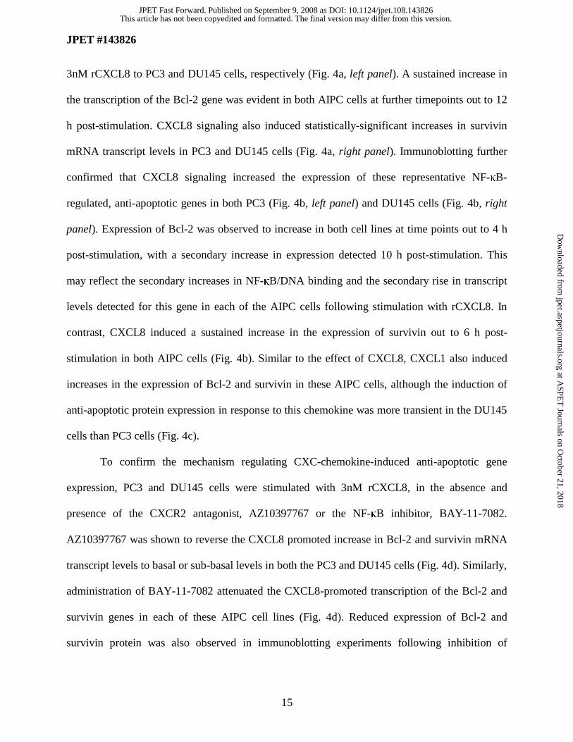
3nM rCXCL8 to PC3 and DU145 cells, respectively (Fig. 4a, left panel). A sustained increase in
the transcription of the Bcl-2 gene was evident in both AIPC cells at further timepoints out to 12
h post-stimulation. CXCL8 signaling also induced statistically-significant increases in survivin
mRNA transcript levels in PC3 and DU145 cells (Fig. 4a, right panel). Immunoblotting further
confirmed that CXCL8 signaling increased the expression of these representative NF-κB-
regulated, anti-apoptotic genes in both PC3 (Fig. 4b, left panel) and DU145 cells (Fig. 4b, right
panel). Expression of Bcl-2 was observed to increase in both cell lines at time points out to 4 h
post-stimulation, with a secondary increase in expression detected 10 h post-stimulation. This
may reflect the secondary increases in NF-κB/DNA binding and the secondary rise in transcript
levels detected for this gene in each of the AIPC cells following stimulation with rCXCL8. In
contrast, CXCL8 induced a sustained increase in the expression of survivin out to 6 h post-
stimulation in both AIPC cells (Fig. 4b). Similar to the effect of CXCL8, CXCL1 also induced
increases in the expression of Bcl-2 and survivin in these AIPC cells, although the induction of
anti-apoptotic protein expression in response to this chemokine was more transient in the DU145
cells than PC3 cells (Fig. 4c).
To confirm the mechanism regulating CXC-chemokine-induced anti-apoptotic gene
expression, PC3 and DU145 cells were stimulated with 3nM rCXCL8, in the absence and
presence of the CXCR2 antagonist, AZ10397767 or the NF-κB inhibitor, BAY-11-7082.
AZ10397767 was shown to reverse the CXCL8 promoted increase in Bcl-2 and survivin mRNA
transcript levels to basal or sub-basal levels in both the PC3 and DU145 cells (Fig. 4d). Similarly,
administration of BAY-11-7082 attenuated the CXCL8-promoted transcription of the Bcl-2 and
survivin genes in each of these AIPC cell lines (Fig. 4d). Reduced expression of Bcl-2 and
survivin protein was also observed in immunoblotting experiments following inhibition of
This article has not been copyedited and formatted. The final version may differ from this version.JPET Fast Forward. Published on September 9, 2008 as DOI: 10.1124/jpet.108.143826
at ASPE
T Journals on O
ctober 21, 2018jpet.aspetjournals.org
Dow
nloaded from
JPET #143826
16
CXCL8-signaling using AZ10397767 or BAY-11-7082 (data not shown). Accordingly, our data
indicates that the CXCR2 receptor primarily couples CXCL8 to NF-κB activation and that
CXCR2/NF-κB signaling underpins CXC-chemokine-promoted regulation of anti-apoptotic
protein expression in these cells.
Inhibition of CXCR2 signaling attenuates oxaliplatin-induced activation of NF-κB.
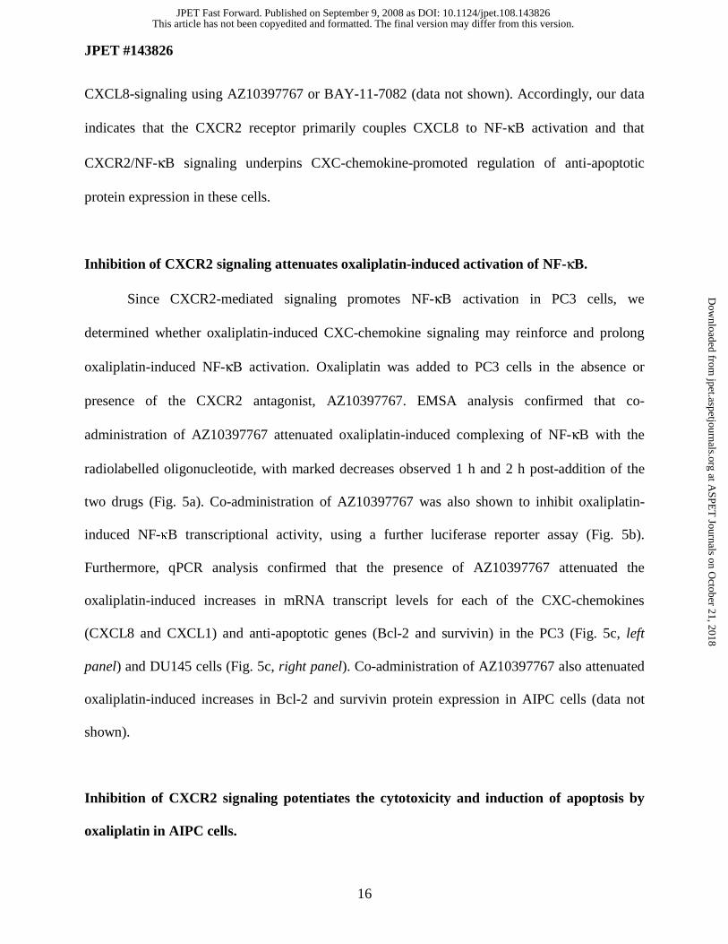
Since CXCR2-mediated signaling promotes NF-κB activation in PC3 cells, we
determined whether oxaliplatin-induced CXC-chemokine signaling may reinforce and prolong
oxaliplatin-induced NF-κB activation. Oxaliplatin was added to PC3 cells in the absence or
presence of the CXCR2 antagonist, AZ10397767. EMSA analysis confirmed that co-
administration of AZ10397767 attenuated oxaliplatin-induced complexing of NF-κB with the
radiolabelled oligonucleotide, with marked decreases observed 1 h and 2 h post-addition of the
two drugs (Fig. 5a). Co-administration of AZ10397767 was also shown to inhibit oxaliplatin-
induced NF-κB transcriptional activity, using a further luciferase reporter assay (Fig. 5b).
Furthermore, qPCR analysis confirmed that the presence of AZ10397767 attenuated the
oxaliplatin-induced increases in mRNA transcript levels for each of the CXC-chemokines
(CXCL8 and CXCL1) and anti-apoptotic genes (Bcl-2 and survivin) in the PC3 (Fig. 5c, left
panel) and DU145 cells (Fig. 5c, right panel). Co-administration of AZ10397767 also attenuated
oxaliplatin-induced increases in Bcl-2 and survivin protein expression in AIPC cells (data not
shown).
Inhibition of CXCR2 signaling potentiates the cytotoxicity and induction of apoptosis by
oxaliplatin in AIPC cells.
This article has not been copyedited and formatted. The final version may differ from this version.JPET Fast Forward. Published on September 9, 2008 as DOI: 10.1124/jpet.108.143826
at ASPE
T Journals on O
ctober 21, 2018jpet.aspetjournals.org
Dow
nloaded from
JPET #143826
17
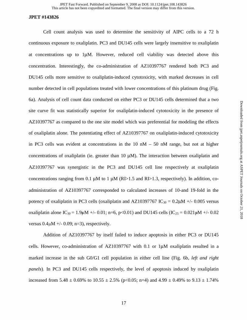
Cell count analysis was used to determine the sensitivity of AIPC cells to a 72 h
continuous exposure to oxaliplatin. PC3 and DU145 cells were largely insensitive to oxaliplatin
at concentrations up to 1μM. However, reduced cell viability was detected above this
concentration. Interestingly, the co-administration of AZ10397767 rendered both PC3 and
DU145 cells more sensitive to oxaliplatin-induced cytotoxicity, with marked decreases in cell
number detected in cell populations treated with lower concentrations of this platinum drug (Fig.
6a). Analysis of cell count data conducted on either PC3 or DU145 cells determined that a two
site curve fit was statistically superior for oxaliplatin-induced cytotoxicity in the presence of
AZ10397767 as compared to the one site model which was preferential for modeling the effects
of oxaliplatin alone. The potentiating effect of AZ10397767 on oxaliplatin-induced cytotoxicity
in PC3 cells was evident at concentrations in the 10 nM – 50 nM range, but not at higher
concentrations of oxaliplatin (ie. greater than 10 μM). The interaction between oxaliplatin and
AZ10397767 was synergistic in the PC3 and DU145 cell line respectively at oxaliplatin
concentrations ranging from 0.1 μM to 1 μM (RI>1.5 and RI>1.3, respectively). In addition, co-
administration of AZ10397767 corresponded to calculated increases of 10-and 19-fold in the
potency of oxaliplatin in PC3 cells (oxaliplatin and AZ10397767 IC30 = 0.2μM +/- 0.005 versus
oxaliplatin alone IC30 = 1.9μM +/- 0.01; n=6, p<0.01) and DU145 cells (IC25 = 0.021μM +/- 0.02
versus 0.4μM +/- 0.09; n=3), respectively.
Addition of AZ10397767 by itself failed to induce apoptosis in either PC3 or DU145
cells. However, co-administration of AZ10397767 with 0.1 or 1μM oxaliplatin resulted in a
marked increase in the sub G0/G1 cell population in either cell line (Fig. 6b, left and right
panels). In PC3 and DU145 cells respectively, the level of apoptosis induced by oxaliplatin
increased from 5.48 ± 0.69% to 10.55 ± 2.5% (p<0.05; n=4) and 4.99 ± 0.49% to 9.13 ± 1.74%
This article has not been copyedited and formatted. The final version may differ from this version.JPET Fast Forward. Published on September 9, 2008 as DOI: 10.1124/jpet.108.143826
at ASPE
T Journals on O
ctober 21, 2018jpet.aspetjournals.org
Dow
nloaded from
JPET #143826
18
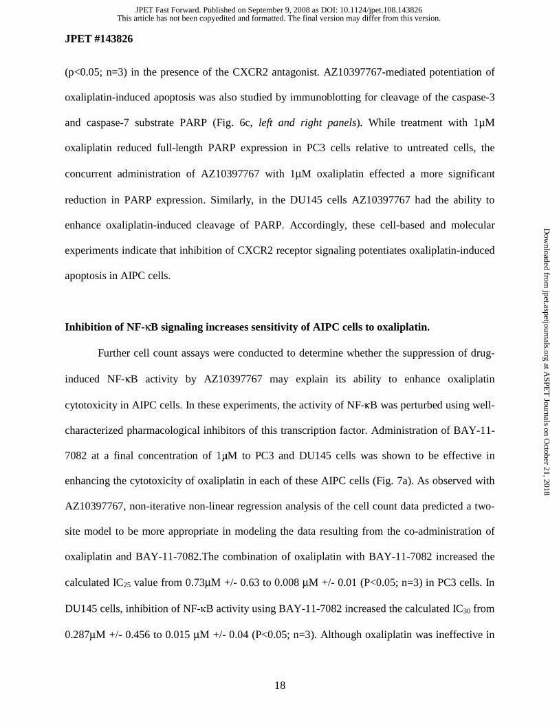
(p<0.05; n=3) in the presence of the CXCR2 antagonist. AZ10397767-mediated potentiation of
oxaliplatin-induced apoptosis was also studied by immunoblotting for cleavage of the caspase-3
and caspase-7 substrate PARP (Fig. 6c, left and right panels). While treatment with 1µM
oxaliplatin reduced full-length PARP expression in PC3 cells relative to untreated cells, the
concurrent administration of AZ10397767 with 1μM oxaliplatin effected a more significant
reduction in PARP expression. Similarly, in the DU145 cells AZ10397767 had the ability to
enhance oxaliplatin-induced cleavage of PARP. Accordingly, these cell-based and molecular
experiments indicate that inhibition of CXCR2 receptor signaling potentiates oxaliplatin-induced
apoptosis in AIPC cells.
Inhibition of NF-κB signaling increases sensitivity of AIPC cells to oxaliplatin.
Further cell count assays were conducted to determine whether the suppression of drug-
induced NF-κB activity by AZ10397767 may explain its ability to enhance oxaliplatin
cytotoxicity in AIPC cells. In these experiments, the activity of NF-κB was perturbed using well-
characterized pharmacological inhibitors of this transcription factor. Administration of BAY-11-
7082 at a final concentration of 1μM to PC3 and DU145 cells was shown to be effective in
enhancing the cytotoxicity of oxaliplatin in each of these AIPC cells (Fig. 7a). As observed with
AZ10397767, non-iterative non-linear regression analysis of the cell count data predicted a two-
site model to be more appropriate in modeling the data resulting from the co-administration of
oxaliplatin and BAY-11-7082.The combination of oxaliplatin with BAY-11-7082 increased the
calculated IC25 value from 0.73μM +/- 0.63 to 0.008 μM +/- 0.01 (P<0.05; n=3) in PC3 cells. In
DU145 cells, inhibition of NF-κB activity using BAY-11-7082 increased the calculated IC30 from
0.287μM +/- 0.456 to 0.015 μM +/- 0.04 (P<0.05; n=3). Although oxaliplatin was ineffective in
This article has not been copyedited and formatted. The final version may differ from this version.JPET Fast Forward. Published on September 9, 2008 as DOI: 10.1124/jpet.108.143826
at ASPE
T Journals on O
ctober 21, 2018jpet.aspetjournals.org
Dow
nloaded from
JPET #143826
19
inducing apoptosis in either cell line, treatment with 1µM BAY-11-7082 did induce apoptosis in
PC3 (Fig. 7b, left panel) and DU145 cells (Fig. 7b, right panel). Furthermore, the inhibition of
NF-κB activity using BAY-11-7082 further enhanced the apoptosis induced by exposure of PC3
or DU145 cells to 0.1 μM and/or 1µM oxaliplatin (Fig. 7b). These results were verified by use of
a second inhibitor of NF-κB, the IκK-2 inhibitor, SC-514 which was administered to PC3 cells at
a final concentration of 2μM. SC-514 inhibited oxaliplatin (1µM)-induced NF-κB binding in
EMSA analysis, sensitized PC3 cells to sub-μM concentrations of oxaliplatin in cell count
analysis and when co-administered with 0.1 μM oxaliplatin, SC-514 increased the accumulation
of apoptotic-cells detected by FACS analysis in the sub Go/G1 peak relative to that observed
when treated with 0.1 μM oxaliplatin alone (data not shown).
Suppression of Bcl-2 and survivin sensitizes PC3 cells to oxaliplatin.
The functional significance of the downstream transcriptional targets of NF-κB, Bcl-2 and
survivin, in underpinning the resistance of AIPC cells to oxaliplatin was studied using gene-
targeted RNAi-based approaches to selectively suppress the expression of these genes in AIPC
cells. PC3 cells were initially transfected using 50nM of a single gene-specific oligonucleotide
(oligo) against either Bcl-2 (Bcl2-T) or survivin (Sur-T) or with a scrambled oligonucleotide
(Sc), used at an identical concentration. At this concentration, transfection of PC3 cells with the
Bcl2-T oligo was shown to decrease but not abrogate Bcl-2 expression in these cells 72 h post-
transfection. In contrast, transfection with Sur-T oligonucleotide was shown to be markedly more
effective in depleting survivin expression in the PC3 cells (Figure 8a, top and bottom panels).
The effect of transiently reducing anti-apoptotic gene expression upon the induction of
apoptosis in PC3 cells in the absence and presence of oxaliplatin was determined using FACS
This article has not been copyedited and formatted. The final version may differ from this version.JPET Fast Forward. Published on September 9, 2008 as DOI: 10.1124/jpet.108.143826
at ASPE
T Journals on O
ctober 21, 2018jpet.aspetjournals.org
Dow
nloaded from
JPET #143826
20
analysis. Initially, PC3 cells were treated with either 50nM of Bcl2-T oligo, Sur-T oligo or Sc
oligo for 48 h, following which cells were exposed to increasing concentrations of oxaliplatin for
a further 72 h. In control experiments, transfection of PC3 cells using concentrations up to
200nM of the Sc oligo did not increase the level of apoptosis over that observed in control PC3
cells (data not shown).
Transfection with 50nM Bcl2-T alone increased the magnitude of the sub G0/G1 fraction
of the cell population (8.53 ± 0.57%, p<0.01, n=4) relative to both mock-transfected control (4.68
± 0.18%) and scrambled-oligo transfected cells (4.75 ± 0.18%) (Fig. 7c), suggesting that even
sub-optimal reductions in the expression of this protein initiates spontaneous apoptosis in PC3
cells. In contrast, abrogation of survivin expression using the Sur-T oligo only effected a modest
increase in the apoptotic fraction (7.55 ± 0.4% of the cell population; p<0.01), indicating that
PC3 cells are not critically dependent upon survivin for viability. Although, treatment with 1 μM
oxaliplatin alone was largely ineffective in increasing the level of apoptosis in Sc-oligo
transfected PC3 cells, oxaliplatin did increase the apoptotic cell population to 10.58% ± 0.72%
and 8.3 ± 0.4% in 50nM Bcl2-T-transfected PC3 cells (p<0.001; n=4) and 50nM Sur-T oligo-
transfected PC3 cells (p<0.05, n=4), respectively (Fig. 8b). The relative importance of these anti-
apoptotic proteins in modulating PC3 cell viability and the sensitivity of these cells to oxaliplatin
was reaffirmed using PARP cleavage as a marker of apoptosis induction. In Sc oligo-transfected
cells, treatment with 1μM oxaliplatin resulted in a modest decrease in PARP expression. In
contrast, transfection of PC3 cells with 50nM Bcl2-T had a pronounced effect in promoting
PARP degradation in both the absence and presence of 1 μM oxaliplatin, shown by the reduced
expression of the full-length protein. Suppression of survivin alone had no effect on PARP
expression but did potentiate the degradation of PARP observed when cells were co-treated with
This article has not been copyedited and formatted. The final version may differ from this version.JPET Fast Forward. Published on September 9, 2008 as DOI: 10.1124/jpet.108.143826
at ASPE
T Journals on O
ctober 21, 2018jpet.aspetjournals.org
Dow
nloaded from
JPET #143826
21
1 μM oxaliplatin (Fig. 8c). Therefore, decreasing Bcl-2 expression in PC3 cells appears to be of
major importance in sensitizing cells to oxaliplatin while suppressing survivin expression
contributes to an enhanced response.
To determine the effect of increasing the effectiveness of targeting Bcl-2 expression, PC3
cells were treated with increasing concentrations of the Bcl2-T oligo. Transfection with 200nM
of this oligo was shown to deplete endogenous Bcl-2 expression in these cells in immunoblots
conducted on protein lysates extracted from transfected cells (Fig. 8d). Suppression of Bcl-2
expression alone was observed to co-incide with a spontaneous loss of full-length PARP
expression and the emergence of PARP cleavage, indicative of apoptosis. Furthermore, use of
this Bcl2-T oligo at this higher concentration induced a more marked increase in the apoptotic
cell fraction detected by FACS analysis, increasing the level to 20.4 ± 2.9% (p<0.001) (Fig. 8e).
A further increase in the apoptotic cell fraction was observed following the addition of 1μM
oxaliplatin to 200nM Bcl2-T-transfected PC3 cells. However, due to the high level of
spontaneous apoptosis resulting from treatment with the Bcl2-T oligo at this higher
concentration, there was no statistically significant enhancement when 200nM Bcl2-T was
combined with 1μM oxaliplatin.
Concurrent suppression of Bcl-2 and survivin potentiates L-OHP cytotoxicity in PC3 cells.
Bcl-2 primarily antagonizes Bax/Bak-induced mitochondrial membrane
permeabilization while the survivin/IAP gene family directly inhibits the activation of
downstream caspases, including caspase-9 (Reed, 2001). To determine whether oxaliplatin and
CXCL8-promoted increases in Bcl-2 and survivin expression describe two complementary
mechanisms of resistance to oxaliplatin, we determined the effect of concurrently suppressing
This article has not been copyedited and formatted. The final version may differ from this version.JPET Fast Forward. Published on September 9, 2008 as DOI: 10.1124/jpet.108.143826
at ASPE
T Journals on O
ctober 21, 2018jpet.aspetjournals.org
Dow
nloaded from
JPET #143826
22
Bcl-2 and survivin expression on oxaliplatin-induced apoptosis. The sensitivity of Bcl2-T/Sur-T
co-transfected cells to undergo apoptosis was compared to the effect of transfecting PC3 cells
with equivalent concentrations of either the Bcl2-T or Sur-T oligos alone or with a Sc oligo (Fig.
8b). All transfections were conducted using a final oligonucleotide concentration of 50nM, given
our observation of high levels of spontaneous apoptosis when anti-apoptotic gene oligos were
used at increased concentrations. Co-transfection with 50nM Bcl2-T and 50nM Sur-T oligos was
shown to induce a more pronounced accumulation of cells in the sub Go/G1 peak (15.0 ± 1.3%;
n=4) relative to transfection with Bcl2-T (8.53 ± 0.58 %, p<0.05) or Sur-T (7.55 ± 0.41) alone. In
the context of sensitizing PC3 cells to oxaliplatin, the combined suppression of Bcl-2 and
survivin potentiated the level of apoptosis detected in PC3 cells in response to oxaliplatin (sub
Go/G1 fraction of 18.77 ± 0.89%) in Bcl2-T/Sur-T co-transfected cells relative to that observed
in Bcl2-T (10.58 ± 0.72 %, p<0.05, n=4) or Sur-T transfected cells (8.3 ± 0.39 %, p<0.01, n=4).
This article has not been copyedited and formatted. The final version may differ from this version.JPET Fast Forward. Published on September 9, 2008 as DOI: 10.1124/jpet.108.143826
at ASPE
T Journals on O
ctober 21, 2018jpet.aspetjournals.org
Dow
nloaded from
JPET #143826
23
DISCUSSION
Deregulation of NF-κB has been proposed as a major factor contributing to the resistance
of AIPC to chemotherapy. Nuclear localization of this transcription factor, indicative of
constitutive activity, has been detected in biopsy tissues of AIPC (Fradet et al., 2004; Sweeney et
al., 2004). Further studies conducted in cell lines have shown that inhibition of NF-κB renders
CaP cells more susceptible to apoptosis mediated in part by the ability of this transcription factor
to regulate anti-apoptotic gene expression (Huang et al., 2005; Li et al., 2005). The sensitivity of
colorectal cancer cells to oxaliplatin-induced death is adversely affected by elevated NF-κB
activity (Rakitina et al., 2003), suggesting that the dysregulation of this transcription factor in
AIPC may underpin the low sensitivity of this disease to oxaliplatin (Droz et al., 2003).
We have shown that administration of oxaliplatin further potentiates NF-κB activity in
AIPC cells, increasing transcriptional regulation and expression of anti-apoptotic genes.
Furthermore, we have shown that oxaliplatin induced a dynamic process of increased CXCL8
gene transcription, intracellular protein expression and ultimately secretion from PC3 cells, that is
predominantly mediated downstream of NF-κB activation. Oxaliplatin was also shown to
increase the transcription and expression of the CXCL8 receptors, CXCR1 and CXCR2 in AIPC
cells. To our knowledge this is the first report demonstrating an effect of chemotherapy drugs
upon the expression of these CXC-chemokine receptors in cancer cells. Furthermore, these
observations are consistent with our recent demonstration that hypoxia-induced transcription of
the CXCR1 and CXCR2 receptors is mediated in part by increased NF-κB transcriptional activity
(Maxwell et al., 2007). Consequently, our demonstration of increased ligand and receptor
expression suggests that exposure of AIPC cells to oxaliplatin increases the magnitude of the
autocrine CXCL8 signaling stimulus received by these cells.
This article has not been copyedited and formatted. The final version may differ from this version.JPET Fast Forward. Published on September 9, 2008 as DOI: 10.1124/jpet.108.143826
at ASPE
T Journals on O
ctober 21, 2018jpet.aspetjournals.org
Dow
nloaded from
JPET #143826
24
To determine whether drug-induced or constitutive CXCL8 signaling may alter the
sensitivity of the cells to the chemotherapy drug, we characterized the effect of stimulating AIPC
cells with rCXCL8, focusing on the NF-κB cell survival pathway. Administration of
pharmacologically-relevant concentrations of rCXCL8 induced NF-κB activation, enhancing the
transcription and expression of the anti-apoptotic genes, Bcl-2 and survivin in AIPC cells. These
responses were attenuated by co-administration of the CXCR2 receptor antagonist, AZ10397767,
at a concentration that selectively blocks CXCR2 activation. Furthermore, pharmacological
inhibition of NF-κB attenuated the rCXCL8- induced increase in Bcl-2 expression, suggesting
that the CXCR2 receptor primarily but not exclusively couples CXCL8 to the activation of NF-
κB and transcriptional regulation of these anti-apoptotic genes in AIPC cells.
The selective increase in CXCR2 expression suggests that signaling through this receptor
may pre-dominate over CXCR1 in dictating the response of AIPC cells to the administration of
oxaliplatin. While CXCR2 binds and is activated by CXCL8, this receptor also binds several
related CXC-chemokines including the growth related oncogenes (CXCL1, CXCL2, CXCL3)
whose expression is also regulated at the level of transcription by NF-κB (Brat et al., 2005).
Consequently, we have shown that oxaliplatin treatment also induces expression of CXCL1 in
AIPC cells. As observed for CXCL8, stimulation of these AIPC cells with CXCL1-potentiates a
CXCR2-dependent activation of NF-κB and increases anti-apoptotic protein expression.
Irrespective of the ligand activating the receptor, the significance of CXCR2-induced signaling in
modulating the sensitivity of AIPC cells to oxaliplatin was illustrated by the ability of the
CXCR2 antagonist AZ10397767 to attenuate oxaliplatin-induced NF-κB activation and reduce
oxaliplatin-induced transcription of the CXCL8, CXCL1, Bcl-2 and survivin genes. Co-
This article has not been copyedited and formatted. The final version may differ from this version.JPET Fast Forward. Published on September 9, 2008 as DOI: 10.1124/jpet.108.143826
at ASPE
T Journals on O
ctober 21, 2018jpet.aspetjournals.org
Dow
nloaded from
JPET #143826
25
administration of AZ10397767 also markedly increased the sensitivity of AIPC cells to
oxaliplatin and enhanced oxaliplatin-induced apoptosis.
Oxaliplatin cytotoxicity in AIPC cells was also increased by direct targeting of NF-κB
activation or its downstream transcriptional targets. Pharmacological inhibition of NF-κB using
the BAY-11-7082 or SC-514 compounds sensitized AIPC cells to low but not high
concentrations of oxaliplatin while RNAi-mediated suppression of Bcl-2 and survivin was shown
to increase the level of oxaliplatin-induced apoptosis detected in PC3 cells. Interestingly,
reduction of Bcl-2 and survivin expression alone was shown to induce apoptosis in PC3 cells,
suggesting that this AIPC cell line may have a marked dependence on the expression of these
anti-apoptotic proteins for maintenance of cell viability. The concurrent suppression of both Bcl-
2 and survivin was also shown to result in a significantly greater induction of apoptosis in PC3
cells in response to oxaliplatin. Therefore, we propose that the potentiation of CXCL8 and
CXCL1-induced CXCR2 signaling underpins a novel and contributing mode of resistance to
oxaliplatin that is mediated at least in part by sustaining NF-κB transcription and reinforcing the
expression of anti-apoptotic proteins in AIPC cells (Fig. 8).
Prior studies have shown that the combination of oxaliplatin with the heat shock protein-
90 inhibitor 17-AAG results in a more effective suppression of cellular NF-κB activation and
promotion of anti-cancer activity (Rakitina et al., 2003). Therefore, the ability of AZ10397767 to
also suppress NF-κB activity and reduce anti-apoptotic protein expression in the presence of
oxaliplatin is a likely explanation of its capacity to increase the cytotoxicity of this drug in AIPC
cells. Our observations that co-administration of either the CXCR2 antagonist or the NF-κB
inhibitor BAY11-7082 sensitizes the AIPC cells to low rather than high concentrations of
oxaliplatin suggests that CXCL8/CXCL1-induced NF-κB-signaling in AIPC cells confers a
This article has not been copyedited and formatted. The final version may differ from this version.JPET Fast Forward. Published on September 9, 2008 as DOI: 10.1124/jpet.108.143826
at ASPE
T Journals on O
ctober 21, 2018jpet.aspetjournals.org
Dow
nloaded from
JPET #143826
26
surmountable resistance to a specific mode of oxaliplatin-induced cytotoxicity. Further detailed
studies will be required to establish the precise mechanism (mitochondrial or death receptor
mediated) by which CXCR2- and NF-κB-signaling impact on oxaliplatin-induced apoptosis in
AIPC cells. Prior studies have shown that oxaliplatin induces apoptosis in colorectal cancer cells
through the activation of caspase-8 and Bid cleavage, suggesting a role for the extrinsic apoptosis
pathway (Griffiths et al., 2004; Longley et al., 2006). Our current observations that CXCL8
signaling regulates Bcl-2 gene family expression together with our recently published data
demonstrating that this chemokine promotes a NF-κB-driven expression of the endogenous
caspase-8 inhibitor c-FLIP in CaP cells (Wilson et al., 2008) suggests that CXCL8 signaling may
attenuate the capacity of oxaliplatin to initiate mitochondrial-driven apoptosis in these cells by
altering the ratio of pro-apoptotic cleaved Bid to the mitochondrial stabilizing effect of increased
Bcl-2 expression. Furthermore, our observation that CXCL8/CXCL1 signaling increases survivin
expression in AIPC cells suggests a further mechanism downstream of the mitochondria by
which AIPC cells may withstand the apoptosis-inducing stress experienced following the
administration of oxaliplatin.
Several final issues warrant mention. Firstly, oxaliplatin is currently employed in the
treatment of metastatic colorectal cancer. Experiments conducted in our laboratory have
characterized a similar induction of CXCL8 signaling in colorectal cancer cells and demonstrated
that the inhibition of chemokine signaling also has a similar effect in sensitizing colorectal cancer
cells to oxaliplatin (C. Purcell and D. Waugh, unpublished observations). Therefore, these
ongoing studies point to a clinical relevance of CXC-chemokine signaling in determining the
response of malignant cells to oxaliplatin in this disease setting. Furthermore, in conducting
experiments in the parental p53 wild-type HCT116 and the matched p53-deficient HCT116 cells,
This article has not been copyedited and formatted. The final version may differ from this version.JPET Fast Forward. Published on September 9, 2008 as DOI: 10.1124/jpet.108.143826
at ASPE
T Journals on O
ctober 21, 2018jpet.aspetjournals.org
Dow
nloaded from
JPET #143826
27
we will be able to undertake a direct investigation of how p53 status influences the magnitude of
the CXC-chemokine promoted anti-apoptotic response in cancer cells. This has not been possible
in the current studies given the p53 deficient and p53 mutant status in the PC3 and DU145 cell
lines, respectively. Finally, we have also conducted experiments to determine whether other
platinum agents induce a similar response in prostate cancer cells. Administration of cisplatin
was also shown to induce NF-κB activity and increase CXC-chemokine expression in AIPC cells.
However, inhibition of CXCR2 signaling did not increase the sensitivity of these cells to cisplatin
(C. Wilson and D. Waugh, unpublished observations). We suspect that this reflects the inability
of this pharmacological intervention to overcome the primary mode of resistance ie. that of
mismatch repair enzyme deficiency in AIPC cells (Chen et al., 2001) that drives resistance to
cisplatin but not oxaliplatin adducts (Zdraveski et al., 2002; Chaney et al., 2004).
In summary, we indicate a role for NF-κB signaling and two of its transcriptional targets,
Bcl-2 and survivin in attenuating oxaliplatin cytotoxicity in AIPC cells. Furthermore, we report
the significance of oxaliplatin-induced CXC-chemokine signaling in reinforcing NF-κB
transcriptional activity and consequently potentiating the resistance of these cells to this platinum
drug. Accordingly, direct targeting of NF-κB, its transcriptional targets or inactivating CXCR2-
mediated signaling may be appropriate strategies to increase the effectiveness of using oxaliplatin
to treat cancer (Fig. 9). The demonstration that CXCL8 signaling potentiates anti-apoptotic
protein expression and attenuates chemotherapy-induced apoptosis further links CXCL8
signaling to another described “hallmark” of cancer cells (Hanahan and Weinberg, 2000), that of
evading apoptosis, in addition to its proposed roles in promoting angiogenesis, invasion and
metastasis.
This article has not been copyedited and formatted. The final version may differ from this version.JPET Fast Forward. Published on September 9, 2008 as DOI: 10.1124/jpet.108.143826
at ASPE
T Journals on O
ctober 21, 2018jpet.aspetjournals.org
Dow
nloaded from
JPET #143826
28
References.
Abdel-Latif MM, O'Riordan JM, Ravi N, Kelleher D and Reynolds JV (2005) Activated nuclear
factor-kappa B and cytokine profiles in the esophagus parallel tumor regression following
neoadjuvant chemoradiotherapy. Dis Esophagus 18: 246-252.
Abdollahi T, Robertson NM, Abdollahi A and Litwack G (2003) Identification of interleukin 8 as
an inhibitor of tumor necrosis factor-related apoptosis-inducing ligand-induced apoptosis in the
ovarian carcinoma cell line OVCAR3. Cancer Res 63: 4521-4526.
Brat DJ, Bellail AC and Van Meir EG (2005) The role of interleukin-8 and its receptors in
gliomagenesis and tumoral angiogenesis. Neuro-Oncology 7: 122-133.
Brennecke S, Deichmann M, Naeher H and Kurzen H (2005) Decline in angiogenic factors, such
as interleukin-8, indicates response to chemotherapy of metastatic melanoma. Melanoma Res 15:
515-522.
Chaney SG, Campbell SL, Temple B, Bassett E, Wu Y and Faldu M (2004) Protein Interations
with platinum-DNA adducts; from structure to function. J. Inorg Biochem. 98: 1551-1559.
Chen Y, Wang J, Fraig MM, Metcalf J, Turner WR, Bissada NK, Watson DK and Schweinfest
CW (2001) Defects of DNA mismatch repair in human prostate cancer. Cancer Res 61: 4112–
4121.
This article has not been copyedited and formatted. The final version may differ from this version.JPET Fast Forward. Published on September 9, 2008 as DOI: 10.1124/jpet.108.143826
at ASPE
T Journals on O
ctober 21, 2018jpet.aspetjournals.org
Dow
nloaded from
JPET #143826
29
Collins TS, Lee LF and Ting JP (2000) Paclitaxel up-regulates interleukin-8 synthesis in human
lung carcinoma through an NF-kappaB- and AP-1-dependent mechanism. Cancer Immunol
Immunother 49: 78-84.
De Larco JE, Wuertz BR, Manivel JC and Furcht LT (2001) Progression and enhancement of
metastatic potential after exposure of tumor cells to chemotherapeutic agents. Cancer Res 61:
2857-2861.
Droz JP, Muracciole X, Mottet N, Ould Kaci M, Vannetzel JM, Albin N, Culine S, Rodier JM,
Misset JL, Mackenzie S, Cvitkovic E and Benoit G (2003) Phase II study of oxaliplatin versus
oxaliplatin combined with infusional 5-fluorouracil in hormone refractory metastatic prostate
cancer patients. Ann Oncol 14: 1291-1298.
Duan Z, Lamendola DE, Penson RT, Kronish KM and Seiden MV (2002) Overexpression of IL-
6 but not IL-8 increases paclitaxel resistance of U-2OS human osteosarcoma cells. Cytokine 17:
234-242.
Fradet V, Lessard L, Begin LR, Karakiewicz P, Masson AM and Saad F (2004) Nuclear factor-
kappaB nuclear localization is predictive of biochemical recurrence in patients with positive
margin prostate cancer. Clin Cancer Res 10, 8460-8464.
Griffiths GJ, Koh MY, Brunton VG, Cawthorne C, Reeves NA, Greaves M, Tilby MJ, Pearson
DG, Ottley CJ, Workman P, Frame MC and Dive C (2004) Expression of kinase-defective
This article has not been copyedited and formatted. The final version may differ from this version.JPET Fast Forward. Published on September 9, 2008 as DOI: 10.1124/jpet.108.143826
at ASPE
T Journals on O
ctober 21, 2018jpet.aspetjournals.org
Dow
nloaded from
JPET #143826
30
mutants of c-Src in human metastatic colon cancer cells decreases Bcl-xL and increases
oxaliplatin- and Fas-induced apoptosis. J Biol Chem 279: 46113-46121.
Hanahan D and Weinberg RA (2000) The Hallmarks of Cancer. Cell 100: 57-70.
Huang J, Yao JL, Zhang L, Bourne PA, Quinn AM, di Sant'Agnese PA and Reeder JE (2005)
Differential expression of Interleukin-8 and its receptors in the neuroendocrine and non-
neuroendocrine compartments of prostate cancer. Am J Pathol 166: 1807-1815.
Huang YT, Pan SL, Guh JH, Chang YL, Lee FY, Kuo SC and Teng CM (2005) YC-1 suppresses
constitutive nuclear factor-kappaB activation and induces apoptosis in human prostate cancer
cells. Mol Cancer Ther 4: 1628-1635.
Inoue K, Slaton JW, Eve BY, Kim SJ, Perrotte P, Balbay MD, Yano S, Bar-Eli M, Radinsky R,
Pettaway CA and Dinney CPN (2000) Interleukin 8 expression regulates tumorigenicity and
metastases in androgen-independent prostate cancer. Clin Cancer Res 6: 2104-2119.
Kim SJ, Uehara H, Karashima T, McCarty M, Shih N and Fidler IJ (2001) Expression of
interleukin-8 correlates with angiogenesis, tumorigenicity, and metastasis of human prostate
cancer cells implanted orthotopically in nude mice. Neoplasia 3: 33-42.
This article has not been copyedited and formatted. The final version may differ from this version.JPET Fast Forward. Published on September 9, 2008 as DOI: 10.1124/jpet.108.143826
at ASPE
T Journals on O
ctober 21, 2018jpet.aspetjournals.org
Dow
nloaded from
JPET #143826
31
Lev DC, Ruiz M, Mills L, McGary EC, Price JE and Bar-Eli M. (2003) Dacarbazine causes
transcriptional up-regulation of interleukin 8 and vascular endothelial growth factor in melanoma
cells: a possible escape mechanism from chemotherapy. Mol Cancer Ther 2: 753-763.
Li Y, Ahmed F, Ali S, Philip PA, Kucuk O and Sarkar FH (2005) Inactivation of nuclear factor
kappaB by soy isoflavone genistein contributes to increased apoptosis induced by
chemotherapeutic agents in human cancer cells. Cancer Res 65: 6934-6942.
Longley DB, Allen WL, McDermott U, Wilson TR, Latif T, Boyer J, Lynch M and Johnston PG
(2004) The roles of thymidylate synthase and p53 in regulating Fas-mediated apoptosis in
response to antimetabolites. Clin Cancer Res 10: 3562-3571.
Longley DB, Wilson TR, McEwan M, Allen WL, McDermott U, Galligan L and Johnston PG
(2006) c-FLIP inhibits chemotherapy-induced colorectal cancer cell death. Oncogene 25: 838-
848.
McCarron SL, Edwards S, Evans PR, Gibbs R, Dearnaley DP, Dowe A, Southgate C, Easton
DF, Eeles RA and Howell WM (2002) Influence of cytokine gene polymorphisms on the
development of prostate cancer. Cancer Res 62: 3369-72.
MacManus CF, Pettigrew J, Seaton A, Wilson C, Maxwell PJ, Berlingeri S, Purcell C, McGurk
M, Johnston PG and Waugh DJJ (2007) Interleukin-8 signaling promotes translational regulation
of cyclin D in androgen-independent prostate cancer cells. Mol Cancer Res 5: 737-748.
This article has not been copyedited and formatted. The final version may differ from this version.JPET Fast Forward. Published on September 9, 2008 as DOI: 10.1124/jpet.108.143826
at ASPE
T Journals on O
ctober 21, 2018jpet.aspetjournals.org
Dow
nloaded from
JPET #143826
32
Maxwell PJ, Gallagher R, Seaton A, Wilson C, Scullin P, Pettigrew J, Stratford IJ, Williams KJ,
Johnston PG and Waugh DJJ (2007) HIF-1 and NF-κB-mediated upregulation of CXCR1 and
CXCR2 expression promotes cell survival in hypoxic prostate cancer cells. Oncogene 26: 7333-
7345.
Murphy C, McGurk M, Pettigrew J, Mazzucchelli R, Santonelli A, Johnston PG, Montironi R
and Waugh DJJ (2005) Non-apical and cytoplasmic expression of interleukin-8, CXCR1 and
CXCR2 correlates with cell proliferation and microvessel density in prostate cancer. Clin Cancer
Res 11: 4117-4127.
Orditura M, De Vita F, Catalano G, Infusino S, Lieto E, Martinelli E, Morgillo F, Castellano P,
Pignatelli C and Galizia G (2002) Elevated serum levels of interleukin-8 in advanced non-small
cell lung cancer patients: relationship with prognosis. J Interferon Cytokine Res 22: 1129-1135.
Penson RT, Kronish K, Duan Z, Feller AJ, Stark P, Cook SE, Duska LR, Fuller AF, Goodman
AK, Nikrui N, MacNeill KM, Matulonis UA, Preffer FI and Seiden MV (2000) Cytokines IL-
1beta, IL-2, IL-6, IL-8, MCP-1, GM-CSF and TNFalpha in patients with epithelial ovarian cancer
and their relationship to treatment with paclitaxel. Int J Gynecol Cancer 10: 33-41.
Rakitina TV, Vasilevskaya IA and O'Dwyer PJ (2003) Additive interaction of oxaliplatin and 17-
allylamino-17-demethoxygeldanamycin in colon cancer cell lines results from inhibition of
nuclear factor kappaB signaling. Cancer Res 63: 8600-8605.
This article has not been copyedited and formatted. The final version may differ from this version.JPET Fast Forward. Published on September 9, 2008 as DOI: 10.1124/jpet.108.143826
at ASPE
T Journals on O
ctober 21, 2018jpet.aspetjournals.org
Dow
nloaded from
JPET #143826
33
Reed JC (2001) The survivin saga goes in vivo. J Clin Invest 108: 965-969.
Shibakura M, Niiya K, Kiguchi T, Kitajima I, Niiya M, Asaumi N, Huh NH, Nakata Y, Harada
M and Tanimoto M (2003) Induction of IL-8 and monoclyte chemoattractant protein-1 by
doxorubicin in human small cell lung carcinoma cells. Int J Cancer 103: 380-386.
Sweeney C, Li L, Shanmugam R, Bhat-Nakshatri P, Jayaprakasan V, Baldridge LA, Gardner T,
Smith M, Nakshatri H and Cheng L (2004). Nuclear factor-kappaB is constitutively activated in
prostate cancer in vitro and is overexpressed in prostatic intraepithelial neoplasia and
adenocarcinoma of the prostate. Clin Cancer Res 10: 5501-5507.
Tamatani T, Azuma M, Ashida Y, Motegi K, Takashima R, Harada K, Kawaguchi S and Sato M.
(2004) Enhanced radiosensitization and chemosensitization in NF-kappaB-suppressed human
oral cancer cells via the inhibition of gamma-irradiation- and 5-FU-induced production of IL-6
and IL-8. Int J Cancer 108: 912-921.
Uehara H, Troncoso P, Johnston D, Bucana CD, Dinney C, Dong Z, Fidler IJ and Pettaway CA
(2005) Expression of interleukin-8 gene in radical prostatectomy specimens is associated with
advanced pathologic stage. Prostate 64: 40-49.
This article has not been copyedited and formatted. The final version may differ from this version.JPET Fast Forward. Published on September 9, 2008 as DOI: 10.1124/jpet.108.143826
at ASPE
T Journals on O
ctober 21, 2018jpet.aspetjournals.org
Dow
nloaded from
JPET #143826
34
Uslu R, Sanli UA, Dikmen Y, Karabulut B, Ozsaran A, Sezgin C, Muezzinoglu GG, Omay SB
and Goker E (2005) Predictive value of serum interleukin-8 levels in ovarian cancer patients
treated with paclitaxel-containing regimens. Int J Gynecol Cancer 15: 240-245.
Veltri RW, Miller MC, Zhao G, Ng A, Marley GM, Wright GL Jr, Vessella RL and Ralph D
(1999) Interleukin-8 serum levels in patients with benign prostatic hyperplasia and prostate
cancer. Urology 53: 139-47.
Walters IA, Austin C, Austin R, Bonnert R, Cage P, Christie M, Ebden M, Gardiner S, Grahames
C, Hill S, Hunt F, Jewell R, Lewis S, Martin I, Nicholls D and Robinson D (2007) Structure-
activity relationships of a series of thiazolopyrimidine-based CXCR2 antagonists. Bioorg Med
Chem Lett 18: 798-803.
Wilson C, Wilson T, Johnston PG, Longley DB and Waugh DJJ (2008) Interleukin-8 signaling
attenuates TRAIL and chemotherapy-induced apoptosis through transcriptional regulation of c-
FLIP in prostate cancer cells. Mol Cancer Ther 7: in press
Woynarowski JM, Faivre S, Herzig MC, Arnett B, Chapman WG, Trevino AV, Raymond E,
Chaney SG, Vaisman A, Varchenko M and Juniewicz PE (2000) Oxaliplatin-induced damage of
cellular DNA. Mol Pharmacol. 58: 920-927.
Zdraveski ZZ, Mello JA, Farinelli CK, Essigmann JM and Marinus MG (2002) MutS
preferentially recognizes cisplatin- over oxaliplatin-modified DNA. J Biol Chem 277:1255-1260.
This article has not been copyedited and formatted. The final version may differ from this version.JPET Fast Forward. Published on September 9, 2008 as DOI: 10.1124/jpet.108.143826
at ASPE
T Journals on O
ctober 21, 2018jpet.aspetjournals.org
Dow
nloaded from
JPET #143826
35
Footnotes.
a) This work was supported by the Ulster Cancer Foundation (PGJ and DJJW), the Prostate
Research Campaign UK (DJJW), the Northern Ireland HPSS R&D Office (Grants
RRG9.14 (DJJW) and EAT/2546/03 (PS, RHW and DJJW)), Cancer Research UK
(C212/A5720 to PGJ, RHW and DJJW) and the Association for International Cancer
Research (AICR-04-516).
b) Address reprint requests to: Dr. David Waugh, Centre for Cancer Research and Cell
Biology, Queens University Belfast, 97 Lisburn Road, Belfast BT9 7BL, Northern
Ireland. Tel: +44-(0)2890-972942; Fax: +44-(0)2890-972776; e-mail:
c) The authors gratefully acknowledge Drs. Simon T. Barry and David Blakey of
AstraZeneca (Alderley Park, Cheshire) for provision of AZ10397767. We extend our
thanks to Stephen McNiece and the Dispensing Pharmacy of the Bridgewater Suite,
Belfast City Hospital for consultation and for provision of the chemotherapy drugs used
in this study.
This article has not been copyedited and formatted. The final version may differ from this version.JPET Fast Forward. Published on September 9, 2008 as DOI: 10.1124/jpet.108.143826
at ASPE
T Journals on O
ctober 21, 2018jpet.aspetjournals.org
Dow
nloaded from
JPET #143826
36
Legends to Figures
Figure 1. Oxaliplatin potentiates NF-κB-promoted transcription of anti-apoptotic genes in
AIPC cells. (a) EMSA blot demonstrating the promotion of NF-κB binding to its consensus
binding site in PC3 cells in response to the addition of 1μM oxaliplatin (L-OHP). (b) Bar graph
illustrating the magnitude and time-dependent induction of NF-κB-induced luciferase activity in
PC3 and DU145 cells following stimulation with 1μM L-OHP. Data shown is the mean ± S.E.M.
values, calculated from four independent experiments. (c) Bar graphs illustrating the time-
dependent increase in the mRNA transcript levels detected for Bcl-2 (left panel) and survivin
(right panel) in PC3 cells in response to addition of 1 μM L-OHP. All values were normalized to
the levels detected in unstimulated cells. Data shown is the mean ± S.E.M. values, calculated
from four and three independent experiments, respectively. (d) Immunoblots demonstrating an
increase in Bcl-2 and survivin expression in PC3 cells (left panel) and DU145 cells (right panel)
in response to the addition of 1 μM L-OHP. Equal protein loading was confirmed by reprobing
the membrane for GAPDH expression.
Figure 2. Oxaliplatin potentiates CXC-chemokine expression in AIPC cells. (a) Bar graphs
illustrating the time-dependent increase in mRNA transcript levels detected for CXCL8 (left) and
CXCL1 (right) in PC3 and DU145 cells over a 12 h timecourse, in response to stimulation with
1μM L-OHP. Data shown is the mean ± S.E.M. values, calculated from five independent
experiments. (b) Bar graph illustrating the effect of the NF-κB inhibitor, BAY-11-7082 on L-
OHP-induced CXCL8 mRNA levels in PC3 and DU145 cells, measured 24 h post-treatment with
1μM L-OHP. Data shown is the mean ± S.E.M. values, calculated from four independent
This article has not been copyedited and formatted. The final version may differ from this version.JPET Fast Forward. Published on September 9, 2008 as DOI: 10.1124/jpet.108.143826
at ASPE
T Journals on O
ctober 21, 2018jpet.aspetjournals.org
Dow
nloaded from
JPET #143826
37
experiments. (c) Immunoblot illustrating the time-dependent increase in intracellular CXCL8
expression following the addition of 1μM L-OHP to PC3 cells. (d) Bar graph illustrating the
time-dependent increase in the secretion of CXCL8 (left panel) and CXCL1 (right panel) from
PC3 and DU145 cells following stimulation with 1μM L-OHP. Data shown is the mean ± S.E.M.
values, calculated from three independent experiments. (e) Bar graph illustrating the time-
dependent increase in mRNA transcript levels detected for CXCR1 (left panel) and CXCR2
(right panel) in PC3 and DU145 cells in response to stimulation with 1 μM L-OHP. Data shown
is the mean ± S.E.M. values, calculated from four independent experiments. (f) Immunoblots
illustrating the effect of L-OHP-stimulation upon the expression of CXCR1 and CXCR2
receptors in PC3 (left panel) and DU145 cells (right panel). All immunoblots shown are
representative of three independent experiments and equal protein loading was confirmed by re-
probing the membranes for GAPDH expression. All values shown for QPCR analysis were
normalized to the respective levels detected in unstimulated cells. Statistically significant
differences were determined using Students t-test; *, p<0.05; **, p<0.01; ***p<0.001.
Figure 3. Characterization of CXC-chemokine-induced NF-κB activation in AIPC cells. (a)
EMSA blots demonstrating the promotion of NF-κB binding to its consensus binding site in PC3
cells in response to short-term (left panel) or long-term [right panel] stimulation with 3nM
rCXCL8. (b) EMSA blot illustrating the effect of co-administration of 20nM AZ10397767
(AZ767), a CXCR2 receptor antagonist, upon the time-dependent DNA binding of NF-κB
induced by stimulation of PC3 cells with 3nM CXCL8. (c) Bar graph illustrating the magnitude
of NF-κB-induced luciferase activity in PC3 and DU145 cells, 24 h post-stimulation with 3nM
CXCL8 (left panel) or 3nM CXCL1 (right panel), in the absence and presence of the CXCR2
This article has not been copyedited and formatted. The final version may differ from this version.JPET Fast Forward. Published on September 9, 2008 as DOI: 10.1124/jpet.108.143826
at ASPE
T Journals on O
ctober 21, 2018jpet.aspetjournals.org
Dow
nloaded from
JPET #143826
38
antagonist AZ10397767 (AZ767; 20nM). Data shown is the mean ± S.E.M. values, calculated
from five and three independent experiments, respectively. Statistically significant differences
were determined using Students t-test; *, p<0.05; **, p<0.01.
Figure 4. Characterization of CXC-chemokine-induced anti-apoptotic protein expression
in AIPC cells. (a) Bar graphs illustrating the effect of 3nM rCXCL8-induced signaling upon the
mRNA transcript levels detected for Bcl-2 (left panel) and survivin (right panel) in PC3 and
DU145 cells. All values were normalized to the levels detected in unstimulated cells. Data shown
is the mean ± S.E.M. values, calculated from three independent experiments. (b) Immunoblots
demonstrating the time-dependent increases in Bcl-2 and survivin expression in PC3 cells (left
panel) and DU145 cells (right panel) following stimulation with 3nM rCXCL8. (c) Immunoblots
demonstrating the time-dependent increases in Bcl-2 and survivin expression in PC3 cells (left
panel) and DU145 cells (right panel) following stimulation with 3nM rCXCL1. All immunoblots
shown in the Figure are representative of two to three experiments. (d) Bar graph illustrating the
effect of the CXCR1 antagonist AZ10397767 (AR767) or the NF-κB antagonist BAY-11-7082
on rCXCL8-induced increases in the mRNA transcript levels detected for Bcl-2 (left panel) and
survivin (right panel) in PC3 and DU145 cells. Data shown is the mean ± S.E.M. values,
calculated from five independent experiments. Statistically significant differences in mRNA
transcript level were determined using Students t-test; *, p<0.05; **, p<0.01; ***p<0.001.
Figure 5. Inhibition of CXCR2 signaling attenuates oxaliplatin-induced NF-κB activation.
(a) EMSA blot illustrating the oxaliplatin (L-OHP)-stimulated binding of NF-κB to an
oligonucleotide containing the consensus nucleotide sequence for this transcription factor, in the
This article has not been copyedited and formatted. The final version may differ from this version.JPET Fast Forward. Published on September 9, 2008 as DOI: 10.1124/jpet.108.143826
at ASPE
T Journals on O
ctober 21, 2018jpet.aspetjournals.org
Dow
nloaded from
JPET #143826
39
absence and presence of the CXCR2 antagonist, AZ10397767 (AR767). In these experiments
cells were treated with 1μM L-OHP and AZ10397767 was co-administered where indicated at a
concentration of 20 nM. (b) Bar graph illustrating the magnitude and time-dependent induction of
NF-κB-induced luciferase activity in PC3 and DU145 cells following a 24 h stimulation with
1μM L-OHP, in the absence and presence of the CXCR2 antagonist, AZ10397767 (AZ767). Data
shown is the mean ± S.E.M. values, calculated from four independent experiments. (c) Bar graph
illustrating the time-dependent alterations in mRNA transcript levels detected for CXCL8,
CXCL1, Bcl-2 and survivin in PC3 cells (left panel) and DU145 cells (right panel) following a
24 h stimulation with 1μM oxaliplatin in the absence or presence of AZ10397767 (AZ767). Data
shown is the mean ± S.E.M. values, calculated from five and four independent experiments,
respectively. Statistically significant differences in mRNA transcript level were determined using
Students t-test; *, p<0.05; **, p<0.01; ***p<0.001.
Figure 6. Inhibition of CXCR2 signaling potentiates oxaliplatin cytotoxicity and oxaliplatin-
induced apoptosis in PC3 cells. (a) Graph illustrating the effect of increasing concentrations of
oxaliplatin (L-OHP) upon the viability of PC3 cell populations (left panel) and DU145 (right
panel) cell populations in the absence or presence of 20 nM AZ10397767 (AZ767), calculated
over the course of 72h. (b) Bar graph illustrating the percentage of apoptotic cells detected in PC3
cell populations (left panel) or DU145 cell populations (right panel) following treatment with
1μM L-OHP in the absence or presence of co-administering AZ10397767 (AZ767; 20 nM).
Values shown represent the percentage of cells detected in the sub G0/G1 fraction as determined
by FACS analysis. (c) Immunoblots illustrating loss of full-length PARP expression in PC3 cells
(left panel) and PARP cleavage in DU145 cells (right panel) in response to addition of 1μM
This article has not been copyedited and formatted. The final version may differ from this version.JPET Fast Forward. Published on September 9, 2008 as DOI: 10.1124/jpet.108.143826
at ASPE
T Journals on O
ctober 21, 2018jpet.aspetjournals.org
Dow
nloaded from
JPET #143826
40
oxaliplatin in the absence and presence of AZ10397767 (AZ767). Equal protein loading was
confirmed by re-probing the membrane for GAPDH expression. Statistically significant
differences in cell viability or apoptotic cell populations were determined using Students t-test; *,
p<0.05; **, p<0.01.
Figure 7. Inhibition of the NF-κB signaling pathway sensitizes AIPC cells to oxaliplatin. (a)
Graphs illustrating the effect of co-administering the NF-κB inhibitor BAY-11-7082 at a
concentration of 1μM upon the cytotoxicity of oxaliplatin (L-OHP) in PC3 (left panel) and
DU145 cells (right panel). Cells were exposed to both agents for 72 h. Data shown is the mean ±
S.E.M. of four independent experiments. (b) Bar graphs presenting the level of apoptosis detected
by FACS analysis in untreated, L-OHP treated, BAY-11-702 treated and L-OHP/BAY-11-7082
treated PC3 cells (left panel) and DU145 cells (right panel). Data shown is the mean ± S.E.M. of
four independent experiments. Statistically significant differences in the apoptotic cell
populations were determined using Students t-test; *, p<0.05; **, p<0.01.
Figure 8. Attenuation of anti-apoptotic gene expression induces spontaneous apoptosis and
increases oxaliplatin-induced apoptosis in PC3 cells. (a) Immunoblots demonstrating the
relative suppression of Bcl-2 or survivin in PC3 cells 72 h-post transfection with either the Bcl2-
T oligonucleotide or Sur-T oligonucleotide, respectively, at a final concentration of 50 nM. The
effect of the gene-targeted oligonucleotides is shown relative to that observed following
transfection of PC3 cells with a scrambled oligonucleotide (Sc) at 50nM. (b) Graph illustrating
the percentage of apoptotic cells detected as the sub G0/G1 fraction by FACS analysis in PC3
cell populations following transfection with the Bcl2-T or Sur-T oligonucleotides at a
This article has not been copyedited and formatted. The final version may differ from this version.JPET Fast Forward. Published on September 9, 2008 as DOI: 10.1124/jpet.108.143826
at ASPE
T Journals on O
ctober 21, 2018jpet.aspetjournals.org
Dow
nloaded from
JPET #143826
41
concentration of 50nM, singly or in combination, in the absence or presence of 1μM L-OHP. The
effect of transfection cells with the scrambled oligonucleotide at a similar concentration is shown
for comparison. Values shown represent the mean ± SEM percentage, determined in four
independent experiments. (c) Immunoblots illustrating the effect of suppressing Bcl-2 or survivin
expression using 50nM of Bcl2-T or Sur-T respectively upon PARP cleavage in PC3 cells in the
absence and presence of 1μM L-OHP. The response is shown relative to the effect of transfecting
PC3 cells with 50nM of the Sc-oligo. Equal protein loading was confirmed by re-probing the
membrane for GAPDH expression. (d) Immunoblots reporting the expression of full-length
PARP, cleaved PARP and Bcl-2 protein expression following the transfection of PC3 cells with
200nM Bcl2-T oligo, in the absence and presence of 1μM oxaliplatin. Equal protein loading was
confirmed by re-probing the membrane for GAPDH expression. (e) Bar graph illustrating the
percentage of apoptotic cells detected as the sub G0/G1 fraction by FACS analysis in PC3 cell
populations following transfection with the Bcl2-T oligonucleotide at a concentration of 200nM,
in the absence or presence of 1μM L-OHP. The effect of transfection cells with the scrambled
oligonucleotide at a similar concentration (200nM) is shown for comparison. Values shown
represent the mean ± SEM percentage, determined in four independent experiments. Statistical
analysis of data presented in (b) or (e) was performed using a Students t-test, comparing the
effect of the combined oligonucleotide transfection (Combo) against that of Sc-oligo treated cells
(directly above column), or against individual gene-targeted transfections as indicated. *, p<0.05;
**, p<0.01; ***, p<0.001.
Figure 9. Schematic representation depicting the importance of NF-κB in oxaliplatin
resistance. (a) Oxaliplatin promotes activation of NF-κB in AIPC cells resulting in increased
This article has not been copyedited and formatted. The final version may differ from this version.JPET Fast Forward. Published on September 9, 2008 as DOI: 10.1124/jpet.108.143826
at ASPE
T Journals on O
ctober 21, 2018jpet.aspetjournals.org
Dow
nloaded from
JPET #143826
42
transcription/expression of anti-apoptotic protein expression, CXCL8, CXCL1 and CXCR2. The
resultant autocrine/paracrine signaling reinforces NF-κB activation, promoting a survival
phenotype in the cells. (b) Inhibition of CXCR2 signaling, NF-κB activation, Bcl-2 and/or
survivin expression attenuates the survival phenotype and potentiates oxaliplatin-induced
apoptosis in AIPC cells.
This article has not been copyedited and formatted. The final version may differ from this version.JPET Fast Forward. Published on September 9, 2008 as DOI: 10.1124/jpet.108.143826
at ASPE
T Journals on O
ctober 21, 2018jpet.aspetjournals.org
Dow
nloaded from
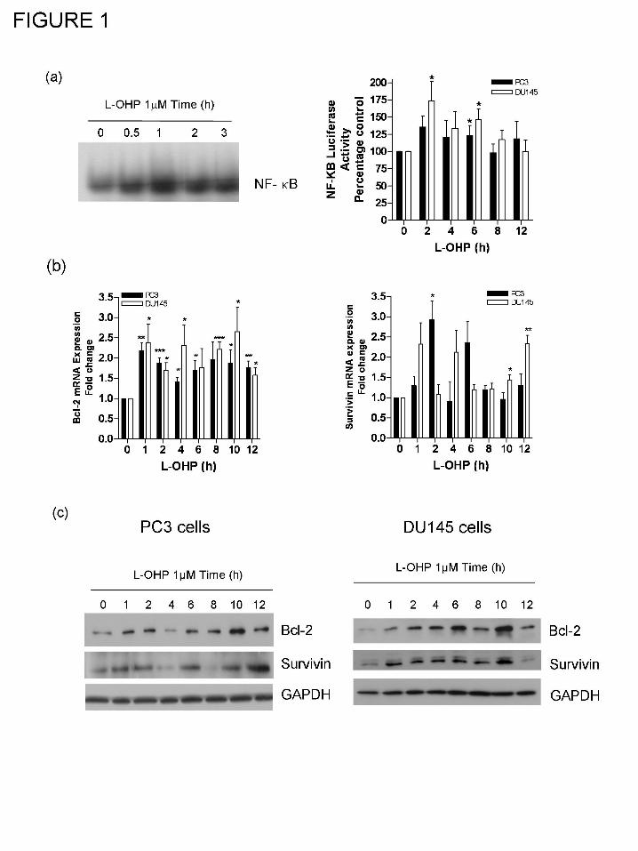
This article has not been copyedited and formatted. The final version may differ from this version.JPET Fast Forward. Published on September 9, 2008 as DOI: 10.1124/jpet.108.143826
at ASPE
T Journals on O
ctober 21, 2018jpet.aspetjournals.org
Dow
nloaded from
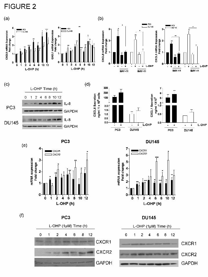
This article has not been copyedited and formatted. The final version may differ from this version.JPET Fast Forward. Published on September 9, 2008 as DOI: 10.1124/jpet.108.143826
at ASPE
T Journals on O
ctober 21, 2018jpet.aspetjournals.org
Dow
nloaded from
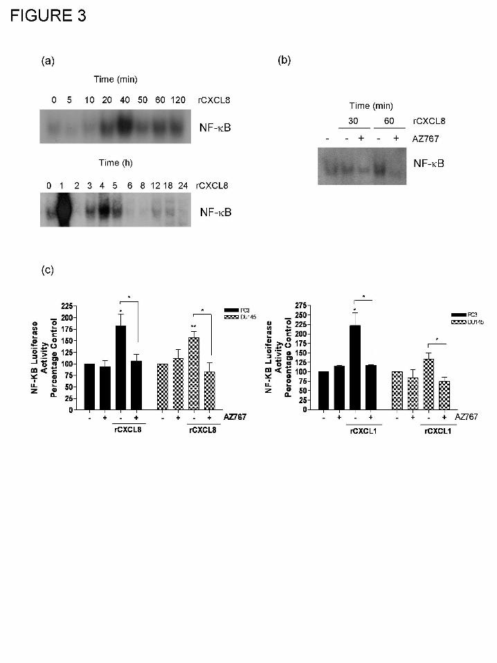
This article has not been copyedited and formatted. The final version may differ from this version.JPET Fast Forward. Published on September 9, 2008 as DOI: 10.1124/jpet.108.143826
at ASPE
T Journals on O
ctober 21, 2018jpet.aspetjournals.org
Dow
nloaded from
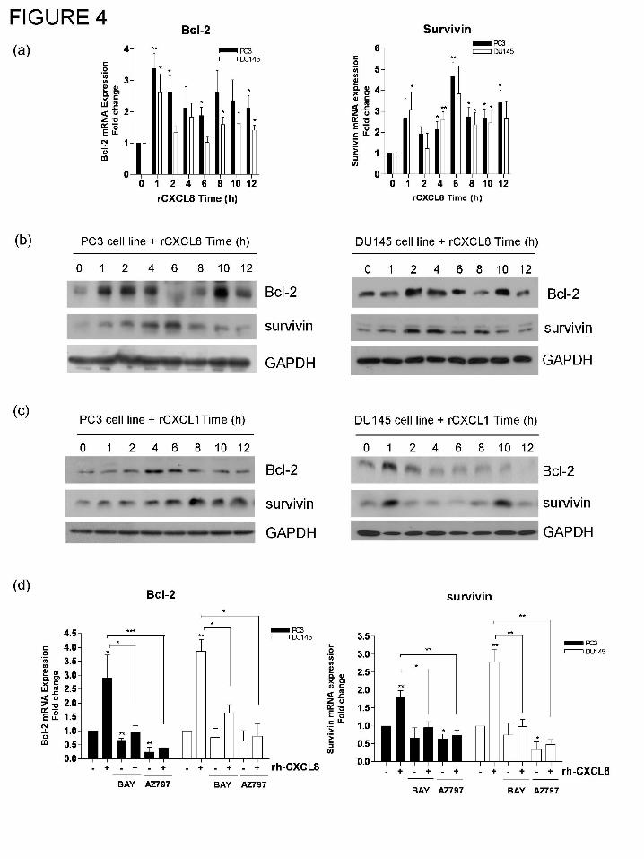
This article has not been copyedited and formatted. The final version may differ from this version.JPET Fast Forward. Published on September 9, 2008 as DOI: 10.1124/jpet.108.143826
at ASPE
T Journals on O
ctober 21, 2018jpet.aspetjournals.org
Dow
nloaded from
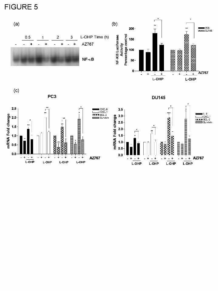
This article has not been copyedited and formatted. The final version may differ from this version.JPET Fast Forward. Published on September 9, 2008 as DOI: 10.1124/jpet.108.143826
at ASPE
T Journals on O
ctober 21, 2018jpet.aspetjournals.org
Dow
nloaded from
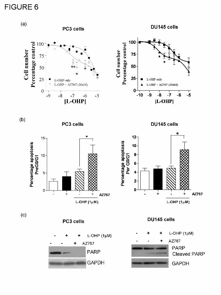
This article has not been copyedited and formatted. The final version may differ from this version.JPET Fast Forward. Published on September 9, 2008 as DOI: 10.1124/jpet.108.143826
at ASPE
T Journals on O
ctober 21, 2018jpet.aspetjournals.org
Dow
nloaded from
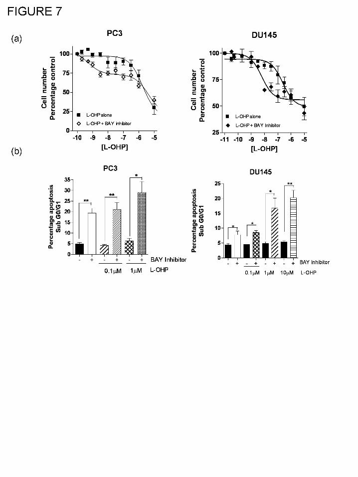
This article has not been copyedited and formatted. The final version may differ from this version.JPET Fast Forward. Published on September 9, 2008 as DOI: 10.1124/jpet.108.143826
at ASPE
T Journals on O
ctober 21, 2018jpet.aspetjournals.org
Dow
nloaded from
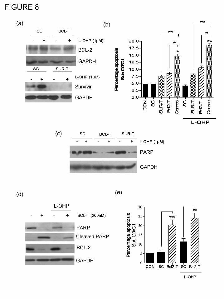
This article has not been copyedited and formatted. The final version may differ from this version.JPET Fast Forward. Published on September 9, 2008 as DOI: 10.1124/jpet.108.143826
at ASPE
T Journals on O
ctober 21, 2018jpet.aspetjournals.org
Dow
nloaded from
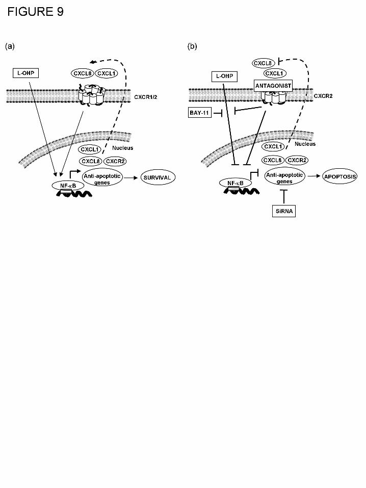
This article has not been copyedited and formatted. The final version may differ from this version.JPET Fast Forward. Published on September 9, 2008 as DOI: 10.1124/jpet.108.143826
at ASPE
T Journals on O
ctober 21, 2018jpet.aspetjournals.org
Dow
nloaded from