3-IKK Facilitates the Activation of NF- B by Hepatitis C Virus Core … · 2018-09-27 · Hepatitis...
9
www.ksmkorea.org / www.ksov.org 93 ORIGINAL ARTICLE J Bacteriol Virol. Vol 48. No 3. September 2018; 48(3): 93-101 http://dx.doi.org/10.4167/jbv.2018.48.3.93 eISSN 2093-0429 JBV IKKγ Facilitates the Activation of NF-κB by Hepatitis C Virus Core Protein Bo Yeong Kang 1 , So Yeong Lee 1 , Jin Ik Kim 1 , Hye Jung Choi 2 , Woo Hong Joo 2 and Dong Wan Kim 1* 1 Department of Biohealth Sciences, Changwon National University, Changwon, Korea 2 Department of Biology and Chemistry, Changwon National University Changwon, Korea Corresponding Dong Wan Kim Department of Biohealth Sciences, Changwon National University, Changwon, 51140, Korea. Phone : +82-55-213-3482 Fax : +82-55-213-3480 E-mail : [email protected] Received : July 24, 2018 Revised : August 28, 2018 Accepted : September 06, 2018 No potential conflict of interest relevant to this article was reported. Copyright © 2018 Journal of Bacteriology and Virology ©This is an Open Access article distributed under the terms of the Creative Commons Attribution Non-Commercial License (http://creativecommons.org/ license/by-nc/3.0/). Hepatitis C virus (HCV) is a major cause of chronic hepatitis, liver cirrhosis and hepatocellular carcinoma. HCV core protein has been shown to modulate various cellular signaling pathways including the nuclear factor κB (NF-κB) pathway which is associated with inflammation, cell proliferation and apoptosis. However, there have been conflicting reports about the effect of HCV core protein on NF-κB pathway, and the mechanism by which the core protein affects NF-κB activity remains nuclear. In this study, the functional interaction of HCV core protein and I κB kinase γ (IKKγ) was investigated using the expression plasmids of core and the components of IKK complex. The data revealed that HCV core protein activates NF-κB. Also, HCV core protein up-regulated the phosphorylation and degradation of IκBα. The activating effect of HCV core protein on NF-κB was synergistically elevated by IKKγ. It was noticed that the N-terminal IKKβ binding site, C-terminal leucine zipper, and zinc finger domains of IKKγ are not necessary for its synergistic effect. HCV core protein and IKKγ appeared to activate NF-κB by up-regulating the IKKβ activity resulting in the degradation of IκBα. As expected, HCV core protein induced the expression of NF-κB-targeted pro-inflammatory genes such as iNOS, IL-1β and IL-6 in the transcription level. These results suggest that HCV core protein induces NF-κB through the interaction with IKKγ and may play a critical role in the development of inflammation and related liver diseases. Key Words: Hepatitis C virus, Core protein, NF-κB, IKKγ, Inflammation INTRODUCTION Hepatitis C virus (HCV)는 Flaviviridae과에 속하는 이러스로서 급성 간염의 15~20% 차지하고 있으, HCV 감염환자의 50~80%는 성간염으로 진행되어 간경화(cirrhosis) 와 간세포암(hepatocellular carcinoma)이 생하는 것으로 알려져 있다 (1). HCV의 genome 은 약 9,600 nucleotide의 양성가닥 RNA (positive-sense RNA)이 약 3,000개의 아 노산으로 구성되어 있는 polyprotein을 생산한다. 이 polyprotein은 N-단에서부터 core, E1, E2/P7의 구조단질과 6개의 비구조단질(NS2, 3, 4a, 4b, 5a, 5b)로 구성되어 있다 (2, 3). Core는 191개의 아노산으로 된 21 kDa의 단질로 capsid 형성하는 기본 단 위이 감염세포에서 다양한 생학적 성질을 나타내는 것으로 알려져 있다 (4, 5). 특히, core는 nuclear factor-kappa B (NF-κB)의 활성화 경로에도 연관되어 있으, Inhibitor of kappa B (IκB) kinase β (IKKβ) 활성화시켜 NF-κB 활성화시킨다는 보고 (6)와 이러한 core의 NF-κB 활성화 작용이 Fas와 tumor necrosis factor (TNF)-α에 의한 apoptosis 억제하는 결과 가져온다는 보고도 있다 (7). , core는 TNF receptor-1에 작용하여 apoptosis 유도한다는 보고 (8)와 core가 NF-κB 억제하여 apoptosis 유도한다는
Transcript of 3-IKK Facilitates the Activation of NF- B by Hepatitis C Virus Core … · 2018-09-27 · Hepatitis...
www.ksmkorea.org / www.ksov.org 93
ORIGINAL ARTICLE J Bacteriol Virol. Vol 48. No 3. September 2018; 48(3): 93-101 http://dx.doi.org/10.4167/jbv.2018.48.3.93 eISSN 2093-0429
JBV
IKKγ Facilitates the Activation of NF-κB by Hepatitis C Virus Core Protein
Bo Yeong Kang1, So Yeong Lee1, Jin Ik Kim1, Hye Jung Choi2, Woo Hong Joo2 and Dong Wan Kim1*
1Department of Biohealth Sciences, Changwon National University, Changwon, Korea 2Department of Biology and Chemistry, Changwon National University Changwon, Korea
Corresponding Dong Wan Kim
Department of Biohealth Sciences,
Changwon National University,
Changwon, 51140, Korea. Phone : +82-55-213-3482 Fax : +82-55-213-3480 E-mail : [email protected]
Received : July 24, 2018
Revised : August 28, 2018
Accepted : September 06, 2018
No potential conflict of interest relevant to this article was reported.
Copyright © 2018 Journal of Bacteriologyand Virology
©This is an Open Access article distributed under the terms of the Creative Commons Attribution Non-Commercial License (http://creativecommons.org/ license/by-nc/3.0/).
Hepatitis C virus (HCV) is a major cause of chronic hepatitis, liver cirrhosis and
hepatocellular carcinoma. HCV core protein has been shown to modulate various
cellular signaling pathways including the nuclear factor κB (NF-κB) pathway which
is associated with inflammation, cell proliferation and apoptosis. However, there
have been conflicting reports about the effect of HCV core protein on NF-κB
pathway, and the mechanism by which the core protein affects NF-κB activity
remains nuclear. In this study, the functional interaction of HCV core protein and I
κB kinase γ (IKKγ) was investigated using the expression plasmids of core and the
components of IKK complex. The data revealed that HCV core protein activates
NF-κB. Also, HCV core protein up-regulated the phosphorylation and degradation
of IκBα. The activating effect of HCV core protein on NF-κB was synergistically
elevated by IKKγ. It was noticed that the N-terminal IKKβ binding site, C-terminal
leucine zipper, and zinc finger domains of IKKγ are not necessary for its synergistic
effect. HCV core protein and IKKγ appeared to activate NF-κB by up-regulating the
IKKβ activity resulting in the degradation of IκBα. As expected, HCV core protein
induced the expression of NF-κB-targeted pro-inflammatory genes such as iNOS,
IL-1β and IL-6 in the transcription level. These results suggest that HCV core protein
induces NF-κB through the interaction with IKKγ and may play a critical role in the
development of inflammation and related liver diseases.
Key Words: Hepatitis C virus, Core protein, NF-κB, IKKγ, Inflammation
INTRODUCTION
Hepatitis C virus (HCV)는 Flaviviridae과에 속하는 바이러스로서 급성 간염의 15~20%를
차지하고 있으며, HCV 감염환자의 50~80%는 만성간염으로 진행되어 간경화(cirrhosis)와
간세포암(hepatocellular carcinoma)이 발생하는 것으로 알려져 있다 (1). HCV의 genome
은 약 9,600 nucleotide의 양성가닥 RNA (positive-sense RNA)이며 약 3,000개의 아미
노산으로 구성되어 있는 polyprotein을 생산한다. 이 polyprotein은 N-말단에서부터 core,
E1, E2/P7의 구조단백질과 6개의 비구조단백질(NS2, 3, 4a, 4b, 5a, 5b)로 구성되어 있다
(2, 3). Core는 191개의 아미노산으로 된 21 kDa의 단백질로 capsid를 형성하는 기본 단
위이며 감염세포에서 다양한 생물학적 성질을 나타내는 것으로 알려져 있다 (4, 5). 특히,
core는 nuclear factor-kappa B (NF-κB)의 활성화 경로에도 연관되어 있으며, Inhibitor of
kappa B (IκB) kinase β (IKKβ)를 활성화시켜 NF-κB를 활성화시킨다는 보고 (6)와 이러한
core의 NF-κB 활성화 작용이 Fas와 tumor necrosis factor (TNF)-α에 의한 apoptosis를
억제하는 결과를 가져온다는 보고도 있다 (7). 반면, core는 TNF receptor-1에 작용하여
apoptosis를 유도한다는 보고 (8)와 core가 NF-κB를 억제하여 apoptosis를 유도한다는
JBV VOL 48. NO 3. SEPTEMBER 2018
94 Copyright © 2018 Journal of Bacteriology and Virology
Journal of Bacteriology and Virology
상반된 보고도 있다 (9). 하지만 HCV의 core와 NF-κB와의 연관성 및 apoptosis에 미치는 영향에 대해서는 정확한 결론이 내려지지 않고
있는 실정이다 (6, 10).
NF-κB는 모든 유형의 세포에서 광범위하게 발현되는 유도성 전사인자로 세포 외부로부터 가해지는 TNF-α, lipopolysaccharide (LPS),
irradiation, reactive oxygen species 등 다양한 자극에 대한 방어 작용과 면역세포의 활성화를 일으키며 세포의 증식 및 사멸, 염증반응
등과 관련된 다양한 유전자의 발현을 조절하는 것으로 알려져 있다 (11~13). NF-κB는 유도자극이 없는 경우 IκB에 결합되어 세포질 속에
머물러 있으나 세포를 TNF-α, LPS, Interleukin (IL)-1β 등으로 자극하면 IκB가 인산화 되고 IκB의 분해가 일어나서 NF-κB의 신호전달회로가
활성화된다 (12). IκB는 IκB kinase인 IKKα와 IKKβ에 의해 serine 잔기가 인산화 되는데 인산화된 IκB는 ubiquitination이 촉진되어 세포질
내의 26S proteasome에 의해 분해된다. 따라서 남은 NF-κB는 자유로이 핵 안으로 이동하여 target DNA에 결합함으로써 전사를 촉진
한다 (14~16). IκB의 인산화는 IKK complex에 의해 일어나며 IKK complex (700~900 kDa)는 catalytic subunit인 IKKα (87 kDa), IKKβ
(85 kDa)와 효소활성은 없지만 NF-κB 활성에 필수적 조절 역할을 하는 IKKγ (48 kDa)를 포함한다. IKKγ는 NF-κB essential modulator
(NEMO)라고도 불리는 것으로 kinase 활성을 가지지 않으며 IKKβ와 IκBα의 interaction을 증가시키고 IKKβ의 kinase 활성 증가 및 IKK
complex 형성에 중요한 역할을 한다 (17, 18). 그러나 아직 IKKγ가 다른 protein과 결합하는 부위와 결합하는 protein의 종류, N-말단
과 C-말단을 포함한 부위별 기능, NF-κB inducing kinase (NIK) 또는 mitogen activated protein kinase kinase kinase (MEKK)와 같은
NF-κB의 upstream kinase와의 연관성 등에 대해서는 상세히 알려져 있지 않으며, 특히 HCV core에 의한 NF-κB의 활성 변화에서 IKKγ
의 역할에 관해서는 아직 보고된 바가 없다.
본 연구에서는 아직 명확하게 밝혀져 있지 않은 HCV core에 의한 NF-κB의 활성 변화에서 IKKγ의 역할을 IKKγ의 deletion mutant와 IKKβ
의 활성형 mutant인 IKKβ-EE를 사용한 NF-κB upstream kinase 기능 연구를 통해 규명하고자 하였다.
METERIALS AND METHODS
Cell Culture 및 transfection
원숭이의 신장세포인 COS-7 세포와 마우스 macrophage인 RAW264.7 세포는 fetal bovine serum (FBS, Gibco BRL Co, Grand Island,
NY, USA)이 10% 함유된 Dulbecco's Modified Eagle Medium (DMEM, Gibco BRL Co.)을 사용하여 37℃, 5% CO2 농도가 유지되는
incubator에서 배양하였다. 세포에 plasmid DNA를 도입하기 위해 FuGENE HD (Roche, Basel, Switzerland)를 사용한 lipofection 방
법을 사용하였으며 sample 당 10~20 μg의 DNA를 serum free DMEM과 FuGENE HD의 혼합액에 섞고 실온에서 20분간 반응시킨 후
70~80%의 밀도로 배양된 recipient cell에 적하하고 배양하였다. Transfection에 사용한 발현 plasmid는 pCMV5 발현백터에 myc이 결
합된 사람의 wild type NIK의 유전자가 cloning된 것과, pCMV5 발현백터에 haemagglutinin (HA)이 결합된 사람의 wild type IKKβ 또
는 사람의 IKKβ의 활성형 mutant인 IKKβ-EE 유전자가 cloning된 것과, pCMV5 발현백터에 myc이 결합된 mouse의 wild type IKKγ 또
는 mutant IKKγ 유전자가 cloning된 것을 사용하였다. HCV core의 발현 plasmid는 pSRα 발현백터에 HCV의 core 발현영역의 cDNA
를 cloning한 것을 사용하였다.
Luciferase assay
Luciferase assay는 Nano-Glo Luciferase Assay Kit (Promega, Madison, WI, USA)를 사용하였다. Nano-Glo substrate를 Nano-Glo
buffer에 100배 희석하여 사용하였으며 cell lysate는 96-well plate에 20 μl씩 분주하여 사용하였다. Luciferase activity는 Centro LB960
luminometer (Berthold Technologies, Bad Wildbad, Germany)와 MikroWin2000 (Mikrotek Laborsysteme GmbH, Overath, Germany)
소프트웨어를 사용하여 측정하였다.
Western blotting
단백질 분석을 위한 세포추출물(whole cell extract)은 세포를 Phosphate buffered saline (PBS)로 두 번 세척 후 harvest 하고 4℃에서
1,500 rpm으로 5분 동안 원심분리하여 상등액을 제거한 후, Total lysis buffer (20 mM hepes (pH 7.9), 25% glycerol, 450 mM NaCl,
0.4 mM ethylenediaminetetraacetic acid (EDTA), 0.5 mM dithiothreitol (DTT), 1% NP-40)로 suspension 하여 ice에서 1시간 동안 반
응시켰다. 이 후 4℃에서 12,000 rpm으로 15분 동안 원심분리하여 상등액을 수거하였다 (18, 19). 세포질추출물(cytoplasmic extract)은
HCV Core and IKKγ Synergistically Activate NF-κB BY Kang, et al.
www.ksmkorea.org / www.ksov.org 95
세포를 PBS로 두 번 세척 후 harvest 하고, buffer A (10 mM HEPES (pH 7.9), 1 mM EDTA, 10 mM KCl, 1 mM DTT)로 suspension
후 ice에서 15분 동안 반응시켰다. 이 후 25G needle을 장착한 1 ml 주사기를 이용해 20번 stroke 하여 세포막을 파괴시키고, 4℃에서
14,000 rpm으로 15분 동안 원심분리하여 상등액을 수거하였다 (18, 19).
Western blotting은 시료 단백질을 10% Sodium dodecyl sulfate (SDS) polyacrylamide gel에서 전기 영동한 후 gel 상의 단백질을 실온
에서 90 V로 2시간 동안 nitrocellulose membrane (GE Healthcare Life Science, Munich, Germany)에 electrotransfer 한 뒤 membrane
을 5% 탈지분유를 포함한 TBS-T (20 mM Tris-HCl (pH 7.6), 137 mM NaCl, 0.1% Tween 20) 용액으로 1시간 동안 실온에서 blocking
하였다. Membrane을 1차 항체와 실온에서 2시간 반응시키고 TBS-T 용액으로 3번 세척한 후 peroxidase-conjugated 2차 항체로 1시
간 동안 실온에서 반응시켰다. 반응시킨 membrane을 TBS-T 용액으로 3번 세척한 후 enhanced chemiluminescence (ECL, Amersham
Corp. Arlington Heights, IL, USA)로 발광시킨 후 Amersham Imager 600 (GE Healthcare)을 이용하여 단백질 발현을 확인하였다.
Membrane의 strip은 stripping buffer (6.25 mM Tris-HCl (pH 6.7), 2% SDS, 100 mM 2-mercaptoethanol)에 membrane을 넣고 50℃
에서 30분간 처리한 뒤 TBS-T 용액으로 10분씩 3회 세척하였다. 실험에 사용한 anti-HCV core monoclonal (C7-50), anti-IκBα polyclonal
(C-21), anti-actin polyclonal (I-19), anti-HA monoclonal (F-7), anti-cMyc monoclonal (9E10) antibody는 Santa Cruz Bio Technology
(Santa Cruz, CA, USA) 제품을 사용하였고, anti-STAT3 polyclonal (#9139), anti-p-STAT3 polyclonal (#9145), anti-p38 monoclonal
(#8690), anti-p-p38 monoclonal (#4511) antibody는 Cell Signaling Technology, Inc. (Danvers, MA, USA) 제품을 제조사의 사용법에
따라 사용하였다. 2차 항체는 Horseradish peroxidase가 결합된 anti-mouse IgG와 anti-rabbit IgG (Bethyl Laboratories, Inc., Montgomery,
TX, USA)를 사용하였다.
RT-PCR
세포를 TRIzol (Invitrogen, Carlsbad, CA, USA) 용액으로 total RNA를 분리한 후 Accupower Roketscript cycle RT premix (Bioneer
Corp., Daejeon, Korea)을 이용하여 cDNA를 합성하였다. PCR을 위한 primer는 iNOS: 5'-GGAGCAGGTGGAAGACTATTT-3' (sense),
5'-GGGAGGAGAGGAGAGAGATTTA-3' (antisense); IL-1β: 5'-GTGTGTGACGTTCCCATTAGA-3' (sense), 5'-TTAGAAACAGTCCA-
GCCCATAC-3' (antisense); IL-6: 5'-GATAAGCTGGAGTCACAGAAGG-3' (sense), 5'-GAAACCATCTGGCTAGGTAACA-3' (antisense);
GAPDH: 5'-AACAGCAACTCCCACTCTTC-3' (sense), 5'-TGGGTGCAGCGAACTTTAT-3' (antisense)를 사용하였으며, PCR 반응은 94℃
에서 3분간 pre-denature하고, 94℃에서 30초 denature, 60℃에서 1분 annealing, 72℃에서 1분 extension 반응을 30 cycle 진행한 후,
72℃에서 10분간 post-extension을 실시하였다. PCR의 결과는 2.5% agarose gel에서 전기 영동하여 확인하였다.
Statistical analysis
실험은 3회 반복하였고, 평균(mean) ± 표준편차(SD)로 나타내었다. Student's t-test를 이용하여 통계적으로 분석하였으며, p값이 0.05
이하이면 유의성이 있다고 판단하였다.
RESULTS
HCV의 core 단백질에 의한 NF-κB의 활성 변화
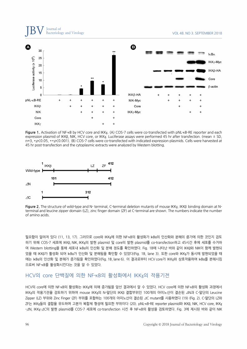
NF-κB의 활성에 대한 HCV core의 영향을 검토하기 위해 pNL-κB-RE reporter plasmid (Promega, code N1111)와 IKKβ, NIK, IKKγ의 발
현 plasmid 및 HCV core 발현 plasmid를 COS-7 세포에 co-transfection하고 45시간 후 세포를 수거하여 luciferase assay를 실시하였
다. pNL-κB-RE는 5개의 NF-κB 결합부위를 가진 luciferase assay reporter plasmid이다. 실험 결과 Fig. 1A에 나타난 바와 같이 IKKβ만
발현되었을 경우(Fig. 1A, lane 3)에는 NF-κB가 활성화되지 않았지만 NF-κB의 활성화 kinase인 NIK의 co-transfection에 의해 NF-κB의
활성이 증가하였으며(Fig. 1A, lane 4), 여기에 HCV core를 발현시켰을 때 NF-κB의 활성은 더욱 증가하는 것으로 나타났다(Fig. 1A, lane
5). 또한 IKKγ를 co-transfection 시켰을 때에도 NF-κB의 활성은 증가하는 것으로 나타났다(Fig. 1A, lane 6). 이 결과로부터 core와 IKKγ
는 각각 NIK에 의한 NF-κB의 활성화를 유의적인 수준으로 증가시킨다는 것을 알 수 있었다. 특히, core와 IKKγ를 동시에 발현시켰을 경
우에는 각각에 의한 NF-κB의 활성 증가보다 2.5배 이상 활성이 증가하는 것으로 나타났다(Fig. 1A, lane 7). 이 결과는 core에 의한 NF-
κB의 활성화가 IKKγ를 경유하거나 IKKγ와 상호협력적으로 일어남을 나타내는 것으로 해석된다. 한편, NF-κB의 활성화는 세포질 내 NF-κB
의 저해제인 IκBα가 인산화된 후 ubiquitination을 거쳐 분해된 후 활성화됨이 알려져 있어 IκBα의 인산화와 분해가 NF-κB의 활성화에
JBV VOL 48. NO 3. SEPTEMBER 2018
96 Copyright © 2018 Journal of Bacteriology and Virology
Journal of Bacteriology and Virology
필요함이 알려져 있다 (11, 13, 17). 그러므로 core와 IKKγ에 의한 NF-κB의 활성화가 IκBα의 인산화와 분해의 증가에 의한 것인지 검토
하기 위해 COS-7 세포에 IKKβ, NIK, IKKγ의 발현 plasmid 및 core의 발현 plasmid를 co-transfection하고 45시간 후에 세포를 수거하
여 Western blotting을 통해 세포내 IκBα의 인산화 및 분해 정도를 확인하였다. Fig. 1B에 나타난 바와 같이 IKKβ와 NIK이 함께 발현되
었을 때 IKKβ가 활성화 되어 IκBα가 인산화 및 분해됨을 확인할 수 있었다(Fig. 1B, lane 3). 또한 core와 IKKγ가 동시에 발현되었을 때
에는 IκBα의 인산화 및 분해가 증가됨을 확인하였다(Fig. 1B, lane 6). 이 결과로부터 HCV core가 IKKγ와 상호작용하여 IκBα를 분해시킴
으로써 NF-κB를 활성화시킨다는 것을 알 수 있었다.
HCV의 core 단백질에 의한 NF-κB의 활성화에서 IKKγ의 작용기전
HCV의 core에 의한 NF-κB의 활성화는 IKKγ에 의해 증가됨을 앞선 결과에서 알 수 있었다. HCV core에 의한 NF-κB의 활성화 과정에서
IKKγ의 작용기전을 검토하기 위하여 mouse IKKγ의 N-말단의 IKKβ 결합부위인 100개의 아미노산이 결손된 ΔN과 C-말단의 Leucine
Zipper (LZ) 부위와 Zinc Finger (ZF) 부위를 포함하는 100개의 아미노산이 결손된 ΔC mutant를 사용하였다 (19) (Fig. 2). C-말단의 LZ와
ZF는 IKKγ들의 결합을 유도하며 고분자 복합체 형성에 필요한 부위이다 (20). pNL-κB-RE reporter plasmid와 IKKβ, NIK, HCV core, IKKγ
-ΔN, IKKγ-ΔC의 발현 plasmid를 COS-7 세포에 co-transfection 시킨 후 NF-κB의 활성을 검토하였다. Fig. 3에 제시된 바와 같이 NIK
Figure 1. Activation of NF-κB by HCV core and IKKγ. (A) COS-7 cells were co-transfected with pNL-κB-RE reporter and each expression plasmid of IKKβ, NIK, HCV core, or IKKγ. Luciferase assays were performed 45 hr after transfection. (mean ± SD, n=3, *p<0.05, **p<0.001). (B) COS-7 cells were co-transfected with indicated expression plasmids. Cells were harvested at 45 hr post-transfection and the cytoplasmic extracts were analyzed by Western blotting.
Figure 2. The structure of wild-type and N- terminal, C-terminal deletion mutants of mouse IKKγ. IKKβ binding domain at N-terminal and leucine zipper domain (LZ), zinc finger domain (ZF) at C-terminal are shown. The numbers indicate the number of amino acids.

HCV Core and IKKγ Synergistically Activate NF-κB BY Kang, et al.
www.ksmkorea.org / www.ksov.org 97
은 NF-κB를 활성화 시켰고(Fig. 3A, lane 4) IKKγ-ΔN과 ΔC도 각각 NF-κB의 활성을 유의적으로 증가시켰다(Fig. 3A, lane 5, 7). 또한
IKKγ-ΔN과 ΔC는 모두 HCV core에 의한 NF-κB의 활성화를 더욱 증가시켜 wild type의 효과와 거의 동일하게 나타났다 (Fig. 3, lane 6,
8). 이 결과로부터 IKKγ의 N말단에 의한 IKKγ와 IKKβ의 결합 및 C-말단에 의한 IKKγ의 고분자 형성은 core의 NF-κB 활성화 작용에 관
여하지 않음을 알 수 있었다. 아울러 HCV core와 IKKγ-ΔN과 ΔC에 의한 NF-κB의 활성화가 IκBα의 인산화 및 분해의 증가에 의한 것인
지 검토하기 위해 IKKβ, NIK, HCV core, IKKγ-ΔN, ΔC의 발현 plasmid를 COS-7 세포에 co-transfection 하고 Western blotting을 수행
하였다. Fig. 3B에 제시된 바와 같이 IKKγ-ΔN, IKKγ-ΔC는 모두 HCV core와 함께 co-transfection 되었을 때 IκBα의 분해를 현저하게 증
가시켰다(Fig. 3B, lane 4, 6). 결과적으로 IKKγ-ΔN, ΔC는 wild type IKKγ와 동일하게 HCV core와 상호작용하여 IκBα의 인산화 및 분해
를 촉진함으로써 NF-κB의 활성을 증가시킨다는 것을 알 수 있었으며, Fig. 3B의 2번째 panel에 제시된 바와 같이, IKKγ의 ΔN은 C-말단
의 인산화 부위를 포함하므로 인산화된 것과 인산화 되지 않은 것으로 표현되는 2개의 band로 검출되었고, ΔC는 C-말단의 인산화 부
위가 제거되어 단일 band로 검출되어 정상적으로 발현되었음을 알 수 있었다.
IKKβ의 mutant를 이용한 HCV core 및 IKKγ의 작용기전 검토
HCV core와 IKKγ에 의한 NF-κB의 활성화가 NIK에 의한 활성화에 특이적으로 작용하는 것인지 알아보기 위해 IKKβ의 활성형 mutant
인 IKKβ-EE를 이용하여 HCV core와 IKKγ가 NF-κB의 활성에 어떠한 영향을 미치는지 검토하였다. IKKβ-EE는 IKKβ의 activation loop의
Ser177과 Ser181 부위를 glutamate로 치환시켜 NIK, MEKK 등 upstream kinase가 없어도 항상 활성화된 상태를 나타내는 IKKβ의 변
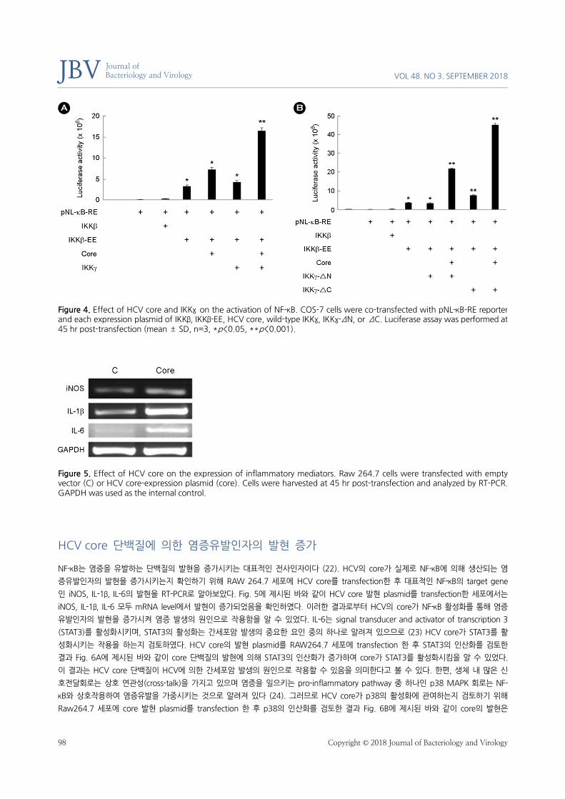
이형이다 (21). COS-7 세포에 IKKβ, IKKβ-EE, HCV core, IKKγ 발현 plasmid와 pNL-κB-RE report plasmid를 co-transfection 시킨 후 45시
간 후에 세포를 수거하여 NF-κB 활성에 미치는 HCV core 와 IKKγ의 영향을 검토한 결과, Fig. 4A에 제시된 바와 같이 IKKβ-EE는 NIK이
없어도 NF-κB를 활성화시킴을 알 수 있었고(Fig. 4A, lane 4), Fig. 1A의 NIK에 의한 활성화의 경우와 마찬가지로 HCV core와 IKKγ는 각
각 IKKβ-EE에 의한 NF-κB의 활성화를 증가시켰고(Fig. 4A, lane5, 6), HCV core와 IKKγ를 동시에 발현시켰을 때 NF-κB의 활성은 가장
크게 증가되었다(Fig. 4A, lane 7). 이 결과로부터 HCV core와 IKKγ에 의한 NF-κB 활성 증가는 NIK에 의한 NF-κB의 활성화에만 특이
적으로 작용하는 것이 아니라 IKKβ를 경유하는 NF-κB의 활성화 경로 모두에 작용할 가능성이 높다는 것을 알 수 있었다. 이러한 결과는
IKKγ의 deletion mutant인 IKKγ-ΔN과 ΔC에서도 동일하게 나타났으며(Fig. 4B), 이는 HCV core와 IKKγ가 NIK 등의 upstream kinase에
작용하는 것이 아니라 IKKβ의 활성을 증가시킴으로써 IKK complex를 활성화하여 NF-κB를 활성화시킨다는 것을 알 수 있었다.
Figure 3. Activation of NF-κB by HCV core and IKKγ mutants. (A) COS-7 cells were co-transfected with pNL-κB-RE reporter andeach expression plasmid of IKKβ, NIK, HCV core or IKKγ mutant. Luciferase assay was performed at 45 hr post-transfection. (mean ± SD, n=3, *p<0.05, **p<0.001). (B) COS-7 cells were co-transfected with expression plasmids as indicated. Cells wereharvested at 45 hr post-transfection and the cytoplasmic extracts were analyzed by western blotting.
JBV VOL 48. NO 3. SEPTEMBER 2018
98 Copyright © 2018 Journal of Bacteriology and Virology
Journal of Bacteriology and Virology
HCV core 단백질에 의한 염증유발인자의 발현 증가
NF-κB는 염증을 유발하는 단백질의 발현을 증가시키는 대표적인 전사인자이다 (22). HCV의 core가 실제로 NF-κB에 의해 생산되는 염
증유발인자의 발현을 증가시키는지 확인하기 위해 RAW 264.7 세포에 HCV core를 transfection한 후 대표적인 NF-κB의 target gene
인 iNOS, IL-1β, IL-6의 발현을 RT-PCR로 알아보았다. Fig. 5에 제시된 바와 같이 HCV core 발현 plasmid를 transfection한 세포에서는
iNOS, IL-1β, IL-6 모두 mRNA level에서 발현이 증가되었음을 확인하였다. 이러한 결과로부터 HCV의 core가 NF-κB 활성화를 통해 염증
유발인자의 발현을 증가시켜 염증 발생의 원인으로 작용함을 알 수 있었다. IL-6는 signal transducer and activator of transcription 3
(STAT3)를 활성화시키며, STAT3의 활성화는 간세포암 발생의 중요한 요인 중의 하나로 알려져 있으므로 (23) HCV core가 STAT3를 활
성화시키는 작용을 하는지 검토하였다. HCV core의 발현 plasmid를 RAW264.7 세포에 transfection 한 후 STAT3의 인산화를 검토한
결과 Fig. 6A에 제시된 바와 같이 core 단백질의 발현에 의해 STAT3의 인산화가 증가하여 core가 STAT3를 활성화시킴을 알 수 있었다.
이 결과는 HCV core 단백질이 HCV에 의한 간세포암 발생의 원인으로 작용할 수 있음을 의미한다고 볼 수 있다. 한편, 생체 내 많은 신
호전달회로는 상호 연관성(cross-talk)을 가지고 있으며 염증을 일으키는 pro-inflammatory pathway 중 하나인 p38 MAPK 회로는 NF-
κB와 상호작용하여 염증유발을 가중시키는 것으로 알려져 있다 (24). 그러므로 HCV core가 p38의 활성화에 관여하는지 검토하기 위해
Raw264.7 세포에 core 발현 plasmid를 transfection 한 후 p38의 인산화를 검토한 결과 Fig. 6B에 제시된 바와 같이 core의 발현은
Figure 4. Effect of HCV core and IKKγ on the activation of NF-κB. COS-7 cells were co-transfected with pNL-κB-RE reporter and each expression plasmid of IKKβ, IKKβ-EE, HCV core, wild-type IKKγ, IKKγ-ΔN, or ΔC. Luciferase assay was performed at 45 hr post-transfection (mean ± SD, n=3, *p<0.05, **p<0.001).
Figure 5. Effect of HCV core on the expression of inflammatory mediators. Raw 264.7 cells were transfected with empty vector (C) or HCV core-expression plasmid (core). Cells were harvested at 45 hr post-transfection and analyzed by RT-PCR. GAPDH was used as the internal control.

HCV Core and IKKγ Synergistically Activate NF-κB BY Kang, et al.
www.ksmkorea.org / www.ksov.org 99
p38의 인산화를 증가시켜 p38을 활성화함을 알 수 있었다. 이 결과로부터 HCV core 단백질이 NF-κB뿐 아니라 다양한 염증유발인자의
활성화에 관여한다는 것을 알 수 있었다.
DISCUSSION
HCV는 간세포뿐 아니라 B-cell, T-cell, monocyte, macrophage 등 다양한 면역세포에서 증식하며 면역세포의 기능을 변화시킨다 (25,
26). HCV의 core 단백질은 감염세포에서 세포증식, 대사, 세포사멸에 영향을 미치며 간세포 암(hepatocellular carcinoma) 발생의 원인
으로 작용하는 것으로 알려져 있다 (27~29). 그러나 아직 HCV core에 의한 숙주세포의 면역기능변화 및 발암에 관한 구체적인 기전은
알려져 있지 않다.
NF-κB는 다양한 면역응답반응의 핵심전사인자로 알려져 있으며 다양한 pro-inflammatory cytokine의 분비가 NF-κB의 활성화에 의해
일어난다 (30, 31). Pro-inflammatory cytokine인 IL-1β, IL-6, TNF-α와 chemokine인 CCL2, CXCL8 등이 NF-κB에 의해 발현되는 것으
로 알려져 있다 (32). 또한 지속적인 NF-κB의 활성화는 세포사멸을 억제하고 세포증식을 촉진하여 암을 유발하고 암 치료에 대한 내성
을 일으키는 것으로 알려져 있다 (33, 34). 선행보고에 따르면 HCV의 core는 NF-κB의 활성을 변화시키고, 이러한 core의 작용이 HCV
감염세포에서 염증을 유발하고 나아가 암을 일으킬 수 있다는 견해가 다수 제시되었다 (6~10). 그러나 아직 HCV core에 의한 NF-κB의
활성조절 기전이 명확하게 제시되고 있지 않다.
이러한 관점에서 본 연구에서는 HCV core에 의한 NF-κB의 활성조절 기전을 규명하고자 HCV core와 IKKγ의 상호 연관성을 검토하였으
며, HCV core는 IKKγ를 경유하거나 IKKγ와 상호협력적으로 NF-κB를 활성화한다는 결론을 얻었다(Fig. 1, Fig. 3). 또한 core에 의한 NF-
κB의 활성화는 NF-κB 신호전달 기전에서 NIK 등의 upstream kinase 활성에 작용하는 것이 아니라 IKKβ의 활성을 증가시킴으로써 NF-
κB를 활성화한다는 것을 알 수 있었다(Fig. 4). 또한 HCV core는 NF-κB의 대표적인 target gene인 iNOS, IL-1β, IL-6의 발현을 증가시켰
으며(Fig. 5), 이러한 결과로부터 HCV의 core 단백질이 HCV 감염세포에서 염증을 유발하는 핵심단백질임을 확인할 수 있었다. 간암은
대표적인 염증성 암이며 HCV 감염환자의 약 60%가 만성간염으로 진행되고 그 중 20~30%에서 간암이 발생하는 것으로 볼 때, HCV
core 단백질이 염증뿐 아니라 간암의 원인으로 작용할 가능성이 높음을 추측할 수 있다. 실제로 core 단백질은 간암 발생의 중요한 원인
으로 알려져 있는 STAT3를 활성화시키는 것으로 본 연구에서 나타났으며(Fig. 6A) (23), NF-κB에 의한 염증을 악화시키는 것으로 알려
져 있는 p38 MAPK를 활성화시키는 것으로 나타나(Fig. 6B) (24) HCV의 만성 간염환자에서 core 단백질은 반복된 염증유발 뿐 아니라
발암 단백질의 활성을 촉진시켜 간암을 유발할 가능성이 있음을 알 수 있었다. 그러나 HCV core 단백질에 의한 발암의 기전규명은 아
직 거의 알려져 있지 않기 때문에 core 단백질과 발암관련 신호전달회로와의 연관성 연구는 앞으로 진행되어야 할 중요 연구과제로 남아
있다.
Figure 6. Effect of HCV core on the phosphorylation of signaling molecules in the proinflammatory pathway. Raw 264.7 cells were transfected with empty vector (C) or HCV core expression plasmid. The whole cell extracts were prepared at 45 hr post-transfection and analyzed by western blotting.
JBV VOL 48. NO 3. SEPTEMBER 2018
100 Copyright © 2018 Journal of Bacteriology and Virology
Journal of Bacteriology and Virology
ACKNOWLEDGEMENTS
This research was supported by Changwon National University in 2017-2018.
REFERENCES
1) Tanaka K, Hirohata T, Koga S, Sugimachi K, Kanematsu T, Ohryohji F, et al. Hepatitis C and hepatitis B in the etiology of
hepatocellular carcinoma in the Japanese population. Cancer Res 1991;51:2842-7.
2) Grakoui A, Wychowski C, Lin C, Feinstone SM, Rice CM. Expression and identification of hepatitis C virus polyprotein
cleavage products. J Virol 1993;67:1385-95.
3) Hijikata M, Kato N, Ootsuyama Y, Nakagawa M, Shimotohno K. Gene mapping of the putative structural region of the
hepatitis C virus genome by in vitro processing analysis. Proc Natl Acad Sci USA 1991;88:5547-51.
4) Mizushima H, Hijikata M, Tanji Y, Kimura K, Shimotohno K. Analysis of N-terminal processing of hepatitis C virus nonstructural
protein 2. J Virol 1994;68:2731-4.
5) Shrivastava A, Manna SK, Ray R, Aggarwal BB. Ectopic expression of hepatitis C virus core protein differentially regulates
nuclear transcription factors. J Virol 1998;72:9722-8.
6) Yoshida H, Kato N, Shiratori Y, Otsuka M, Maeda S, Kato J, et al. Hepatitis C virus core protein activates nuclear factor κ
B-dependent signaling through tumor necrosis factor receptor-associated factor. J Biol Chem 2001;276:16399-405.
7) Marusawa H, Hijicata M, Chiba T, Shimotohno K. Hepatitis C virus core protein inhibits Fas- and tumor necrosis factor alpha-
mediated apoptosis via NF-κB activation. J Virol 1999;73:4713-20.
8) Zhu N, Ware CF, Lai MM. Hepatitis C virus core protein enhances FADD-mediated apoptosis and suppresses TRADD signaling
of tumor necrosis factor receptor. Virology 2001;283:178-87.
9) Park J, Kang W, Ryu SW, Kim WI, Chang DY, Lee DH, et al. Hepatitis C virus infection enhances TNFα-induced cell death via
suppression of NF-κB. Hepatology 2012;56:831-40.
10) Ray RB, Meyer K, Ray R. Suppression of apoptotic cell death by hepatitis C virus core protein. Virology 1996;226:176-82.
11) Ghosh G, Wang VY, Huang DB, Fusco A. NF-κB regulation: lessons from structures. Immunol Rev 2012;246:36-58.
12) Hayden MS, Ghosh S. Shared principles in NF-κB signaling. Cell 2008;132:344-62.
13) Oeckinghaus A, Hayden MS, Ghosh S. Crosstalk in NF-κB signaling pathways. Nat Immunol 2011;12:695-708.
14) Ghosh S, Febin Prabhu Dass J. Non-canonical pathway network modelling and ubiquitination site prediction through
homology modelling of NF-κB. Gene 2016;581:48-56.
15) Hinz M, Arslan SC, Scheidereit C. It takes two to tango: IκBs, the multifunctional partners of NF-κB. Immunol Rev 2012;
246:59-76.
HCV Core and IKKγ Synergistically Activate NF-κB BY Kang, et al.
www.ksmkorea.org / www.ksov.org 101
16) Morgan MJ, Liu ZG. Crosstalk of reactive oxygen species and NF-κB signaling. Cell Res 2011;21:103-15.
17) Mauro C, Pacifico F, Lavorgna A, Mellone S, Iannetti A, Acquaviva R, et al. ABIN-1 binds to NEMO/IKKγ and co-operates
with A20 in inhibiting NF-κB. J Biol Chem 2006;281:18482-8.
18) Yamamoto Y, Kim DW, Kwak YT, Parajapati S, Verma U, Gaynor RB. IKKγ/NEMO facilitates the recruitment of the IκB
proteins into the IκB kinase complex. J Biol Chem 2001;276:36327-36.
19) Kwon WJ, Kim SH, Park YO, Cho M, Kang CD, Lee G, et al. IKKγ inhibits activation of NF-κB by NIK. Mol cells 2004;18:200-6.
20) Li XH, Fang X, Gaynor RB. Role of Ikkγ/NEMO in assembly of the ikappa B kinase complex. J Biol Chem 2001;276:4494-500.
21) Mercurio F, Zhu H, Murray BW, Shevchenko A, Bennett BL, Li J, et al. Ikk-1 and Ikk-2: Cytokine-activated ikappa B kinase
essential for NF-κB activation. Science 1997;278:860-6.
22) Tang H, Chen H, Jia Y, Liu X, Han Z, Wang A, et al. Effect of inhibitors of endocytosis and NF-κB signal pathway on
folate-conjugated nanoparticle endocytosis by rat Kupffer cells. Int J Nanomedicine 2017;12:6937-47.
23) He G, Karin M. NF-κB and STAT3 - key players in liver inflammation and cancer. Cell Res 2011;21:159-68.
24) Craig R, Larkin A, Mingo AM, Thuerauf DJ, Andrews C, McDonough PM, et al. p38 MAPK and NF-κB collaborate to induce
interleukin-6 gene expression and release. J Biol Chem 2000;275:23814-24.
25) Bouffard P, Hayashi PH, Acevedo R, Levy N, Zeldis JB. Hepatitis C virus is detected in a monocyte/macrophage subpopulation
of peripheral blood mononuclear cells of infected patients. J Infect Dis 1992;166:1276-80.
26) Heydtmann M, Adams DH. Chemokines in the immunopathogenesis of hepatitis C infection. Hepatology 2009;49:676-88.
27) Saito K, Meyer K, Warner R, Basu A, Ray RB, Ray R. Hepatitis C virus core protein inhibits tumor necrosis factor alpha-
mediated apoptosis by a protective effect involving cellular FLICE inhibitory protein. J virol 2006;80:4372-9.
28) McLauchlan J. Properties of the hepatitis C virus core protein: a structural protein that modulates cellular processes. J viral
hepat 2000;7:2-14.
29) Bühler S, Bartenschlager R. Promotion of hepatocellular carcinoma by hepatitis C virus. Dig Dis 2012;30:445-52.
30) Hayden MS, Ghosh S. NF-κB in immunology. Cell Res 2011;21:223-44.
31) Xing Y, Wang X, Jameson SC, Hogquist KA. Late stage of T cell maturation in the thymus involve NF-κB and tonic type I
interferon signaling. Nat Immunol 2016;17:565-73.
32) Colotta F, Allavena P, Sica A, Garlanda C, Mantovani A. Cancer-related inflammation, the seventh hallmark of cancer: Links
to genetic instability. Carcinogenesis 2009;30:1073-81.
33) Bours V, Bentires-Alj M, Hellin AC, Viatour P, Robe P, Delhalle S, et al. Nuclear factor-kappa B, cancer, and apoptosis.
Biochem Pharmacol 2000; 60:1085-9.
34) Wu JT, Kral JG. The NF-kappaB/IkappaB signaling system: A molecular target in breast cancer therapy. J Surg Res 2005;
123:158-69.