Feline herpes virus
31
Feline herpes virus Dr. Tara Richards BSc DVM Dip ACVO PhD
-
Upload
truongthuan -
Category
Documents
-
view
240 -
download
1
Transcript of Feline herpes virus
Feline herpes virus Dr. Tara Richards BSc DVM Dip ACVO PhD

Photos courtesy of Dr. A LaBelle
Feline herpes virus (FHV-1)
Cause of Feline Viral Rhinotracheitis
DNA α- herpesvirus… herpes is “forever”
80% become carriers
45% of these “re-activation” = asymptomatic shedding vs. chronic disease
Clinical signs:
Many subclinical cases
Acute vs. Chronic
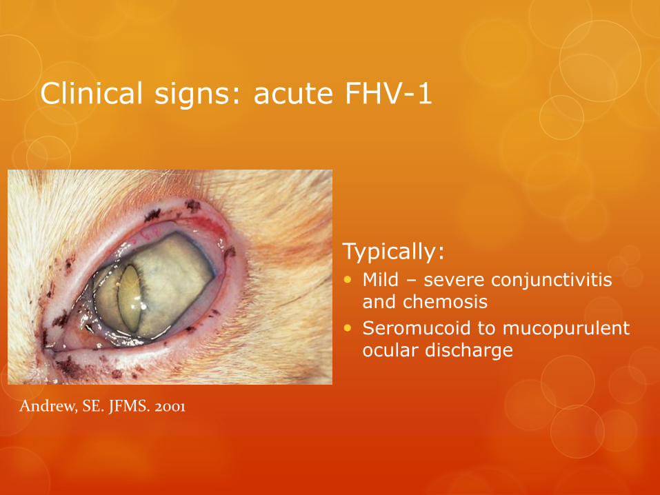
Clinical signs: acute FHV-1
Typically:
Mild – severe conjunctivitis and chemosis
Seromucoid to mucopurulent ocular discharge
Andrew, SE. JFMS. 2001
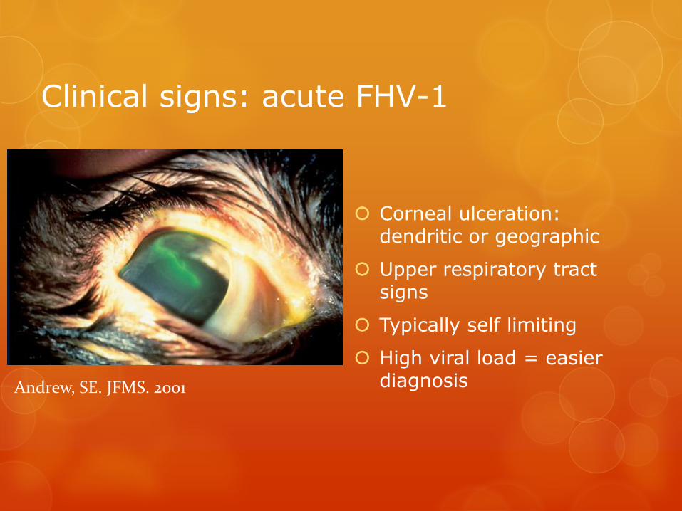
Clinical signs: acute FHV-1
Corneal ulceration: dendritic or geographic
Upper respiratory tract signs
Typically self limiting
High viral load = easier diagnosis
Andrew, SE. JFMS. 2001
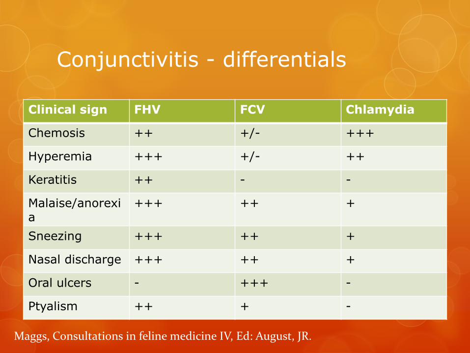
Conjunctivitis - differentials
Clinical sign FHV FCV Chlamydia
Chemosis ++ +/- +++
Hyperemia +++ +/- ++
Keratitis ++ - -
Malaise/anorexia
+++ ++ +
Sneezing +++ ++ +
Nasal discharge +++ ++ +
Oral ulcers - +++ -
Ptyalism ++ + -
Maggs, Consultations in feline medicine IV, Ed: August, JR.
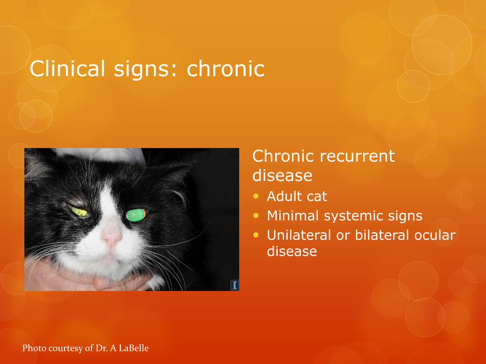
Clinical signs: chronic
Chronic recurrent disease
Adult cat
Minimal systemic signs
Unilateral or bilateral ocular disease
Photo courtesy of Dr. A LaBelle
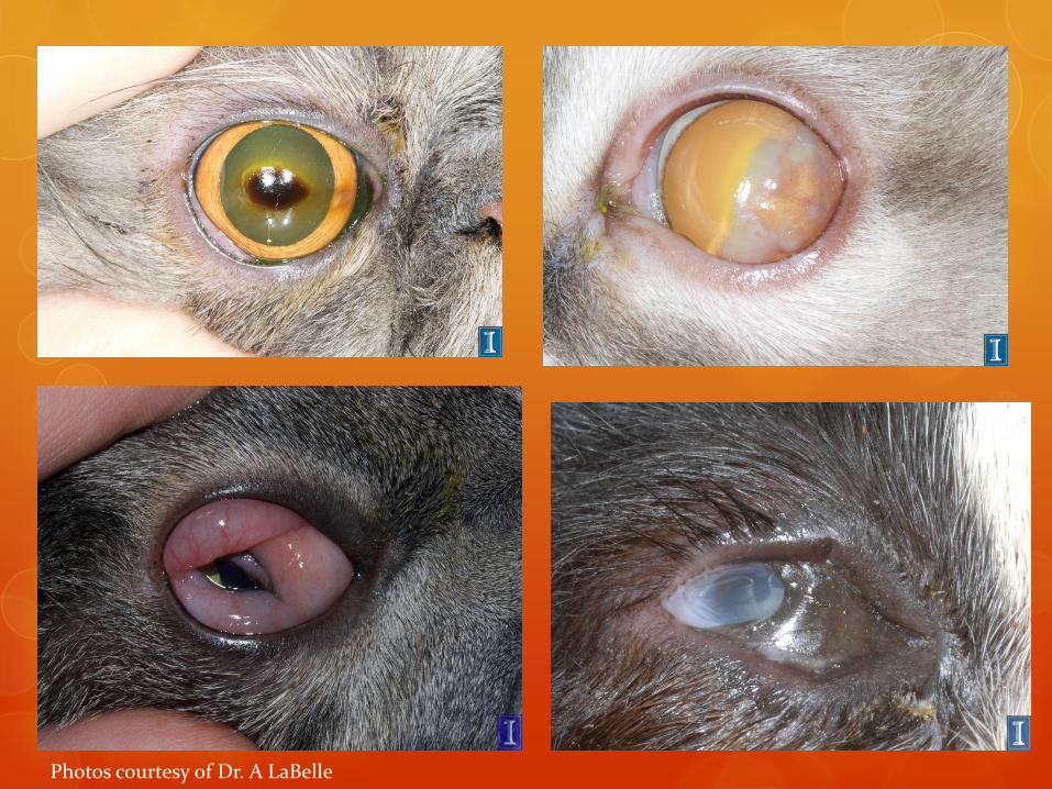
Photos courtesy of Dr. A LaBelle
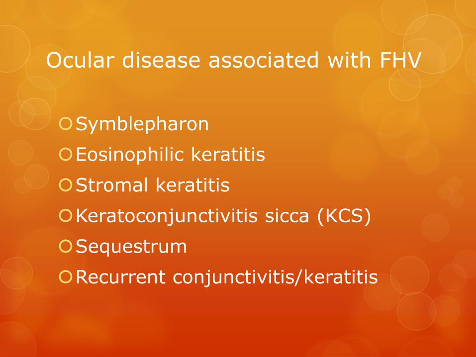
Ocular disease associated with FHV
Symblepharon
Eosinophilic keratitis
Stromal keratitis
Keratoconjunctivitis sicca (KCS)
Sequestrum
Recurrent conjunctivitis/keratitis
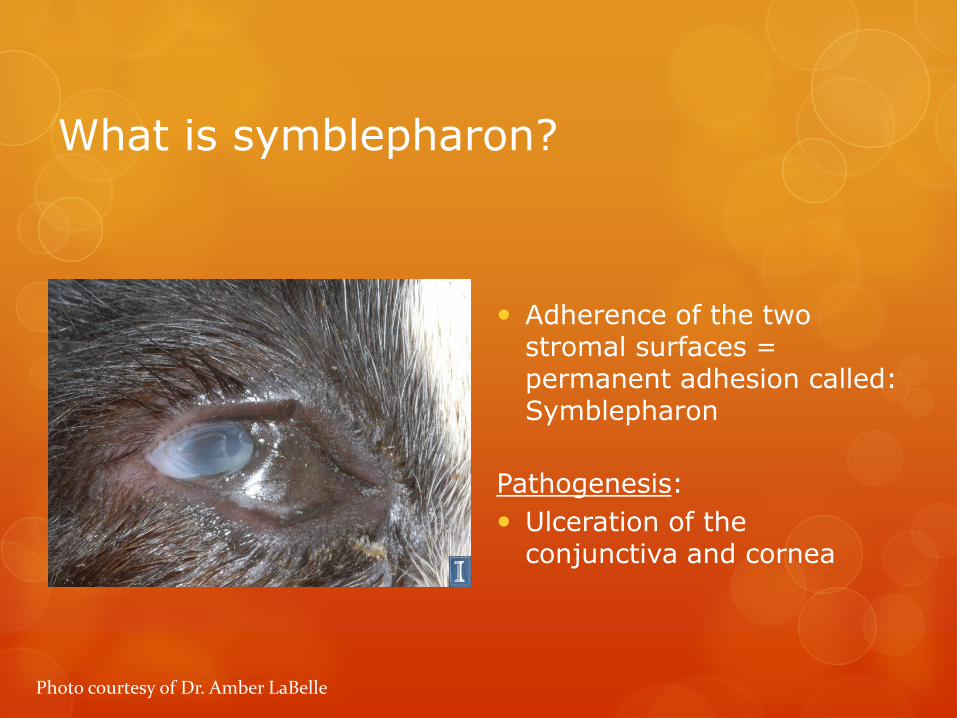
What is symblepharon?
Adherence of the two stromal surfaces = permanent adhesion called: Symblepharon
Pathogenesis:
Ulceration of the conjunctiva and cornea
Photo courtesy of Dr. Amber LaBelle
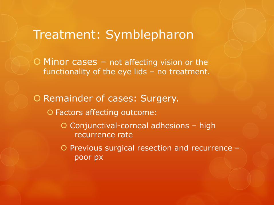
Treatment: Symblepharon
Minor cases – not affecting vision or the
functionality of the eye lids – no treatment.
Remainder of cases: Surgery.
Factors affecting outcome:
Conjunctival-corneal adhesions – high recurrence rate
Previous surgical resection and recurrence – poor px
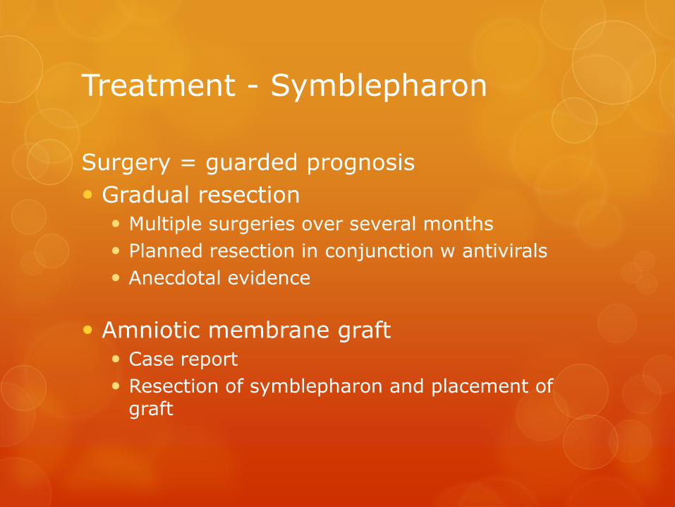
Treatment - Symblepharon
Surgery = guarded prognosis
Gradual resection
Multiple surgeries over several months
Planned resection in conjunction w antivirals
Anecdotal evidence
Amniotic membrane graft
Case report
Resection of symblepharon and placement of graft
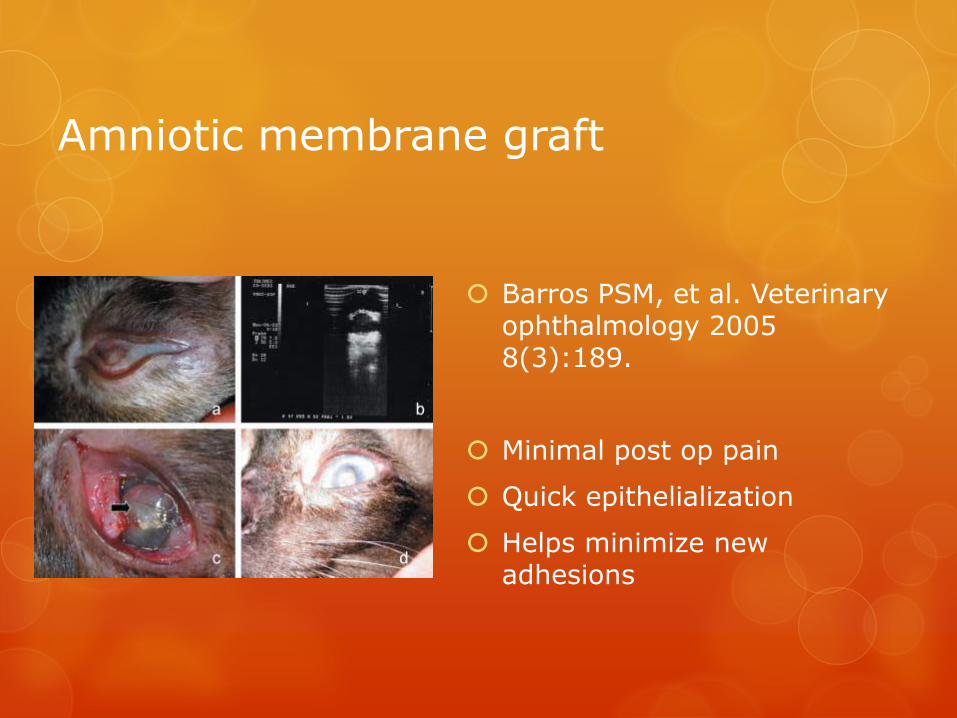
Amniotic membrane graft
Barros PSM, et al. Veterinary ophthalmology 2005 8(3):189.
Minimal post op pain
Quick epithelialization
Helps minimize new adhesions
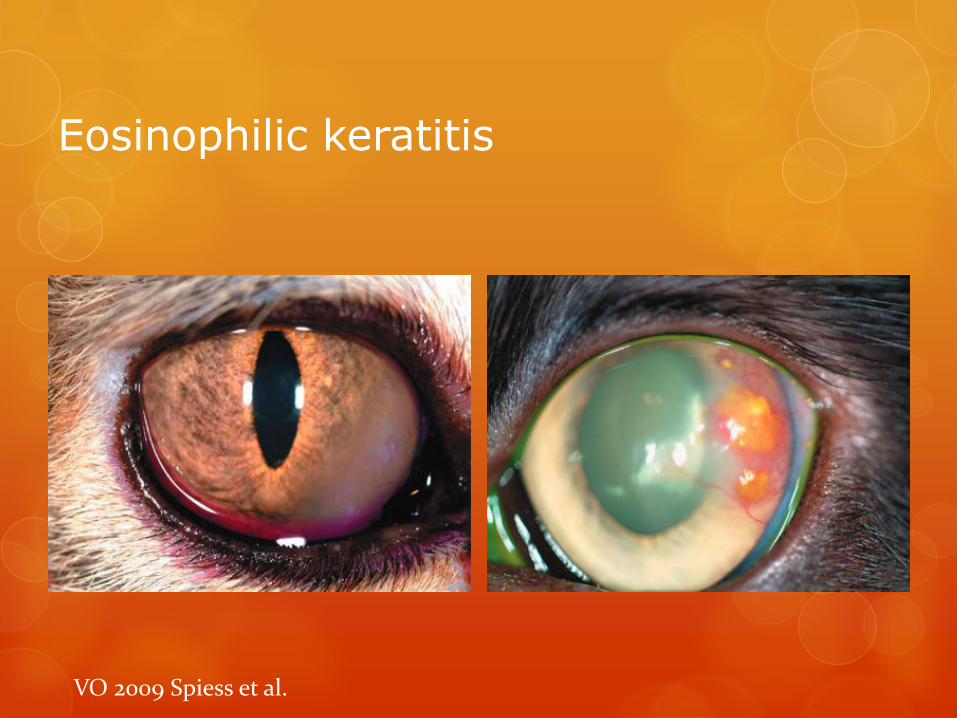
Eosinophilic keratitis
VO 2009 Spiess et al.
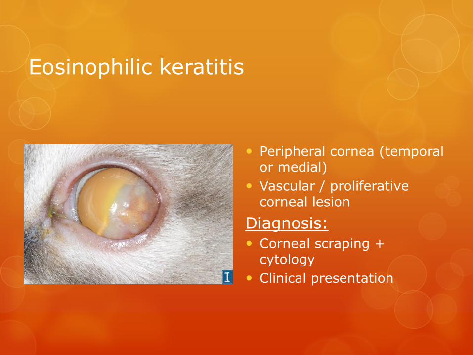
Eosinophilic keratitis
Peripheral cornea (temporal or medial)
Vascular / proliferative corneal lesion
Diagnosis:
Corneal scraping + cytology
Clinical presentation
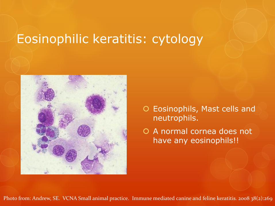
Eosinophilic keratitis: cytology
Eosinophils, Mast cells and neutrophils.
A normal cornea does not have any eosinophils!!
Photo from: Andrew, SE. VCNA Small animal practice. Immune mediated canine and feline keratitis. 2008 38(2):269
Eosinophilic keratitis: treatment
33-79% incidence of FHV-1
Immunomodulatory
Topical steroid (dexamethasone) 4x daily
(1.5% cyclosporine 2-3 times daily – VO 2009)
If corneal ulceration or conjunctivitis
Topical antibiotics (Chloramphenicol, triple antibiotic)
Anti-viral medication (topically or orally)
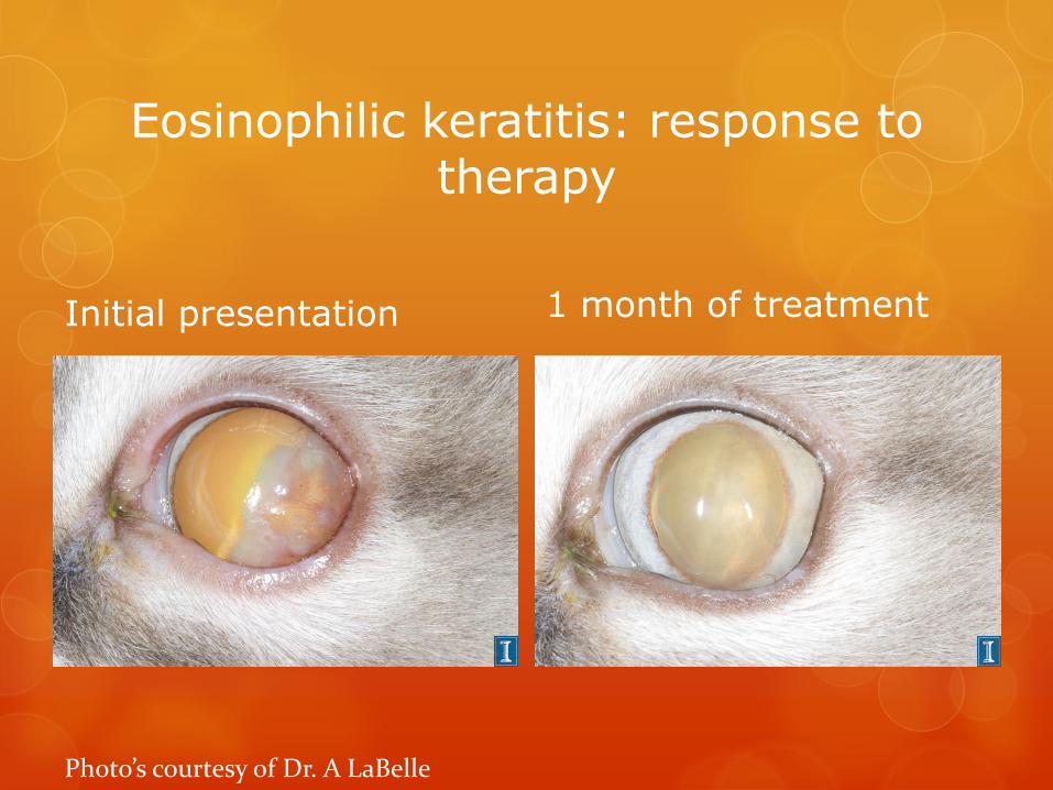
Eosinophilic keratitis: response to therapy
Initial presentation 1 month of treatment
Photo’s courtesy of Dr. A LaBelle
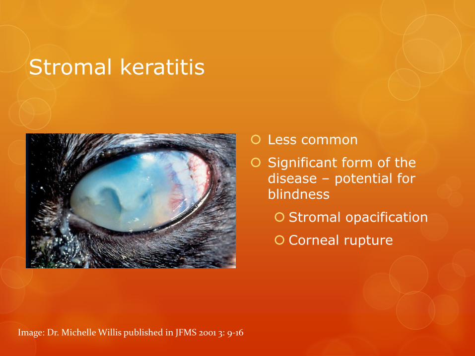
Stromal keratitis
Less common
Significant form of the disease – potential for blindness
Stromal opacification
Corneal rupture
Image: Dr. Michelle Willis published in JFMS 2001 3: 9-16
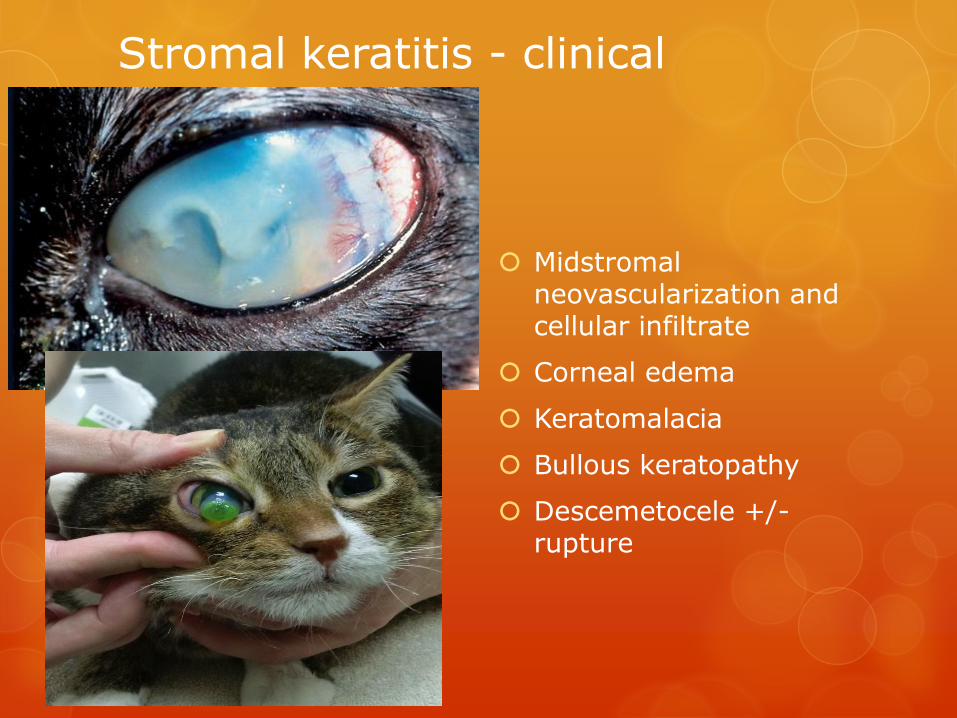
Stromal keratitis - clinical
Image: Dr. Michelle Willis published in JFMS 2001 3: 9-16
Midstromal neovascularization and cellular infiltrate
Corneal edema
Keratomalacia
Bullous keratopathy
Descemetocele +/- rupture
Stromal keratitis - treatment
If deep corneal ulceration or marked corneal edema/ bulla – consider referral.
Medical management Anti-viral therapy (topical +/-oral)
Ganciclovir interferron 4-6 times daily
Antibiotics
Ciprofloxacin, Ofloxacin 4-6 times daily
Cefazolin 4-6 times daily
Anti-collagenase
Serum or EDTA (1-2%) 4 -6 times daily
Ecollar
Stromal keratitis - treatment
Surgical management
Bullous keratopathy – third eyelid flap
NOTE: do not do this if the cornea is melting!
Conjunctival flap or biosis graft placement
Provide support to the cornea
Provide vasculature – conjunctival flap
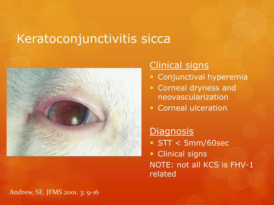
Keratoconjunctivitis sicca
Clinical signs
Conjunctival hyperemia
Corneal dryness and neovascularization
Corneal ulceration
Diagnosis
STT < 5mm/60sec
Clinical signs
NOTE: not all KCS is FHV-1 related
Andrew, SE. JFMS 2001. 3: 9-16
KCS: treatment
Lubricants
0.15% Hylashield (iMed Pharma) – long lasting
Preservative free artificial tears (gel / ointment) – as often as possible
(Pilocarpine) Oral 0.25% Pilocarpine – titrate to effect (beware
parasympathomimetic toxicity)
Cyclosporine (Optimmune) – not licensed in cats
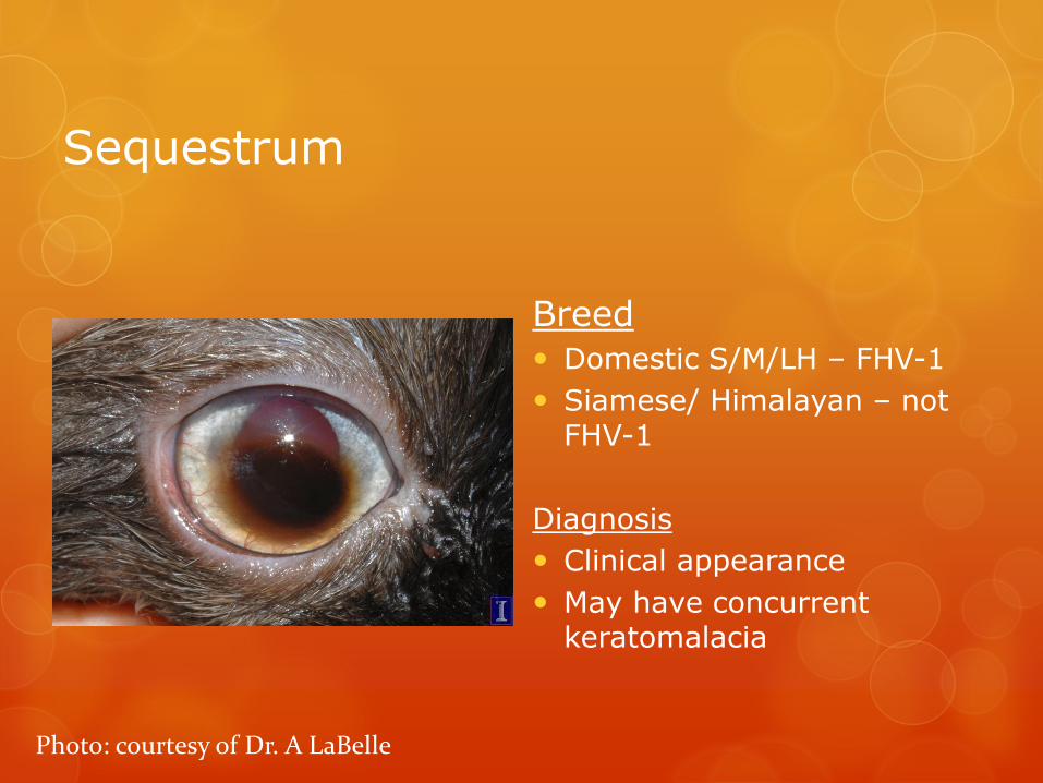
Sequestrum
Breed
Domestic S/M/LH – FHV-1
Siamese/ Himalayan – not FHV-1
Diagnosis
Clinical appearance
May have concurrent keratomalacia
Photo: courtesy of Dr. A LaBelle
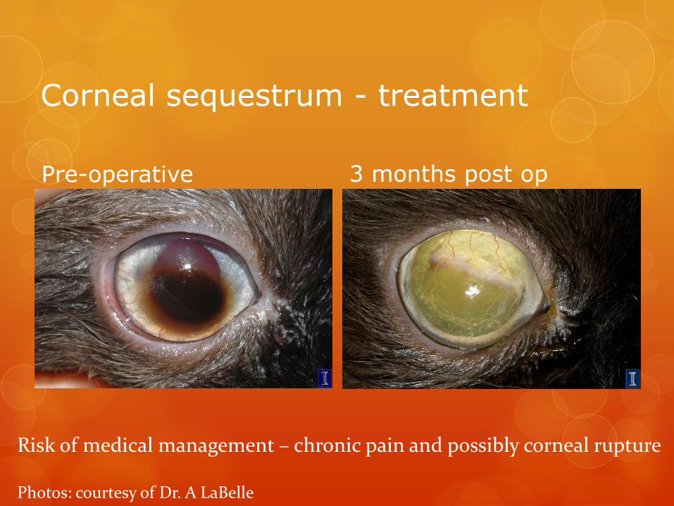
Corneal sequestrum - treatment
Pre-operative 3 months post op
Photos: courtesy of Dr. A LaBelle
Risk of medical management – chronic pain and possibly corneal rupture
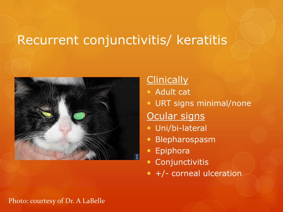
Recurrent conjunctivitis/ keratitis
Clinically
Adult cat
URT signs minimal/none
Ocular signs
Uni/bi-lateral
Blepharospasm
Epiphora
Conjunctivitis
+/- corneal ulceration
Photo: courtesy of Dr. A LaBelle
Recurrent conjunctivitis/ keratitis
Treatment:
Topical antiviral – Ganciclovir interferron 4-6 times daily for 10-14 days (or until the clinical signs have been resolved for 1 week at least)
Tear gel
If marked conjunctivitis or repeated “flare ups” – consider systemic anti-viral (Famciclovir)
Treat other concurrent pathologies (low tear values, corneal ulceration)
Famciclovir…
Oral anti-viral
“pro-drug” of penciclovir
Blood work should be done prior to starting this medication – should not be given in cats with renal compromise.
Dose: 40mg/kg BID-TID (Thomasy SM 2012)
What about L-Lysine?
Excellent adjunct therapy!
Not virucidal – thus cannot be the sole therapy
Some evidence to support - life long use
500mg PO BID

Thank you for your time.