α-Methylacyl-CoA racemase. An enzyme at crossroads in lipid
73
α-METHYLACYL-COA RACEMASE. AN ENZYME AT CROSSROADS IN LIPID METABOLISM KALLE SAVOLAINEN Faculty of Medicine; Faculty of Science, Department of Biochemistry; Biocenter Oulu, University of Oulu OULU 2004
Transcript of α-Methylacyl-CoA racemase. An enzyme at crossroads in lipid
α-METHYLACYL-COA RACEMASE. AN ENZYME AT CROSSROADS IN LIPID METABOLISM
KALLESAVOLAINEN
Faculty of Medicine;Faculty of Science,
Department of Biochemistry;Biocenter Oulu,
University of Oulu
OULU 2004

KALLE SAVOLAINEN
α-METHYLACYL-COA RACEMASE. AN ENZYME AT CROSSROADS IN LIPID METABOLISM
Academic Dissertation to be presented with the assent ofthe Faculty of Medicine, University of Oulu, for publicdiscussion in Raahensali (Auditorium L10), Linnanmaa, onNovember 19th, 2004, at 12 noon.
OULUN YLIOPISTO, OULU 2004
Copyright © 2004University of Oulu, 2004
Supervised byProfessor J. Kalervo HiltunenDoctor Tiina Kotti
Reviewed byDocent Matti JauhiainenProfessor J. Peter Slotte
ISBN 951-42-7468-7 (nid.)ISBN 951-42-7469-5 (PDF) http://herkules.oulu.fi/isbn9514274695/
ISSN 0355-3221 http://herkules.oulu.fi/issn03553221/
OULU UNIVERSITY PRESSOULU 2004
Savolainen, Kalle, α-Methylacyl-CoA racemase. An enzyme at crossroads in lipidmetabolism Faculty of Medicine and Biocenter Oulu, P.O.Box 5000, FIN-90014 University of Oulu, Finland;Faculty of Science, Department of Biochemistry, P.O.Box 3000, FIN-90014 University of Oulu,Finland, 2004Oulu, Finland
Abstractα-Methylacyl-CoA racemase (Amacr) is an enzyme at the merging point of two important pathwaysof lipid metabolism: elimination of methyl-branched fatty acids and synthesis of bile acids. Amacr isregarded as obligatory for these processes. Patients with Amacr-deficiency suffer from adult onsetsensory motor neuropathy and/or severe neonatal cholestasis with coagulopathy and fat-solublevitamin malabsorption. Amacr is also linked to cancer and so far has been proposed as a new markerfor diagnosis of at least prostate and colon cancers. Common sources of phytol derived branched-chain fatty acids for man are ruminant fats, meat and dairy products. The bile acid synthesis is themain pathway for cholesterol catabolism. Amacr is considered to be a member of family III of theCoA transferases (L-carnitine dehydratase - bile acid inducible protein F (CaiB-BaiF) family) andlocalized to two subcellular compartments, mitochondria and peroxisomes.
In this work the mouse gene encoding Amacr was characterized, the gene was inactivated andmutational and structural studies were used to determine the loop and the active site structure of theenzyme. It was shown that mouse Amacr which locates both to mitochondria and peroxisomes, is anidentical product of a single gene, which is located at chromosome 15, region 15B1. Neitheralternative replication, splicing, or any post-translational modifications of the enzyme occur.
The mouse model for Amacr-deficiency indicated a role of Amacr in detoxification of methyl-branched fatty acids, and suggested that a diet free from these phytol metabolites may function as atreatment for the deficiency. Furthermore, major changes were observed in the bile acid pool of theknock-out mice compared to wild type mice. However, the study suggests that there is an Amacr-independent pathway for synthesis of bile acids albeit of low capacity, which provides a way forAmacr-deficient individuals to survive.
The mutational and structural studies confirmed Amacr as a member of family III of the CoAtransferases. Furthermore, according to comparisons of the structural data of Amacr and othermembers of the family (FRC, YfdW), the superfamily can be divided into two subgroups, racemasesand transferases. Proteins in the subfamilies share the CoA-binding mode, but the substratespecificities as well as the catalysed reaction differ greatly.
Keywords: α-methylacyl-CoA racemase, bile acids, cholesterol, fatty acids, lipidmetabolism, mouse model, physiological function


To Emilia

Acknowledgemets
The present study was carried out at the Department of Biochemistry and Biocenter Oulu, University of Oulu, during the years 1998-2004.
I owe my most sincere thanks to my supervisor and friend, Dr. Tiina Kotti, without whom I certainly could not have managed. I wish to express my deepest gratitude to my supervisor, Professor Kalervo Hiltunen for his guidance and enthusiastic attitude towards research. I am deeply grateful to Dr. Werner Schmitz and Professor Ernst Conzelmann for their contribution to this work and also for hosting me in Würzburg. I wish to thank Professors Seppo Vainio and Rik Wierenga for their help and knowledge. I thank all the group leaders and staff at the Department for providing nice environment for research. I am grateful to Docent Matti Jauhiainen and Professor J. Peter Slotte for their valuable comments on the manuscript and Dr. Peter Cura for revision of the language.
I owe my thanks to all the co-authors and collaborators. Especially, I wish to thank Eija Selkälä for frisky attitude and irreplaceable work in the lab, and Prasenjit Bhaumik for his efforts in racemase business. Dr. Raija Sormunen and Dr. Ahmed Yagi deserve my warmest thanks for all the help with electron microscopy. My special thanks go to past and present members of KH-team. Particularly Aare Rokka, Jukka Taskinen, Ilkka Miinalainen, Antti Haapalainen and Juha Torkko are acknowledged for participating in stimulating conversations about researchers state and of course afterwork activities. I am grateful to Dr. Ari-Pekka Kvist for all the help with unwilling computers. I also wish to thank the members of Oulu bioscience community who I have learned to know during these years. Especially, I thank the brothers, Mika Ilves (Anttu) and Seppo Kilpeläinen, who have helped a lot with little defects, which are easy to fix.
I am most grateful to my friends, who helped me to forget the work from time to time. Special thanks go to Tuomo, Marko and Kimmo for being part of the zipper, and also to Mika, Riikonen and Kangis. A lot have we experienced during the years. I thank the guys from Heinola for good times. I owe my thanks to Mika, Suvi, Teemu, Tuomo, Mako, Timppa, Tumppi and all the other taidokas, especially deceased Saiko Shihan, Seiken Shukumine, the founder of Taido, who have given me strength, bruises and fun moments on tatami and outside.
I wish to thank my brothers Janne and Harri for all the things, especially the old and English ones, during so many years. I also thank Harri and his wife Maria for

reproducing themselves and bringing up wonderful children, my godson Volmari, his sisters Vilhelmiina and Magdaleena, and those yet to come. My warmest thanks go to my parents who have given me love and support throughout my life. I further thank my father for his contribution to this work and brilliant insight on pathology.
Finally, I owe my heartfelt thanks to my love, Emilia. Your presence and love has kept me going. I know that you have a heart of gold.
This study has been supported financially by the Academy of Finland, Sigrid Juselius Foundation, Scientific foundations of Farmos and Instrumentarium, and Finnish medical and cultural foundations.
Oulu, November 2004 Kalle Savolainen
Abbreviations
ACOX1/AOX straight chain acyl-CoA oxidase ACOX2/BOX branched chain acyl-CoA oxidase AKR1D1 ∆4-3-oxosteroid 5β-reductase ALAT alanine amino transferase Amacr α-methylacyl-CoA racemase from mammals APHOS alkaline phosphatase BAC bacterial artificial chromosome BaiF bile acid inducible protein F C27 3β-HSD 3β-hydroxy-∆5-C27-steroid oxidoreductase CaiB L-carnitine dehydratase CD circular dichroism CH25H cholesterol 25-hydroxylase CHO Chinese hamster ovary CoA coenzyme A CPT carnitine palmitoyltransferase CTX cerebrotendinous xanthomatosis CYP7A1 cholesterol 7α-hydroxylase CYP8B1 sterol 12α-hydroxylase CYP27A1 sterol 27-hydroxylase CYP39A1 oxysterol 7α-hydroxylase CYP46A1 cholesterol 24-hydroxylase D/THCA di/trihydroxycoprostanoic acid EM electron microscopy ER endoplasmic reticulum ES embryonic stem FRC formyl-CoA: oxalate CoA-transferase FXR farnesoid X receptor HMG-CoA hydroxymethylglutaryl-CoA IPP isopentenyl pyrophosphate LACS long-chain fatty acyl-CoA synthetase LXR liver X receptor
MCR α-methylacyl-CoA racemase from Mycobacterium tuberculosis MFE multifunctional enzyme MTS mitochondrial targeting signal PBD peroxisome biogenesis defect PCR polymerase chain reaction PGK phosphoglycerine kinase PhyH phytanoyl-CoA 2-hydroxylase PVDF polyvinylidene difluoride PPARα,β,γ peroxisome proliferator activated receptor type α,β,γ PTS peroxisomal targeting signal R rectus RT-PCR reverse transcriptase-PCR SCP sterol carrier protein SDS-PAGE sodium dodecyl sulphate polyacrylamide gel electrophoresis S sinister VLACS very long chain acyl-CoA synthetase VLCFA very long chain fatty acids XALD X-linked adrenoleukodystrophy
List of original articles
This thesis is based on the following articles, which are referred to in the text by their Roman numerals:
I Kotti TJ, Savolainen K, Helander HM, Yagi A, Novikov DK, Kalkkinen N,
Conzelmann E, Hiltunen JK & Schmitz W (2000) In mouse α-methylacyl-CoA racemase – the same gene product is simultaneously located in mitochondria and peroxisomes. J Biol Chem 275: 20887-20895.
II Savolainen K, Kotti TJ, Schmitz W, Savolainen TI, Sormunen RT, Ilves M, Vainio
SJ, Conzelmann E & Hiltunen JK (2004) A mouse model for α-methylacyl-CoA racemase deficiency: adjustment of bile acid synthesis and intolerance to dietary methyl-branched lipids. Hum Mol Genet 13:955-965.
III Savolainen K, Bhaumik P, Schmitz W, Kotti TJ, Conzelmann E, Wierenga RK and
Hiltunen JK α-Methylacyl-CoA racemase from Mycobacterium tuberculosis – mutational and structural characterization of the fold and the active site (submitted)

Contents
Acknowledgements Abbreviations List of original articles Contents 1 Introduction ...................................................................................................................15 2 Review of the literature .................................................................................................16
2.1 Isoprenoids .............................................................................................................16 2.2 α-Oxidation ............................................................................................................17
2.2.1 The α-oxidation pathway.................................................................................18 2.2.2 Genetic defects associated with phytanic acid metabolism .............................20
2.2.2.1 Refsum disease .........................................................................................20 2.2.2.2 Peroxisome biogenesis defects .................................................................21 2.2.2.3 Other defects.............................................................................................21
2.3 Formation of bile acid intermediates from cholesterol ...........................................22 2.3.1 Hydroxylation of cholesterol ...........................................................................22 2.3.2 Modifications to the ring structure and side chain...........................................23 2.3.3 Deficiencies in the bile acid synthesis pathway...............................................24
2.3.3.1 CYP7A1-deficiency..................................................................................24 2.3.3.2 Cerebrotendinous xanthomatosis ..............................................................24 2.3.3.3 CYP7B1-deficiency..................................................................................25 2.3.3.4 C27 3β-HSD-deficiency.............................................................................25 2.3.3.5 AKR1D1-deficiency .................................................................................25
2.4 α-Methylacyl-CoA racemase..................................................................................25 2.4.1 Substrates for Amacr .......................................................................................26 2.4.2 Properties of Amacr .........................................................................................27 2.4.3 Subcellular localization of Amacr....................................................................28 2.4.4 The reaction catalyzed by Amacr.....................................................................28 2.4.5 Evolution of Amacr .........................................................................................29 2.4.6 Amacr-deficiency.............................................................................................30 2.4.7 Amacr in cancer ...............................................................................................30
2.5 β-Oxidation.............................................................................................................31 2.5.1 β-Oxidation of branched chain compounds.....................................................33 2.5.2 Conjugation of bile acid intermediates ............................................................33 2.5.3 Complete degradation of pristanoyl-CoA........................................................34 2.5.4 MFE-2-deficiency............................................................................................35
3 Outlines of the present study .........................................................................................36 4 Materials and Methods ..................................................................................................37
4.1 Sequences (I, II, III)................................................................................................37 4.2 Animal care (I, II) ...................................................................................................37 4.3 Protein analysis (I, II, III) .......................................................................................38 4.4 Activity measurements (I, II, III) ............................................................................38 4.5 Expression of recombinant proteins (I, III) ............................................................38 4.6 Histological analyses (I, II) ....................................................................................39 4.7 Separation of organelles and immunoaffinity isolation of Amacr (I) .....................39 4.8 Southern hybridization (I, II)..................................................................................39 4.9 cDNA and mRNA analyses (I, II)...........................................................................40 4.10 Characterization of the Amacr gene (I).................................................................40 4.11 Promoter activity (I) .............................................................................................41 4.12 Generation of Amacr-/- mice (II) ...........................................................................41 4.13 Analyses of Amacr-/- mice (II) ..............................................................................41 4.14 Circular dichroism spectroscopy (III)...................................................................42 4.15 Crystallization and structure characterization of MCR (III) .................................42
5 Results ...........................................................................................................................43 5.1 Amacr is localized to both peroxisomes and mitochondria (I) ...............................43 5.2 cDNA analysis reveals an extension to cAMACR (I) ............................................44 5.3 A single Amacr with an operable promoter region encoding one gene product (I).44 5.4 Alterations in the bile acid pool of Amacr-/- mice (II).............................................45 5.5 The effects of phytol (II).........................................................................................46 5.6 The expression of various genes upon inactivation of Amacr (II) ..........................47 5.7 Mutations identifying the active site of Amacr (III) ...............................................47 5.8 Overall structure of the fold (III) ............................................................................48
6 Discussion .....................................................................................................................49 6.1 Dual localization of Amacr.....................................................................................49 6.2 Mouse model for Amacr-deficiency .......................................................................51 6.3 Studies on the active site of Amacr.........................................................................53
7 Conclusions ...................................................................................................................55 References
1 Introduction
Methyl-branched fatty acids and other isoprenoid-derived compounds such as cholesterol and its metabolites must be degraded in order to prevent their accumulation in the body, which can result in various diseases. The α-oxidation cycle converts 3-methyl-branched fatty acids, which are usually derived from phytol, to 2/α-methyl-branched fatty acids. Phytol is a degradation product of chlorophyll and its metabolites are obtained mostly from dietary sources in ruminant fats, meat and dairy products. 25/α-Methyl-branched bile acid intermediates are derived from cholesterol through a multi-step pathway. The products from both of these pathways can be further processed to shortened fatty acids or primary bile acids by β-oxidation, which is the main degradation pathway for fatty acids.
The β-oxidation cycle in higher eukaryotes occurs in both mitochondria and peroxisomes. Straight and short-chained fatty acids are usually degraded in mitochondrial β-oxidation, while peroxisomal β-oxidation is responsible of degradation of long and branched chain compounds. The α-oxidation cycle yields both rectus (R)- and sinister (S)-isoforms of α-methyl-branched fatty acids and furthermore, all bile acid intermediates are in (R)-configuration. However, the first enzymes in the β-oxidation cycles are stereo-specific for (S)-isoforms of their CoA-activated substrates. Therefore, (R)-isoforms have to be converted to the (S)-orientation by a peroxisomal and mitochondrial enzyme, α-methylacyl-CoA racemase (Amacr), before β-oxidation.
Amacr is considered to be a member of family III of the CoA transferases, also referred to as the CaiB-BaiF family. The enzyme is important in i) bile acid synthesis from cholesterol, ii) degradation and detoxification of methyl-branched fatty acids, and iii) metabolism of 2-arylpropionic acid (Ibuprofen®). Patients with Amacr-deficiency suffer from adult onset sensory motor neuropathy and/or severe neonatal cholestasis with coagulopathy and fat-soluble vitamin malabsorption. Since Amacr has so far been found to be overexpressed in prostate and colon cancers, it is also linked to cancer and proposed as a relatively specific cancer marker.
2 Review of the literature
2.1 Isoprenoids
Isoprenoids are distributed throughout the animal, plant, and bacterial kingdoms. Important isoprenoids in mammals are fat-soluble vitamins A (retinoid), E (tocopherol) and K (quinone), acyclic fatty acids and dolichols, and cholesterol and its derivatives. A feature common to all isoprenoids is that they are formed from isoprene units and include many methyl-branches in their structures (Wright 1961, West 1997).
Isoprenoids are derived from one or more isoprene (2-methyl-1,3-butadiene) units. Isopentenyl pyrophosphate (IPP) is a universal biological five carbon containing (C5) precursor of isoprenoids (Wright 1961) It is generally accepted that in the cells of most of living organisms IPP is synthesized via the acetate/mevalonate pathway, in which two molecules of acetyl-CoA form an acetoacetyl-CoA, which accepts an additional acetyl-CoA to form hydroxymethylglutaryl-CoA (HMG-CoA), which is then reduced to mevalonic acid by HMG-CoA reductase, a principal regulatory enzyme in isoprenoid biosynthesis (Siperstein 1970). Mevalonic acid is phosphorylated twice to yield mevalonic-5-pyrophosphate from which IPP is formed via a decarboxylation/elimination step (Spurgeon & Porter 1983) (Figure 1).
In plant cells there is also an additional mevalonate independent pathway for IPP synthesis. Sterols are synthesized via the general acetate/mevalonate pathway in the cytoplasm and endoplasmic reticulum (ER) but the chloroplast-bound isoprenoids like β-carotene, lutein, phytol and plastoquinone-9, are synthesized in plastids via the glyceraldehyde phosphate/pyruvate pathway. In this pathway a C2-precursor is derived from pyruvate by adding thiamine pyrophosphate and removing one carbon as CO2. Addition of this C2 precursor to the C3 precursor glyceraldehyde-3-phosphate and removing thiamine pyrophosphate yields the C5 precursor D-1-deoxyxylulose-5-phosphate which undergoes rearrangements yielding a branched carbon chain of IPP (Lichtenthaler et al. 1997). IPP can undergo isomerization and form dimethylallyl pyrophosphate which condensates with another IPP molecule to yield geranyl pyrophosphate (C10). Additions of C5 units to geranyl pyrophosphate yield farnesyl pyrophosphate (C15) and geranylgeranyl pyrophosphate (C20). These longer prenyl
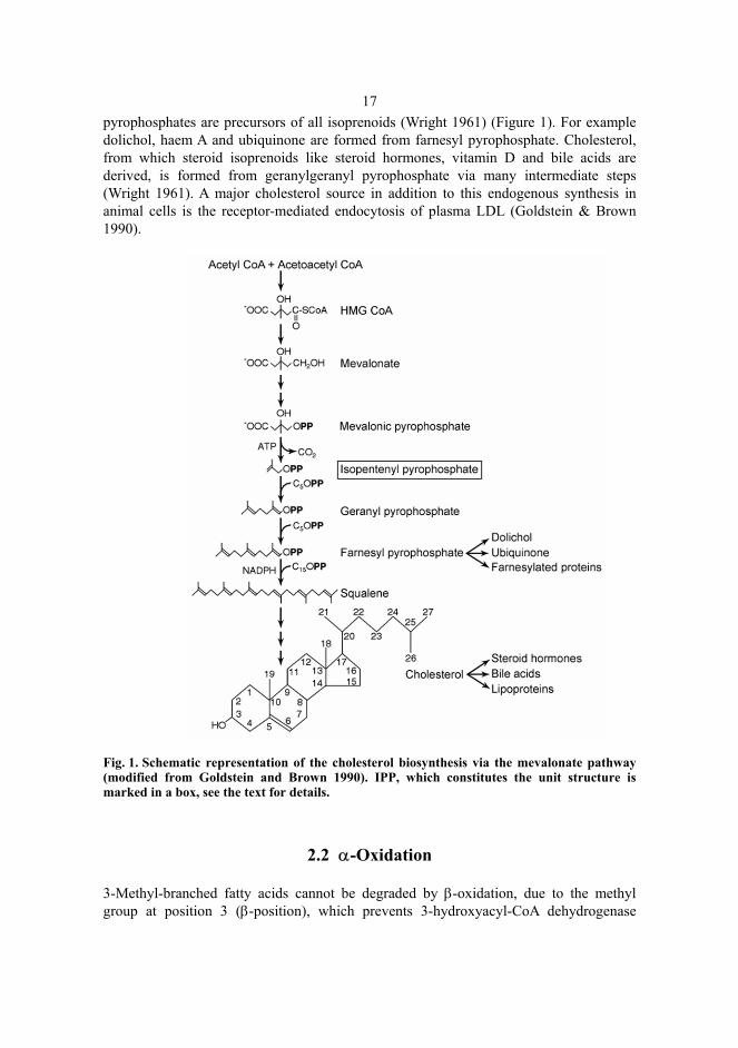
17pyrophosphates are precursors of all isoprenoids (Wright 1961) (Figure 1). For example dolichol, haem A and ubiquinone are formed from farnesyl pyrophosphate. Cholesterol, from which steroid isoprenoids like steroid hormones, vitamin D and bile acids are derived, is formed from geranylgeranyl pyrophosphate via many intermediate steps (Wright 1961). A major cholesterol source in addition to this endogenous synthesis in animal cells is the receptor-mediated endocytosis of plasma LDL (Goldstein & Brown 1990).
Fig. 1. Schematic representation of the cholesterol biosynthesis via the mevalonate pathway (modified from Goldstein and Brown 1990). IPP, which constitutes the unit structure is marked in a box, see the text for details.
2.2 α-Oxidation
3-Methyl-branched fatty acids cannot be degraded by β-oxidation, due to the methyl group at position 3 (β-position), which prevents 3-hydroxyacyl-CoA dehydrogenase
18functioning and the formation of a 3-ketoacyl-CoA intermediate, in the third step of β-oxidation (Van Veldhoven et al. 1991). The terminal carboxyl-group must first be removed by α-oxidation, which yields 2-methyl-branched fatty acids (methyl group at α-position). 3-Methyl-branched fatty acids can also be degraded by ω-oxidation, but at least with phytanic acid (3,7,11,15-tetramethylhexadecanoic acid) this occurs only at low rate in healthy individuals (Try 1968, Billimoria et al. 1982). Since phytanic acid was found to accumulate in patients with Refsum disease (Klenk & Kahike 1963) numerous studies have been carried out on its metabolism and most of the information about α-oxidation has been obtained from these studies (Wanders et al. 2003).
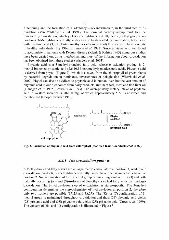
Phytanic acid is a 3-methyl-branched fatty acid, whose α-oxidation product is 2-methyl-branched pristanic acid (2,6,10,14-tetramethylpentadecanoic acid). Phytanic acid is derived from phytol (Figure 2), which is cleaved from the chlorophyll of green plants by bacterial degradation in ruminants, invertebrates or pelagic fish (Wierzbicki et al. 2002). Phytol can also be oxidized to phytanic acid in human liver, but the vast amount of phytanic acid in our diet comes from dairy products, ruminant fats, meat and fish liver oil (Flanagan et al. 1975, Brown et al. 1993). The average daily dietary intake of phytanic acid in western societies is 50-100 mg, of which approximately 50% is absorbed and metabolized (Dhopeshwarkar 1980).
Fig. 2. Formation of phytanic acid from chlorophyll (modified from Wierzbicki et al. 2002).
2.2.1 The α-oxidation pathway
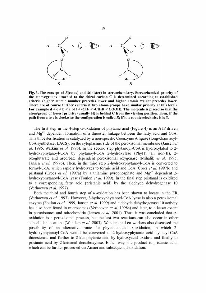
3-Methyl-branched fatty acids have an asymmetric carbon atom at position 3, while their α-oxidation products, 2-methyl-branched fatty acids have the asymmetric carbon at position 2. No racemization of the 3-methyl group occurs (Fingerhut et al. 1993) and both naturally occurring (R)- and (S)-isoforms of 3-methyl-branched fatty acids can undergo α-oxidation. The 2-hydroxylation step of α-oxidation is stereo-specific. The 3-methyl configuration determines the stereochemistry of hydroxylation at position 2, therefore only two isomers are possible (3R,2S and 3S,2R). The (R)- or (S)-configuration of 3-methyl group is maintained throughout α-oxidation and thus, (3S)-phytanic acid yields (2S)-pristanic acid and (3R)-phytanic acid yields (2R)-pristanic acid (Croes et al. 1999). The concept of (R)- and (S)-configuration is illustrated in Figure 3.
19
Fig. 3. The concept of R(ectus) and S(inister) in stereochemistry. Stereochemical priority of the atoms/groups attached to the chiral carbon C is determined according to established criteria (higher atomic number precedes lower and higher atomic weight precedes lower. There are of course further criteria if two atoms/groups have similar priority at this level). For example d < c < b < a (-H < -CH3 < -CH2R < COOH). The molecule is placed so that the atom/group of lowest priority (usually H) is behind C from the viewing position. Then, if the path from a to c is clockwise the configuration is called R; if it is counterclockwise it is S.
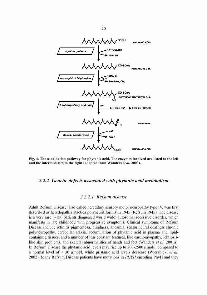
The first step in the 4-step α-oxidation of phytanic acid (Figure 4) is an ATP driven
and Mg2+ dependent formation of a thioester linkage between the fatty acid and CoA. This thioesterification is catalyzed by a non-specific Coenzyme A ligase (long-chain acyl-CoA synthetase, LACS), on the cytoplasmic side of the peroxisomal membrane (Jansen et al. 1996, Watkins et al. 1996). In the second step phytanoyl-CoA is hydroxylated to 2-hydroxyphytanoyl-CoA by phytanoyl-CoA 2-hydroxylase (PhyH), an iron(II), 2-oxoglutarate and ascorbate dependent peroxisomal oxygenase (Mihalik et al. 1995, Jansen et al. 1997b). Then, in the third step 2-hydroxyphytanoyl-CoA is converted to formyl-CoA, which rapidly hydrolyzes to formic acid and CoA (Croes et al. 1997b) and pristanal (Croes et al. 1997a) by a thiamine pyrophosphate and Mg2+ dependent 2-hydroxyphytanoyl-CoA lyase (Foulon et al. 1999). In the final step pristanal is oxidized to a corresponding fatty acid (pristanic acid) by the aldehyde dehydrogenase 10 (Verhoeven et al. 1997).
Both the third and fourth step of α-oxidation has been shown to locate in the ER (Verhoeven et al. 1997). However, 2-hydroxyphytanoyl-CoA lyase is also a peroxisomal enzyme (Foulon et al. 1999, Jansen et al. 1999) and aldehyde dehydrogenase 10 activity has also been found in microsomes (Verhoeven et al. 1998a) and later, to a lesser extent in peroxisomes and mitochondria (Jansen et al. 2001). Thus, it was concluded that α-oxidation is a peroxisomal process, but the last two reactions can also occur in other subcellular locations (Wanders et al. 2003). Wanders and co-workers also discussed the possibility of an alternative route for phytanic acid α-oxidation, in which 2-hydroxyphytanoyl-CoA would be converted to 2-hydroxyphytanic acid by acyl-CoA thioesterase and further to 2-ketophytanic acid by hydroxyacid oxidase and finally to pristanic acid by 2-ketoacid decarboxylase. Either way, the product is pristanic acid, which can be further processed via Amacr and subsequent β-oxidation.
20
Fig. 4. The α-oxidation pathway for phytanic acid. The enzymes involved are listed to the left and the intermediates to the right (adapted from Wanders et al. 2003).
2.2.2 Genetic defects associated with phytanic acid metabolism
2.2.2.1 Refsum disease
Adult Refsum Disease, also called hereditary sensory motor neuropathy type IV, was first described as heredopathia atactica polyneuritiformis in 1945 (Refsum 1945). The disease is a very rare (~150 patients diagnosed world wide) autosomal recessive disorder, which manifests in late childhood with progressive symptoms. Clinical symptoms of Refsum Disease include retinitis pigmentosa, blindness, anosmia, sensorineural deafness chronic polyneuropathy, cerebellar ataxia, accumulation of phytanic acid in plasma and lipid-containing tissues, and a number of less constant features, like cardiomyopathy, ichtiosis-like skin problems, and skeletal abnormalities of hands and feet (Wanders et al. 2001a). In Refsum Disease the phytanic acid levels may rise up to 200-2500 µmol/l, compared to a normal level of < 30 µmol/l, while pristanic acid levels decrease (Wierzbicki et al. 2002). Many Refsum Disease patients have mutations in PHYH encoding PhyH and they
21are unable to detoxify phytanic acid by α-oxidation. Recently it has been shown that quite a number of Refsum Disease patients actually have a defect in the peroxisomal import of PhyH and are in fact atypical cases of rhizomelic chondrodysplasia punctata (van den Brink et al. 2003). Because α-oxidation of phytanic acid is restricted in Refsum Disease patients, the ω-oxidation, producing 3-methyl-adipic acid is a degradation pathway, which enables these patients to cope with limited increases in phytanic acid levels (Steinberg et al. 1967, Wierzbicki et al. 2003).
2.2.2.2 Peroxisome biogenesis defects
Rhizomelic chondrodysplasia punctata is a peroxisome biogenesis defect (PBD) characterized by skeletal, eye and brain abnormalities. It is caused by a genetic defect in the HsPEX7 gene encoding the peroxisomal targeting signal (PTS)2 receptor (Braverman et al. 1997, Motley et al. 1997, Purdue et al. 1997). Phytanic acid accumulates in rhizomelic chondrodysplasia punctata patients due to a deficiency in PhyH (Jansen et al. 1997a). This is probably due to mislocation of PhyH along with the other PTS2 proteins. Other PBDs also affect phytanic acid oxidation, causing a milder phenotype, as in neonatal adrenoleukodystrophy and infantile Refsum disease or a more severe version like Zellweger syndrome. The latter is a polymalformation condition affecting the brain, liver, kidney and skeleton. The patients develop characteristic dysmorphic features, severe muscular hypotonia, and intractable seizures, and they die within the first year of life (Gould et al. 2001). Zellweger syndrome can be caused by at least ten different gene defects, in all of which the import of PTS1 proteins is prevented. It is obvious that these PBDs also affect other peroxisomal processes requiring PTS1 proteins, such as β-oxidations of very long chain fatty acids, pristanic acid and bile acid intermediates, causing their accumulation in tissues of affected individuals (Gould et al. 2001).
2.2.2.3 Other defects
It has been proposed that the Sjögren-Larsson syndrome may also be associated with phytanic acid metabolism (Verhoeven et al. 1998a). The syndrome includes visual deterioration and eventual blindness, disturbance of the central nervous system, mental handicap and spastic di- or tetraplegia with disturbances in fatty alcohol metabolism (Rizzo 1998). Sjögren-Larsson syndrome patients are aldehyde dehydrogenase 10 deficient, but recent studies have shown that they do not accumulate phytanic acid (Jansen et al. 2001), which may suggest the existence of another enzyme with similar activity. Interestingly, a recent study has provided evidence that the substrate for PhyH is actually the phytanoyl-CoA/sterol carrier protein 2 (SCP2) complex instead of phytanoyl-CoA (Mukherji et al. 2002). This finding may explain the abnormalities in phytanic acid metabolism observed in SCP2-deficient mice (Seedorf et al. 1998) and link SCP2 to α-oxidation in addition to β-oxidation.
22
2.3 Formation of bile acid intermediates from cholesterol
Bile acid synthesis through α-methyl-branched intermediates, dihydroxycoprostanoic acid (DHCA; 3α,7α-dihydroxy-5β-cholestanoic acid) and trihydroxycoprostanoic acid (THCA; 3α,7α,12α-trihydroxy-5β-cholestanoic acid), comprise the major pathway for cholesterol catabolism (~90%) and is also a major biosynthesis route for bile acids in mammals (Danielsson & Sjovall 1975). Approximately 0.5 g of cholesterol is converted into bile acids in our liver every day (Glickman & Sabesin 1988). Bile acids are secreted into the bile, in which they prevent cholesterol precipitation, and further into the lumen of the small intestine, where they act as emulsifying detergents of dietary cholesterol, triacylglycerols and fat-soluble vitamins during digestion. Bile acids undergo enterohepatic circulation as they are reabsorbed from the gut. Only about 5% is lost into feces per day (Carey & Cahaland 1988). The bile acid synthesis in man involves at least 17 different enzymes. The products are primary bile acids such as cholic acid and chenodeoxycholic acid. The composition of the bile acid pool, the relative contribution of different enzymes to the maintenance of the pool and the regulation of the enzymes varies between species (Haslewood 1967); the present review concentrates mainly upon the bile acid synthesis route in humans with no special emphasis on regulation. Intestinal anaerobic bacteria process primary bile acids further into secondary (deoxycholic acid, litocholic acid) and tertiary bile acids (ursodeoxycholic acid), which also contribute to the bile acid pool (Hylemon & Harder 1998). The first phase in bile acid synthesis from cholesterol is the multi-step formation of THCA and DHCA by ring structure and side chain modifications.
2.3.1 Hydroxylation of cholesterol
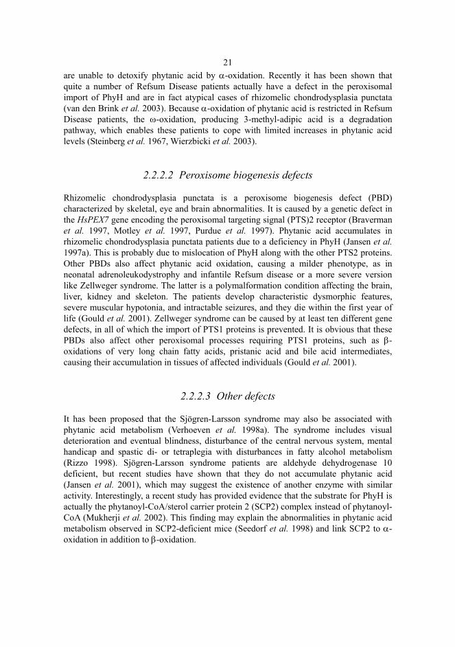
The classical bile acid synthesis pathway is initiated by cholesterol 7α-hydroxylase (CYP7A1) (Figure 5), a microsomal cytochrome P450 enzyme, expressed only in the liver (Myant & Mitropoulos 1977, Noshiro et al. 1989, Noshiro & Okuda 1990). The CYP7A1-catalyzed reaction is a rate limiting step in the pathway (Shefer et al. 1970) and therefore it is subject to a number of regulatory inputs from cholesterol, oxysterols and bile acids (Chiang 1998) as well as from recently described transcription factors, nuclear orphan receptors, like liver X receptor (LXR) (Chawla et al. 2001). CYP7A1 hydroxylates cholesterol to 7α-hydroxycholesterol, which can undergo subsequent ring structure modifications of bile acid synthesis.
Alternatively, cholesterol can be hydroxylated to oxysterols by cholesterol 24-hydroxylase (CYP46A1), cholesterol 25-hydroxylase (CH25H) or sterol 27-hydroxylase (CYP27A1) (Figure 5). The contribution of the first two to overall bile acid synthesis is minor (Duane et al. 1988, Bjorkhem et al. 1998). CYP46A1, which is expressed in the brain and at a low level in the liver, produces 24-hydroxycholesterol and has a role in elimination of endogenous cholesterol from the brain (Lund et al. 1999). CH25H, which produces 25-hydroxycholesterol and is expressed at low levels in multiple tissues (Lund et al. 1998), may also have a role in tissue-specific cholesterol catabolism (Russell 2003). CYP27A1 is a mitochondrial cytochrome P450 enzyme (Andersson et al. 1989), which is
23present in liver and many other tissues. Its main oxysterol product, 27-hydroxycholesterol, is the most abundant oxysterol in plasma (Dzeletovic et al. 1995). In mice CYP27A1 is responsible for about 25% of bile acids synthesized (Schwarz et al. 1998, Schwarz et al. 2001). However, bile acid analysis of healthy and CYP7A1-deficient humans suggests that all oxysterol pathways together account for only 5-10% of the bile acid production (Bjorkhem et al. 1998, Babiker et al. 1999, Duane & Javitt 1999). Before oxysterols can be converted to bile acids they have to undergo 7α-hydroxylation. A microsomal oxysterol 7α-hydroxylase (CYP39A1) catalyzes this in the case of 24-hydroxycholesterol (Li-Hawkins et al. 2000) and another microsomal oxysterol 7α-hydroxylase CYP7B1 in the cases of 25- and 27-hydroxycholesterols (Martin et al. 1997, Schwarz et al. 1997)(Figure 5).
Fig. 5. Hydroxylation of cholesterol by various hydroxylases. Different products shown are directed to ring structure and side chain modifications, see text for details (modified from Russell 2003).
2.3.2 Modifications to the ring structure and side chain
The 7α-hydroxylated cholesterol and oxysterol derivates from previous steps are then converted to their 3-oxo∆4 forms by a microsomal 3β-hydroxy-∆5-C27-steroid oxidoreductase (C27 3β-HSD) (Wikvall 1981). Because there is only one enzyme that possesses this activity, the synthesis of all bile acids is blocked in its absence (Clayton et al. 1987). The products from this reaction can then either be hydroxylated by sterol 12α-hydroxylase, a microsomal cytochrome P450 (CYP8B1) or skip this reaction and be subjected directly to reduction of the double bond in the A-ring by a cytosolic ∆4-3-
24oxosteroid 5β-reductase (AKR1D1) (Russell & Setchell 1992). In the first case the product is directed to the AKR1D1 catalyzed reaction after the CYP8B1 reaction and becomes an intermediate for cholic acid synthesis and in the latter case the product is an intermediate for chenodeoxycholic acid synthesis. Like CYP7A1, this branch point is a target of many regulatory inputs (del Castillo-Olivares & Gil 2000, del Castillo-Olivares & Gil 2001). The last of the ring modifications is the reduction of the 3-oxo group to an alcohol with an α-configuration, which is catalyzed by 3α-hydroxysteroid dehydrogenase, a cytoplasmic enzyme, which is abundant in liver (Penning 1997).
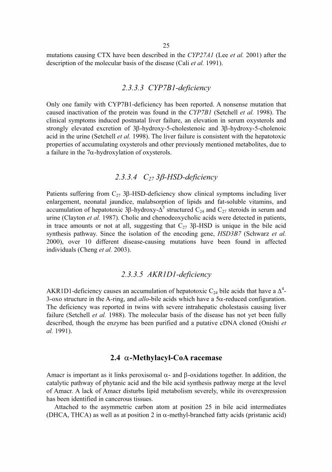
The products from ring modifications, 5β-cholestan-3α,7α-diol (chenodeoxycholic acid synthesis) and 5β-cholestan-3α,7α,12α-triol (cholic acid synthesis) are further subjected to side chain modifications. CYP27A1, the very same mitochondrial enzyme that operates in oxysterol synthesis catalyzes the first three steps, yielding DHCA and THCA (see Figure 7) i.e. the introduction of a hydroxyl group at carbon 27, oxidation of this group to an aldehyde and further to a carboxylic acid (Andersson et al. 1989, Dahlback & Holmberg 1990, Pikuleva et al. 1998). Further modifications are analogous to β-oxidation of branched-chain fatty acids, starting with activation and subsequent interconversion by Amacr (Figure 6).
2.3.3 Deficiencies in the bile acid synthesis pathway
2.3.3.1 CYP7A1-deficiency
A study of a family with a two base pair deletion in the CYP7A1, leading to a truncated inactive CYP7A1, revealed that CYP7A1-deficiency results in a hypercholesterolemic phenotype (Pullinger et al. 2002). Individuals with homozygous mutation had elevated cholesterol and triacylglycerol levels, decreased bile acid excretion and they accumulated cholesterol in the liver. These alterations lead to symptoms, which are commonly linked to hypercholesterolemia, such as premature coronary and peripheral vascular disease and cholesterol gallstones. Also heterozygous individuals were hyperlipidemic, suggesting that this is a codominant disorder (Pullinger et al. 2002). These findings are consistent with earlier population studies which revealed an association between polymorphisms at the CYP7A1 locus and levels of cholesterol in the plasma (Wang et al. 1998, Couture et al. 1999).
2.3.3.2 Cerebrotendinous xanthomatosis
A neuropathological disorder cerebrotendinous xanthomatosis (CTX) is characterized by synthesis of abnormal bile alcohols, reduced synthesis of normal bile acids and accumulation of cholesterol, a 5α-reduced cholesterol derivative, and cholestanol in serum and tissues. CTX is caused by a deficiency of CYP27A1 (Bjorkhem et al. 1998). Progressive neurological dysfunction and eventual death is caused by the accumulation of cholesterol derivatives in the myelin sheaths surrounding the neurons in brain. Over 40
25mutations causing CTX have been described in the CYP27A1 (Lee et al. 2001) after the description of the molecular basis of the disease (Cali et al. 1991).
2.3.3.3 CYP7B1-deficiency
Only one family with CYP7B1-deficiency has been reported. A nonsense mutation that caused inactivation of the protein was found in the CYP7B1 (Setchell et al. 1998). The clinical symptoms induced postnatal liver failure, an elevation in serum oxysterols and strongly elevated excretion of 3β-hydroxy-5-cholestenoic and 3β-hydroxy-5-cholenoic acid in the urine (Setchell et al. 1998). The liver failure is consistent with the hepatotoxic properties of accumulating oxysterols and other previously mentioned metabolites, due to a failure in the 7α-hydroxylation of oxysterols.
2.3.3.4 C27 3β-HSD-deficiency
Patients suffering from C27 3β-HSD-deficiency show clinical symptoms including liver enlargement, neonatal jaundice, malabsorption of lipids and fat-soluble vitamins, and accumulation of hepatotoxic 3β-hydroxy-∆5 structured C24 and C27 steroids in serum and urine (Clayton et al. 1987). Cholic and chenodeoxycholic acids were detected in patients, in trace amounts or not at all, suggesting that C27 3β-HSD is unique in the bile acid synthesis pathway. Since the isolation of the encoding gene, HSD3B7 (Schwarz et al. 2000), over 10 different disease-causing mutations have been found in affected individuals (Cheng et al. 2003).
2.3.3.5 AKR1D1-deficiency
AKR1D1-deficiency causes an accumulation of hepatotoxic C24 bile acids that have a ∆4-3-oxo structure in the A-ring, and allo-bile acids which have a 5α-reduced configuration. The deficiency was reported in twins with severe intrahepatic cholestasis causing liver failure (Setchell et al. 1988). The molecular basis of the disease has not yet been fully described, though the enzyme has been purified and a putative cDNA cloned (Onishi et al. 1991).
2.4 α-Methylacyl-CoA racemase
Amacr is important as it links peroxisomal α- and β-oxidations together. In addition, the catalytic pathway of phytanic acid and the bile acid synthesis pathway merge at the level of Amacr. A lack of Amacr disturbs lipid metabolism severely, while its overexpression has been identified in cancerous tissues.
Attached to the asymmetric carbon atom at position 25 in bile acid intermediates (DHCA, THCA) as well as at position 2 in α-methyl-branched fatty acids (pristanic acid)
26is a methyl group, which may be either in (S)- or (R)-configuration. Mitochondrial CYP27A1 produces only (25R)-isoforms of the bile acid intermediates (Gustafsson & Sjostedt 1978, Shefer et al. 1978, Batta et al. 1983) and half of the pristanic acid produced in α-oxidation are also (2R)-isoforms (Ackman & Hansen 1967, Croes et al. 1999). Since the first enzymes in the β-oxidation of branched-chain substrates (branched chain acyl-CoA oxidase (ACOX2) in humans and mice and THCA-CoA oxidase and pristanoyl-CoA oxidase in rats) are strictly specific for (S)-isoforms, the substrates have to be converted into the (S)-configuration before they can be β-oxidized (Pedersen et al. 1996, Van Veldhoven et al. 1996, Ikegawa et al. 1998). This conversion or racemization is catalyzed by Amacr, a peroxisomal and mitochondrial enzyme first described in 1994 (Schmitz et al. 1994) (Figure 6).
Fig. 6. The central role of Amacr in degradation of branched-chain fatty acids and in bile acid synthesis.
2.4.1 Substrates for Amacr
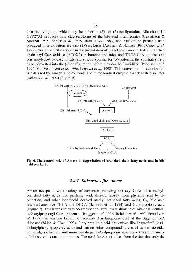
Amacr accepts a wide variety of substrates including the acyl-CoAs of α-methyl-branched fatty acids like pristanic acid, derived mostly from phytanic acid by α-oxidation, and other isoprenoid derived methyl branched fatty acids, C27 bile acid intermediates like THCA and DHCA (Schmitz et al. 1994) and 2-arylpropionic acid (Figure 7). This latter substrate became evident after it was shown that Amacr is identical to 2-arylpropionyl-CoA epimerase (Brugger et al. 1996, Reichel et al. 1997, Schmitz et al. 1997), an enzyme known to racemize 2-arylpropionic acid at the stage of CoA thioester (Shieh & Chen 1993). 2-arylpropionic acid derivatives like Ibuprofen® (2-(4-isobutylphenyl)propionic acid) and various other compounds are used as non-steroidal anti-analgesic and anti-inflammatory drugs. 2-Arylpropionic acid derivatives are usually administered as racemic mixtures. The need for Amacr arises from the fact that only the
27(2S)-isoforms are pharmaceutically active in inhibitng cyclo-oxygenases, the key enzymes in prostaglandin synthesis (Caldwell et al. 1988). Additionally, the esterification and subsequent secretion is stereospecific for (2R)-isoforms (Knihinicki et al. 1991, Shieh & Chen 1993).
Fig. 7. Various substrates for Amacr.
Before Amacr-catalyzed racemization the substrates have to be activated to their CoA thioesters. The intraperoxisomal activation of pristanic acid obtained from α-oxidation is activated by the very long chain acyl-CoA synthetase (VLACS), an enzyme localized to both peroxisomes and the ER (Steinberg et al. 1999a, Steinberg et al. 2000). Dietary pristanic acid, which is lesser in amount is assumed to be activated by LACS in the cytoplasm, in a similar way to phytanic acid (Wanders et al. 1992, Mukherji et al. 2003). Peroxisomal VLACS activates most of the bile acid intermediates from mitochondrial CYP27A1 (Uchiyama et al. 1996, Berger et al. 1998). Another synthetase in the ER, VLACS homolog 2 (also called bile acyl-CoA synthetase), is involved in the activation of deconjugated C24 bile acids, after their return to the liver via the enterohepatic circulation (Steinberg et al. 1999b, Mihalik et al. 2002).
2.4.2 Properties of Amacr
Amacr is responsible for the rapid interconversion between (R)- and (S)-isoforms of its substrates. The enzyme was first purified from rat liver as a monomer of 45 kDa with an isoelectric point at pH 6.1 and optimal activity between pH 6 and 7 (Schmitz et al. 1994). Human Amacr was found to cross-react with the polyclonal antiserum produced in rabbits against rat Amacr and it was purified from human liver as a 47 kDa monomer with an isoelectric point at pH 6.1 and optimal activity between pH 7 and 8 (Schmitz et al. 1995). Subsequently the cDNA was cloned from rats and mice and the encoded protein had a molecular mass of approximately 39.7 kDa (Schmitz et al. 1997). Recently it was shown that the native Amacr exists as a dimer in solution (Bhaumik et al. 2003). In rats the highest Amacr-activity is found in the liver, followed by the Harderian gland, kidneys and intestinal mucosa (Schmitz et al. 1997, Van Veldhoven et al. 1997). In
28humans, Amacr expression has been detected in liver, the kidneys and at lower levels in the intestinal tract and salivary glands (Schmitz et al. 1995, Zha et al. 2003). The substrate specificities of human and rat Amacrs are very similar. Amacr with only minor differences in physical and functional properties have also been identified in mice and other species like Chinese hamsters (Schmitz et al. 1997). The main difference between the Amacrs of different species seems to be in their subcellular distribution.
2.4.3 Subcellular localization of Amacr
Since Amacr is involved in the formation of primary bile acids and degradation of pristanic acid by peroxisomal β-oxidation (Prydz et al. 1986, Verhoeven et al. 1998c), one would expect that Amacr is located in peroxisomes. However, the subcellular distribution of Amacr is not similar among different species. In humans and rats Amacr activity is associated mainly (75-90%) with peroxisomes and the rest of the activity is present in mitochondria (Schmitz et al. 1995, Van Veldhoven et al. 1997). In mice, the Amacr activity is evenly distributed between peroxisomes and mitochondria (Schmitz et al. 1997). There is general agreement that Amacr shows a bimodal distribution, but the factor(s) that determine the final subcellular location is not fully established.
Another enzyme that shows different subcellular distribution among different species and also a bimodal distribution in some species is alanine/glyoxylate aminotransferase I. The enzyme is peroxisomal in humans, rabbits and guinea pigs, mitochondrial in cats and dogs and evenly distributed between both of these organelles in rats, mice and hamsters (Noguchi et al. 1978, Noguchi & Takada 1979, Okuno et al. 1979, Danpure et al. 1990). The bimodal distribution of alanine/glyoxylate aminotransferase I is caused by alternative initiation sites for transcription. The longer transcript in rat liver encodes a 22-amino-acid mitochondrial targeting signal (MTS) at the 5' end. Due to a point mutation the MTS has disappeared from the shorter transcript found in humans (Takada et al. 1990, Purdue et al. 1992).
2.4.4 The reaction catalyzed by Amacr
The mechanism of the Amacr catalyzed reaction is not fully understood in detail. However, the enzyme does not apparently require any co-factors or co-substrates and the reaction is assumed to proceed via a carbanion adjacent to the thioester with enol/keto tautomerization as an intermediate step (Schmitz et al. 1994, Reichel et al. 1997, Mukherji et al. 2003) (Figure 8). Thioesterification of the substrates increases the acidity of the C-2 hydrogen sufficiently for deprotonation and reprotonation to occur (Reichel et al. 1997). C-2 deuterated analogues of Ibuprofen® show loss of the label during chiral inversion, consistent with the proposed mechanism (Baillie et al. 1989, Chen et al. 1991).
Schmitz et al. (1994) proposed that the mechanism proceeds via abstraction of the hydrogen atom from the α-position of the substrate by a basic group on the enzyme. This creates a resonance-stabilized carbanion intermediate, which very rapidly accepts a proton from water to the α-position similar to the mechanism proposed for
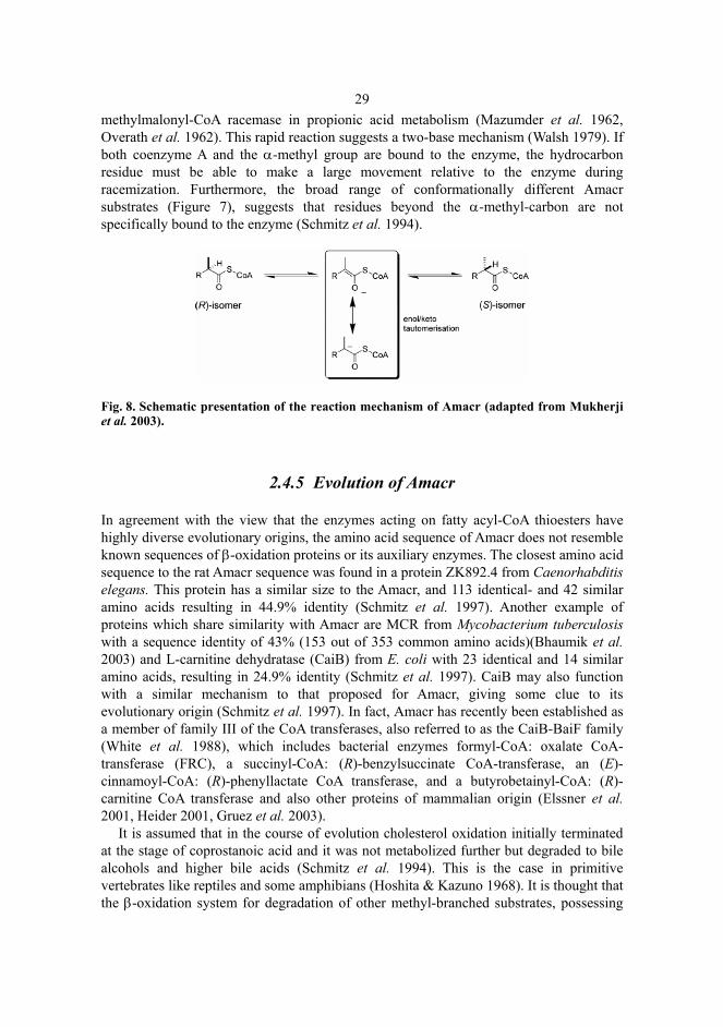
29methylmalonyl-CoA racemase in propionic acid metabolism (Mazumder et al. 1962, Overath et al. 1962). This rapid reaction suggests a two-base mechanism (Walsh 1979). If both coenzyme A and the α-methyl group are bound to the enzyme, the hydrocarbon residue must be able to make a large movement relative to the enzyme during racemization. Furthermore, the broad range of conformationally different Amacr substrates (Figure 7), suggests that residues beyond the α-methyl-carbon are not specifically bound to the enzyme (Schmitz et al. 1994).
Fig. 8. Schematic presentation of the reaction mechanism of Amacr (adapted from Mukherji et al. 2003).
2.4.5 Evolution of Amacr
In agreement with the view that the enzymes acting on fatty acyl-CoA thioesters have highly diverse evolutionary origins, the amino acid sequence of Amacr does not resemble known sequences of β-oxidation proteins or its auxiliary enzymes. The closest amino acid sequence to the rat Amacr sequence was found in a protein ZK892.4 from Caenorhabditis elegans. This protein has a similar size to the Amacr, and 113 identical- and 42 similar amino acids resulting in 44.9% identity (Schmitz et al. 1997). Another example of proteins which share similarity with Amacr are MCR from Mycobacterium tuberculosis with a sequence identity of 43% (153 out of 353 common amino acids)(Bhaumik et al. 2003) and L-carnitine dehydratase (CaiB) from E. coli with 23 identical and 14 similar amino acids, resulting in 24.9% identity (Schmitz et al. 1997). CaiB may also function with a similar mechanism to that proposed for Amacr, giving some clue to its evolutionary origin (Schmitz et al. 1997). In fact, Amacr has recently been established as a member of family III of the CoA transferases, also referred to as the CaiB-BaiF family (White et al. 1988), which includes bacterial enzymes formyl-CoA: oxalate CoA-transferase (FRC), a succinyl-CoA: (R)-benzylsuccinate CoA-transferase, an (E)-cinnamoyl-CoA: (R)-phenyllactate CoA transferase, and a butyrobetainyl-CoA: (R)-carnitine CoA transferase and also other proteins of mammalian origin (Elssner et al. 2001, Heider 2001, Gruez et al. 2003).
It is assumed that in the course of evolution cholesterol oxidation initially terminated at the stage of coprostanoic acid and it was not metabolized further but degraded to bile alcohols and higher bile acids (Schmitz et al. 1994). This is the case in primitive vertebrates like reptiles and some amphibians (Hoshita & Kazuno 1968). It is thought that the β-oxidation system for degradation of other methyl-branched substrates, possessing
30opposite stereospecificities than cholesterol derivatives, have evolved independently and Amacr have appeared later to link these two pathways (Schmitz et al. 1994). This theory is also supported by the finding that trihydroxycoprostanoyl conjugates, isolated from the bile of the evolutionarily quite primitive Alligator mississippiensis, are all in (25R)-configuration (Batta et al. 1979).
2.4.6 Amacr-deficiency
Amacr-deficiency was first described in 2000 in a study including two adults and one child (Ferdinandusse et al. 2000a). The adults suffered from adult onset sensory motor neuropathy. One also had pigmentary retinopathy and the other signs of upper motor neuron problems in the legs. The first case resembles adult Refsum Disease and the latter adrenomyeloneuropathy (Ferdinandusse et al. 2000a). The third patient was a child without neuropathy and was previously diagnosed with Niemann-Pick type C (Sequeira et al. 1998). All of the patients had somewhat elevated phytanic acid and markedly elevated pristanic acid, THCA and DHCA plasma levels. Two mutations, S52P and L107P likely to cause Amacr-deficiency were reported (Ferdinandusse et al. 2000a). The same subjects were studied more thoroughly later and the mass spectrometrical analysis of accumulation of (25R)-isomers of free and taurine-conjugated DHCA and THCA in the plasma was presented as a method for diagnosis of Amacr-deficiency (Ferdinandusse et al. 2001, McLean et al. 2002).
In a more recent study, Amacr-deficiency was linked also to neonatal cholestasis with coagulopathy and fat-soluble vitamin malabsorption in a study of a 2 week-old girl (Setchell et al. 2003). The patient carried the missense mutation S52P in AMACR and had markedly elevated pristanic acid, THCA and DHCA plasma, urine and bile levels, but normal phytanic acid levels. Interestingly, the composition of the bile acid pool of the subject was qualitatively identical to that of Alligator mississippiensis. Liver biopsy showed giant cell transformations, hepatocyte necrosis and a reduced number of peroxisomes (Setchell et al. 2003). The Amacr-deficiency has also been linked to vitamin K deficiency in a fibroblast study (Van Veldhoven et al. 2001).
It seems that Amacr-deficiency results in different clinical manifestations. This is true also in some other disorders such as X-linked adrenoleukodystrophy (XALD), in which differences are observed even within the same family (Moser 1997). It is also possible that various patients, such as those diagnosed with a possible deficiency at the level of branched-chain fatty acid and THCA oxidation or uptake (Christensen et al. 1990, Przyrembel et al. 1990, Clayton et al. 1996) or with atypical Refsum disease (Wierzbicki et al. 2002) are actually Amacr-deficient patients, but are not diagnosed as such, due to lack of proper knowledge or methods for diagnosis.
2.4.7 Amacr in cancer
Recently, microarray screenings have been used to identify differentially expressed genes in normal prostate tissue and prostatic carcinoma. Several independent groups found that
31Amacr (also referred to as P504S in cancer studies) is overexpressed in prostate cancer (Xu et al. 2000, Dhanasekaran et al. 2001, Luo et al. 2001, Welsh et al. 2001). This was verified with immunohistochemistry and the overexpression of Amacr was introduced as a new marker for prostate cancer (Jiang et al. 2001, Rubin et al. 2002). The expression of Amacr in prostate carcinoma was shown by quantitative reverse transcriptase polymerase chain reaction (RT-PCR) to be increased up to 9 fold compared to that in benign prostate tissue (Luo et al. 2002).
A number of studies have been carried out in order to establish the reliability of immunohistochemical staining with Amacr in cancer diagnostics from different types of prostate samples (radical prostatectomy, transurethral resection, needle biopsy). Overall the studies showed sensitivities that range from 82% to 100% and specificities ranging from 79% to 100% (Evans 2003) indicating that the method is quite reliable. The variation in results is likely due to the different sample materials, antibodies (monoclonal vs polyclonal) and staining conditions used.
Overexpression of Amacr as high as in prostate cancer was found also in colorectal cancer by microarray screening (Zhou et al. 2002). Furthermore, various other cancers like breast, ovarian, bladder, lung and renal carcinomas showed overexpression of Amacr using the same screening method. In a study of 242 cases of colonic tumors the expression of Amacr was also proposed as a potential marker for colonic cancer (Jiang et al. 2003). It was even proposed that in vivo visualization of the expression of Amacr, using magnetic resonance imaging (Louie et al. 2000), might be applied. In addition, increased Amacr mRNA levels have been proposed as useful in differential diagnosis of renal carcinomas as they are elevated in papillary renal carcinomas, but not in other renal subtypes (Tretiakova et al. 2004).
The activity of Amacr is also elevated in prostate cancer and therefore measurement of the enzyme’s activity has been proposed for use in cancer diagnostics (Zha et al. 2003, Kumar-Sinha et al. 2004). Amacr is an androgen-independent growth modifier in prostate cancer (Kuefer et al. 2002, Zha et al. 2003) and its role and the role of β-oxidation of branched-chain fatty acids in initiation and development of carcinomas has been speculated upon widely. Interestingly, the epidemiological studies revealed a correlation between the consumption of beef, milk and dairy products, the main sources of branched-chain fatty acids (Flanagan et al. 1975, Brown et al. 1993), and increased risk of both prostate (Giovannucci et al. 1993) and colon (Giovannucci & Willett 1994, Truswell 2002) cancers. Furthermore, Amacr expression was enhanced in prostate cancer cells by branched-chain fatty acids in vitro (Mobley et al. 2003). Unfortunately, Mobley and co-workers did not measure the activity of Amacr.
2.5 β-Oxidation
Universally, in one cycle of mitochondrial and peroxisomal β-oxidation the fatty acids are shortened by two carbon fragments, the fragments being either acetyl-CoA or propionyl-CoA. Regardless of the location of occurrence, the β-oxidation cycles are analogous and consists of four enzymatic steps 1) dehydrogenation/oxidation, 2) hydration, 3) dehydrogenation and 4) thiolytic cleavage (Figure 9), which however, are
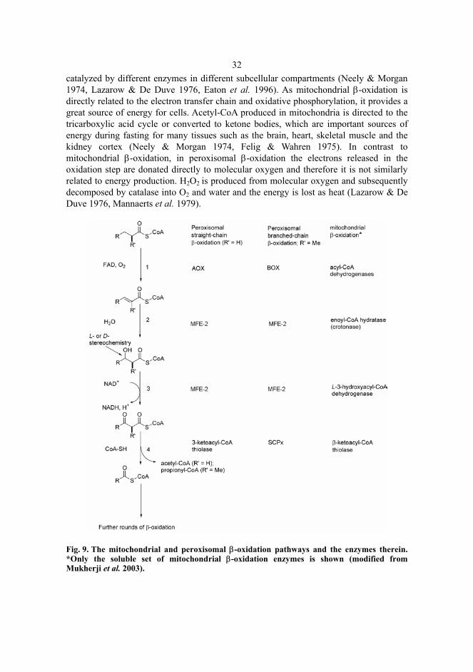
32catalyzed by different enzymes in different subcellular compartments (Neely & Morgan 1974, Lazarow & De Duve 1976, Eaton et al. 1996). As mitochondrial β-oxidation is directly related to the electron transfer chain and oxidative phosphorylation, it provides a great source of energy for cells. Acetyl-CoA produced in mitochondria is directed to the tricarboxylic acid cycle or converted to ketone bodies, which are important sources of energy during fasting for many tissues such as the brain, heart, skeletal muscle and the kidney cortex (Neely & Morgan 1974, Felig & Wahren 1975). In contrast to mitochondrial β-oxidation, in peroxisomal β-oxidation the electrons released in the oxidation step are donated directly to molecular oxygen and therefore it is not similarly related to energy production. H2O2 is produced from molecular oxygen and subsequently decomposed by catalase into O2 and water and the energy is lost as heat (Lazarow & De Duve 1976, Mannaerts et al. 1979).
Fig. 9. The mitochondrial and peroxisomal β-oxidation pathways and the enzymes therein. *Only the soluble set of mitochondrial β-oxidation enzymes is shown (modified from Mukherji et al. 2003).
33
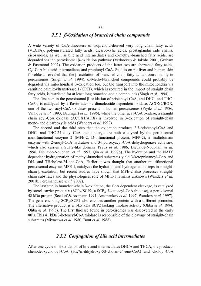
2.5.1 β-Oxidation of branched chain compounds
A wide variety of CoA-thioesters of isoprenoid-derived very long chain fatty acids (VLCFA), polyunsaturated fatty acids, dicarboxylic acids, prostaglandin side chains, eicosanoids, as well as bile acid intermediates and α-methyl-branched fatty acids, are degraded via the peroxisomal β-oxidation pathway (Verhoeven & Jakobs 2001, Graham & Eastmond 2002). The oxidation products of the latter two are shortened fatty acids, C24-CoA bile acid intermediates and propionyl-CoA. Studies on rat liver and human skin fibroblasts revealed that the β-oxidation of branched chain fatty acids occurs mainly in peroxisomes (Singh et al. 1994). α-Methyl-branched compounds could probably be degraded via mitochondrial β-oxidation too, but the transport into the mitochondria via carnitine palmitoyltransferase I (CPTI), which is required in the import of straight chain fatty acids, is restricted for at least long branched-chain compounds (Singh et al. 1994).
The first step in the peroxisomal β-oxidation of pristanoyl-CoA, and DHC- and THC-CoAs, is catalyzed by a flavin adenine dinucleotide dependent oxidase, ACOX2/BOX, one of the two acyl-CoA oxidases present in human peroxisomes (Prydz et al. 1986, Vanhove et al. 1993, Baumgart et al. 1996), while the other acyl-CoA oxidase, a straight chain acyl-CoA oxidase (ACOX1/AOX) is involved in β-oxidation of straight-chain mono- and dicarboxylic acids (Wanders et al. 1992).
The second and the third step that the oxidation products 2,3-pristenoyl-CoA and DHC- and THC-24-enoyl-CoA then undergo are both catalyzed by the peroxisomal multifunctional enzyme 2 (MFE-2, D-bifunctional protein, MFP-2), a multidomain enzyme with 2-enoyl-CoA hydratase and 3-hydroxyacyl-CoA dehydrogenase activities, which also carries a SCP2-like domain (Prydz et al. 1986, Dieuaide-Noubhani et al. 1996, Dieuaide-Noubhani et al. 1997, Qin et al. 1997b). The hydration and the NAD+ dependent hydrogenation of methyl-branched substrates yield 3-ketopristanoyl-CoA and DH- and THcholest-24-one-CoA. Earlier it was thought that another multifunctional peroxisomal enzyme, MFE-1, catalyzes the hydration and hydrogenation steps in straight-chain β-oxidation, but recent studies have shown that MFE-2 also processes straight-chain substrates and the physiological role of MFE-1 remains unknown (Wanders et al. 2001b, Ferdinandusse et al. 2002).
The last step in branched-chain β-oxidation, the CoA dependent cleavage, is catalyzed by sterol carrier protein x (SCPX/SCP2, a SCPX 3-ketoacyl-CoA thiolase), a peroxisomal 48 kDa protein (Seedorf & Assmann 1991, Antonenkov et al. 1997, Wanders et al. 1997). The gene encoding SCPX/SCP2 also encodes another protein with a different promoter. The alternative product is a 14.5 kDa SCP2 lacking thiolase activity (Ohba et al. 1994, Ohba et al. 1995). The first thiolase found in peroxisomes was discovered in the early 80’s. This 41 kDa 3-ketoacyl-CoA thiolase is responsible of the cleavage of straight-chain substrates (Miyazawa et al. 1980, Bout et al. 1988).
2.5.2 Conjugation of bile acid intermediates
After one cycle of β-oxidation of bile acid intermediates DHCA and THCA, the products chenodeoxycholoyl-CoA (3α,7α-dihydroxy-5β-cholan-24-one-CoA) and choloyl-CoA
34(3α,7α,12α-trihydroxy-5β-cholan-24-one-CoA) are conjugated. A peroxisomal bile acid conjugating enzyme (N-acyl-transferase) catalyzes the conjugation of amino acids to carbon 24 of the C24 substrates (Kase & Bjorkhem 1989). Over 98% of primary bile acids are conjugated with taurine or glycine (Falany et al. 1994) before they are exported and secreted into the bile with help from various membrane transporters, such as ABCG5 and ABCG8 (Love & Dawson 1998, Borst & Elferink 2002).
2.5.3 Complete degradation of pristanoyl-CoA
Pristanoyl-CoA (2,6,10,14-tetramethylpentadecanoyl-CoA) contains four methyl-branches, of which the three innermost are in the (R)-configuration. The first cycle of mammalian β-oxidation produces propionyl-CoA and 4,8,12-trimethyltridecanoyl-CoA, and the latter then undergoes a second cycle of β-oxidation. The second cycle produces acetyl-CoA and 2,6,10-trimethylundecanoyl-CoA, which, before entering the third cycle has to be racemized by Amacr. At least in human fibroblasts the product of the third cycle (along with propionyl-CoA), 4,8-dimethylnonaoyl-CoA (Verhoeven et al. 1998b), is converted to a corresponding carnitine ester by peroxisomal carnitine octanoyl transferase (Ferdinandusse et al. 1999) and imported to mitochondria via the carnitine acylcarnitine carrier and converted back into 4,8-dimethylnonaoyl-CoA via carnitine palmitoyltransferase II (CPTII) (Verhoeven & Jakobs 2001). In mitochondria 4,8-dimethylnonaoyl-CoA undergoes a cycle of β-oxidation and then the product 2,6-dimethylheptanoyl has to again be racemized by Amacr. 2,6-Dimethylheptanoyl undergoes two further rounds of mitochondrial β-oxidation and the product of the final round (in addition to acetyl CoA) is isobutyryl-CoA (Figure 10).
Fig. 10. Complete degradation of pristanic acid in mammals. 4,8-Dimethylnonaoyl-CoA is transported from peroxisome into the mitochondrion as a carnitine ester (modified from Wanders et al. 2003), see the text for details. Propionyl-CoA from peroxisomal β-oxidation and most likely also acetyl-CoA are converted to their carnitine esters in the peroxisome, after which they are transported into the mitochondria, where they are oxidized to CO2 and water (Jakobs & Wanders 1995).
35
2.5.4 MFE-2-deficiency
MFE-2-deficiency, first described in 1986 as a Pseudo-Zellweger syndrome (Goldfischer et al. 1986) is a severe defect disturbing the β-oxidation of at least VLCFA and branched-chain metabolites. There are other defects like ACOX1-deficiency (Poll-The et al. 1988), XALD (Hashmi et al. 1986) and a deficiency in a 41 kDa 3-ketoacyl thiolase (Goldfischer et al. 1986, Schram et al. 1987), that affect peroxisomal β-oxidation of straight-, but not branched-chain metabolites.
The MFE-2-deficiency resembles Zellweger syndrome in many respects: clinical findings include neonatal hypotonia, craniofacial dysmorphia, seizures, severe developmental failure and early death. Biochemically the patients accumulate VLCFAs, bile acid intermediates, pristanic acid and to a lesser extent phytanic acid (Goldfischer et al. 1986, Watkins et al. 1989). In contrast, in PBDs both branched-chain fatty acids accumulate at the same level (ten Brink et al. 1992). The cause of the defect was only later identified as a defect in the level of MFE-2 (van Grunsven et al. 1999b, Ferdinandusse et al. 2002).
Patients with defects in only one of the functions of MFE-2 have also been identified. Both groups of patients with defects either in the 3-hydroxyacyl-CoA dehydrogenase or in the enoyl-CoA hydratase domain of the MFE-2 showed similar severe clinical findings to those with total MFE-2-deficiency and died in their first years of life (van Grunsven et al. 1998, van Grunsven et al. 1999a). However, in the enoyl-CoA hydratase group no accumulation of bile acid intermediates was found, while in the plasma of the dehydrogenase group the main accumulating intermediate was identified as varanic acid (24S,25S)-3α,7α,12α,24-tetrahydroxy-5β-cholestanoic acid). The different biochemical phenotype among the three subgroups of MFE-2-deficiency is proposed to be due to the combined action of MFE-1 and Amacr on the different diastereoisomers of the intermediates present in each of the different groups (Qin et al. 1997a, Cuebas et al. 2002).
3 Outlines of the present study
Amacr is an enzyme acting at least i) in bile acid synthesis from cholesterol, ii) in branched chain fatty acid degradation, as well as iii) in the metabolism of ibuprofen®. In mice (and in man and rats) Amacr is localized to both mitochondria and peroxisomes. Amacr belongs to a family of CoA-transferases which possess diverse functions. The structural relations among proteins within the group are unclear. Amacr-deficient patients have been reported to show somewhat different symptoms among themselves. The purpose of this work was to characterize the gene and study the physiological function and significance of Amacr and to create an animal model for Amacr-deficiency. More specifically we aimed: 1. To characterize mouse Amacr, determine its genomic localization and study further the subcellular localization of Amacr. 2. To study the physiological importance of Amacr and to create an animal model for Amacr-deficiency via generation of a knock-out mouse. 3. To characterize the active site of Amacr by mutational and structural studies on the Amacr ortholog MCR, purified from Mycobacterium tuberculosis.
4 Materials and Methods
Detailed description of the methods used can be found in the original articles.
4.1 Sequences (I, II, III)
Nucleotide and amino acid sequence analyses and alignments were done using the DNASIS program (Hitachi Software Engineering Co., Yokohama, Japan) or Clustal X program (Thompson et al. 1997), respectively. The transcription factor binding sites were located using a MatInspector V2.2 database (Quandt et al. 1995). Amino acid sequences were collected from the SWISS-PROT database using Blast and Fasta algorithms.
DNA sequencing was done with an automated ABI Prism 377 DNA sequencer (Perkin-Elmer, Boston, MA, USA). Oligonucleotides were synthesized with an ABI DNA synthesizer Model 394 (Perkin-Elmer), or were purchased from Carl Roth (Karlsruhe, Germany) or Amersham Pharmacia Biotech (Buckinghamshire, England). The sequences of the oligonucleotides used in sequencing or in PCR are presented in I, II, and III.
4.2 Animal care (I, II)
Animal experimentation was done in the germ free Barrier of the University of Oulu and in the animal facility of the Department of Biochemistry, University of Oulu. The research plan and use of animals were approved by the University of Oulu committee of animal experimentation (Licence numbers 75/00, 044/02 and 035/03). The mice used in all experiments were adults (4.5 months) unless stated otherwise. Before vascular perfusion, blood collection or further tissue harvesting the animals were anesthetized and subsequently sacrificed by cervical dislocation. For feeding experiments, mice were stressed with chows supplemented with 0.5% (w/w) clofibrate (ICN Biomedicals, Aurora, OH, USA), 0.5% (w/w) phytol or 5% (w/w) triolein (both from Sigma Aldrich Steinheim, Germany).
38
4.3 Protein analysis (I, II, III)
Protein contents were determined photometrically at 595 nm with the Bradford method (Bradford 1976) using BIO-RAD (Hercules, CA, USA) chemistry or at 280 nm for purified protein samples. Sodium dodecyl sulphate polyacrylamide gel electrophoreses (SDS-PAGE) were performed on 12.5% gels (Laemmli 1970). For Western blotting (I, II) and N-terminal sequence analysis (I) the samples from Coomassie Brilliant Blue stained SDS-PAGE gels were electroblotted into PROTRAN, nitrocellulose transfer membrane (Schleicher & Schuell, Dassel, Germany) or ProBlott, polyvinylidene difluoride membrane (PVDF, Applied Biosystems, Foster City, CA, USA)(Towbin et al. 1979, Matsudaira 1987). Polyclonal anti-rabbit antibody against rat Amacr (Schmitz et al. 1995) was used in immunostaining of the Western blot. N-terminal sequencing of the bands of interest was performed with a Procise 494A sequencer (Applied Biosystems).
4.4 Activity measurements (I, II, III)
The Amacr activity measurements with [2-3H]pristanoyl-CoA or [24,25-3H]THCA-CoA were performed as previously described (Schmitz et al. 1994). Samples containing Amacr, including mitochondria and peroxisomes (I), tissue samples (II) and the cell extracts (III) were incubated with [3H]-labeled substrate, after which [3H]-H2O was separated from unreacted substrate and quantified by liquid scintillation counting. Catalase activity was determined by the TiO(SO4) method (Huebl & Bretschneider 1964) and succinate dehydrogenase activity as described (Banerjee et al. 1984).
4.5 Expression of recombinant proteins (I, III)
pET3a (pET Expression System 3, Novagen, Madison, WI, USA) was used in expressions of complete rat cAMACR, produced by PCR using rat liver Marathon-Ready cDNA (Clontech, Palo Alto, CA, USA) (I), MCR (an α-methylacyl-CoA racemase from Mycobacterium tuberculosis) and several MCR-variants, produced from MCR-pET3a by a QuickChange site-directed mutagenesis kit (Stratagene, La Jolla, CA, USA) (III). The plasmids, containing correctly oriented inserts or sequence verified mutations, were transformed into competent BL21(DE3)-LysS cells for overexpression as described (Bhaumik et al. 2003).
For the expression of Amacr in yeast cells, the Pichia Expression Kit (Invitrogen, De Schelp, the Netherlands) was used. Coding cAMACR was amplified by PCR, using plasmid C1—9 (I) as a template. The product was cloned through pCR2.1 and INV-α into the pHilD2 vector, which was eventually transformed into Pichia pastoris strain GS115, in which the expression of recombinant Amacr was analyzed.
39
4.6 Histological analyses (I, II)
For immuno electron microscopy (EM; I) livers were harvested from mice after vascular perfusion fixation (Filppula et al. 1998) and processed for thin sectioning (Carlemalm et al. 1985). For regular transmission EM (II) the samples were fixed after harvesting the tissue. The samples were embedded either in MicroBed resin or Epon Embed 812 (Electron Microscopy Sciences, Fort Washington, PA, USA) and the thin sections were prepared. Immuno EM samples were immunogold labeled with antiserum against rat Amacr (Schmitz et al. 1995), peroxisomal catalase (a gift from Dr. Stefan Alexson, Karolinska Institutet, Huddinge, Sweden) or mitochondrial ∆2-∆4-dienoyl-CoA reductase (Hakkola et al. 1989), respectively. The samples were analyzed with a Philips CM100 transmission electron microscope equipped with TCL-EM-Menu version 3 (Tietz Video and Image Processing Systems, Gaunting, Germany). The distribution of antibodies was identified by gold particles conjugated to protein A.
Samples for light microscopy (II) were fixed, embedded in paraffin and prepared sections were stained with haematoxylin and eosin. Frozen sections were prepared and Oil red O-stained. The Sections were analyzed with an Olympus BX50 light microscope equipped with Analysis® software (Soft Imaging Systems, Münster, Germany).
4.7 Separation of organelles and immunoaffinity isolation of Amacr (I)
Subcellular fractionation of mouse liver and normal and CHRS-like (peroxisome-deficient mutant Chinese hamster ovarian (CHO) cells (Tsukamoto et al. 1991) supplied by Dr. Jutta Gärtner, Düsseldorf University Children’s Hospital, Germany) was performed with Nycodenz gradients as described (Schmitz & Conzelmann 1997).
For immunopurification of Amacr, livers were homogenized and applied into a Percoll-Suspension (Amersham Pharmacia Biotech) to establish the Percoll gradient. Fractions showing succinate dehydrogenase (mitochondrial marker) or catalase (peroxisomal marker) activities were treated with ultra sound. The supernates were applied onto the immuno affinity column, prepared by binding HiTrap ProteinA column (Amersham Pharmacia Biotech) purified IgGs from anti-Amacr-antiserum (Schmitz et al. 1995) to CNBr-activated Sepharose 4B (Amersham Pharmacia Biotech). Bound proteins were eluted from the column with either acidic or alkaline elution. The alkaline eluates were immediately neutralized and concentrated on centriprep tube equipped with a 30 kDa cutoff membrane (Amicon, Witten, Germany).
4.8 Southern hybridization (I, II)
Genomic DNA was extracted from mouse kidneys (Sambrook et al. 1989) (I) or from embryonic stem (ES) cells and mouse tail tip samples with a Blood & Cell Culture DNA Midi Kit (Qiagen, Hilden, Germany) (II) and digested with restriction endonucleases.
40Southern blotting and hybridization were done on nitrocellulose blotting membranes (Schleicher & Schuell) or Hybond-N+ nylon membranes (Amersham Pharmacia Biotech) as described (Reed & Mann 1985, Sambrook et al. 1989) using 32P-labeled (Random Primed Oligonucleotide Labeling Kit, Amersham Pharmacia Biotech) mouse cAMACR (10) (I) or the 1 kb fragment before the 5'-flanking region (II) as probes. The membranes were analyzed with either Kodak X-omat AR film or Phosphocassettes and Phosphorimager apparatus (Molecular Dynamics, Buckinhamshire, UK).
4.9 cDNA and mRNA analyses (I, II)
Total RNA was extracted from rat liver using the Biozym PUREscript RNA isolation kit (Biozym Diagnostik, Hess Oldendorf, Germany) (I) or from mouse tissues using the QuickPrep™ Total RNA Extraction Kit (Amersham Pharmacia Biotech) (II). The Titan™
One Tube RT-PCR system (Boehringer Mannheim, Mannheim, Germany) was used for the first strand synthesis of rat cAMACR, which was polyadenylated and amplified by PCR, and a further nested PCR was performed. The products were ligated into pCR2.1 (Original TA Cloning Kit, Invitrogen) and transformed into competent INV-α cells. Isolated plasmids were cleaved, the sizes of the inserts were determined and the 3 largest were sequenced.
Mouse cDNA was synthesized using the First Strand cDNA Synthesis Kit (MBI Fermentas, Heidelberg, Germany) and the expression levels of various genes were measured by Real-Time Quantitative PCR using ABI PRISM 7700 Sequence Detection System (Perkin-Elmer) as described (Majalahti-Palviainen et al. 2000). The Mouse Multiple Tissue Northern Blot™ (Clontech) was used for Northern hybridization to detect the Amacr expression in various tissues (I) with 32P-labeled mouse cAMACR (10) as a probe.
4.10 Characterization of the Amacr gene (I)
The mouse ES-129/SvJ I genomic library was screened by Genome Systems (St. Louis, MO, USA) with the specific primers from the exon-intron boundary of Amacr. The presence of Amacr in the resulting bacterial artificial chromosome (BAC) clones was detected by digestion and southern hybridization. 3' end and upstream sequencing of the Amacr were performed on the clone. PCR reactions were used to determine the size of the introns.
A fluorescent in situ hybridization method was performed by Genome Systems to reveal the chromosomal localization of the Amacr. Hybridization to normal metaphase chromosomes, derived from mouse embryo fibroblasts, was done using a digoxigenin-labeled BACM-13L1 clone as a probe. The identification of the mouse chromosomes was based on their G-banding pattern. In addition, mouse chromosome 15 was identified with a probe specific for the telomeric region of that chromosome. Only double spot signals were considered as specific.
41
4.11 Promoter activity (I)
A 1670 bp fragment of the 5' noncoding region of Amacr was amplified by PCR and cloned into pGL3-Basic reporter vector (Promega, Madison, WI, USA). HepG2 cells (American Type Culture Collection, Rockville, MD) were transfected, using Lipofect Amine-transfection reagent (Life Technologies, Gaithesburg, MD, USA), with a solution containing luciferase reporter plasmid with amplified promoter, the same region in inverted orientation, plain luciferase reporter plasmid or pGL3-control vector (Promega). pSV-β-galactosidase control plasmid (Promega) was used as normalization vector. Cell extracts were prepared using Reporter Lysis Buffer (Promega) and luciferase activities were assayed using a Luminoscan RS luminometer (Labsystems, Helsinki, Finland) and normalized for β-galactosidase activity.
4.12 Generation of Amacr-/- mice (II)
The 5' and 3' flanking regions for the phosphoglycerine kinase (PGK)-neo cassette were cleaved from the BACM-13L1-clone and ligated to the targeting vector, which contained the genes for double selection and was used for homologous recombination. The vector was linearized and electroporated into R1 ES-cells. The cells were cultured under a selection medium containing G418 (GIBCO/BRL, Gaithersburgh, MD, USA) and ganciclovir (Syntex, Palo Alto, CA, USA). The surviving colonies were screened by Southern blot analysis for correct replacement of the exons 1—3 and introns 1—2 with the neomycin-resistance gene (13 kb band for wild type and 7 kb for knock-out allele; Figure 1 in II). Chimeric mice were produced by microinjecting Amacr+/- cell aggregates into C57BL/6 blastocysts and implementing them into foster mothers. Mouse genotypes were identified with PCR analysis as described in more detail in II. Heterozygous offspring were further interbred to produce Amacr-/-, Amacr+/-and Amacr+/+ mice.
4.13 Analyses of Amacr-/- mice (II)
The levels of serum cholesterol and triacylglycerols, and the activities of alanine amino transferase (ALAT) and alkaline phosphatase (APHOS) were determined by TG-diagnostics (Oulu, Finland), the prothrombin complex assay was done by the clinical laboratory of the University Hospital of Oulu and titrimetrical determination of total fatty acids from stools was done by Yhtyneet Laboratoriot (Helsinki, Finland). Careful analysis of bile acids, bile acid derivatives and fatty acids were performed on serum, bile and liver tissue samples by mass spectrometry. The samples were treated and the mass spectral data were obtained either in negative or positive mode using an APEX II FT-ICR mass spectrometer controlled by Xmass 6.0.0 software and equipped with a 7T active shielded magnet, an APOLLO electrospray ion source and a syringe pump (Bruker Daltonics, Billerica, MA, USA).
42
4.14 Circular dichroism spectroscopy (III)
MCR and its mutated variants were overexpressed, purified from cell extracts as described (Bhaumik et al., 2003) and a size exclusion chromatography was performed (Superdex 200® HR 10/30 column). The far-UV (200—250 nm) circular dichroism (CD) spectra for purified proteins in the measuring buffer were determined using a Jasco J715 spectropolarimeter (Tokyo, Japan).
4.15 Crystallization and structure characterization of MCR (III)
The crystals, belonging to monoclinic space group C2, were grown by the hanging-drop, vapor-diffusion method. Assuming four subunits (two dimers) per asymmetric unit, a VM value of 2.6 Å3 / Da was obtained, corresponding to a solvent content of 55% (Matthews 1968). The heavy atom derivatives were obtained as described (III). The datasets were collected by the oscillation method using a synchrotron radiation source (EMBL-Hamburg, DESY, Germany) and a CCD detector. The collected datasets were processed using the program packages DENZO (Otwinowski & Minor 1997), XDS (Kabsch 1993) and CCP4 (Collaborative Computational Project 1994).
The initial phases of structure determination were obtained with the MIRAS method and the programs SOLVE (Terwilliger & Berendzen 1999), RESOLVE (Terwilliger 2000), DM (Cowtan 1994), REFMAC5 (Murshudov et al. 1997) and O (Jones et al. 1991), and were used for positioning the heavy atoms and model building at 1.8 Å resolution. The program PROCHECK (Laskowski et al. 1993) was used to monitor the stereochemistry of the model. There are no differences among the four monomers. The refinement statistics are shown in Table II in study III. The secondary structure assignment was done with DSSP (Kabsch & Sander 1983). Structure superpositions were done with the LSQ option of O, using corresponding Cα-atoms.
5 Results
5.1 Amacr is localized to both peroxisomes and mitochondria (I)
Amacr activity was found in three regions of a Nycodenz gradient of mouse liver homogenate (Figure 1 in I). Two were comigrated with peroxisomal and mitochondrial marker enzymes, respectively and one was at the top of the gradient. The bands of the same size with intensities that correlated with the distribution of Amacr activity were visualized in a Western blot (Figure 1 in I). The fractionation of CHO fibroblasts also gave similar results (Figure 2a in I). In the case of peroxisome-deficient CHO fibroblasts, Amacr activity comigrated with the mitochondrial fractions with similar activity to the mitochondrial fractions of the control cells (Figure 2b in I).
Mouse liver homogenates, and peroxisomes and mitochondria, isolated from them by Percoll gradient centrifugation, were applied separately to the affinity column, prepared with anti-Amacr antibodies. When the bound proteins were eluted with acidic and alkaline buffer, respectively, SDS-PAGE and Western blot analyses gave a size of 42 kDa corresponding the size of mouse Amacr (Figure 4 in I). No Amacr activity was detected in the acidic elution, but very low Amacr activity in the alkaline elution was detected. The results show that all Amacrs behave similarly in the affinity column, but are rapidly inactivated under stringent conditions. Alkaline eluted proteins from peroxisomes, mitochondria and cytosol were blotted on a hydrophilic PVDF membrane, and the Amacr bands were subjected to N-terminal sequencing. Each of the samples gave the same single homogeneous N-terminal sequence: Val-Leu-Arg-Gly-Val-Arg-Val-Val-Asp-Leu-Ala-Gly-Leu-Ala-Pro (NAmacr).
Using immuno electron microscopy with the anti-Amacr antiserum, Amacr was localized to both mitochondria and peroxisomes, supporting the dual localization of the Amacr (Figure 3 in I). The control antibodies against mitochondrial and peroxisomal enzymes recognized the expected compartments.
44
5.2 cDNA analysis reveals an extension to cAMACR (I)
Since NAmacr was not present in the polypeptide encoded by the known mouse cAMACR (Schmitz et al. 1997), the data banks were searched and Expressed Sequence Tag clones, including AA085247, were identified to encode NAmacr. The cDNA of AA085247 contained an ATG in a Kozak consensus sequence followed by a sequence encoding NAmacr and 24 additional base pairs, of which the last 10 bp matched with the published cAMACR (Figure 5 in I). cAMACR was extended by 62 bp altogether at the 5'end and the open reading frame of the revised cAMACR encodes a polypeptide of 381 amino acids with a predicted molecular mass of 41718 Da.
As rat and mouse Amacrs have been shown to be of the same size, the revised mouse cAMACR also suggests that the previously published rat cAMACR was incomplete. Indeed, a cDNA fragment of 570 bp containing an additional 246 bp, preceding the 5' end of the published rat cAMACR (Schmitz et al. 1997) was obtained from the RT-PCR using rat total RNA as a template. The extended rat liver cAMACR includes an ATG as a putative initial codon, followed by an open reading frame that corresponds quite well to that for mice (Figure 5 in I). The open reading frame of rat liver cAMACR was overexpressed in P. pastoris. The resulting product showed Amacr activity and its size corresponded to the size of Amacr found in rat liver.
5.3 A single Amacr with an operable promoter region encoding one gene product (I)
A screening of the mouse genomic library ES-129/SvJ I by Genome Systems revealed 3 positive BAC clones (BACM-235K4, BACM-13L1 and BACM-151H13). The clone BACM-13L1 was digested with HindIII, the fragments were ligated to pUC18, transformed into E.coli and screened with the mouse 32P labeled cAMACR probe. A ~2 kb insert contained a sequence identical to the 3' end of cAMACR up to the poly (A+) tail. The sequence was interrupted by intronic sequences 766 nucleotides upstream and the exon/intron junction was confirmed with sequencing. Further sequencing and PCR revealed that Amacr contains altogether 4 introns with sizes of 1.3, 1.2, 4 and 5 kb (Figure 5 in I) and that the gene covers 13 kb of the mouse genome (Figure 6 in I). Altogether 1680 bp were sequenced upstream of the putative initial ATG; a GC-rich region, which can serve as a SP1 binding site was located at the positions -53 to -90. No TATA box was found, but consensus sequences for transcription factors such as sites for GATA1, NF1, AP-1, AP-2 and AP-4 were present. Furthermore, the region -1670 to -1 was cloned to the 5' end of the luciferase gene in the reporter vector, which, in human HepG2 cells, exhibited 135 ± 49 (mean ± SD, n = 10) fold higher luciferase values compared to negative controls, indicating that the fragment encompasses sequences that can function as operable promoter elements in intact cells. Chromosomal localization of the mouse Amacr revealed that the genomic location of the gene is at chromosome 15, region 15B1 (Figure 8 in I).
45Southern blot analysis of digested genomic mouse DNA resulted in fragments of
approximately 13 and 5 kb with EcoRI digestion, 6.2, 5.8, 4.0, 2.3 and 1.7 kb with HindIII digestion and 6.0, 4.0, 2.1, 1.7 and 0.9 kb with double digestion (Figure 9 in I). According to this and the restriction map (Figure 6 in I) the size of Amacr is about 15 kb, which is not enough to encompass multiple 13 kb genes in a row. The hybridization of mouse Multiple Tissue Northern Blot™, which contained mRNA from heart, brain, spleen, lung, liver, skeletal muscle, kidney and testis, with labeled cAMACR as a probe, showed a signal at 1.9 kb only on the lanes corresponding to liver and kidneys (Figure 10A in I), suggesting that Amacr is expressed in these tissues.
5.4 Alterations in the bile acid pool of Amacr-/- mice (II)
The correct disruption of the Amacr and the absence of the gene products thereof in the knock-out mice were confirmed. Southern blot analysis of genomic DNA (7 kb band in Figure 1B in II), Real-Time Quantitative PCR analysis of mRNA from liver (and other tissues) (Table 1 in II), and immunoblotting of the samples from the same tissues with anti-Amacr-antibody (Figure 1C in II) were performed. These and the disappearance of the Amacr activity in the liver and kidneys of Amacr-/- mice suggest that Amacr is the only enzyme responsible for this activity.
Heterozygous Amacr+/- mice produced litters of normal size with normal Mendelian distribution. This suggests that the intrauterine and postnatal survival rate is not reduced. The knock-out offspring were normal in appearance, generally healthy, fertile and the growth and bodyweights were comparable to age and sex matched littermate controls. The motor coordination and muscular performance of mice were tested by hanging and grid tests (II). There was no differences between performances of Amacr+/+ and Amacr-/- mice in either of the tests, not even after phytol treatment.
Bile, serum and liver tissue were analyzed for contents of taurine- and glycine conjugated, and unconjugated forms of 35 bile acid metabolites with negative ion mass spectrometry. The wild type and Amacr+/- mice had almost exclusively C24 bile acids (198 ± 103 mM and 179 ± 84 mM) in the bile and only negligible amounts of C27 precursors (approximately 1 mM), while the Amacr-/- animals had significantly reduced C24 species (98 ± 34 mM), but a strongly increased concentration of C27 precursors (44 ± 11 mM) (Figure 2 in II). The concentrations of taurine conjugated bile acid metabolites were 100—200 and 15—75 folds higher than the concentrations of glycine conjugates or unconjugated metabolites, respectively. Similar bile acid patterns were also reflected in serum and liver tissues.
Because of the decline in the bile acid pool, the intestinal lipid absorption and possible steatorrhea in Amacr-/- mice were studied. There was no difference in stool triacylglycerol content between Amacr-/- and Amacr+/+ mice on regular chow or on the triolein supplemented diet, suggesting that the absorption of triacylglycerols was not altered upon disruption of Amacr. The prothrombin complex assay, which was used as an indicator for absorption of lipid soluble vitamins and the function of the vitamin K dependent coagulation cascade, did not reveal differences between animal groups either.
46
5.5 The effects of phytol (II)
As there was dicrepancy between the condition of mice and the reported symptoms of Amacr-deficient patients (Ferdinandusse et al. 2000a, Setchell et al. 2003), mice were fed a diet supplemented with phytol (0.5%), which is metabolized to phytanic acid and further to pristanic acid, a substrate for Amacr (Schmitz et al. 1994). One out of seven Amacr-/- mice died during the six-week feeding period, which resulted in accumulation of both phytanic and pristanic acids in the serum of surviving Amacr-/- mice, compared to wild type mice (0.52 ± 0.28 vs 0.10 ± 0.01 mM and 0.23 ± 0.07 vs 0.022 ± 0.002 mM; Figure 3 in II). Phytol feeding also resulted in differences in straight-chain fatty acid (C16:0, C18:1, C18:0) concentrations, namely they were lower in Amacr-/- mice. When comparing mice on regular and phytol chows there was a 72 and 84% decrease in stearic acid in Amacr+/+ and Amacr-/- mice, respectively, whereas oleic acid increased by 44% in Amacr+/+ mice (Figure 3 in II).
The total serum concentration of cholesterol was reduced to about half in Amacr-/- mice on both diets. Serum triacylglycerol concentration remained unchanged with the regular chow diet but the phytol diet reduced the concentration in Amacr-/- mice by 52% (Figure 4A in II). While there was no elevation in the serum concentration of APHOS in Amacr-/- mice on the normal diet, the phytol diet increased it by 175%. However, liver tissue specific (ALAT) remained unchanged in Amacr-/- mice on both diets, as well as ALAT and APHOS in Amacr+/+ mice upon phytol feeding (Figure 4B in II).
At the initiation of the phytol diet the bodyweights of Amacr-/- and Amacr+/+ mice were the same, but after treatment Amacr-/- mice weighed 10% less than Amacr+/+ mice (Figure 4C in I), although no differences in food intake were observed. The weights of the epididymal fatpads of the Amacr-/- mice on the phytol diet were 62% less than those of Amacr-/- mice on regular chow (Figure 4D in I). The phytol diet caused hepatomegaly in both animal groups, but the effect was stronger in Amacr-/- mice (38% vs 17%).
Lobular accumulation of lymphocytes and Kupffer’s cells, as well as multivacuolar degeneration and coagulation necrosis, indicating the liver damage, were observed in light microscopy of the livers of Amacr-/- mice fed with phytol (Figure 5A in II). Oil red O staining of frozen liver sections suggested that the observed vacuolization was not due to accumulation of lipids. Electron microscopy revealed proliferation of enlarged and unevenly shaped peroxisomes and smooth ER (Figure 5B in II) in Amacr-/- mice fed with phytol.
Since the phytol metabolites phytanic acid and pristanic acid are peroxisome proliferators (Ellinghaus et al. 1999, Zomer et al. 2000), clofibrate feeding was used to rule out peroxisome proliferation as solely responsible for the development of the disease state in mice. Indeed the effects of a clofibrate (another peroxisome proliferator) supplemented diet (0.5%) were not similar to a phytol diet. No changes were observed in liver morphology, but electron microscopy revealed proliferation of normal peroxisomes upon clofibrate feeding in Amacr-/- and Amacr+/+ mice (Figure 5B in II). Slight elevations in APHOS and in liver weight-percentage were observed, but the other parameters remained unchanged in mice on the clofibrate diet (Figure 3 and 4 in II).
47
5.6 The expression of various genes upon inactivation of Amacr (II)
Real-Time Quantitative PCR analysis of cDNA from livers of mice on normal and phytol diets revealed alterations in expression ratios (Amacr-/- / Amacr+/+ mice) in some of the 25 representative genes, encoding peroxisomal proteins of lipid metabolism (Abcd2, Acox1, Acox2, Amacr, pMfe1, pMfe2), proteins involved in cholesterol, oxysterol and bile acid transportations and lipid hydroxylations (Abca1, Abcb4, Abcb11, Cyp26a1, Cyp4b1, Cyp7a1, Cyp3a11, Cyp46a1, Cyp39a1, Chol25OH, Cyp7b1) and transcription factors regulating lipid metabolisms (Fxr, Lxrα, Lxrβ, Pparα, Pparβ/δ, Pparγ, Rarα, Rarβ, Rarγ, Rxrα, Rxrβ, Srebp1, Srebp2) (Table I in II).
5.7 Mutations identifying the active site of Amacr (III)
The alignment of amino acid sequences of 28 Amacr related proteins revealed 26 conserved protic amino acid residues, which were Asp28, Arg38, Arg52, Arg54, Asp60, Lys62, Asp78, Glu82, Arg85, Glu90, Arg91, Glu97, His126, Asp127, Tyr130, Asp156, Arg175, Asp185, Asp190, Tyr224, Tyr227, Asp231, Glu241, Cys297, Glu306 and His312 in the MCR sequence (Table I in III). MCR was used in these experiments, due to its stability and amenability to purification. Assuming acid-base catalysis as the reaction mechanism for Amacr, the protic residues were mutated to alanine, respectively, to inactivate the resulting variants and identify catalytically critical residues. Additionally, two mutations, I56P and M111P, corresponding the disease causing mutations in human Amacr (Ferdinandusse et al. 2000a, Setchell et al. 2003), were introduced.
The appearance of polypeptide bands of 39 kDa in SDS-PAGE indicated the production of MCR and variants thereof in E. coli cells. Six out of 10 overexpressed variants that had less than 20% Amacr activity compared to wild type MCR, could be purified as wild type MCR (Table I in III), and were subjected to further characterization. Size exclusion chromatography gives 89 kDa as the native molecular mass of MCR, whereas SDS-PAGE suggests a molecular mass of 39 kDa, indicating that the recombinant protein is a dimer. The dimerism was verified with dynamic light scattering measurement. The inactivity of the variants that could not be purified was assumed to be due to changes in protein conformation.
The spectra in the far UV-region (200-250 nm) for MCR and variants R91A, H126A, D156A and E241A MCRs were practically identical when the six purified MCR variants were subjected to CD-spectroscopy (Figure 3 in III). This suggests that the protic residues Arg91, His126, Arg156 and Glu241 are critical for optimal enzymatic activity. Two of the variants that could be purified, C297A and H312A, had different CD-spectra compared to the others (Figure 3 in III), indicating changes in their secondary structure elements. Preliminary calorimetric substrate-binding data supports the importance of Asp156 as the potential active site, as well as the role of Arg91, His126 and Glu241 in the catalytic mechanism of the racemization (results not shown).
48
5.8 Overall structure of the fold (III)
MCR, FRC and the YfdW protein from E. coli share a similar fold (Gruez et al., 2003, Shapiro et al., 2004, Ricagno et al., 2003). Figure 4 in III shows the overall fold and the secondary structure of the MCR monomer. The sequence and secondary structure comparison with FRC is shown in Figure 5 in III. MCR monomers comprise large and small domains, and two linker regions which connect the two domains. The C-terminal region (residues 336—360, α14) is associated with the large domain, while the small domain consists of residues 224—300, β7—β9. The open α/β sheet structure of the large domain comprises a Rossmann fold, which consists of a central six-stranded parallel β-sheet (β1—β6) surrounded by three helices. A protrusion towards the small domain is made by α5 and α6, two of the helices that connect β5 and β6. After that a long 13 residue loop goes back to the large domain and α7, while β6 completes the α/β structure. The large and small domains are connected with a α8 linker region. Three stranded anti-parallel β-sheet, which starts and ends with β strands (β7 and β9), forms the core of the small domain. Three helices (α9, α10 and α11) inserted between the second and the third strand, cover one side of the sheet. The small domain is connected back to the large domain by the second linker region (α12—β11) after β9 in such a way that the folded polypeptide chain of one subunit forms a large hole (Figure 4 in III). A very tight dimer, with a local 2-fold axis, is formed as the connecting helix α8, as well as helices α5 and α6 of the second subunit pass through the hole.
The sequence identities between MCR with FRC and YfdW, other members of the family III of CoA transferases, are 24% and 25%, respectively. The sequence identity between FRC and YfdW is 61% (Gruez et al. 2003) and they are both about 60 residues longer than MCR. Both amino acid sequence and structure comparisons revealed that FRC has two loops, which are not present in MCR (13 residues following β2 and 27 residues following α8; Figures 5 and 6 in III). FRC has also an additional C-terminal helix (α20). The two extra loops are near the active site region of FRC. The quaternary structure of the interlocked dimer is well preserved between MCR and FRC (Figure 6 in III). The tertiary structure of the large domain is also conserved, but the helical part in the small domain is different. The sequences of the large domains are also more conserved than the sequences of the small domains (31% vs 13% identity).
6 Discussion
6.1 Dual localization of Amacr
This work shows that Amacr, localized to both mitochondria and peroxisomes, is identical in these subcellular organelles. The mouse genome contains only one Amacr gene as revealed by chromosomal fluorescent in situ hybridization on mouse metaphase chromosomes and Southern analysis of EcoRI/HindIII digested genomic DNA. Southern analysis gave an estimated gene size of 15 kb, which is sufficient to accommodate one but not multiple copies of the complete Amacr, the size of which, according to direct sequencing and PCR analysis, is about 13.5 kb.
This single Amacr gives rise to a single mRNA, since only one cAMACR was identified by the screening of mouse Expressed Sequence Tag and liver cDNA (Schmitz et al. 1997) libraries and PCR analysis. Furthermore, Northern blot analysis of mouse liver and kidney RNA visualized only one band of about 1.9 kb, which is in line with the size of mouse cAMACR. The identified extended cAMACR is likely complete as the luciferase assay demonstrated that the 1671 bp genomic fragment, preceding the open reading frame, is a functional promoter. 22 stop-codons were encountered when the 5'-flanking nucleotide sequence was translated in frame and the identified putative initial ATG was embedded in a Kozak consensus sequence.
Also at the protein level, only one gene product was found in different subcellular compartments (some alternative splicing occurs at least in human cancers, see below). Western blot analysis of tissue homogenate, cytosol, and purified mitochondria and peroxisomes detected only one protein band of the same size. Further, N-terminal amino acid sequencing of the immuno isolated Amacrs from different subcellular compartments resulted in the same NAmacr, which matched with the sequence predicted by the cAMACR. Finally, the possibility of the existence of Amacr activities, which are not detected by the anti-Amacr-antibody, was excluded as the anti-Amacr-affinity column bound all the Amacr activity from tissue extracts.
Similar bimodal localization of unmodified Amacr, encoded by a single gene, AMACR, in the genome has also been established in humans and rats (Amery et al. 2000, Ferdinandusse et al. 2000b). Since many other proteins, for example ∆3,5-∆2,4-dienoyl-
50CoA isomerase (Filppula et al. 1998) and peroxisomal carnitine acyltransferase (Corti et al. 1994), show dual localization to mitochondria and peroxisomes, the feature itself is not unique to Amacr. However, Amacr is exceptional because, unlike other enzymes present in various subcellular compartments, the same Amacr can be targeted to two subcellular compartments without modifications in mice (I) and in humans and rats (Amery et al. 2000, Ferdinandusse et al. 2000b). Most proteins with different compartmentalization acquire different localization signals by alternative transcription or splicing or, in some cases, different translation starting points, or proteolytic modifications (Danpure 1995). For example the dual localizations of the carnitine acyltransferase and ∆3,5-∆2,4-dienoyl-CoA isomerase mentioned above are due to alternative splicing and cleavage of the N-terminal amino acid sequence, respectively (Corti et al. 1994, Filppula et al. 1998).
It is still unknown which factor determines the alternative targeting of Amacr either to mitochondria or to peroxisomes. Mouse and rat Amacrs contain the polypeptide -KANL at the C-terminus, which has previously been shown to act as functional PTS1 in catalase (Purdue & Lazarow 1996). It has also been demonstrated that the C-terminus of human Amacr, -KASL, acts as PTS1 (Amery et al. 2000). The N-terminus of mouse Amacr shows some typical features of MTS. Although it contains a negatively charged residue (glutamate) at position 11, it could be accepted as MTS similar to that in the β subunit of human pyruvate dehydrogenase (Koike et al. 1988). It has been suggested that in human Amacr weak potential MTS is present at positions 1-34 (Ferdinandusse et al. 2000b), and somewhat contrary that the topogenic information is located between amino acids 22-84 (Amery et al. 2000). Conceivably, the MTS in Amacr is poorly recognized by the mitochondrial import machinery, allowing the folding of part of the proteins in the cytoplasm, in which case the enzyme is imported into the peroxisome. This hypothesis might determine the relative distribution of the Amacr protein between the two compartments. This would also explain why Amacr activity in the cells of Zellweger patients, who lack functional peroxisomes, is reduced to 10—20%, normally found in mitochondria (Schmitz et al. 1995). The Amacrs not sequestered by the mitochondrial import system would be degraded in the cytosol.
Very recently, four other sequences with similar N-terminal but different C-terminal regions with original Amacr have been published (Shen-Ong et al. 2003, Mubiru et al. 2004). The polypeptides encoded by these sequences are expressed only at a level of ~10% of original Amacr and were first allocated by sensitive methods from cancerous samples overexpressing Amacr. As all four alternatively spliced variants have lost the PTS domain, the authors have suggested this as a possible factor behind the dual localization of Amacr. However, this remains to be established.
The physiological role of Amacr in mammalian peroxisomes is easily inferred due to its involvement in peroxisomal processes. The physiological significance in mitochondria has been established to be in the racemization of a pristanic acid derivative, 4,8-dimethylnonanoic acid, imported from peroxisomes (Ferdinandusse et al. 2000b). Amacr is required because the methyl branches in 4,8-dimethylnonanoic acid have the (R)-configuration (Ackman & Hansen 1967), and the dehydrogenases in mitochondria appear to be specific for the (2S)-enantiomers (Schmitz & Conzelmann 1997, Battaile et al. 1998).
51
6.2 Mouse model for Amacr-deficiency
The generation of Amacr knock-out mouse by targeted disruption of Amacr via homologous recombination in ES-cells is described (study II). Inactivation of the gene resulted in loss of Amacr mRNA and a polypeptide recognizable by the anti-Amacr-antibody in the tissues. This together with undetectable Amacr activity indicated that the knock-out was complete. The loss of activity indicated also that Amacr is the only protein with Amacr activity in the tissues tested.
In view of the essential role of Amacr in bile acid synthesis (Pedersen et al. 1996, Van Veldhoven et al. 1996, Ikegawa et al. 1998), as well as the severe symptoms of Amacr-deficient patients (Ferdinandusse et al. 2000a, Setchell et al. 2003), the symptomless phenotype of Amacr-/- mice was unexpected. However, the mice had clear disturbances in the bile acid pool. While the serum bile acid pool consists of C24 bile acids in healthy humans and C27 metabolites are undetectable, the proportion of C24 bile acids drops to 14—18% and C27 precursors count for 82—86% of the pool in Amacr-deficient patients (McLean et al. 2002, Setchell et al. 2003). The corresponding shares of C24 and C27 metabolites in mice are 97% vs 3% and 7% vs 93% in Amacr+/+ and Amacr-/- mice, respectively. The levels demonstrate that the effect of inactivity of Amacr upon bile acid synthesis is stronger in mouse than in man. However, the mice suffered neither from malabsorption of lipids nor deficiency of the vitamin K-dependent coagulation factors. This could in part be explained by the reduced, but still considerable, amount of C24 bile acids in Amacr-/- mice and by the substitution of bile acid function with C27 precursors, which are the only bile acids in many amphibians and reptiles (Hoshita & Kazuno 1968).
Bile acids, among other factors, regulate the feedback from the bile acid pool to bile acid synthesis from cholesterol by acting as ligand(s) for transcription factors, such as farnesoid X receptor (FXR) (Makishima et al. 1999, Wang et al. 1999, Li-Hawkins et al. 2002). The concentrations of primary bile acids were decreased in Amacr-/- mice explaining the enhanced expression of Cyp7a1, which encodes CYP7A1, a rate limiting enzyme in bile acid synthesis (Shefer et al. 1970), and which is downregulated by FXR (Table I in II). Therefore, the ~50% reduction in serum cholesterol in Amacr-/- mice on all diets can be explained at least partially in terms of accelerated channeling of cholesterol towards bile acids. An isolated defect in intestinal cholesterol absorption is also possible, although the mice did not show general defects in lipid absorption. Interestingly, a moderate reduction in serum cholesterol has also been described in Amacr-deficient patients (McLean et al. 2002).
Only after stressing the mice with phytol, Amacr-/- but not Amacr+/+ mice developed hepatomegaly, histological liver injuries with concomitant accumulation of large deformed peroxisomes and elevated serum levels of APHOS. Significant decreases in serum triacylglycerols and in the epididymal fatpads were also observed. The induced proliferation of peroxisomes is probably transmitted via peroxisome proliferator activated receptor α (PPARα), since the accumulating phytol metabolites, phytanic and pristanic acids, are ligands of PPARα (Ellinghaus et al. 1999, Zomer et al. 2000). Furthermore, the expressions of Abcb4, Abcd2, pMfe1 and Acox1, which are regulated by PPARα (Aoyama et al. 1998, Fourcade et al. 2001, Kok et al. 2003), were induced (Table I in II). Since no liver damage was observed in mice fed with clofibrate, the effects of phytol
52cannot be ascribed solely to metabolic changes associated with peroxisome proliferation (e.g. production of free oxygen radicals). Therefore the acute toxic effects are assumed to be mediated by phytol metabolites.
Peroxisomal chain-shortening of the cholesterol tail is suggested to be dependent on MFE-2, as bile acid precursors (D/THCAs) accumulate in its absence (Watkins et al. 1989, van Grunsven et al. 1999b, Baes et al. 2000). Usually, the precursors are oxidized to (24E)-∆24-D/THCA-CoAs, substrates for MFE-2, which then hydrate and oxidize them into 3-keto-D/THCA-CoAs. The peroxisomal MFE-1 can also catalyze the first hydration (Qin et al. 1997a), but the resulting product is (24S,25S)-hydroxy-D/THCA-CoA, which is not a substrate for either MFE-1 or MFE-2. However, Amacr can convert (24S,25S)-Hydroxy-D/THCA-CoA into the (24S,25R)-form, which is a substrate for MFE-1 (Qin et al. 1997a, Cuebas et al. 2002). This finding has been used to explain the C24 bile acids still found in subjects with MFE-2-deficiency. It is noteworthy that the MFE-1-deficient mice are symptomless (Qi et al. 1999, Wanders et al. 2001b).
However, at least two Amacr-independent syntheses of (24S,25R)-hydroxy-D/THCA-CoA through oxysterols can be predicted (Figure 6 in II). CYP3A4, which corresponds to CYP3A11 in mice, catalyzes (24S)-hydroxylation of oxysterols (Honda et al. 2001) resulting in the generation of (24S,25R)-metabolites, potential precursors of MFE-1 substrates. (24S,25R)-Metabolites can also be generated from cholesterol by CYP46A1, which is expressed in the brain, liver and testis (Lutjohann et al. 1996, Lund et al. 1999). In line with the proposed contribution of CYP3A11 to bile acid synthesis, the expression of Cyp3a11 was moderately increased in livers of Amacr-/- mice, but the contribution of CYP46A1 was challenged by the observation that the expression level of Cyp46A1 was decreased. Additionally, increased expression of Mfe1 upon disruption of Amacr supports the proposed involvement of MFE-1 in bile acid synthesis.
There are many enzyme-catalyzed steps in bile acid synthesis and in the degradation of branched-chain fatty acids. Moreover, there exist at least two parallel, although partially overlapping, pathways with different substrate specificities for their common peroxisomal β-oxidation. Thus, the different deficiencies in these pathways result in highly variable clinical presentations, which may also vary between species as with Amacr-deficiency. The MFE-2-deficiency in mice and in man causes severe growth retardation and premature death with accumulation of methyl-branched fatty acids and bile acid intermediates combined with impeded peroxisomal oxidation of VLCFAs (Watkins et al. 1989, van Grunsven et al. 1999b, Baes et al. 2000). No effective therapy has been reported for MFE-2-deficiency. Although, the biochemistry of both Amacr-/- and Mfe2-/- mice show many common features, such as accumulation of phytanic and pristanic acids and bile acid intermediates, the severe phenotype of the latter mice can at least be partially explained in terms of accumulation of very long chain fatty acids, which was not detected in Amacr-/- animals.
The phenotypes of SCPX/SCP2- and Amacr-deficiencies in mice show evident similarities. The ScpX/Scp2-/- mice developed normally up to six months of age and the morphology of their major organs appeared to be normal. However, ScpX/Scp2-/- mice are sensitive to phytol, developing liver disease ultimately resulting in death within three weeks on a phytol diet (Seedorf et al. 1998). Peroxisomal β-oxidation pathways merge at the stage of 3-ketoacyl intermediates, substrates for SCPX/SCP2, which in bile acid synthesis, regardless of operating MFE, are 3-ketoD/THCA-CoAs. Although, ScpX/Scp2-/-
53mice show accumulation of abnormal bile acid intermediates, normal bile acids are still synthesized (Kannenberg et al. 1999), suggesting that the specificity of other peroxisomal thiolase(s) may not be as strict as generally thought. The SCPX/SCP2-deficiency has not been described in man.
CYP7A1-deficient mice appeared normal at birth, but died within the first 18 days of life, suffering from symptoms such as fat malabsorption (Ishibashi et al. 1996). However, the postnatal lethality was reversed by bile acid and vitamin supplementation. Adult mice showed the normolipidemic phenotype in contrast to humans suffering from CYP7A1-deficiency, who show a hyperlipidemic phenotype with reduced bile acid synthesis and accumulation of cholesterol (Pullinger et al. 2002). Bile acid therapy, which is known to ameliorate patients with cholesterol gallstones, has not been reported for this deficiency in humans, but CYP7A1-deficiency appears to be resistant to statins (HMG-CoA reductase inhibitors). In humans, CYP27A1-deficiency results in CTX, which leads to death in the long run, and is characterized by the synthesis of abnormal bile alcohols, reduced synthesis of normal bile acids and the accumulation of cholesterol (Bjorkhem et al. 2001). CTX can be effectively treated with oral bile acid therapy if started at an early age. Althought the bile acid synthesis of CYP27A1-deficient mice is reduced, the cholesterol levels and lipid absorption appears to be normal and the mice do not develop CTX (Rosen et al. 1998). This is propably due to the induced expression of CYP3A11, which is known to metabolize the abnormal bile alcohols, which accumulate in CTX (Honda et al. 2001).
As indicated, there are evident differences between the effects of Amacr-deficiency in mice and man. Nevertheless, since Amacr-/- mice developed symptoms only after feeding with phytol, the present study suggests a diet with low or no branched-chain fatty acids as a treatment for Amacr-deficiency (Ferdinandusse et al. 2000a). Oral bile acid therapy is also known to be effective in treatment of an Amacr-patient with postnatal liver deficiency and fat-soluble vitamin malabsorption (Setchell et al. 2003) i.e. there are no defects in absorption in enterocytes.
6.3 Studies on the active site of Amacr
The mutagenesis studies on MCR, a bacterial ortholog for Amacr, identified four residues as important for efficient catalysis: Arg91, His126, Asp156 and Glu241 (study III). Their location in the structure of MCR is shown in Figure 7 in III. Arg91 and Asp156 are conserved across family III of CoA the transferases, but His126 and Glu241 are unique residues for Amacrs. Arg91 and Asp156 correspond to Arg104 and Asp169 of FRC, respectively. Based on the structure of the FRC-CoA complex, Arg104 stabilizes the binding of the CoA moiety and Asp169 is the catalytic aspartate residue (Ricagno et al. 2003). The mode of CoA binding is similar in FRC and YfdW, and the CoA binding pocket appears to be essentially conserved between Amacr and FRC/YfdW. From the FRC-MCR comparison, it is also predicted that Asp156 is the catalytic residue in MCR. This agrees well with the mutagenesis data, which also predicts Arg91, His126 and Glu241 to be important in the catalytic mechanism. His126 is in the large domain (N-terminus of α5), while Glu241 is in the small domain just after β8. Apparently, the
54catalytic site is at the interface between the large and the small domains of two different subunits of the dimer (Figure 7 in III).
Based on the sequence comparison of Amacr and the four alternatively spliced variants, it is suggested that the segment corresponding to residues Glu141—Arg233 in MCR, may be important for the physiological function of the enzyme (Mubiru et al. 2004). However, since Asp152 (corresponding to Asp156 in MCR) was absent in two of the variants, neither of the residues found in this study was within the segment.
As seen in YfdW the bound acetyl-CoA shows two modes of binding (Gruez et al. 2003). In the structure of MCR the corresponding binding mode may occur so that the acetyl moiety points either upwards (to the N-loop) or downwards (to His126) (Figure 7 in III). The precise mode of the binding in Amacr is not yet fully established. However, a small rearrangement of the downwards orientation of the acetyl moiety brings the thioester oxygen to a potential oxyanion hole formed by His126 and Glu127 at the N-terminus of α5. Commonly, the thioester oxygen atom of CoA dependent enzymes binds in an oxyanion hole to facilitate catalysis. The possible relevance of this oxyanion hole to catalysis is entirely consistent with the observed importance of His126 and Glu241 for the efficient catalysis. The residues Glu241 of subunit B and His126 of subunit A are bonded to each other via a relevant hydrogen bond of 2.7 Å. Apparently, the catalytic properties of the side chain of histidine are enhanced by the side chain of Glu241. In FRC the corresponding residues are Tyr139 and Ala285, which in this respect are not functional homologues for His126 and Glu241. As Amacr is a racemase and FRC is a CoA transferase, the differences in the active site properties are in line with the differences in the catalytic properties of the enzymes. Also, the very different substrate specificities of the enzymes can be explained in terms of the binding pocket being much more extended and solvent exposed in Amacr, due to the shorter loops near the active site (Figure 6 in III). This agrees with the large size of the substrate molecules of Amacr i.e. steroid and pristanoyl moieties. The data challenge the suggestion that Amacr is a bifunctional protein with an additional CoA-transferase activity, which has not been detected in the assays used (Heider 2001).
As the C-terminal tail of one subunit folds back on its own N-terminal large domain after a small domain is formed, the dimeric assembly of the two subunits results in an interlocked mode of interaction between the two polypeptide chains (Figure 6 in III). The structural studies also allow us to understand the structural impact of the disease causing point mutations in Amacr-deficient patients, S52P and L107P. Interestingly the mutation S52P in human Amacr has been considered as single nucleotide polymorphism (Zheng et al. 2002). The corresponding residues in MCR are Ile56 and Met111. The residual activity of the MCR variant I56P may be due to differences between MCR and Amacr, or to the fact that the low activity of the human variant has been considered as inactivity in earlier studies (Ferdinandusse et al. 2000a). Ile56 is located in the large domain in the outer β-strand of its parallel β-sheet, interacting with the C-terminal tail (Figure 7 in III). The mutation I56P causes structural differences that may affect the interaction between the large domain and the C-terminal tail, which further points to the necessity of these interactions for full stability of the native MCR. Met111 locates inside the folded protein very close to the active site (C-terminal end of β5). This positioning explains the importance of Met111 for the proper function of the protein. Moreover, the mutant M111P could not be purified as wild type MCR.
7 Conclusions
Amacr, localized to both mitochondria and peroxisomes, is identical in both subcellular compartments, and thus it is concluded that there are no alternative splicing or post-translational modifications (at least in mice). The causes of the dual localization will be studied with mutated Amacr variants in fibroblasts derived from the knock-out mice.
According to the mouse model, the key physiological role of Amacr is the detoxification of methyl-branched fatty acids. The regular laboratory-chow, low in phytol, is actually a therapeutic diet explaining the symptomless phenotype of Amacr-/- mice. Similar elimination of phytol from the diet is also beneficial for patients suffering from Amacr-deficiency. Amacr remains important in the synthesis of bile acids. However, the proposed oxysterol-MFE-1 route may provide a fall-back system that suffices for survival of Amacr-deficient individuals by circumventing the Amacr-dependent pathway. In the future, the contribution of MFE-1 to the metabolism of oxysterols and the synthesis of bile acids in an Amacr-independent manner can be tested by generation of Amacr-/-
/Mfe1-/- double knock-out mouse. Although the Amacr-/- mice did not show defects in intestinal triacylglycerol absorption, an isolated defect in cholesterol absorption is still possible. This and the contribution of the possible changes in endogenous cholesterol synthesis to the low serum cholesterol are currently under investigation.
This study confirms that Amacr belongs to superfamily III of the CoA transferases. Apparently this superfamily covers a wide range of substrate and catalytic specificities as Amacr and, for example, YfdW and FRC are different in this respect. The superfamily may even be divided into two subgroups, racemases and transferases. Arg91, His126, Asp156 and Glu241 are suggested as being important residues in the catalytic mechanism of MCR (Amacr). Furthermore Asp156 is likely the catalytic residue. The unique properties of Amacr can be understood due to its much more extended and solvent exposed binding pocket and the differences near the catalytic site. Current crystallographic and calorimetric binding studies are aimed at understanding the binding of the substrates and the reaction mechanism of Amacr at the atomic level.
References
Ackman RG & Hansen RP (1967) The occurence of diastereomers of phytanic and pristanic acids and their determination by gas-liquid chromatography. Lipids 2: 357-362.
Amery L, Fransen M, De Nys K, Mannaerts GP & Van Veldhoven PP (2000) Mitochondrial and peroxisomal targeting of 2-methylacyl-CoA racemase in humans. J Lipid Res 41: 1752-1759.
Andersson S, Davis DL, Dahlback H, Jornvall H & Russell DW (1989) Cloning, structure, and expression of the mitochondrial cytochrome P-450 sterol 26-hydroxylase, a bile acid biosynthetic enzyme. J Biol Chem 264: 8222-8229.
Antonenkov VD, Van Veldhoven PP, Waelkens E & Mannaerts GP (1997) Substrate specificities of 3-oxoacyl-CoA thiolase A and sterol carrier protein 2/3-oxoacyl-CoA thiolase purified from normal rat liver peroxisomes. Sterol carrier protein 2/3-oxoacyl-CoA thiolase is involved in the metabolism of 2-methyl-branched fatty acids and bile acid intermediates. J Biol Chem 272: 26023-26031.
Aoyama T, Peters JM, Iritani N, Nakajima T, Furihata K, Hashimoto T & Gonzalez FJ (1998) Altered constitutive expression of fatty acid-metabolizing enzymes in mice lacking the peroxisome proliferator-activated receptor α (PPARα). J Biol Chem 273: 5678-5684.
Babiker A, Andersson O, Lindblom D, van der Linden J, Wiklund B, Lutjohann D, Diczfalusy U & Bjorkhem I (1999) Elimination of cholesterol as cholestenoic acid in human lung by sterol 27-hydroxylase: evidence that most of this steroid in the circulation is of pulmonary origin. J Lipid Res 40: 1417-1425.
Baes M, Huyghe S, Carmeliet P, Declercq PE, Collen D, Mannaerts GP & Van Veldhoven PP (2000) Inactivation of the peroxisomal multifunctional protein-2 in mice impedes the degradation of not only 2-methyl-branched fatty acids and bile acid intermediates but also of very long chain fatty acids. J Biol Chem 275: 16329-16336.
Baillie TA, Adams WJ, Kaiser DG, Olanoff LS, Halstead GW, Harpootlian H & Van Giessen GJ (1989) Mechanistic studies of the metabolic chiral inversion of (R)-ibuprofen in humans. J Pharmacol Exp Ther 249: 517-523.
Banerjee A, Burg J, Conzelmann E, Carroll M & Sandhoff K (1984) Enzyme-linked immunosorbent assay for the ganglioside GM2-activator protein. Screening of normal human tissues and body fluids, of tissues of GM2 gangliosidosis, and for its subcellular localization. Hoppe Seylers Z Physiol Chem 365: 347-356.
57Batta AK, Salen G, Blount JF & Shefer S (1979) Configuration at C-25 in 3α,7α,12α-trihydroxy-
5β-cholestan-26-oic acid by X-ray crystallography. J Lipid Res 20: 935-940. Batta AK, Salen G, Shefer S, Dayal B & Tint GS (1983) Configuration at C-25 in 3α,7α,12α-
trihydroxy-5β-cholestan-26-oic acid isolated from human bile. J Lipid Res 24: 94-96. Battaile KP, McBurney M, Van Veldhoven PP & Vockley J (1998) Human long chain, very long
chain and medium chain acyl-CoA dehydrogenases are specific for the S-enantiomer of 2-methylpentadecanoyl-CoA. Biochim Biophys Acta 1390: 333-338.
Baumgart E, Vanhooren JC, Fransen M, Marynen P, Puype M, Vandekerckhove J, Leunissen JA, Fahimi HD, Mannaerts GP & van Veldhoven PP (1996) Molecular characterization of the human peroxisomal branched-chain acyl-CoA oxidase: cDNA cloning, chromosomal assignment, tissue distribution, and evidence for the absence of the protein in Zellweger syndrome. Proc Natl Acad Sci USA 93: 13748-13753.
Berger J, Truppe C, Neumann H & Forss-Petter S (1998) cDNA cloning and mRNA distribution of a mouse very long-chain acyl-CoA synthetase. FEBS Lett 425: 305-309.
Bhaumik P, Kursula P, Ratas V, Conzelmann E, Hiltunen JK, Schmitz W & Wierenga RK (2003) Crystallization and preliminary X-ray diffraction studies of an α-methylacyl-CoA racemase from Mycobacterium tuberculosis. Acta Crystallogr D Biol Crystallogr 59: 353-355.
Billimoria JD, Clemens ME, Gibberd FB & Whitelaw MN (1982) Metabolism of phytanic acid in Refsum's disease. Lancet 1: 194-196.
Bjorkhem I, Boberg KM & Leitersdorf E (2001) Cerebrotendinous xantomatosis. In: B. Childs (ed) In: The Metabolic and Molecular Bases of Inherited Disease, McGraw-Hill, p 2961-2988.
Bjorkhem I, Lutjohann D, Diczfalusy U, Stahle L, Ahlborg G & Wahren J (1998) Cholesterol homeostasis in human brain: turnover of 24S-hydroxycholesterol and evidence for a cerebral origin of most of this oxysterol in the circulation. J Lipid Res 39: 1594-1600.
Borst P & Elferink RO (2002) Mammalian ABC transporters in health and disease. Annu Rev Biochem 71: 537-592.
Bout A, Teunissen Y, Hashimoto T, Benne R & Tager JM (1988) Nucleotide sequence of human peroxisomal 3-oxoacyl-CoA thiolase. Nucleic Acids Res 16: 10369.
Bradford MM (1976) A rapid and sensitive method for the quantitation of microgram quantities of protein utilizing the principle of protein-dye binding. Anal Biochem 72: 248-254.
Braverman N, Steel G, Obie C, Moser A, Moser H, Gould SJ & Valle D (1997) Human PEX7 encodes the peroxisomal PTS2 receptor and is responsible for rhizomelic chondrodysplasia punctata. Nat Genet 15: 369-376.
Brown PJ, Mei G, Gibberd FB, Burston D, Mayne PD, McClinchy JE & Sidey MC (1993) Diet and Refsum's disease. The determination of phytanic acid and phytol in certain foods and application of thesis knowledge to the choice of suitable convenience foods for patients with Refsum's disease. J Hum Nutr Diet 6: 295-305.
Brugger R, Garcia Alia B, Reichel C, Waibel R, Menzel S, Brune K & Geisslinger G (1996) Isolation and characterization of rat liver microsomal R-ibuprofenoyl-CoA synthetase. Biochem Pharmacol 52: 1007-1013.
Caldwell J, Hutt AJ & Fournel-Gigleux S (1988) The metabolic chiral inversion and dispositional enantioselectivity of the 2-arylpropionic acids and their biological consequences. Biochem Pharmacol 37: 105-114.
Cali JJ, Hsieh CL, Francke U & Russell DW (1991) Mutations in the bile acid biosynthetic enzyme sterol 27-hydroxylase underlie cerebrotendinous xanthomatosis. J Biol Chem 266: 7779-7783.
58Carey MC & Cahaland MJ (1988) Enterohepatic circulation. In: H. Popper (ed) In: The liver:
Biology and Pathobiology. New York, Raven, p 573-616. Carlemalm E, Villiger W, Hobot JA, Acetarin JD & Kellenberger E (1985) Low temperature
embedding with Lowicryl resins: two new formulations and some applications. J Microsc 140 ( Pt 1): 55-63.
Chawla A, Repa JJ, Evans RM & Mangelsdorf DJ (2001) Nuclear receptors and lipid physiology: opening the X-files. Science 294: 1866-1870.
Chen CS, Shieh WR, Lu PH, Harriman S & Chen CY (1991) Metabolic stereoisomeric inversion of ibuprofen in mammals. Biochim Biophys Acta 1078: 411-417.
Cheng JB, Jacquemin E, Gerhardt M, Nazer H, Cresteil D, Heubi JE, Setchell KD & Russell DW (2003) Molecular genetics of 3β-hydroxy-∆5-C27-steroid oxidoreductase deficiency in 16 patients with loss of bile acid synthesis and liver disease. J Clin Endocrinol Metab 88: 1833-1841.
Chiang JYL (1998) Regulation of bile acid synthesis. Front Biosci 3: D176-193. Christensen E, Van Eldere J, Brandt NJ, Schutgens RB, Wanders RJ & Eyssen HJ (1990) A new
peroxisomal disorder: di- and trihydroxycholestanaemia due to a presumed trihydroxycholestanoyl-CoA oxidase deficiency. J Inherit Metab Dis 13: 363-366.
Clayton PT, Johnson AW, Mills KA, Lynes GW, Wilson J, Casteels M & Mannaerts G (1996) Ataxia associated with increased plasma concentrations of pristanic acid, phytanic acid and C27 bile acids but normal fibroblast branched-chain fatty acid oxidation. J Inherit Metab Dis 19: 761-768.
Clayton PT, Leonard JV, Lawson AM, Setchell KD, Andersson S, Egestad B & Sjovall J (1987) Familial giant cell hepatitis associated with synthesis of 3β,7α-dihydroxy- and 3β,7α,12α-trihydroxy-5-cholenoic acids. J Clin Invest 79: 1031-1038.
Collaborative Computational Project Number 4 (1994) Acta Crystallogr D Biol Crystallogr 50: 760-763.
Corti O, DiDonato S & Finocchiaro G (1994) Divergent sequences in the 5' region of cDNA suggest alternative splicing as a mechanism for the generation of carnitine acetyltransferases with different subcellular localizations. Biochem J 303 (Pt 1): 37-41.
Couture P, Otvos JD, Cupples LA, Wilson PW, Schaefer EJ & Ordovas JM (1999) Association of the A-204C polymorphism in the cholesterol 7α-hydroxylase gene with variations in plasma low density lipoprotein cholesterol levels in the Framingham Offspring Study. J Lipid Res 40: 1883-1889.
Cowtan K (1994) 'dm': An automated procedure for phase improvement by density modification. Joint CCP4 and ESF-EACBM Newsletter on Protein Crystallography 31: 34-38.
Croes K, Casteels M, Asselberghs S, Herdewijn P, Mannaerts GP & Van Veldhoven PP (1997a) Formation of a 2-methyl-branched fatty aldehyde during peroxisomal α-oxidation. FEBS Lett 412: 643-645.
Croes K, Casteels M, Dieuaide-Noubhani M, Mannaerts GP & Van Veldhoven PP (1999) Stereochemistry of the α-oxidation of 3-methyl-branched fatty acids in rat liver. J Lipid Res 40: 601-609.
Croes K, Van Veldhoven PP, Mannaerts GP & Casteels M (1997b) Production of formyl-CoA during peroxisomal α-oxidation of 3-methyl-branched fatty acids. FEBS Lett 407: 197-200.
Cuebas DA, Phillips C, Schmitz W, Conzelmann E & Novikov DK (2002) The role of α-methylacyl-CoA racemase in bile acid synthesis. Biochem J 363: 801-807.
59Dahlback H & Holmberg I (1990) Oxidation of 5β-cholestane-3α,7α,12α-triol into 3α,7α,12α-
trihydroxy-5β-cholestanoic acid by cytochrome P-450(26) from rabbit liver mitochondria. Biochem Biophys Res Commun 167: 391-395.
Danielsson H & Sjovall J (1975) Bile acid metabolism. Annu Rev Biochem 44: 233-253. Danpure CJ (1995) How can the products of a single gene be localized to more than one
intracellular compartment? Trends Cell Biol 5: 230-238. Danpure CJ, Guttridge KM, Fryer P, Jennings PR, Allsop J & Purdue PE (1990) Subcellular
distribution of hepatic alanine:glyoxylate aminotransferase in various mammalian species. J Cell Sci 97 (Pt 4): 669-678.
del Castillo-Olivares A & Gil G (2000) α1-fetoprotein transcription factor is required for the expression of sterol 12α-hydroxylase, the specific enzyme for cholic acid synthesis. Potential role in the bile acid-mediated regulation of gene transcription. J Biol Chem 275: 17793-17799.
del Castillo-Olivares A & Gil G (2001) Suppression of sterol 12α-hydroxylase transcription by the short heterodimer partner: insights into the repression mechanism. Nucleic Acids Res 29: 4035-4042.
Dhanasekaran SM, Barrette TR, Ghosh D, Shah R, Varambally S, Kurachi K, Pienta KJ, Rubin MA & Chinnaiyan AM (2001) Delineation of prognostic biomarkers in prostate cancer. Nature 412: 822-826.
Dhopeshwarkar GA (1980) Naturally occurring food toxicants: toxic lipids. Prog Lipid Res 19: 107-118.
Dieuaide-Noubhani M, Novikov D, Baumgart E, Vanhooren JC, Fransen M, Goethals M, Vandekerckhove J, Van Veldhoven PP & Mannaerts GP (1996) Further characterization of the peroxisomal 3-hydroxyacyl-CoA dehydrogenases from rat liver. Relationship between the different dehydrogenases and evidence that fatty acids and the C27 bile acids di- and tri-hydroxycoprostanic acids are metabolized by separate multifunctional proteins. Eur J Biochem 240: 660-666.
Dieuaide-Noubhani M, Novikov D, Vandekerckhove J, Veldhoven PP & Mannaerts GP (1997) Identification and characterization of the 2-enoyl-CoA hydratases involved in peroxisomal β-oxidation in rat liver. Biochem J 321 ( Pt 1): 253-259.
Duane WC & Javitt NB (1999) 27-hydroxycholesterol: production rates in normal human subjects. J Lipid Res 40: 1194-1199.
Duane WC, Pooler PA & Hamilton JN (1988) Bile acid synthesis in man. In vivo activity of the 25-hydroxylation pathway. J Clin Invest 82: 82-85.
Dzeletovic S, Breuer O, Lund E & Diczfalusy U (1995) Determination of cholesterol oxidation products in human plasma by isotope dilution-mass spectrometry. Anal Biochem 225: 73-80.
Eaton S, Bartlett K & Pourfarzam M (1996) Mammalian mitochondrial β-oxidation. Biochem J 320 ( Pt 2): 345-357.
Ellinghaus P, Wolfrum C, Assmann G, Spener F & Seedorf U (1999) Phytanic acid activates the peroxisome proliferator-activated receptor α (PPARα) in sterol carrier protein 2-/sterol carrier protein x-deficient mice. J Biol Chem 274: 2766-2772.
Elssner T, Engemann C, Baumgart K & Kleber HP (2001) Involvement of coenzyme A esters and two new enzymes, an enoyl-CoA hydratase and a CoA-transferase, in the hydration of crotonobetaine to L-carnitine by Escherichia coli. Biochemistry 40: 11140-11148.
Evans AJ (2003) α-methylacyl CoA racemase (P504S): overview and potential uses in diagnostic pathology as applied to prostate needle biopsies. J Clin Pathol 56: 892-897.
60Falany CN, Johnson MR, Barnes S & Diasio RB (1994) Glycine and taurine conjugation of bile
acids by a single enzyme. Molecular cloning and expression of human liver bile acid CoA:amino acid N-acyltransferase. J Biol Chem 269: 19375-19379.
Felig P & Wahren J (1975) Fuel homeostasis in exercise. N Engl J Med 293: 1078-1084. Ferdinandusse S, Denis S, Clayton PT, Graham A, Rees JE, Allen JT, McLean BN, Brown AY,
Vreken P, Waterham HR & Wanders RJ (2000a) Mutations in the gene encoding peroxisomal α-methylacyl-CoA racemase cause adult-onset sensory motor neuropathy. Nat Genet 24: 188-191.
Ferdinandusse S, Denis S, L IJ, Dacremont G, Waterham HR & Wanders RJ (2000b) Subcellular localization and physiological role of α-methylacyl-CoA racemase. J Lipid Res 41: 1890-1896.
Ferdinandusse S, Mulders J, L IJ, Denis S, Dacremont G, Waterham HR & Wanders RJ (1999) Molecular cloning and expression of human carnitine octanoyltransferase: evidence for its role in the peroxisomal β-oxidation of branched-chain fatty acids. Biochem Biophys Res Commun 263: 213-218.
Ferdinandusse S, Overmars H, Denis S, Waterham HR, Wanders RJ & Vreken P (2001) Plasma analysis of di- and trihydroxycholestanoic acid diastereoisomers in peroxisomal α-methylacyl-CoA racemase deficiency. J Lipid Res 42: 137-141.
Ferdinandusse S, van Grunsven EG, Oostheim W, Denis S, Hogenhout EM, L IJ, van Roermund CW, Waterham HR, Goldfischer S & Wanders RJ (2002) Reinvestigation of peroxisomal 3-ketoacyl-CoA thiolase deficiency: identification of the true defect at the level of d-bifunctional protein. Am J Hum Genet 70: 1589-1593.
Filppula SA, Yagi AI, Kilpelainen SH, Novikov D, FitzPatrick DR, Vihinen M, Valle D & Hiltunen JK (1998) ∆3,5-∆2,4-dienoyl-CoA isomerase from rat liver. Molecular characterization. J Biol Chem 273: 349-355.
Fingerhut R, Schmitz W & Conzelmann E (1993) Accumulation of phytanic acid α-oxidation intermediates in Zellweger fibroblasts. J Inherit Metab Dis 16: 591-594.
Flanagan VP, Ferretti A, Schwartz DP & Ruth JM (1975) Characterization of two steroidal ketones and two isoprenoid alcohols in dairy products. J Lipid Res 16: 97-101.
Foulon V, Antonenkov VD, Croes K, Waelkens E, Mannaerts GP, Van Veldhoven PP & Casteels M (1999) Purification, molecular cloning, and expression of 2-hydroxyphytanoyl-CoA lyase, a peroxisomal thiamine pyrophosphate-dependent enzyme that catalyzes the carbon-carbon bond cleavage during α-oxidation of 3-methyl-branched fatty acids. Proc Natl Acad Sci USA 96: 10039-10044.
Fourcade S, Savary S, Albet S, Gauthe D, Gondcaille C, Pineau T, Bellenger J, Bentejac M, Holzinger A, Berger J & Bugaut M (2001) Fibrate induction of the adrenoleukodystrophy-related gene (ABCD2): promoter analysis and role of the peroxisome proliferator-activated receptor PPARα. Eur J Biochem 268: 3490-3500.
Giovannucci E, Rimm EB, Colditz GA, Stampfer MJ, Ascherio A, Chute CC & Willett WC (1993) A prospective study of dietary fat and risk of prostate cancer. J Natl Cancer Inst 85: 1571-1579.
Giovannucci E & Willett WC (1994) Dietary factors and risk of colon cancer. Ann Med 26: 443-452.
Glickman RM & Sabesin SM (1988) Lipoprotein metabolism. In: H. Popper (ed) In: The liver: Biology and Pathobiology. New York, p 331-354.
Goldfischer S, Collins J, Rapin I, Neumann P, Neglia W, Spiro AJ, Ishii T, Roels F, Vamecq J & Van Hoof F (1986) Pseudo-Zellweger syndrome: deficiencies in several peroxisomal oxidative activities. J Pediatr 108: 25-32.
61Goldstein JL & Brown MS (1990) Regulation of the mevalonate pathway. Nature 343: 425-430. Gould SJ, Raymond GV & Valle D (2001) The peroxisome biogenesis disorders. In: D. Valle (ed)
In: The Metabolic and Molecular Bases of Inherited Disease. New York, McGraw-Hill, p 3181-3217.
Graham IA & Eastmond PJ (2002) Pathways of straight and branched chain fatty acid catabolism in higher plants. Prog Lipid Res 41: 156-181.
Gruez A, Roig-Zamboni V, Valencia C, Campanacci V & Cambillau C (2003) The crystal structure of the Escherichia coli YfdW gene product reveals a new fold of two interlaced rings identifying a wide family of CoA transferases. J Biol Chem 278: 34582-34586.
Gustafsson J & Sjostedt S (1978) On the stereospecificity of microsomal "26"-hydroxylation in bile acid biosynthesis. J Biol Chem 253: 199-201.
Hakkola EH, Autio-Harmainen HI, Sormunen RT, Hassinen IE & Hiltunen JK (1989) The known purified mammalian 2,4-dienoyl-CoA reductases are mitochondrial isoenzymes. J Histochem Cytochem 37: 1863-1867.
Hashmi M, Stanley W & Singh I (1986) Lignoceroyl-CoASH ligase: enzyme defect in fatty acid β-oxidation system in X-linked childhood adrenoleukodystrophy. FEBS Lett 196: 247-250.
Haslewood GA (1967) Bile salt evolution. J Lipid Res 8: 535-550. Heider J (2001) A new family of CoA-transferases. FEBS Lett 509: 345-349. Honda A, Salen G, Matsuzaki Y, Batta AK, Xu G, Leitersdorf E, Tint GS, Erickson SK, Tanaka N
& Shefer S (2001) Side chain hydroxylations in bile acid biosynthesis catalyzed by CYP3A are markedly up-regulated in Cyp27-/- mice but not in cerebrotendinous xanthomatosis. J Biol Chem 276: 34579-34585.
Hoshita T & Kazuno T (1968) Chemistry and metabolism of bile alcohols and higher bile acids. Adv Lipid Res 6: 207-254.
Huebl P & Bretschneider R (1964) [the Titanyl Sulfate Method for the Determination of Catalase in Blood, Serum and Urine]. Hoppe Seylers Z Physiol Chem 335: 146-155.
Hylemon PB & Harder J (1998) Biotransformation of monoterpenes, bile acids, and other isoprenoids in anaerobic ecosystems. FEMS Microbiol Rev 22: 475-488.
Ikegawa S, Goto T, Mano N & Goto J (1998) Substrate specificity of THCA-CoA oxidases from rat liver light mitochondrial fractions on dehydrogenation of 3α,7α,12α-trihydroxy-5β-cholestanoic acid CoA thioester. Steroids 63: 603-607.
Ishibashi S, Schwarz M, Frykman PK, Herz J & Russell DW (1996) Disruption of cholesterol 7α-hydroxylase gene in mice. I. Postnatal lethality reversed by bile acid and vitamin supplementation. J Biol Chem 271: 18017-18023.
Jakobs BS & Wanders RJ (1995) Fatty acid β-oxidation in peroxisomes and mitochondria: the first, unequivocal evidence for the involvement of carnitine in shuttling propionyl-CoA from peroxisomes to mitochondria. Biochem Biophys Res Commun 213: 1035-1041.
Jansen GA, Mihalik SJ, Watkins PA, Moser HW, Jakobs C, Denis S & Wanders RJ (1996) Phytanoyl-CoA hydroxylase is present in human liver, located in peroxisomes, and deficient in Zellweger syndrome: direct, unequivocal evidence for the new, revised pathway of phytanic acid α-oxidation in humans. Biochem Biophys Res Commun 229: 205-210.
Jansen GA, Mihalik SJ, Watkins PA, Moser HW, Jakobs C, Heijmans HS & Wanders RJ (1997a) Phytanoyl-CoA hydroxylase is not only deficient in classical Refsum disease but also in rhizomelic chondrodysplasia punctata. J Inherit Metab Dis 20: 444-446.
62Jansen GA, Ofman R, Ferdinandusse S, Ijlst L, Muijsers AO, Skjeldal OH, Stokke O, Jakobs C,
Besley GT, Wraith JE & Wanders RJ (1997b) Refsum disease is caused by mutations in the phytanoyl-CoA hydroxylase gene. Nat Genet 17: 190-193.
Jansen GA, van den Brink DM, Ofman R, Draghici O, Dacremont G & Wanders RJ (2001) Identification of pristanal dehydrogenase activity in peroxisomes: conclusive evidence that the complete phytanic acid α-oxidation pathway is localized in peroxisomes. Biochem Biophys Res Commun 283: 674-679.
Jansen GA, Verhoeven NM, Denis S, Romeijn G, Jakobs C, ten Brink HJ & Wanders RJ (1999) Phytanic acid α-oxidation: identification of 2-hydroxyphytanoyl-CoA lyase in rat liver and its localisation in peroxisomes. Biochim Biophys Acta 1440: 176-182.
Jiang Z, Fanger GR, Banner BF, Woda BA, Algate P, Dresser K, Xu J, Reed SG, Rock KL & Chu PG (2003) A dietary enzyme: α-methylacyl-CoA racemase/P504S is overexpressed in colon carcinoma. Cancer Detect Prev 27: 422-426.
Jiang Z, Woda BA, Rock KL, Xu Y, Savas L, Khan A, Pihan G, Cai F, Babcook JS, Rathanaswami P, Reed SG, Xu J & Fanger GR (2001) P504S: a new molecular marker for the detection of prostate carcinoma. Am J Surg Pathol 25: 1397-1404.
Jones TA, Zou JY, Cowan SW & Kjeldgaard (1991) Improved methods for building protein models in electron density maps and the location of errors in these models. Acta Crystallogr A 47 (Pt 2): 110-119.
Kabsch W (1993) Automatic Processing of Rotation Diffraction Data from Crystals of Initially Unknown Symmetry and Cell Constants. J. Appl. Cryst. 26: 795-800.
Kabsch W & Sander C (1983) Dictionary of protein secondary structure: pattern recognition of hydrogen-bonded and geometrical features. Biopolymers 22: 2577-2637.
Kannenberg F, Ellinghaus P, Assmann G & Seedorf U (1999) Aberrant oxidation of the cholesterol side chain in bile acid synthesis of sterol carrier protein-2/sterol carrier protein-x knockout mice. J Biol Chem 274: 35455-35460.
Kase BF & Bjorkhem I (1989) Peroxisomal bile acid-CoA:amino-acid N-acyltransferase in rat liver. J Biol Chem 264: 9220-9223.
Klenk E & Kahike W (1963) [on the Presence of 3,7,11,15-Tetramethylhexadecanoic Acid (Phytanic Acid) in the Cholesterol Esters and Other Lipoid Fractions of the Organs in a Case of a Disease of Unknown Origin (Possibly Heredopathia Atactica Polyneuritiformis, Refsum's Syndrome)]. Hoppe Seylers Z Physiol Chem 333: 133-139.
Knihinicki RD, Day RO & Williams KM (1991) Chiral inversion of 2-arylpropionic acid non-steroidal anti-inflammatory drugs--II. Racemization and hydrolysis of (R)- and (S)-ibuprofen-CoA thioesters. Biochem Pharmacol 42: 1905-1911.
Koike K, Ohta S, Urata Y, Kagawa Y & Koike M (1988) Cloning and sequencing of cDNAs encoding α and β subunits of human pyruvate dehydrogenase. Proc Natl Acad Sci USA 85: 41-45.
Kok T, Bloks VW, Wolters H, Havinga R, Jansen PL, Staels B & Kuipers F (2003) Peroxisome proliferator-activated receptor α (PPARα)-mediated regulation of multidrug resistance 2 (Mdr2) expression and function in mice. Biochem J 369: 539-547.
Kuefer R, Varambally S, Zhou M, Lucas PC, Loeffler M, Wolter H, Mattfeldt T, Hautmann RE, Gschwend JE, Barrette TR, Dunn RL, Chinnaiyan AM & Rubin MA (2002) α-Methylacyl-CoA racemase: expression levels of this novel cancer biomarker depend on tumor differentiation. Am J Pathol 161: 841-848.
63Kumar-Sinha C, Shah RB, Laxman B, Tomlins SA, Harwood J, Schmitz W, Conzelmann E, Sanda
MG, Wei JT, Rubin MA & Chinnaiyan AM (2004) Elevated α-methylacyl-CoA racemase enzymatic activity in prostate cancer. Am J Pathol 164: 787-793.
Laemmli UK (1970) Cleavage of structural proteins during the assembly of the head of bacteriophage T4. Nature 227: 680-685.
Laskowski RA, MacArthur MW, Moss DS & Thornton JM (1993) PROCHECK: A program to check the stereochemical quality of protein structures. J. Appl. Cryst. 26: 283-291.
Lazarow PB & De Duve C (1976) A fatty acyl-CoA oxidizing system in rat liver peroxisomes; enhancement by clofibrate, a hypolipidemic drug. Proc Natl Acad Sci USA 73: 2043-2046.
Lee MH, Hazard S, Carpten JD, Yi S, Cohen J, Gerhardt GT, Salen G & Patel SB (2001) Fine-mapping, mutation analyses, and structural mapping of cerebrotendinous xanthomatosis in U.S. pedigrees. J Lipid Res 42: 159-169.
Lichtenthaler HK, Schwender J, Disch A & Rohmer M (1997) Biosynthesis of isoprenoids in higher plant chloroplasts proceeds via a mevalonate-independent pathway. FEBS Lett 400: 271-274.
Li-Hawkins J, Gafvels M, Olin M, Lund EG, Andersson U, Schuster G, Bjorkhem I, Russell DW & Eggertsen G (2002) Cholic acid mediates negative feedback regulation of bile acid synthesis in mice. J Clin Invest 110: 1191-1200.
Li-Hawkins J, Lund EG, Bronson AD & Russell DW (2000) Expression cloning of an oxysterol 7α-hydroxylase selective for 24-hydroxycholesterol. J Biol Chem 275: 16543-16549.
Louie AY, Huber MM, Ahrens ET, Rothbacher U, Moats R, Jacobs RE, Fraser SE & Meade TJ (2000) In vivo visualization of gene expression using magnetic resonance imaging. Nat Biotechnol 18: 321-325.
Love MW & Dawson PA (1998) New insights into bile acid transport. Curr Opin Lipidol 9: 225-229.
Lund EG, Guileyardo JM & Russell DW (1999) cDNA cloning of cholesterol 24-hydroxylase, a mediator of cholesterol homeostasis in the brain. Proc Natl Acad Sci USA 96: 7238-7243.
Lund EG, Kerr TA, Sakai J, Li WP & Russell DW (1998) cDNA cloning of mouse and human cholesterol 25-hydroxylases, polytopic membrane proteins that synthesize a potent oxysterol regulator of lipid metabolism. J Biol Chem 273: 34316-34327.
Luo J, Duggan DJ, Chen Y, Sauvageot J, Ewing CM, Bittner ML, Trent JM & Isaacs WB (2001) Human prostate cancer and benign prostatic hyperplasia: molecular dissection by gene expression profiling. Cancer Res 61: 4683-4688.
Luo J, Zha S, Gage WR, Dunn TA, Hicks JL, Bennett CJ, Ewing CM, Platz EA, Ferdinandusse S, Wanders RJ, Trent JM, Isaacs WB & De Marzo AM (2002) α-methylacyl-CoA racemase: a new molecular marker for prostate cancer. Cancer Res 62: 2220-2226.
Lutjohann D, Breuer O, Ahlborg G, Nennesmo I, Siden A, Diczfalusy U & Bjorkhem I (1996) Cholesterol homeostasis in human brain: evidence for an age-dependent flux of 24S-hydroxycholesterol from the brain into the circulation. Proc Natl Acad Sci USA 93: 9799-9804.
Majalahti-Palviainen T, Hirvinen M, Tervonen V, Ilves M, Ruskoaho H & Vuolteenaho O (2000) Gene structure of a new cardiac peptide hormone: a model for heart-specific gene expression. Endocrinology 141: 731-740.
Makishima M, Okamoto AY, Repa JJ, Tu H, Learned RM, Luk A, Hull MV, Lustig KD, Mangelsdorf DJ & Shan B (1999) Identification of a nuclear receptor for bile acids. Science 284: 1362-1365.
64Mannaerts GP, Debeer LJ, Thomas J & De Schepper PJ (1979) Mitochondrial and peroxisomal
fatty acid oxidation in liver homogenates and isolated hepatocytes from control and clofibrate-treated rats. J Biol Chem 254: 4585-4595.
Martin KO, Reiss AB, Lathe R & Javitt NB (1997) 7α-hydroxylation of 27-hydroxycholesterol: biologic role in the regulation of cholesterol synthesis. J Lipid Res 38: 1053-1058.
Matsudaira P (1987) Sequence from picomole quantities of proteins electroblotted onto polyvinylidene difluoride membranes. J Biol Chem 262: 10035-10038.
Matthews BW (1968) Solvent content of protein crystals. J Mol Biol 33: 491-497. Mazumder R, Sasakawa T, Kaziro Y & Ochoa S (1962) Metabolism of propionic acid in animal
tissues. IX. Methylmalonyl coenzyme A racemase. J Biol Chem 237: 3065-3068. McLean BN, Allen J, Ferdinandusse S & Wanders RJ (2002) A new defect of peroxisomal function
involving pristanic acid: a case report. J Neurol Neurosurg Psychiatry 72: 396-399. Mihalik SJ, Rainville AM & Watkins PA (1995) Phytanic acid α-oxidation in rat liver
peroxisomes. Production of α-hydroxyphytanoyl-CoA and formate is enhanced by dioxygenase cofactors. Eur J Biochem 232: 545-551.
Mihalik SJ, Steinberg SJ, Pei Z, Park J, Kim do G, Heinzer AK, Dacremont G, Wanders RJ, Cuebas DA, Smith KD & Watkins PA (2002) Participation of two members of the very long-chain acyl-CoA synthetase family in bile acid synthesis and recycling. J Biol Chem 277: 24771-24779.
Miyazawa S, Osumi T & Hashimoto T (1980) The presence of a new 3-oxoacyl-CoA thiolase in rat liver peroxisomes. Eur J Biochem 103: 589-596.
Mobley JA, Leav I, Zielie P, Wotkowitz C, Evans J, Lam YW, L'Esperance BS, Jiang Z & Ho SM (2003) Branched fatty acids in dairy and beef products markedly enhance α-methylacyl-CoA racemase expression in prostate cancer cells in vitro. Cancer Epidemiol Biomarkers Prev 12: 775-783.
Moser HW (1997) Adrenoleukodystrophy: phenotype, genetics, pathogenesis and therapy. Brain 120 ( Pt 8): 1485-1508.
Motley AM, Hettema EH, Hogenhout EM, Brites P, ten Asbroek AL, Wijburg FA, Baas F, Heijmans HS, Tabak HF, Wanders RJ & Distel B (1997) Rhizomelic chondrodysplasia punctata is a peroxisomal protein targeting disease caused by a non-functional PTS2 receptor. Nat Genet 15: 377-380.
Mubiru JN, Shen-Ong GL, Valente AJ & Troyer DA (2004) Alternative spliced variants of the α-methylacyl-CoA racemase gene and their expression in prostate cancer. Gene 327: 89-98.
Mukherji M, Kershaw NJ, Schofield CJ, Wierzbicki AS & Lloyd MD (2002) Utilization of sterol carrier protein-2 by phytanoyl-CoA 2-hydroxylase in the peroxisomal α-oxidation of phytanic acid. Chem Biol 9: 597-605.
Mukherji M, Schofield CJ, Wierzbicki AS, Jansen GA, Wanders RJ & Lloyd MD (2003) The chemical biology of branched-chain lipid metabolism. Prog Lipid Res 42: 359-376.
Murshudov GN, Vagin AA & Dodson EJ (1997) Refinement of Macromolecular Structures by the Maximum-Likelihood Method. Acta Cryst. D53: 240-255.
Myant NB & Mitropoulos KA (1977) Cholesterol 7α-hydroxylase. J Lipid Res 18: 135-153. Neely JR & Morgan HE (1974) Relationship Between Carbohydrate and Lipid Metabolism and the
Energy Balance of Heart Muscle. Annu Rev Physiol 36: 413-459. Noguchi T, Minatogawa Y, Takada Y, Okuno E & Kido R (1978) Subcellular distribution of
pyruvate (glyoxylate) aminotransferases in rat liver. Biochem J 170: 173-175.
65Noguchi T & Takada Y (1979) Peroxisomal localization of alanine: glyoxylate aminotransferase in
human liver. Arch Biochem Biophys 196: 645-647. Noshiro M, Nishimoto M, Morohashi K & Okuda K (1989) Molecular cloning of cDNA for
cholesterol 7α-hydroxylase from rat liver microsomes. Nucleotide sequence and expression. FEBS Lett 257: 97-100.
Noshiro M & Okuda K (1990) Molecular cloning and sequence analysis of cDNA encoding human cholesterol 7α-hydroxylase. FEBS Lett 268: 137-140.
Ohba T, Holt JA, Billheimer JT & Strauss JF, 3rd (1995) Human sterol carrier protein x/sterol carrier protein 2 gene has two promoters. Biochemistry 34: 10660-10668.
Ohba T, Rennert H, Pfeifer SM, He Z, Yamamoto R, Holt JA, Billheimer JT & Strauss JF, 3rd (1994) The structure of the human sterol carrier protein X/sterol carrier protein 2 gene (SCP2). Genomics 24: 370-374.
Okuno E, Minatogawa Y, Nakanishi J, Nakamura M, Kamoda N, Makino M & Kido R (1979) The subcellular distribution of alanine-glyoxylate aminotransferase and serine-pyruvate aminotransferase in dog liver. Biochem J 182: 877-879.
Onishi Y, Noshiro M, Shimosato T & Okuda K (1991) Molecular cloning and sequence analysis of cDNA encoding ∆4-3-ketosteroid 5β-reductase of rat liver. FEBS Lett 283: 215-218.
Otwinowski Z & Minor W (1997) Processing of X-ray Diffraction Data Collected in Oscillation Mode. Macromolecular Crystallography, part A. In: R. M. Sweet (ed) Methods in Enzymology, Academic press 276, p 307-326.
Overath P, Kellerman GM, Lynen F, Fritz HP & Keller HJ (1962) [On the mechanism of the rearrangement of methylmalonyl-Co A into succinyl-Co A. II. Experiments on the mechanism of action of methylmalonyl-Co A isomerase and methylmalonyl-Co A racemase]. Biochem Z 335: 500-518.
Pedersen JI, Veggan T & Bjorkhem I (1996) Substrate stereospecificity in oxidation of (25S)-3α,7α,12α-trihydroxy-5β-cholestanoyl-CoA by peroxisomal trihydroxy-5β-cholestanoyl-CoA oxidase. Biochem Biophys Res Commun 224: 37-42.
Penning TM (1997) Molecular endocrinology of hydroxysteroid dehydrogenases. Endocr Rev 18: 281-305.
Pikuleva IA, Babiker A, Waterman MR & Bjorkhem I (1998) Activities of recombinant human cytochrome P450c27 (CYP27) which produce intermediates of alternative bile acid biosynthetic pathways. J Biol Chem 273: 18153-18160.
Poll-The BT, Roels F, Ogier H, Scotto J, Vamecq J, Schutgens RB, Wanders RJ, van Roermund CW, van Wijland MJ, Schram AW & et al. (1988) A new peroxisomal disorder with enlarged peroxisomes and a specific deficiency of acyl-CoA oxidase (pseudo-neonatal adrenoleukodystrophy). Am J Hum Genet 42: 422-434.
Prydz K, Kase BF, Bjorkhem I & Pedersen JI (1986) Formation of chenodeoxycholic acid from 3α,7α-dihydroxy-5β-cholestanoic acid by rat liver peroxisomes. J Lipid Res 27: 622-628.
Przyrembel H, Wanders RJ, van Roermund CW, Schutgens RB, Mannaerts GP & Casteels M (1990) Di- and trihydroxycholestanoic acidaemia with hepatic failure. J Inherit Metab Dis 13: 367-370.
Pullinger CR, Eng C, Salen G, Shefer S, Batta AK, Erickson SK, Verhagen A, Rivera CR, Mulvihill SJ, Malloy MJ & Kane JP (2002) Human cholesterol 7α-hydroxylase (CYP7A1) deficiency has a hypercholesterolemic phenotype. J Clin Invest 110: 109-117.
Purdue PE & Lazarow PB (1996) Targeting of human catalase to peroxisomes is dependent upon a novel COOH-terminal peroxisomal targeting sequence. J Cell Biol 134: 849-862.
66Purdue PE, Lumb MJ & Danpure CJ (1992) Molecular evolution of alanine/glyoxylate
aminotransferase 1 intracellular targeting. Analysis of the marmoset and rabbit genes. Eur J Biochem 207: 757-766.
Purdue PE, Zhang JW, Skoneczny M & Lazarow PB (1997) Rhizomelic chondrodysplasia punctata is caused by deficiency of human PEX7, a homologue of the yeast PTS2 receptor. Nat Genet 15: 381-384.
Qi C, Zhu Y, Pan J, Usuda N, Maeda N, Yeldandi AV, Rao MS, Hashimoto T & Reddy JK (1999) Absence of spontaneous peroxisome proliferation in enoyl-CoA Hydratase/L-3-hydroxyacyl-CoA dehydrogenase-deficient mouse liver. Further support for the role of fatty acyl CoA oxidase in PPARα ligand metabolism. J Biol Chem 274: 15775-15780.
Qin YM, Haapalainen AM, Conry D, Cuebas DA, Hiltunen JK & Novikov DK (1997a) Recombinant 2-enoyl-CoA hydratase derived from rat peroxisomal multifunctional enzyme 2: role of the hydratase reaction in bile acid synthesis. Biochem J 328 ( Pt 2): 377-382.
Qin YM, Poutanen MH, Helander HM, Kvist AP, Siivari KM, Schmitz W, Conzelmann E, Hellman U & Hiltunen JK (1997b) Peroxisomal multifunctional enzyme of β-oxidation metabolizing D-3-hydroxyacyl-CoA esters in rat liver: molecular cloning, expression and characterization. Biochem J 321 ( Pt 1): 21-28.
Quandt K, Frech K, Karas H, Wingender E & Werner T (1995) MatInd and MatInspector: new fast and versatile tools for detection of consensus matches in nucleotide sequence data. Nucleic Acids Res 23: 4878-4884.
Reed KC & Mann DA (1985) Rapid transfer of DNA from agarose gels to nylon membranes. Nucleic Acids Res 13: 7207-7221.
Refsum S (1945) Heredotaxia hemeralopica polyneuritiformis - at tidigere ikke beskrevet familiaert syndrom? En forelobig meddelse. (Heredotaxia hemeralopica polyneuritiformis - a previously non-described familial syndrome? A preliminary report.). Nord. Med. 28: 2682-2686.
Reichel C, Brugger R, Bang H, Geisslinger G & Brune K (1997) Molecular cloning and expression of a 2-arylpropionyl-coenzyme A epimerase: a key enzyme in the inversion metabolism of ibuprofen. Mol Pharmacol 51: 576-582.
Ricagno S, Jonsson S, Richards N & Lindqvist Y (2003) Formyl-CoA transferase encloses the CoA binding site at the interface of an interlocked dimer. Embo J 22: 3210-3219.
Rizzo WB (1998) Inherited disorders of fatty alcohol metabolism. Mol Genet Metab 65: 63-73. Rosen H, Reshef A, Maeda N, Lippoldt A, Shpizen S, Triger L, Eggertsen G, Bjorkhem I &
Leitersdorf E (1998) Markedly reduced bile acid synthesis but maintained levels of cholesterol and vitamin D metabolites in mice with disrupted sterol 27-hydroxylase gene. J Biol Chem 273: 14805-14812.
Rubin MA, Zhou M, Dhanasekaran SM, Varambally S, Barrette TR, Sanda MG, Pienta KJ, Ghosh D & Chinnaiyan AM (2002) α-Methylacyl coenzyme A racemase as a tissue biomarker for prostate cancer. Jama 287: 1662-1670.
Russell DW (2003) The Enzymes, Regulation, and Genetics of Bile Acid Synthesis. Annu Rev Biochem 72: 137-174.
Russell DW & Setchell KD (1992) Bile acid biosynthesis. Biochemistry 31: 4737-4749. Sambrook J, Fritsch EF & Maniatis T (1989) Molecular cloning. A laboratory manual. Cold Spring
Harbor Laboratoty Press, Cold Spring Harbor, NY. Schmitz W, Albers C, Fingerhut R & Conzelmann E (1995) Purification and characterization of an
α-methylacyl-CoA racemase from human liver. Eur J Biochem 231: 815-822.
67Schmitz W & Conzelmann E (1997) Stereochemistry of peroxisomal and mitochondrial β-
oxidation of α-methylacyl-CoAs. Eur J Biochem 244: 434-440. Schmitz W, Fingerhut R & Conzelmann E (1994) Purification and properties of an α-methylacyl-
CoA racemase from rat liver. Eur J Biochem 222: 313-323. Schmitz W, Helander HM, Hiltunen JK & Conzelmann E (1997) Molecular cloning of cDNA
species for rat and mouse liver α-methylacyl-CoA racemases. Biochem J 326 (Pt 3): 883-889. Schram AW, Goldfischer S, van Roermund CW, Brouwer-Kelder EM, Collins J, Hashimoto T,
Heymans HS, van den Bosch H, Schutgens RB, Tager JM & et al. (1987) Human peroxisomal 3-oxoacyl-coenzyme A thiolase deficiency. Proc Natl Acad Sci USA 84: 2494-2496.
Schwarz M, Lund EG, Lathe R, Bjorkhem I & Russell DW (1997) Identification and characterization of a mouse oxysterol 7α-hydroxylase cDNA. J Biol Chem 272: 23995-24001.
Schwarz M, Russell DW, Dietschy JM & Turley SD (1998) Marked reduction in bile acid synthesis in cholesterol 7α-hydroxylase-deficient mice does not lead to diminished tissue cholesterol turnover or to hypercholesterolemia. J Lipid Res 39: 1833-1843.
Schwarz M, Russell DW, Dietschy JM & Turley SD (2001) Alternate pathways of bile acid synthesis in the cholesterol 7α-hydroxylase knockout mouse are not upregulated by either cholesterol or cholestyramine feeding. J Lipid Res 42: 1594-1603.
Schwarz M, Wright AC, Davis DL, Nazer H, Bjorkhem I & Russell DW (2000) The bile acid synthetic gene 3β-hydroxy-∆(5)-C(27)-steroid oxidoreductase is mutated in progressive intrahepatic cholestasis. J Clin Invest 106: 1175-1184.
Seedorf U & Assmann G (1991) Cloning, expression, and nucleotide sequence of rat liver sterol carrier protein 2 cDNAs. J Biol Chem 266: 630-636.
Seedorf U, Raabe M, Ellinghaus P, Kannenberg F, Fobker M, Engel T, Denis S, Wouters F, Wirtz KW, Wanders RJ, Maeda N & Assmann G (1998) Defective peroxisomal catabolism of branched fatty acyl coenzyme A in mice lacking the sterol carrier protein-2/sterol carrier protein-x gene function. Genes Dev 12: 1189-1201.
Sequeira JS, Vellodi A, Vanier MT & Clayton PT (1998) Niemann-Pick disease type C and defective peroxisomal β-oxidation of branched-chain substrates. J Inherit Metab Dis 21: 149-154.
Setchell KD, Heubi JE, Bove KE, O'Connell NC, Brewsaugh T, Steinberg SJ, Moser A & Squires RH, Jr. (2003) Liver disease caused by failure to racemize trihydroxycholestanoic acid: gene mutation and effect of bile acid therapy. Gastroenterology 124: 217-232.
Setchell KD, Schwarz M, O'Connell NC, Lund EG, Davis DL, Lathe R, Thompson HR, Weslie Tyson R, Sokol RJ & Russell DW (1998) Identification of a new inborn error in bile acid synthesis: mutation of the oxysterol 7α-hydroxylase gene causes severe neonatal liver disease. J Clin Invest 102: 1690-1703.
Setchell KD, Suchy FJ, Welsh MB, Zimmer-Nechemias L, Heubi J & Balistreri WF (1988) ∆4-3-oxosteroid-5β-reductase deficiency described in identical twins with neonatal hepatitis. A new inborn error in bile acid synthesis. J Clin Invest 82: 2148-2157.
Shefer S, Cheng FW, Batta AK, Dayal B, Tint GS, Salen G & Mosbach EH (1978) Stereospecific side chain hydroxylations in the biosynthesis of chenodeoxycholic acid. J Biol Chem 253: 6386-6392.
Shefer S, Hauser S, Bekersky I & Mosbach EH (1970) Biochemical site of regulation of bile acid biosynthesis in the rat. J Lipid Res 11: 404-411.
Shen-Ong GL, Feng Y & Troyer DA (2003) Expression profiling identifies a novel α-methylacyl-CoA racemase exon with fumarate hydratase homology. Cancer Res 63: 3296-3301.
68Shieh WR & Chen CS (1993) Purification and characterization of novel "2-arylpropionyl-CoA
epimerases" from rat liver cytosol and mitochondria. J Biol Chem 268: 3487-3493. Singh H, Beckman K & Poulos A (1994) Peroxisomal β-oxidation of branched chain fatty acids in
rat liver. Evidence that carnitine palmitoyltransferase I prevents transport of branched chain fatty acids into mitochondria. J Biol Chem 269: 9514-9520.
Siperstein MD (1970) Regulation of cholesterol synthesis in normal and malignant tissues. Curr Top Cell Regul 2: 74-76.
Spurgeon SL & Porter JW (1983) Biosynthesis of Isoprenoid Compounds, Vol. 1. New York, John Wiley, p 1-46.
Steinberg D, Herndon JH, Jr., Uhlendorf BW, Mize CE, Avigan J & Milne GW (1967) Refsum's disease: nature of the enzyme defect. Science 156: 1740-1742.
Steinberg SJ, Morgenthaler J, Heinzer AK, Smith KD & Watkins PA (2000) Very long-chain acyl-CoA synthetases. Human "bubblegum" represents a new family of proteins capable of activating very long-chain fatty acids. J Biol Chem 275: 35162-35169.
Steinberg SJ, Wang SJ, Kim DG, Mihalik SJ & Watkins PA (1999a) Human very-long-chain acyl-CoA synthetase: cloning, topography, and relevance to branched-chain fatty acid metabolism. Biochem Biophys Res Commun 257: 615-621.
Steinberg SJ, Wang SJ, McGuinness MC & Watkins PA (1999b) Human liver-specific very-long-chain acyl-coenzyme A synthetase: cDNA cloning and characterization of a second enzymatically active protein. Mol Genet Metab 68: 32-42.
Takada Y, Kaneko N, Esumi H, Purdue PE & Danpure CJ (1990) Human peroxisomal L-alanine: glyoxylate aminotransferase. Evolutionary loss of a mitochondrial targeting signal by point mutation of the initiation codon. Biochem J 268: 517-520.
ten Brink HJ, Stellaard F, van den Heuvel CM, Kok RM, Schor DS, Wanders RJ & Jakobs C (1992) Pristanic acid and phytanic acid in plasma from patients with peroxisomal disorders: stable isotope dilution analysis with electron capture negative ion mass fragmentography. J Lipid Res 33: 41-47.
Terwilliger TC (2000) Maximum-likelihood density modification. Acta Crystallogr D Biol Crystallogr 56 (Pt 8): 965-972.
Terwilliger TC & Berendzen J (1999) Automated MAD and MIR structure solution. Acta Crystallogr D Biol Crystallogr 55 (Pt 4): 849-861.
Thompson JD, Gibson TJ, Plewniak F, Jeanmougin F & Higgins DG (1997) The CLUSTAL_X windows interface: flexible strategies for multiple sequence alignment aided by quality analysis tools. Nucleic Acids Res 25: 4876-4882.
Towbin H, Staehelin T & Gordon J (1979) Electrophoretic transfer of proteins from polyacrylamide gels to nitrocellulose sheets: procedure and some applications. Proc Natl Acad Sci USA 76: 4350-4354.
Tretiakova MS, Sahoo S, Takahashi M, Turkyilmaz M, Vogelzang NJ, Lin F, Krausz T, Teh BT & Yang XJ (2004) Expression of α-methylacyl-CoA racemase in papillary renal cell carcinoma. Am J Surg Pathol 28: 69-76.
Truswell AS (2002) Meat consumption and cancer of the large bowel. Eur J Clin Nutr 56 Suppl 1: S19-24.
Try K (1968) The in vitro omega-oxidation of phytanic acid and other branched chain fatty acids by mammalian liver. Scand J Clin Lab Invest 22: 224-230.
Tsukamoto T, Miura S & Fujiki Y (1991) Restoration by a 35K membrane protein of peroxisome assembly in a peroxisome-deficient mammalian cell mutant. Nature 350: 77-81.
69Uchiyama A, Aoyama T, Kamijo K, Uchida Y, Kondo N, Orii T & Hashimoto T (1996) Molecular
cloning of cDNA encoding rat very long-chain acyl-CoA synthetase. J Biol Chem 271: 30360-30365.
Walsh C (1979) Enzymatic reaction mechanisms. W.H. Freeman, San Francisco. van den Brink DM, Brites P, Haasjes J, Wierzbicki AS, Mitchell J, Lambert-Hamill M, de
Belleroche J, Jansen GA, Waterham HR & Wanders RJ (2003) Identification of PEX7 as the second gene involved in Refsum disease. Am J Hum Genet 72: 471-477.
van Grunsven EG, Mooijer PA, Aubourg P & Wanders RJ (1999a) Enoyl-CoA hydratase deficiency: identification of a new type of D-bifunctional protein deficiency. Hum Mol Genet 8: 1509-1516.
van Grunsven EG, van Berkel E, Ijlst L, Vreken P, de Klerk JB, Adamski J, Lemonde H, Clayton PT, Cuebas DA & Wanders RJ (1998) Peroxisomal D-hydroxyacyl-CoA dehydrogenase deficiency: resolution of the enzyme defect and its molecular basis in bifunctional protein deficiency. Proc Natl Acad Sci USA 95: 2128-2133.
van Grunsven EG, van Berkel E, Mooijer PA, Watkins PA, Moser HW, Suzuki Y, Jiang LL, Hashimoto T, Hoefler G, Adamski J & Wanders RJ (1999b) Peroxisomal bifunctional protein deficiency revisited: resolution of its true enzymatic and molecular basis. Am J Hum Genet 64: 99-107.
Van Veldhoven PP, Croes K, Asselberghs S, Herdewijn P & Mannaerts GP (1996) Peroxisomal β-oxidation of 2-methyl-branched acyl-CoA esters: stereospecific recognition of the 2S-methyl compounds by trihydroxycoprostanoyl-CoA oxidase and pristanoyl-CoA oxidase. FEBS Lett 388: 80-84.
Van Veldhoven PP, Croes K, Casteels M & Mannaerts GP (1997) 2-methylacyl racemase: a coupled assay based on the use of pristanoyl-CoA oxidase/peroxidase and reinvestigation of its subcellular distribution in rat and human liver. Biochim Biophys Acta 1347: 62-68.
Van Veldhoven PP, Meyhi E, Squires RH, Fransen M, Fournier B, Brys V, Bennett MJ & Mannaerts GP (2001) Fibroblast studies documenting a case of peroxisomal 2-methylacyl-CoA racemase deficiency: possible link between racemase deficiency and malabsorption and vitamin K deficiency. Eur J Clin Invest 31: 714-722.
Van Veldhoven PP, Vanhove G, Vanhoutte F, Dacremont G, Parmentier G, Eyssen HJ & Mannaerts GP (1991) Identification and purification of a peroxisomal branched chain fatty acyl-CoA oxidase. J Biol Chem 266: 24676-24683.
Wanders RJ, Denis S, van Roermund CW, Jakobs C & ten Brink HJ (1992) Characteristics and subcellular localization of pristanoyl-CoA synthetase in rat liver. Biochim Biophys Acta 1125: 274-279.
Wanders RJ, Denis S, Wouters F, Wirtz KW & Seedorf U (1997) Sterol carrier protein X (SCPx) is a peroxisomal branched-chain β-ketothiolase specifically reacting with 3-oxo-pristanoyl-CoA: a new, unique role for SCPx in branched-chain fatty acid metabolism in peroxisomes. Biochem Biophys Res Commun 236: 565-569.
Wanders RJ, Jacobs C & Skjeldal OH (2001a) Refsum Disease. In: B. Childs (eds) In: The Metabolic and Molecular Bases of Inherited Disease. New York, McGraw-Hill, p 3303-3321.
Wanders RJ, Jansen GA & Lloyd MD (2003) Phytanic acid α-oxidation, new insights into an old problem: a review. Biochim Biophys Acta 1631: 119-135.
70Wanders RJ, Vreken P, Ferdinandusse S, Jansen GA, Waterham HR, van Roermund CW & Van
Grunsven EG (2001b) Peroxisomal fatty acid α- and β-oxidation in humans: enzymology, peroxisomal metabolite transporters and peroxisomal diseases. Biochem Soc Trans 29: 250-267.
Wang H, Chen J, Hollister K, Sowers LC & Forman BM (1999) Endogenous bile acids are ligands for the nuclear receptor FXR/BAR. Mol Cell 3: 543-553.
Wang J, Freeman DJ, Grundy SM, Levine DM, Guerra R & Cohen JC (1998) Linkage between cholesterol 7α-hydroxylase and high plasma low-density lipoprotein cholesterol concentrations. J Clin Invest 101: 1283-1291.
Vanhove GF, Van Veldhoven PP, Fransen M, Denis S, Eyssen HJ, Wanders RJ & Mannaerts GP (1993) The CoA esters of 2-methyl-branched chain fatty acids and of the bile acid intermediates di- and trihydroxycoprostanic acids are oxidized by one single peroxisomal branched chain acyl-CoA oxidase in human liver and kidney. J Biol Chem 268: 10335-10344.
Watkins PA, Chen WW, Harris CJ, Hoefler G, Hoefler S, Blake DC, Jr., Balfe A, Kelley RI, Moser AB, Beard ME & et al. (1989) Peroxisomal bifunctional enzyme deficiency. J Clin Invest 83: 771-777.
Watkins PA, Howard AE, Gould SJ, Avigan J & Mihalik SJ (1996) Phytanic acid activation in rat liver peroxisomes is catalyzed by long-chain acyl-CoA synthetase. J Lipid Res 37: 2288-2295.
Welsh JB, Sapinoso LM, Su AI, Kern SG, Wang-Rodriguez J, Moskaluk CA, Frierson HF, Jr. & Hampton GM (2001) Analysis of gene expression identifies candidate markers and pharmacological targets in prostate cancer. Cancer Res 61: 5974-5978.
Verhoeven NM & Jakobs C (2001) Human metabolism of phytanic acid and pristanic acid. Prog Lipid Res 40: 453-466.
Verhoeven NM, Jakobs C, Carney G, Somers MP, Wanders RJ & Rizzo WB (1998a) Involvement of microsomal fatty aldehyde dehydrogenase in the α-oxidation of phytanic acid. FEBS Lett 429: 225-228.
Verhoeven NM, Roe DS, Kok RM, Wanders RJ, Jakobs C & Roe CR (1998b) Phytanic acid and pristanic acid are oxidized by sequential peroxisomal and mitochondrial reactions in cultured fibroblasts. J Lipid Res 39: 66-74.
Verhoeven NM, Wanders RJ, Poll-The BT, Saudubray JM & Jakobs C (1998c) The metabolism of phytanic acid and pristanic acid in man: a review. J Inherit Metab Dis 21: 697-728.
Verhoeven NM, Wanders RJ, Schor DS, Jansen GA & Jakobs C (1997) Phytanic acid α-oxidation: decarboxylation of 2-hydroxyphytanoyl-CoA to pristanic acid in human liver. J Lipid Res 38: 2062-2070.
West CA (1997) Terpene biosynthesis and metabolism. In: Plant metabolism. England, Addison Wesley Longman Limited, p 430-445.
White WB, Franklund CV, Coleman JP & Hylemon PB (1988) Evidence for a multigene family involved in bile acid 7-dehydroxylation in Eubacterium sp. strain VPI 12708. J Bacteriol 170: 4555-4561.
Wierzbicki AS, Lloyd MD, Schofield CJ, Feher MD & Gibberd FB (2002) Refsum's disease: a peroxisomal disorder affecting phytanic acid α-oxidation. J Neurochem 80: 727-735.
Wierzbicki AS, Mayne PD, Lloyd MD, Burston D, Mei G, Sidey MC, Feher MD & Gibberd FB (2003) Metabolism of phytanic acid and 3-methyl-adipic acid excretion in patients with adult Refsum disease. J Lipid Res 44: 1481-1488.
Wikvall K (1981) Purification and properties of a 3β-hydroxy-∆5-C27-steroid oxidoreductase from rabbit liver microsomes. J Biol Chem 256: 3376-3380.
71Wright DL (1961) Biosynthesis of isoprenoid compounds. Ann Rev Biochem 30: 525-548. Xu J, Stolk JA, Zhang X, Silva SJ, Houghton RL, Matsumura M, Vedvick TS, Leslie KB, Badaro
R & Reed SG (2000) Identification of differentially expressed genes in human prostate cancer using subtraction and microarray. Cancer Res 60: 1677-1682.
Zha S, Ferdinandusse S, Denis S, Wanders RJ, Ewing CM, Luo J, De Marzo AM & Isaacs WB (2003) α-methylacyl-CoA racemase as an androgen-independent growth modifier in prostate cancer. Cancer Res 63: 7365-7376.
Zheng SL, Chang BL, Faith DA, Johnson JR, Isaacs SD, Hawkins GA, Turner A, Wiley KE, Bleecker ER, Walsh PC, Meyers DA, Isaacs WB & Xu J (2002) Sequence variants of α-methylacyl-CoA racemase are associated with prostate cancer risk. Cancer Res 62: 6485-6488.
Zhou M, Chinnaiyan AM, Kleer CG, Lucas PC & Rubin MA (2002) α-Methylacyl-CoA racemase: a novel tumor marker over-expressed in several human cancers and their precursor lesions. Am J Surg Pathol 26: 926-931.
Zomer AW, van Der Burg B, Jansen GA, Wanders RJ, Poll-The BT & van Der Saag PT (2000) Pristanic acid and phytanic acid: naturally occurring ligands for the nuclear receptor peroxisome proliferator-activated receptor α. J Lipid Res 41: 1801-1807.
![Metabolisme Lipid [Recovered]](https://static.fdocument.org/doc/165x107/55cf98ee550346d0339a8594/metabolisme-lipid-recovered.jpg)